蛋白质分析 protein assay
蛋白质定量检测方法

Bradford法蛋白定量(Bradford Protein Assay )Bradford Assay is a rapid and accurate method commonly used to determine the total protein concentration of a sample. The assay is based on the observation that the absorbance maximum for an acidic solution of Coomassie Brilliant Blue G-250 shifts from 465 nm to 595 nm when binding to protein occurs. Both hydrophobic and ionic interactions stabilize the anionic form of the dye, causing a visible color change. Within the linear range of the assay (~5-25 mcg/mL), the more protein present, the more Coomassie binds.ReferenceBradford, M. A rapid and sensitive method for the quantitation of microgram quantities of protein utilizing the principle of protein-dye binding. Anal. Biochem. (1976) 72, 248-254.考马斯亮蓝染色法(Bradford法)测定蛋白质含量原理1976年Bradford建立了用考马斯亮蓝G250与蛋白质结合的原理,迅速、敏感的定量测定蛋白质的方法。
BCA法微量蛋白质浓度测定试剂盒MicroBCAProtein-生工生物工程

BCA 法微量蛋白质浓度测定试剂盒 Micro BCA Protein Assay Kit产品编号:C503061包装规格:250 Assays/1250 Assays 产品简介BCA 法是理想的蛋白质定量方法,在碱性环境下蛋白质分子中肽键结构与Cu 2+络合并将Cu 2+还原成Cu 1+。
BCA 特异地与Cu 1+结合形成稳定的紫蓝色复合物,在562 nM 处有最大的光吸收值并与蛋白质浓度成正比,颜色的深浅与蛋白质的含量成正比,可以根据吸收值测定蛋白质浓度。
该测定方法灵敏度高,操作简单,并且受干扰物质去垢剂等影响小。
本试剂盒适用于微量蛋白质浓度的测定。
若采用分光光度法,标准曲线的线性范围为0.5-20 μg/mL 。
若采用酶标板法,标准曲线的线性范围为2-40 μg/mL 。
产品特点 1. 适用于浓度比较稀的蛋白质样品的定量。
2. 不受样品中离子型和非离子型去污剂影响。
3.检测不同蛋白质分子的变异系数远小于考马氏亮蓝,线性关系良好。
运输和保存条件在常温下运输,收到后将溶液A 、溶液B 和溶液C 于4°C 保存,BSA 蛋白标准液-20°C ,保质期两年。
试剂盒组成 组分 C503061 试剂 A 60 mL 试剂 B 60 mL试剂 C3 mL BSA 标准溶液 5mg/mL 2 mL 操作步骤A 试剂准备 1. 按照以下公式计算所需的BCA 工作液总体积。
ü BCA 工作液总体积=(标准曲线测定点数+样品数)×重复次数×每个样品所需的BCA 工作液体积 2. 根据所需的BCA 工作液总体积,定量取溶液A :溶液B :溶液C=25:24:1,混匀,制成BCA 工作液。
3. 取一定量的BSA 标准溶液,用1XPBS 溶液稀释为40 μg/mL 。
S a ng onB i o te chB. 分光光度法 1.取22支1.5 mL 离心管,分为标准组和样品组。
实验一、protein_assay

光 电 源 件
读 数 单 元
测量完毕,请把光度计的盖打开 !
TU1800 紫外可见分光光度计 波长范围:1100-200nm 测光系统:单光束 钨灯:340-1100nm 氘灯:200-340nm
光度测量
光谱扫描 定量测量
仪器使用完毕需在记录本上登记使用情况
Folin-酚试剂法(Lowry法)的特点: 4、因Lowry反应的显色随时间不断加深,因此各项操 作必须精确控制时间。
5、费时较长,试剂配制较繁琐。
实验步骤
0 标 准 蛋 白 (1mg/ml)ul 待测样品 ul 蒸馏水 ul Folin - 酚 试 剂 甲 ml Folin-试剂乙 ul A640 0 1000 4 1 20 980 4 2 40 960 4 3 80 920 4 4 120 880 4 5 160 840 4 6 200 800 4 0 400 600 4 7
注意事项: 1.测紫外吸收要用石英比色皿 ! 2.定量实验取液要准确。 3. 从低浓度到高浓度依次测量,比色皿 不要润洗。 因此3ml溶液足够测定。 4.测量完毕,请把光度计的盖打开 ! 5.如实填写仪器使用记录本。 6.进实验室后签到,做完离开时再签名. 7.每次实验时提交上一次的实验报告.
操作
图1.蛋白质和核酸的紫外吸收光谱 图A是15μ g /ml蛋白的吸收光谱。图A中插图是1mg/ml的牛免疫球蛋白 IgG(I)、牛血清白蛋白(B)和白明胶(G)的吸收光谱,缓冲液为:0.01% Brij35,0.1M K2SO4,5mM KH2PO4,pH7。 图B是10μ g /ml RNA和DNA的吸收 光谱。
3、所有的样品(包括标准样品),都必须在规定时间 内测试。时间过长,得到的吸光值会有变化,导致测出 的样品浓度与实际的浓度不符。
BCA测蛋白的具体操作步骤
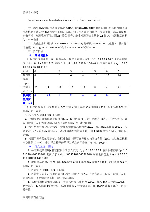
For personal use only in study and research; not for commercial use一. 原理BCA蛋白浓度测定试剂盒(BCA Protein Assay Kit)是根据目前世界上最常用蛋白浓度检测方法之一BCA法研制而成,实现了蛋白浓度测定的简单,高稳定性,高灵敏度和高兼容性。
检测浓度下限达到25 微克/毫升,最小检测蛋白量达到0.5微克,待测样品体积为1~20微升。
二. 试剂盒组份组份Cat: KGPBCA (250 assay酶标板/50assay 1mL比色杯)蛋白标准溶液(0. 5 μg/μL )5 mL BCA试剂A 25 mL×2 BCA试剂B 1mL三. 操作步骤A.酶标板操作1. 标准曲线的绘制:取一块酶标板,按照下表加入试剂孔号0 1 2 3 4 5 6 7 蛋白标准溶液(μL)0 1 2 4 8 12 16 20 去离子水(μL)20 19 18 16 12 8 4 0 对应蛋白含量(μg)0 0.5作液,充分混匀;3. 各孔加入200μL BCA工作液;4. 把酶标板放在振荡器上振荡30sec,37℃放置30分钟,然后在562nm下比色测定。
以蛋白含量(μg)为横坐标,吸光值为纵坐标,绘出标准曲线;5. 稀释待测样品至合适浓度,使样品稀释液总体积为20μL,加入BCA工作液200μL,充分混匀,37℃放置30分钟后,以标准曲线0号管做参比,在562nm波长下比色,记录吸光值;6. 根据所测样品的吸光值,在标准曲线上即可查得相应的蛋白含量(μg),除以样品稀释液总体积(20μL),乘以样品稀释倍数即为样品实际浓度(单位:μg/μL)。
B.分光光度计测定1. 标准曲线的绘制:各管按照下表加入试剂孔号0 1 2 3 4 5 6 7 蛋白标准溶液(μL)0 5 10 20 40 60 80 100 去离子水(μL)100 95 90 80 60 40 20 0 对应蛋白含量(μg)02.5 5.0 10.0 20.0 30.0 40.0 50.02. 根据样品数量,按50体积BCA试剂A加1体积BCA试剂B(50:1)配制适量BCA工作液,充分混匀;3. 各管加入1000μL BCA工作液;4. 各管充分混匀,37℃放置30分钟,然后在562nm下比色测定。
血清蛋白质浓度定量测定

空白孔 0 0.1 — 2.0
1 0.02 0.08 — 2.0 0.05
2
3
4
5 0.1 0 — 2.0 0.5
实 验 步 测定孔 骤
— 0.1 2.0
0.04 0.06 0.08 0.06 0.04 0.02 — 2.0 0.1 — 2.0 0.2 — 2.0 0.4
孔号 试剂(mL) 标准蛋白溶液 (1.5mg/ml) PBS 缓冲液 待测样品 BCA 工作液 蛋白质含量(μ g)
2、 蛋白质标准液:
用结晶牛血清白蛋白(BSA)根据其纯度用生理盐水
配制成1.5mg/ml的蛋白质标准液。(纯度可经凯
氏定氮法测定蛋白质含量而确定)
3.绘制标准曲线:
取6支试管,编号后分别加入0mL、0.02mL 、0.04mL、0.06mL、0.08mL、0.1mL标准 蛋白质溶液(BSA),然后各管分别用去离子 水补充到0.1mL,最后各试管中分别加入 2.0mLBCA工作液,每加完一管,立即在漩 涡混合器上混合(注意不要太剧烈,以免 产生大量气泡而难于消除),以蛋白质的 蛋白含量为横坐标、吸光度A为纵坐标绘制 标准曲线。
试剂盒组成
2.稀释标准品:完全溶解蛋白质标准品 (5mg/mlBSA(牛血清白蛋白),-20℃保存)取10微升 用PBS(pH7.4 的等渗磷酸盐缓冲液)稀释至100微升 (样品一般可用PBS稀释),使终浓度为 0.5mg/ml。 将稀释后的标准品(0.5mg/ml)按0, 2, 4,6,8, 12, 16, 20微升分別加到96孔板的蛋白标准品孔中, 加标准品稀释液补足到20微升。 0 0 2 4 6 8 12 0.3 16 0.4 20 0.5
BCA 法定量蛋白质浓度
BCA测蛋白的具体操作步骤
For personal use only in study and research; not for commercial use一. 原理BCA蛋白浓度测定试剂盒(BCA Protein Assay Kit)是根据目前世界上最常用蛋白浓度检测方法之一BCA法研制而成,实现了蛋白浓度测定的简单,高稳定性,高灵敏度和高兼容性。
检测浓度下限达到25 微克/毫升,最小检测蛋白量达到0.5微克,待测样品体积为1~20微升。
二. 试剂盒组份组份Cat: KGPBCA (250 assay酶标板/50assay 1mL比色杯)蛋白标准溶液(0. 5 μg/μL )5 mL BCA试剂A 25 mL×2 BCA试剂B 1mL三. 操作步骤A.酶标板操作1. 标准曲线的绘制:取一块酶标板,按照下表加入试剂孔号0 1 2 3 4 5 6 7 蛋白标准溶液(μL)0 1 2 4 8 12 16 20 去离子水(μL)20 19 18 16 12 8 4 0 对应蛋白含量(μg)0 0.5作液,充分混匀;3. 各孔加入200μL BCA工作液;4. 把酶标板放在振荡器上振荡30sec,37℃放置30分钟,然后在562nm下比色测定。
以蛋白含量(μg)为横坐标,吸光值为纵坐标,绘出标准曲线;5. 稀释待测样品至合适浓度,使样品稀释液总体积为20μL,加入BCA工作液200μL,充分混匀,37℃放置30分钟后,以标准曲线0号管做参比,在562nm波长下比色,记录吸光值;6. 根据所测样品的吸光值,在标准曲线上即可查得相应的蛋白含量(μg),除以样品稀释液总体积(20μL),乘以样品稀释倍数即为样品实际浓度(单位:μg/μL)。
B.分光光度计测定1. 标准曲线的绘制:各管按照下表加入试剂孔号0 1 2 3 4 5 6 7 蛋白标准溶液(μL)0 5 10 20 40 60 80 100 去离子水(μL)100 95 90 80 60 40 20 0 对应蛋白含量(μg)02.5 5.0 10.0 20.0 30.0 40.0 50.02. 根据样品数量,按50体积BCA试剂A加1体积BCA试剂B(50:1)配制适量BCA工作液,充分混匀;3. 各管加入1000μL BCA工作液;4. 各管充分混匀,37℃放置30分钟,然后在562nm下比色测定。
BCA蛋白检测方法

BCA蛋白检测方法Bicinchoninic acid (BCA )法是近来广为应用的蛋白定量方法。
其原理与Lowery法蛋白定量相似,即在碱性环境下蛋白质与Cu2+络合并将Cu2+还原成Cu1+。
BCA与Cu1+结合形成稳定的紫蓝色复合物,在562 nM处有高的光吸收值并与蛋白质浓度成正比,据此可测定蛋白质浓度。
与Lowery法相比,BCA蛋白测定方法灵敏度高,操作简单,试剂及其形成的颜色复合物稳定性俱佳,并且受干扰物质影响小。
与Bradford法相比,BCA法的显著优点是不受去垢剂的影响。
BCA定量中的A和B液BCA定量中的A和B液的成分是什么,作用是什么?试剂A:BCA(bicinchonininc acid)碱性溶液试剂B:硫酸铜溶液作用:BCA与二价铜离子的硫酸铜溶液等其他试剂组成的试剂,混合后显示苹果绿色。
在碱性条件下,BCA与蛋白质结合时,蛋白质将Cu2+ 还原为Cu+,一个Cu+螯合两个BCA 分子,工作试剂由原来的苹果绿形成紫色复合物。
试剂(1)试剂A:含1%BCA二钠盐、2%无水碳酸钠、0.16%酒石酸钠、0.4%氢氧化钠、0.95%碳酸氢钠,将上述液体混合后调pH至11.25。
(2)试剂B:4%硫酸铜。
(3)BCA工作液:试剂A100mL+试剂B2L,混合。
(4)蛋白质标准液:准确称取150mg牛血清白蛋白,溶于100mL蒸馏水中,即为1.5mg/mL的蛋白质标准液。
(5)待测样品。
1、将上述各管混匀后于37°C保温30分钟,用562nm波长比色测定吸光度值。
2、以蛋白质含量为横坐标,光吸收值为纵坐标绘制标准曲线。
3、用测定管光吸收值在标准曲线上查找相应的蛋白质含量,再计算出待测血清中蛋白质浓度(g/L)。
待测样品浓度在50~2000微克/毫升浓度范围内有较好的线性关系BCA蛋白检测BCA Protein Assay 是基于在碱性条件下,Cu2+ 被蛋白还原成Cu+,进而发生高敏感性和选择性的颜色变化,通过分光测定,精确定量蛋白质浓度。
bsa蛋白定量流程

bsa蛋白定量流程English Answer:Introduction:Bovine serum albumin (BSA) is a widely used protein standard for quantitative protein assays. Its abundance in bovine serum, high solubility, and relatively low cost make it an ideal choice for calibration and standardization purposes. This protocol describes a step-by-step procedure for BSA protein quantification using a spectrophotometer.Materials:BSA standard solution.Protein assay reagent compatible with BCA or Bradford method.Cuvettes.Spectrophotometer.Pipettes.Vortex mixer.Microcentrifuge tubes.Procedure:1. Prepare BSA Standards:Dilute the BSA standard solution to create a series of known concentrations, typically ranging from 0 to 2mg/mL.2. Prepare Protein Assay Mixture:Mix the protein assay reagent with the appropriate volume of water according to the manufacturer's instructions.3. Incubate Reaction:Add equal volumes of the protein assay mixture to each standard and sample.Incubate the reaction at the recommended temperature and time for the specific assay method.4. Measure Absorbance:Transfer the reaction mixtures to cuvettes and measure the absorbance at the appropriate wavelength (usually 562 nm for BCA and 600 nm for Bradford).5. Create Standard Curve:Plot the absorbance values obtained from the BSA standards against their corresponding concentrations.Fit a linear regression line to the standard curve.6. Determine Sample Concentration:Measure the absorbance of the unknown samples.Use the standard curve equation to calculate the protein concentration in the samples.Additional Notes:Use high-quality reagents and equipment for accurate measurements.Calibrate the spectrophotometer regularly to ensure accuracy.Perform replicates for each sample to minimize errors.Refer to the specific protein assay reagent manufacturer's instructions for detailed protocols.中文回答:牛血清白蛋白(BSA)定量流程。
- 1、下载文档前请自行甄别文档内容的完整性,平台不提供额外的编辑、内容补充、找答案等附加服务。
- 2、"仅部分预览"的文档,不可在线预览部分如存在完整性等问题,可反馈申请退款(可完整预览的文档不适用该条件!)。
- 3、如文档侵犯您的权益,请联系客服反馈,我们会尽快为您处理(人工客服工作时间:9:00-18:30)。
– Unique structure
• can be used to produce antibodies to that protein
• antibodies specific for a protein are the basis of immune assays
Introduction
• Samples
– Blood(serum and plasma) – And other body fluids
• Urine • Pleural fluid • Cerebrospinal fluid
Introduction
• Interest proteins
– the immunoglobulins (antibodies)
cathode
The depth of the color
IgG/M/A
Electrophoresis(电泳)
• Serum protein electrophoresis
IgG/M/A???
multiple myeloma Bone marrow morphology
Myeloma cell
Electrophoresis(电泳)
– the concentration of the protein of interest will be crucial to the choice of test
Qualitative techniques
• Electrophoresis(电泳) • Ouchterlony immunodiffusion(Ouchterlony免疫扩
antiserum
Light chains
Ouchterlony immunodiffusion(免疫扩散)
Serum sample Ab
Countercurrent immunoelectrophoresis
(对流免疫电泳)
electric osmosis 电渗作用
?
Immunoelectrophoresis(免疫电泳)
散) • Countercurrent immunoelectrophoresis(对流免疫
电泳) • Immunoelectrophoresis(免疫电泳)
Electrophoresis(电泳)
• Electrophoresis equipment
cathode
5 microliters
negative charge
– theoretically can detect all the proteins
Introduction
• The test purpose
– Deficiencies of specific proteins
• such as in immunoglobulin deficiency.
– Abnormal or inappropriate expression of proteins
• Immunofixation electrophoresis 免疫固定电泳
– Permits the detection and typing of monoclonal antibodies or immunogloblins in serum or urine
Serum protein electrophores
Immunoelectrophoresis(免疫电泳)
The immunoprecipitate arcs in the serum at bottom show a normal pattern; those for IgG, IgA and IgM are labelled. Note the absence of these immunoprecipitates in the upper sample, indicating that this patient was deficient in all three immunoglobulins.
Proliferate unlimitedly
hybridoma technology
Introduction
• The selection of an appropriate assay method
– whether a qualitative or quantitative result is required
Support Medium gels (such as agar, agarose and polyacrylamide)
isoelectric point
anode
sis(电泳)
• Serum protein electrophoresis
anode
Density scan
– polyclonal antibodies by immunizing animals with that human protein
– monoclonal antibodies by hybridoma technology
hybridoma cells
cell fusion
Produce Ab
Protein assay
Jim Liu Department of Clinical Laboratory The Second Affiliated Hospital of Nanchang University
Content
• Introduction • A short historical perspective • Qualitative techniques • Quantitative methods • Conclusion
• HBV, HCV, HIV antibodies
– the proteins of the complement system
• C3, C4, C1q
– Cytokines
• Growth factor, GF • Tumor necrosis factor, TNF • Interferon, IFN
• e.g. in monoclonal gammopathies, autoimmune disease or atopy
Introduction plasma cells myeloma cells
• Principles of the protein analysis methods
– Different electrical charge