腺病毒包装原理 流程全解析
病毒包装实验整体流程及原理(慢病毒、腺病毒)

广州英思特生物科技有限公司为您提供高效快速的病毒包装实验外包服务,病毒感染细胞实验整体流程及原理目的基因不能直接整合到大多数真核细胞,常用的手段是将目的基因包装成病毒来感染细胞,从而得到表达满足实验需求。
1、病毒的种类病毒有很多种,常见的有慢病毒和腺病毒1.1慢病毒1.1.1原理慢病毒(Lentivirus)是逆转录病毒的一种。
构建的siRNA/miRNA慢病毒载体,与化学合成的siRNA和基于瞬时表达载体构建的普通siRNA载体相比,一方面可以扩增替代瞬时表达载体使用,另一方面,Lentivirus-siRNA克隆经过慢病毒包装系统包装后,可用于感染依靠传统转染试剂难于转染的细胞系如原代细胞、悬浮细胞和处于非分裂状态的细胞,并且在感染后可以整合到受感染细胞的基因组,进行长时间的稳定表达。
1.1.2特点1)直接包装成为假病毒颗粒,对分裂和非分裂细胞均有感染作用,适合RNAi研究和体内实验中难于转染的细胞(比如神经元细胞、干细胞或其它原代细胞)。
?2)可以通过简单方式,在短时间内获得稳定表达特定基因的多种细胞株。
3)可用于基因敲除、基因治疗和转基因动物研究。
4)无需任何转染试剂,操作简便。
5)可以根据客户需要制备多种标记。
1.1.3慢病毒包装简要流程:1)含有目的基因的慢病毒RNAi干扰载体的构建和质粒纯化提取。
2)慢病毒载体,包装系统共转染病毒包装细胞293T等。
3)培养48hrs-72hrs左右,收集含有病毒的上清培养液。
4)病毒的纯化和浓缩。
5)分装、-80 ℃保存。
6)滴度测定目的基因检定,并出具检测报告。
1.2、腺病毒1.2.1原理腺病毒(Adenovirus,Ad)是一种无包膜的线状双链DNA病毒,其复制不依赖于宿主细胞的分裂。
有近50个血清型,大多数Ad载体都是基于血清型2和5,通过转基因的方式取代E1和E3基因,降低病毒的复制能力。
这些重组病毒仅在高水平表达E1和E3基因的细胞中复制,因此是一种适用于治疗的高效控制系统。
腺相关病毒包装原理及步骤

腺相关病毒包装原理及步骤采用第2代重组腺相关病毒包装系统制备病毒颗粒。
该包装系统包含3个质粒和一株包装细胞株。
1)1个转移质粒:含有两侧有AAVITR序列的转基因表达盒子。
2)1个Rep-cap表达质粒:表达Rep和Cap两个基因。
3)1个辅助质粒:包含来自腺病毒的一些基因(E4、E2a和VA)。
这些腺病毒的基因可以促进AAV基因组的复制。
4)1株含有腺病毒E1基因的293细胞。
野生型腺相关病毒基因组是一条长约4.7 kb的线性单链DNA (ssDNA)分子。
基因组两末端各有一个145 nt长的Inverted Terminal Repeat (ITR),ITR序列是对称的,自己能形成发夹结构。
病毒感染细胞后,释放出单链病毒基因组,末端发夹结构促使腺相关病毒以单链基因组为模板复制互补链,形成双链DNA。
野生型病毒只表达2个基因:rep (replication) 和cap (capsid),Rep编码非结构蛋白,Cap编码结构蛋白。
腺相关病毒基因组不编码polymerase,基因组DNA的复制需要用到宿主细胞表达的polymerase。
将基因组上的Rep和Cap基因删除,克隆到质粒上,然后将目的基因克隆在两个ITR 之间,这个质粒就成为转移质粒。
而Rep和Cap基因被克隆到另外一个质粒上,成为Rep-cap表达质粒。
除了Rep-cap表达质粒,还需要一个辅助质粒。
这个辅助质粒表达腺病毒的一些基因(E4、E2a和VA),这些腺病毒基因可以促进AAV基因组的复制。
除了3个质粒外,腺相关病毒包装系统还需要一株表达腺病毒E1基因的293细胞。
每次包装重组腺相关病毒,辅助质粒均固定不变,只要血清型不变,Rep-cap质粒也是相对固定,变化的是编码目的基因的转移质粒。
腺病毒包装

腺病毒(Adenovirus)是目前最高效可靠的重组病毒表达系统之一,可用于在哺乳细胞中瞬时及高水平地表达shRNA或目的蛋白。
腺病毒是一种线性双链DNA病毒,由于其具有宿主范围广、可感染分裂和非分裂细胞、高滴度制备、外源基因装载容量大等优点,重组腺病毒已越来越广泛地被应用于基础研究和临床试验中,并成为世界上最常用的基因治疗载体之一。
服务内容
重组腺病毒质粒的构建;重组腺病毒包装制备;制备重组腺病毒。
客户提供
所需包装的目的基因信息或者序列
我们提供
含有目的基因的重组腺病毒质粒;重组腺病毒液;详细完整的实验报告。
腺病毒包装系统是什么?腺病毒包装系统详细介绍

腺病毒包装系统是什么?腺病毒包装系统详细介绍目前根据需要已经开发三代腺病毒载体:E1区或者E1&E3缺失的载体被称作第一代腺病毒载体;E2区或者E2&E4缺失的载体被称作第二代腺病毒载体;依赖于辅助病毒的腺病毒载体(Helper-Dependent Ad,HD-Ad)通常也称作无偿腺病毒或者高容量载体被划分为第三代腺病毒载体。
经典的双质粒共转染法(同源重组)原理是同源重组。
在腺病毒左臂区域(通常缺失E1基因区)插入目的基因表达盒构成腺病毒穿梭质粒,与带有腺病毒全基因组(通常缺失E1基因区)的大质粒共同转染携带E1基因区的细胞如293细胞,两种质粒在293细胞内通过同源重组形成重组腺病毒基因组,并包装成病毒颗粒。
主要缺点是重组效率比较低。
AdEasyTM包装系统AdEasyTM包装系统主要的特点是利用Cre/loxP系统在大肠杆菌中完成外源基因插入腺病毒基因组的过程,获得环状的重组腺病毒基因组,并且具有在细菌中复制的必需元件。
将重组腺病毒基因组酶切线性化后转染293细胞,避免了双质粒共转染得到重组腺病毒。
由于获得重组腺病毒质粒有明显的筛选方法,操作步骤也比较好掌握,因此这个系统一出现就受到极大的欢迎。
使用这个系统需要注意以下问题:要用能表达RecA重组酶的特定的大肠杆菌,建议建立菌种库以免细菌发生变化;由于重组是在细菌中发生的,微小的基因组变化不易发现;要获得高品质的重组腺病毒毒种(高病毒产量、高目的基因表达)应挑选多个重组腺病毒质粒,分别转染293细胞,获得多个重组病毒来筛选。
AdMax包装系统AdMax包装系统是通过Cre/loxP获得重组病毒,这个过程发生在293细胞中,从而避免在细菌中重组。
将克隆了外源基因的腺病毒穿梭质粒与携带了腺病毒大部分基因组的包装质粒共转染293细胞,利用Cre/loxP系统的作用实现重组,产生重组腺病毒。
这个系统有多种优势,包括操作简便、重组效率高、获得的病毒产率高(一般大于104vp/cell)、目的基因的表达水平高(因为mCMV启动子比hCMV 启动子强若干倍)等。
病毒包装实验整体规程及原理(慢病毒、腺病毒)
广州英思特生物科技有限公司为您提供高效快速的病毒包装实验外包服务,病毒感染细胞实验整体流程及原理目的基因不能直接整合到大多数真核细胞,常用的手段是将目的基因包装成病毒来感染细胞,从而得到表达满足实验需求。
1、病毒的种类1.11.1.1体,一方面1.1.21)研。
? 2)可以通过简单方式,在短时间内获得稳定表达特定基因的多种细胞株。
3)可用于基因敲除、基因治疗和转基因动物研究。
4)无需任何转染试剂,操作简便。
5)可以根据客户需要制备多种标记。
1.1.3慢病毒包装简要流程:1)含有目的基因的慢病毒 RNAi 干扰载体的构建和质粒纯化提取。
2)慢病毒载体,包装系统共转染病毒包装细胞293T等。
3)培养 48hrs - 72hrs 左右,收集含有病毒的上清培养液。
4)病毒的纯化和浓缩。
5)分装、- 80 ℃保存。
6)滴度测定目的基因检定,并出具检测报告。
1.21.2.1达E11.2.21)2)3)4)5)1.2.3腺病毒包装简要流程1)构建表达 siRNA/miRNA 的腺病毒载体2)采用 PacI 消化纯化的质粒。
3)消化好的腺病毒表达载体转染 293A 细胞,收获细胞以制备病毒粗提液。
4)将病毒粗提液感染 293A 细胞以扩增病毒。
5)分装,-80℃保存。
1.3、慢病毒和腺病毒的比较2、构建目的基因到载体2.1构建手段一般是根据原始质粒信息确定克隆方案,有以下两种手段。
1)如果原始质粒与载体有匹配酶切位点,采用相应的内切酶切下相应片段,回收并连接到载体,酶切,并测序鉴定DNA分子。
质粒在宿主细胞体内外都可复制。
通过个些特性,人们可以把一些目的DNA片断构建在质粒中,通过转化入大肠杆菌中,利用选择培养基来筛选从而不断的复制,来得到目的产物。
3、质粒DNA在大肠杆菌里转化连接上目的基因的质粒转化大肠杆菌是为了让目的基因在大肠杆菌里扩增,然后提取质粒,以下是质粒DNA在大肠杆菌里转化的三步骤。
3.1大肠杆菌感受态细胞的制备30min2)离心管放到42℃保温90s3)冰浴2min4)每管加800ulLB液体培养基,37℃培养1h(150r/min)5)取适当体积(100ul)的复苏细胞,涂布在选择性培养基上,正置6)倒置平皿37℃,12~16h,出现菌落3.3质粒提取步骤1)取1~4ml在LB培养基中培养过夜的菌液,12000转离心1min,弃上清一次。
腺病毒载体的分类及包装方法

腺病毒载体的分类及包装方法1.腺病毒载体分类第一代腺病毒载体:指E1或E3基因缺失的腺病毒载体。
特点:(1)外源基因容量为6.5-8Kb;(2)适用于大多数基因治疗,且容易获得高滴度的病毒;(3)可用于表达重组蛋白质或将外源基因导入对其他转染方法不敏感的细胞株中;(4)若未经纯化使用,容易引发机体产生强烈的炎症反应和免疫反应,纯化后可安全使用。
第二代腺病毒载体:指E2A或E4基因缺失的腺病毒载体。
特点:(1)外源基因容量增加,可达14Kb;(2)病毒蛋白表达降低,引发机体的免疫反应比第一代弱;(3)外源基因表达时间较长;(4)病毒产量较低,且滴度降低。
第三代腺病毒载体:也称为空壳载体或辅助病毒载体,指全部或大部分腺病毒基因组缺失,仅可保留ITR和包装信号序列的腺病毒载体。
特点:(1)外源基因容量大幅度提高,可达37Kb;(2)无病毒蛋白表达,降低了机体的免疫反应,提高了安全性;(3)外源基因可长期稳定表达;(4)需要一个腺病毒突变体作为辅助病毒;(5)可反复应用,无需考虑机体的抗腺病毒中和抗体的影响。
2.腺病毒载体包装方法传统的双质粒共转染法将插入了外源基因的穿梭载体质粒与携带腺病毒基因组的质粒共转染293细胞,这两种质粒在293细胞内通过随机的同源重组(同源序列的DNA分子之间或者分子之内的重新组合),形成重组腺病毒载体基因组,并包装成病毒颗粒。
局限性:(1) 操作较复杂,因其易被亲代病毒污染,需经过多次纯化和鉴定;(2) 重组效率低,耗费时间较长。
注:此方法适用于临床。
(1)Ad-Easy法将克隆了外源基因的腺病毒穿梭质粒与携带腺病毒大部分基因组的质粒共转染RecA+细菌,在细菌RecA 重组酶的作用下,经抗性筛选获得重组腺病毒基因组质粒,将其酶切纯化后转染293细胞,包装成重组病毒颗粒。
特点:(1) 酶切和连接步骤少,操作简便;(2) 可选择的穿梭载体多;(3) 同源重组率达60%以上,效率相对较高;局限性:(1) 某些腺病毒基因容易发生突变;(2) 设备和技术要求较高。
腺病毒的包装与制备
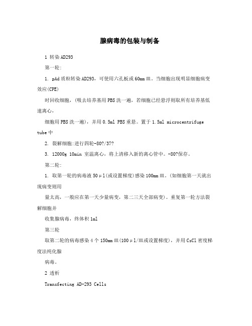
腺病毒的包装与制备1 转染AD293第一轮:1. pAd质粒转染AD293,可使用六孔板或60mm皿。
当细胞出现明显细胞病变效应(CPE)时回收细胞,(吸去培养基用PBS洗一遍,若细胞已经悬浮则取所有培养基低速离心,细胞用PBS洗一遍),并用0.5ml PBS重悬。
置于1.5ml microcentrifuge tube中2. 裂解细胞:进行四轮-80?/37?3. 12000g 10min 室温离心,将上清移入新的离心管中。
-80?保存。
第二轮:1. 取第一轮的病毒液50μl(或设置梯度)感染100mm皿。
(如细胞第一天就出现病变则用量太高,一般应在第一天少量病变,第二三天全部病变)。
重复第一轮方法裂解细胞并收集腺病毒,终体积1ml第三轮取第二轮的病毒感染4个150mm皿(100μl/皿或设置梯度),并用CsCl密度梯度法纯化腺病毒。
2 透析Transfecting AD-293 Cells以Lipofectamine? 2000说明书为本,更改了细胞密度和DNA量51. One day before transfection, plate 4x 10 cells per 6-wellofgrowth medium without antibiotics so that cells will be 50-70% confluent at the time of transfection. 2. For each transfection sample, prepare complexes as follows:a. Dilute 4μg linearized DNA in 250 μl of Opti-MEM I Reduced Serum Medium without serum (or other medium without serum). Mix gently.b. Mix Lipofectamine? 2000 gently before use, then dilute 10μl lipofectaminein250 μl of Opti-MEM I Medium. Incubate for 5 minutes at room temperature. Note:Proceed to Step c within 25 minutes.c. After the 5 minute incubation, combine the diluted DNA withdilutedLipofectamine? 2000 (total volume = 100 μl). Mix gently andincubate for 20 minutes at room temperature (solution may appear cloudy). Note:Complexes are stable for 6 hours at room temperature. 3. Add the 500 μl of complexes to each well containing cells and medium. Mix gently by rocking the plate back and forth.4. Incubate cells at 37?C in a CO2 incubator for 17-10days prior to testing for GFP. Medium may be changed after 4-6 hours.Preparing the Primary Viral Stocks1. Prepare a small dry ice-methanol bath and a small 37?C water bath and placethem in the laminar flow hood.2. Carefully remove growth medium from adenovirus-producing AD-293 plates andwash the cells once with PBS. Take care not to lose any clusters of floating andpartially attached cells during this process.Note :If the cells are already mostly detached, pipet up and down gently in the growth medium until cells become completely resuspended. Transfer cell suspension to a screw cap centrifuge tube and pellet the cells by low speed centrifugation. Aspirate medium, and wash the cells once with 0.5 ml of sterile PBS. Resuspend the cell pellet in a fresh 0.5 ml sterile PBS (per 60-mM dish) and proceed to Step 5. 3. Add 0.5 ml of PBS to each plate of cells to be harvested. Collect the cells by holding the plate at an angle and scraping the cells into the pool of PBS with a cell lifter.4. Transfer the cell suspension to a 1.7-ml screw-capmicrocentrifuge tube. If duplicate DNA samples were transfected, the cells from duplicate samples may be combined in the microcentrifuge tube at this stage.5. Subject the cell suspension to four rounds of freeze/thaw by alternating the tubes between the dry ice-methanol bath and the 37?C water bath,vortexing briefly after each thaw.Note :Each freeze and each thaw will require approximately 5 minutes’ incubationtime.6. Collect cellular debris by microcentrifugation at 12,000 × g for 10 minutes at room temperature.7. Transfer the supernatant (primary virus stock) to a fresh screw-cap microcentrifuge tube. Viral stocks can be stored for more than one year at –80?C.腺病毒扩增1. Plate 5 X 10⁶ QBI-293A cells in a 100 mm culture dish in 10 mL DMEM 5%.2. Remove 50μl(100μl) of the viral stock of the first amplification, complete to 1 mL with DMEM 5% and mix. This dilution will give a MOI of about 5.3. Remove the medium from the 100 mm dish, delicately add the viral particle mix onto the cells taking care not to disturb the monolayer and spread by slowly rocking the dish 3 times in a cross shape. Incubate for 90 minutes at 37?C in 5% CO2.4. Add 9 mL of DMEM 5%.5. Incubate at 37?C in a CO2 incubator for 72 hours. At this point you should have between 5 x91010 and 5 x 10 VP in 10 mL of DMEM 5%. Perform a MOI test (section 5.3) to estimate the titer if desired.If this quantity of virus is sufficient, you can immediately proceed to the titration step (section 5.8). In this case, collect the cells in a centrifuge tube, spin at 600 x g for 5 min, discard supernatant and resuspend the cell pellet in a minimal volume, typically 1:10 of the original volume or about 1mL. Then proceed with 3 freeze/thaw cycles (-20?C / 37?C), spin at maximum speed on a tabletop centrifuge to remove cellular debris, and collect the supernatant. Titer as described in section 5.8. 6. Perform three freeze/thaw cycles at -20?C until completely frozen/37?C until fully thawed. 7. Pellet cell debris in a sterile 15 mL conical tube. Centrifuge at maximum speed in a tabletop centrifuge for 10 minutes, remove and store the supernatant in another sterile 15 mL conical tube at -20?C or -80?C.8. Plate 3 x 10⁷ QBI-293A cells in 3 x 175 cm2(150mm dish) culture flasks (1 X 10⁷cells/flask).9. Mix 3 mL of cell lysate supernatant from step 7 with 12 mL DMEM 5%. Remove the mediumfrom the flask, then delicately add 5 mL of supernatant mix perflask on the cells, taking care notto disturb the monolayer, and spread by slowly rocking the dish 3 times in a cross shape. Incubate for 90 minutes at 37?C in 5% CO2. This should give a MOI of 25.10. Complete to 30 mL/flask with DMEM 5%.11. Incubate at 37?C in a CO2 incubator for 48 to 72 hours. At this point you should have between 3 x 10¹⁰ and 3 x 10¹¹ VP in 90mL of DMEM 5%. Perform a MOI test (section 5.3) to estimate the titer if desired. Again, if this quantity of virus is sufficient, you can immediately proceed to the titration step (section 5.8). In this case, collect the cells in a centrifuge tube, spin at 600 x g for 5 min, discard supernatant and resuspend the cell pellet in a minimal volume, typically 1:10 of the original volume. Then proceed with 3 freeze/thaw cycles (-20?C / 37?C), spin at maximum speed on a tabletop centrifuge to remove cellular debris, and collect the supernatant. Titer as described in section 5.8.12. Perform three freeze/thaw cycles at -20?C/37?C.13. Pellet cell debris in a sterile 50 mL conical tube. Centrifugeat maximum speed in a tabletop centrifuge for 10 minutes. Remove and store the supernatant.14. Plate 3 x 10⁸ QBI-293A cells in 30 x 175 cm2 culture flasks (1 X 10⁷ cells/flask). 15. Mix 45 mL of cell lysate supernatant from step 13 with 105 mL DMEM 5%. Remove the medium from the 175 cm2 flasks, pour 5 mL of the cell lysate mix per flask on the cells, taking care not to disturb the cell monolayer, and spread by slowly rocking the flask 3 times in a cross shape. Incubate for 90 minutes at 37?C in 5% CO2. This should give a MOI of 25.16. Complete to 30 mL/flask with DMEM 5%.17. Put the cells back at 37?C in a CO2 incubator for 2-3 days. You should now have between 3 x 111210 to 3 x 10 VP. Perform a MOI test (section 5.3) to estimate the titer if desired. If you want to produce viral particles, first collect and pellet the infected cells, then resuspend in 5 mL DMEM 5%. Extract viral particles by performing three freeze/thaw cycles at -20?C/37?C; pellet cell debris by centrifugation. At this point you should have between 3 x 10¹¹ to 3 x 10¹² recombinant virus particles in 5 mL DMEM 5%, for a final VP/mL of 6 X 10¹⁰ to 6 x 10¹¹. Your viral particle pre-stock will now be ready to be titrated directly, asdescribed in section 5.8, or purified, using standard cesiumchloride gradients (section 5.7). Further amplifying your recombinant virus on 109 cells will result in the production of a viral stock, while amplification on 1 x 10¹⁰ cells is technically a viral production. You should never use the virus from a production stock to infect additional cells because you will at the same time significantly increase the amount of RCA generated. Instead, use virus from an earlieramplification steps such as the stock, pre-stock or pre-amplification in order to produce more virus particles. You can eventually go back to the purified eluted plaque in order to minimize RCAproduction as much as possible. If a purified eluted plaque is no longer available, you will have to perform a plaque assay of your recombinant virus and start the amplification step again from a purified eluted plaque, after analysis of your clone. If more material than produced in the first 4 passages(see Table 5) is needed, remember to always return to the previous passage as your source for virus. For example, use virus from passage 2 in order to generate passage 3. Note that once you have depleted all of your viruses from passage 2, you must use virus from the first amplification in order to create a new passage 2. Proceeding in this fashion will keep the level of RCA as low as possible. If you want to overexpress a protein, pellet the cells and extract the protein according to its characteristics. Typically, about 1-5 mg of a well-expressed protein can beretrieved from the pellet. It is recommended to first determine the best time pi to extract the protein on a smallscale, then to proceed with large-scale protein production.CsCl密度梯度法纯化腺病毒1 Add either purified Ad or unpurified Ad (medium) to monolayer culture cells (50-100pfu/cell)2 If using one T150 flask total 10-12 ml medium is used to cover the cells and allow cells to culture for one day3 Cells should look swollen and part of the cells may be floating. Another 10ml of complete medium is added into cells and allow another day culture (36-40hrs infection period )4 All of the cells should be floating. Collect all the cells and resuspend them in 0.5-1ml of complete medium . Also save the culture medium and store at -70?.5 Freeze in methanol/dry ice bath and thaw at 37?. Repeat for 3-4times6 Spin at high speed for 5min.7 Make 40% and 15% CsCl in TBS, PH8.1(50ml each and keep at 4?)8 Make CsCl gradient solution(5ml of 15% and 4.5ml of 40%) in Beckman centrifuge tube (14*89mm, which frist sw41 rotor)9 Load the supernatant from step 6 on the top of gradient10 Centrifuge at 4? 30000rpm for 16hr11 Two bands can be seen: the faint top band (mainly defective Ad) and the lower Ad band. Only the lower band is collected.12 the Ad is dialyzed in TBS PH8.1 for 1hr and then in TBS containing 10% glycol twice (1hr each time)13 Determine the Ad concentration, aliquot into microfuge tube(50μl each )and store at -70?TBS: tris 10mM, NaCl 0.9%, PH8.115% CsCl(50ml): 9.085g CsCl+47.69ml TBS40% CsCl(50ml): 28.45g CsCl+42.7ml TBSDialyze:1 透析袋: MW 8000-144002 透析缓冲液: TBS PH8.1 灭菌甘油步骤:1冲洗透析袋,如果是蛋白则需NaHCO-NaEDTA煮沸处理后用蒸馏水洗。
腺病毒的包装系统是什么?

腺病毒的包装系统是什么?腺病毒包装系统按照重组方式主要分为两种,一种在细菌(BJ5183)中重组,为AdEasy TM腺病毒重组系统,另外一种在QBI-293A细胞中重组,为AdMax腺病毒重组系统。
一、 AdEasyTM腺病毒重组系统AdEasyTM腺病毒重组系统1998年由He Tong Chuan等改造的一种非常简单易用的双质粒系统,腺病毒基因组质粒pAdEasy-1和转移质粒pShuttle-CMV,pAdEasy-1质粒是Ad5腺病毒基因组,其中删除了E1基因(1-3533bp)和E3基因(28130-30830bp),E1基因的功能区在293A细胞中得到补充;pShuttle-CMV质粒包含目的基因插入位点、Ad5基因组左右同源臂、Ad5末端重复序列(ITR)、包装信号序列、PacI双酶切位点和卡那霉素抗性。
第一步:将目的基因cDNA插入转移质粒pShuttle-CMV;第二步:将得到的质粒用PmeI或ECORI内切酶线性化;第三步:pShuttle-CMV和pAdEasy-1共同转化大肠杆菌BJ5183,双质粒在BJ5183中发生重组,卡那霉素筛选重组子,并酶切鉴定。
因为BJ5183有recA重组酶活性但同时又缺失介导细菌本身基因组重组的酶,可以用于质粒重组和转化,得到候选重组子;第四步:将酶切鉴定正确的重组子转化DH5a感受态,在DH5a 中扩大培养,质粒提取,鉴定,PacI酶切线性化,暴露其反向末端重复序列(ITR)。
第五步:线性化质粒回收后转染293A细胞,产生重组病毒颗粒,收毒,后续纯化浓缩。
另外,也可以采用单质粒两步法重组,即将AdEasy转入感受态细胞BJ5183中,再将pADEasy-BJ5183制备成感受态细胞,将构建好的穿梭质粒pShuttle-CMV用PmeI线性化后直接转化pADEasy-BJ5183感受态细胞,卡那霉素筛选重组子,并酶切鉴定,线性化后转染293A细胞。
- 1、下载文档前请自行甄别文档内容的完整性,平台不提供额外的编辑、内容补充、找答案等附加服务。
- 2、"仅部分预览"的文档,不可在线预览部分如存在完整性等问题,可反馈申请退款(可完整预览的文档不适用该条件!)。
- 3、如文档侵犯您的权益,请联系客服反馈,我们会尽快为您处理(人工客服工作时间:9:00-18:30)。
腺病毒包装原理
腺病毒(Adenovirus)是一种线性双链DNA无包膜病毒,对分裂期细胞和非分裂期细胞均具有感染能力,且具有嗜上皮细胞性。
重组腺病毒载体是以腺病毒为基础发展起来的工具病毒载体,具有宿主范围广,免疫原性强,基因容量大,不与基因组整合,瞬时表达等特点。
重组腺病毒(AdV)是人血清5型腺病毒(Ad5),一种复制缺陷的腺病毒载体系统,在基因治疗、基础生命科学研究等领域被广泛应用。
腺病毒缺失了早期表达基因序列E1和E3区。
E1是腺病毒复制所必须的,E1的缺失使其不能自身复制,只能依靠包装细胞如293A细胞提供的反式互补进行复制扩增,以此保证腺病毒的安全性。
源井生物所提供的腺病毒颗粒经过超速梯度离心和过滤,并通过壳蛋白免疫法进行滴度测定。
腺病毒的滴度在10^10~10^12pfu/ml,完全可以满足各类实验的使用要求。
产品规格
服务流程及质量控制:
基因表达腺病毒
源井生物通过构建腺病毒表达载体并包装成腺病毒。
使用过表达腺病毒感染细胞后可以实现目的基因瞬时高表达。
腺病毒包装容量大,重组片段可达4.5kb。
基因敲除腺相关病毒载体选择:
基因干扰腺病毒
源井生物通过构建腺病毒干扰载体并包装成腺病毒。
使用干扰腺病毒感染细胞后可以实现干扰RNA瞬时高表达。
基因干扰腺病毒载体选择:
注明| 文章是源井生物原创,转载请注明。