显微镜简易使用手册——【蔡司精品】
Axio Vert A1_LED-Xcite使用简述_河南城建 zgh201809——【蔡司精品】

Vert A1显微镜操作简述说明:一般操作习惯为用透射光找到目标物后,再用荧光观察。
1.开机:打开主机底座左侧的电源开关,底座前方的电源指示灯亮起;2.使用透射光观察2.1 在载物台上放好样品,2.2 选择合适的物镜2.3 调节聚光镜转盘,选择合适的观察模式。
看有色样本时通常用1号位;看无色透明生物样本使用4/ 5号位(相差)观察时,注意与物镜相匹配,40x和100x对应5号位2.4调节荧光滤块位置4号位“BF”2.5 调节底座右侧的光源强度调节开关调节合适的光强,通过粗、细焦螺旋聚焦样品;2.6 调整聚光镜上孔径光阑的大小(切换不同物镜后可能需要重新调整以获得合适对比度)及亮度等,进行观察;(调节清楚后,将下图所示结构拉出,切换到相机,可在ZEN软件进行拍照)113.使用反射光:X-cite 荧光光源3.1 打开X-cite 灯箱电源开关(若确定使用荧光观察,开启显微镜时即可打开), 3.2 将聚光镜上方的插板推至金属挡片位置,防止LED 灯对荧光信号产生干扰;3.3 选择合适的荧光滤片组,调节X-cite 灯箱上转轮选择合适的亮度档位等,进行观察;4. 使用完毕后,将物镜清洁,降至最低位置;5. 关闭X-cite 灯箱上的电源上开关,盖上防尘罩。
注:1、为延长X-cite 灯泡寿命,关闭和打开之间请保证间隔在30min 以上;2、短时间内不使用X-cit 荧光光源时,可将X-cite 灯箱上的亮度旋钮调至0档位(注意最后不用时要关掉电源)。
常用部件简介:透射光部分:光源:3——LED灯4——两孔的滤光片插板,一般安装:A:金属挡片,当使用荧光观察时,使用此挡片遮挡上方的LED灯,来防止LED灯受荧光激发后产生干扰荧光。
在使用透射光时需要移开。
B:色温片,使用LED透射光时,用来改变LED光源的色温,使其更接近日光。
聚光镜:1——孔径光阑,用于调节样本图像的对比度(非亮度,亮度调节请使用底座上的光强调节旋钮);2——调节转盘,更换聚光镜内光阑的档位,如使用相差观察方式时,需要使用与物镜一致的档位(相差环)。
蔡司显微镜操作步骤及注意事项

蔡司显微镜操作步骤1.检查电源线和灯箱接头是否接好,确保各个部件都正常插入光路中。
2.打开机体电源,调节光的亮度至适中。
3.把载物台降到最低,放入样品。
4.首先用低倍物镜观察样品。
先用粗调旋钮升高载物台,等到样品快清晰时改用微调旋钮把样品调清晰,然后再逐步切换到高倍,微调一下,使图像清晰。
5. 打开AxioVision SE64 Rel. 4.9软件,点击“预览”调出图像,微调旋钮把样品调清晰后点击“拍摄”。
6. 点击“控制区域”的比例尺选项,选定放大倍数,之后点击“标尺”进行标尺的添加。
7. 图像保存与拷贝。
8. 关机。
蔡司显微镜注意事项1.只有经过培训且能够独立熟练操作的人员方可使用,避免损坏机器。
2.调焦时注意不要使物镜碰到试样,以免划伤物镜。
3.在载物台垫片圆孔中心的位置远离物镜中心位置时不要切换物镜,以免划伤物镜。
4.亮度调整切忌忽大忽小,也不要过亮,影响灯泡的使用寿命,同时也伤害视力。
5.所有(功能)切换,动作要轻,要到位。
6. 转换倍数时,不要直接拨动物镜;而是拨动转盘;调节焦距,从低倍到高倍找焦平面,禁止直接用高倍找焦平面;高倍观察一定采用微调;7. 载物台支架旋钮、摄像头、加密狗和双滤光轮滑齿不要动。
8. 禁止使用USB、移动硬盘等可移动存储;9. 所有部件禁止用酒精擦拭;10.禁止带腐蚀液进入实验室。
11.关机时要将亮度调到最小。
12.关机不使用时,将物镜通过调焦机构调整到最低状态。
13.关机不使用时,不要立即该盖防尘罩,待冷却后再盖,注意防火。
手术室德国蔡司显微镜
德国蔡司显微镜(VARIO700)一操作流程
1放松底座的刹车装置,收拢各节横臂,旋紧制动手轮,推至手术床边。
2将制动手轮放松,根据手术部位安放显微镜,满意后立刻将底座的刹车刹牢,并将各制动手轮重新旋紧。
3插上电源插座,摆放好足控踏板,开启电源开关。
4按动自平衡按钮机器进行自平衡调节
5开始设置:设置显微镜到最小放大倍数。
5.1将显微镜移动到工作位置并根据物镜的焦距长度选择工作距离。
6调节瞳距:使用旋钮,根据您的瞳距调节目镜之间的距离,以保证两个目镜中的图像重合到一起。
7调节目镜:调节屈光度设置环到刻度0。
7.1如戴眼镜的操作者佩戴眼镜拟实施手术,请将屈光度设置环调节刻度到+5开始调节。
7.2戴眼镜者观察时,不需要旋出眼杯。
8根据机体说明书:自动调节显微镜白平衡。
9手术结束,应将亮度调至最小时才关闭电源开关,拔除电源线。
蔡司显微镜的使用流程

蔡司显微镜的使用流程1. 准备工作•确保蔡司显微镜的电源线已经插入电源插座,并将显微镜的电源打开。
•检查显微镜的镜头是否清洁,并使用布擦拭。
•检查显微镜的目镜和物镜是否正确安装。
2. 样品准备•将需要观察的样品放在显微镜的载物台上。
•确保样品平整,并没有移动或者摇晃。
3. 调整光源•通过显微镜的光源控制按钮,调整光源的亮度,使得样品中的细节能够清晰可见。
•根据需要,可以通过光源控制按钮调整光源的颜色。
4. 调整倍率•通过旋转物镜转盘,选择合适的倍率。
一般来说,较低的倍率可以提供较宽的视野,较高的倍率则可以提供更高的放大倍数。
•注意,在更换物镜时,需要用载物台上的样品进行对焦,以保证观察时的清晰度。
5. 对焦•使用调节轮实施初步对焦,获得大致的清晰图像。
•进一步调节调节轮,进行精细对焦,以便获得最清晰的图像。
•如果需要改变焦点深度,可以通过显微镜上的聚焦控制器进行调节。
6. 观察和记录•使用目镜调整显微镜,使自己的双眼能够同时观察到样品。
•用眼睛凝视样品,尽量维持稳定的观察角度,避免眼睛疲劳。
•如果需要,可以使用显微镜上的调节装置,将目镜与眼睛的距离调整到最舒适的位置。
•在观察过程中,可以通过显微镜上的调节轮来调整焦点。
•如果需要,可以使用显微镜上的标尺或刻度来测量样品的长度、面积等。
7. 关闭显微镜•观察结束后,将倍率调节到最低档位,并关闭光源。
•将样品从载物台上取下,清洁显微镜的各个部分。
•关闭显微镜的电源。
结论以上就是使用蔡司显微镜的基本流程。
在使用显微镜时,需要注意保持样品的干净和稳定,并留意调节光源和倍率的合适性,以获得更好的观察效果。
同时,在使用显微镜时,也要注意保护眼睛,避免长时间用眼造成的疲劳。
蔡司 kinevo 900 手术显微镜使用说明书
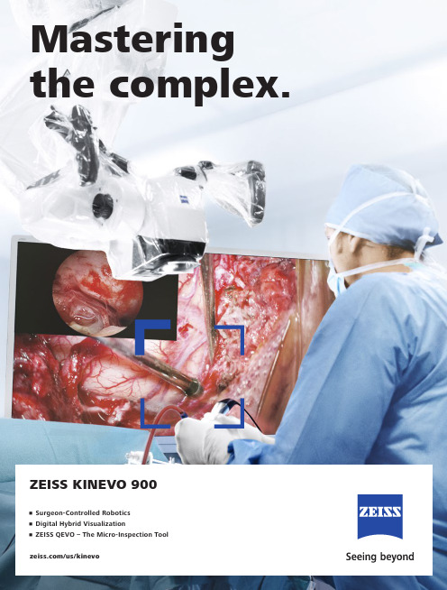
/us/kinevoZEISS KINEVO 900Mastering the complex.• Surgeon-Controlled Robotics • Digital Hybrid Visualization • ZEISS QEVO – The Micro-Inspection ToolZEISS KINEVO 900The Robotic Visualization SystemJust like you, we love challenging the status quo.The result? Over 100 innovations to perfect the already acclaimed surgical visualization platform. KINEVO® 900 from ZEISS is designed to deliver more functionalities than any surgical microscope today. ZEISS KINEVO 900 combines digital and optical visualization modalities, offers a unique Micro-Inspection Tool and will impress you with its Surgeon-Controlled Robotics. All to enable you to gain greater certainty in a virtually disruption-free workflow.Designed to meet real needs. To make a real difference!A lot more. And, a lot less too.When treating complex vascular conditions, you typically work at high magnification. Even the slightest vibrations can cause disruptions. And constant manual repositioning to better visualize structures or precisely approach deep-seated lesions can become extremely tedious. Not anymore! ZEISS KINEVO 900 delivers a lot more positioning precision with a lot less effort.PointLockSurgeon-Controlled Robotics adds a complete new level of ease to precise positioning. Imagine being able to focus and move around a structure to visualize the targeted anatomy – reducing any manual hassle. In addition, PointLock enables you to do a KeyHole movement to observe a larger area inside a cavity – a particular benefit in areas with narrow access. Simply put:Focus. Activate. Swivel.Active vibration dampingYou know the problems that can be created by the tiniest vibrations. The active damping provided by ZEISS KINEVO 900 minimizes collateral system vibrations, ensuring rock-solid stability. Enabling you to completely, and steadily, focus on what matters most: your treatment.Focus Activate Swivel5When you need it. Where you need it. The new navigation interface of ZEISS KINEVO 900 is designed to work in concert with your navigation device. When you require precise repositioning to reexamine previously visualized structures or when you need to align with a pre-mapped trajectory, making use of all six axes, the Robotic Visualization System® delivers precise positioning at the push of a button. Putting you exactly where you need to be – when you need to be there. PositionMemoryWhen working on a tumor case, you may already have identified regions of concern where you want to protect the functional structure. After storing these in PositionMemory, you can come back and visualize them at the exact same magnification, working distance and focus – without losing time for manual repositioning. In a nutshell:Save. Move. Recall.Image-guided surgeryMinimize time-consuming efforts in approaching challenging neurosurgical pathologies. Combine the Surgeon-Controlled Robotics of ZEISS KINEVO 900 with navigation interface to approach deep-seated pathologies in cranial surgery, brain stem or skull base tumor removals – right when you need it.Save Move RecallImage with Brainlab Microscope Navigation Software7New dimensions. Freedom of choice. Working through oculars at extreme angles can sometimes be a pain in the neck. Literally. With no way out, you might have to contend with uncomfortable working positions causing fatigue. Now, relief and revolutionary dimensions in visualization arein sight.The Digital Hybrid Visualization with integrated 4K technology of ZEISS KINEVO 900 welcomes you to a world of heads-up ocular-free surgery, giving you freedom of movement. And freedom of choice to use an optical setup, depending on the application need.Fully integrated 4K camera technologyDuring lateral lumbar or thoracic spine and posterior fossa approaches,ZEISS KINEVO 900’s integrated 4K visualization can be essential. It providesyou with multimodal visualization capabilities – the flexibility to decouple fromthe classic optical approach and to work with outstanding 4K picture qualityand clarity. Even when magnifying tiny details.What’s more… your assistant surgeon, OR staff and residents also benefit from the 4K visual clarity of ZEISS KINEVO 900. They share the same high-resolution, digital image to follow the procedure with comparable fidelity. Delivering indispensable education and training.9Critical challenge. Vital solution.Your challenge: When working from an external perspectiveof a surgical microscope, your visualization of the anatomy is limited to a straight line of sight – missing critical information behind tissue or corners. Efficient and effortless access to this comprehensive information is essential for treatment.Our solution: QEVO® from ZEISSThe unique, proprietary Micro-Inspection Tool from ZEISS complements intraoperative microsurgical visualization, enabling you to discover unexplored areas during the surgical intervention without additional footprint. You can look around corners and eliminate blind spots. And most importantly, you can gain greater insights – for better clinical decisions.To support your surgical workflow, ZEISS QEVO is engineered with an angled design – keeping your hands out of the lineof sight during insertion in the surgical field. And, it allowsfor an easy fit between the ZEISS KINEVO 900 and the situs, eliminating the need to reposition the head of the device. Greater insights, on demand.ZEISS QEVO enables you to inspect the perforator or examine the distal neck of the aneurysm to ensure the clip blades are fully extended.11Ease of use. Peace of mind.Surgical certainty is your imperative. Enabling you to achieve it is ours. That’s why, in the development of the Micro-Inspection Tool, we placed a high priority on its ease of use.ZEISS QEVO is truly integrated. You don’t have to plan for an additional device during surgery. Just plug it into your ZEISS KINEVO 900 for a seamless surgical workflow and to easily switch back and forth between views.ZEISS QEVO is fully autoclavable.So there’s no need forany additional draping. This is another attribute that makes ZEISS QEVO an indispensable tool – always available during surgery. On demand.ZEISS QEVO. Innovation in action.ZEISS QEVO in clinical use during a left mini-pterional approach for clipping an aneurysm.1314For the fluorescence distribution: The Intensity Map enables you to conveniently identify relative fluorescence levels reached during the INFRARED 800 observation period.For the speed of the flow: The Speed Map indicates how fast the fluorescence intensity increased during the observation period – indicating the speed of the blood flow.For the indicative time: The Delay Map (or Summary Map) provides quick information about the time when the fluorescent signal appeared for each image point in the map.1 P ZEISS BLUE 4001ZEISS YELLOW 5601Visualization of fluorescence-stained structures using BLUE 400 during surgery.Visualization of fluorescence-stained structures using YELLOW 560.For a complete picture: The Diagram Function outlines assessment of fluorescence intensity variation over time and fast access to the key indicators for further analysis.BeforeFor no compromises:After 15Setting new benchmarks. Shaping a new future. When we envisioned the all-new Robotic Visualization System,we conceived a design that can deliver so much more withoutlosing its familiarity. With ZEISS KINEVO 900, we continue tolive our vision of supporting you in becoming one with yourvisualization system – of delivering purposeful innovations.ones that matter the most for you.The Robotic Visualization System: The first of its kind.Surgeon-Controlled RoboticsDelivering precise positioning with a lotless effort – with motors in all axes.ZEISS QEVO – The Micro-Inspection ToolComplementing intraoperative microsurgicalvisualization to discover unexplored areasduring surgical intervention. Gain greaterinsight. On demand.16Digital Hybrid VisualizationProviding an opportunity for ocular-free surgery, with the freedom to use a traditional optical setup – depending on the application need.Integrated Intraoperative Fluorescence – The Power of Four.The redesigned intraoperative fluorescence technologies from ZEISS offer you the Power of Four – so you always have the tools you need.17Digital connectivity. Transforming OR’s.ZEISS ConnectZEISS Observe Neurosurgery, in particular, is a technologically intensivesurgical discipline. This has pushed us toward the edge oftransformation: to develop leading digital technologiesenabling you to expand the boundaries of surgical care –to the next level.ZEISS KINEVO 900 offers full digital connectivity.Manage surgical data wherever you are: ZEISS Connect Appenables you to access your surgical data from your iOS device,and also delivers dedicated functionalities for efficient work-flows.Take teaching to new heights: ZEISS Observe App enablesyou to virtually broadcast your procedure in the OR. Yourstudents can follow the live surgery directly on mobile screensor immerse themselves in a rich VR Experience.Gain value with new digital services: ZEISS Smart Servicesenables faster support for you and your team with remoteconnectivity. Benefit from the increased system availabilitypowered by a secure connection to your ZEISS KINEVO 900.18Connecting simplicity and innovation.ZEISS SMARTDRAPEYour visualization needs are paramount to us. And, soare the needs of your team. That’s why we gave a specialfocus to the OR preparation process in the developmentof ZEISS KINEVO 900.Being an integral part of the optical path, the SMARTDRAPEwith VisionGuard® from ZEISS is designed together withZEISS KINEVO 900 so you and your team can have thebenefits of a vivid view, and effective patient protection.At the same time – the new innovations make the drapingprocess simply simple!• Innovative folding: to eliminate guesswork and complexity.• Intuitive attachment: for an effortless and simple self-locking mechanism.• Integrated RFID chip: for easy activation of AutoDrape®.Designed for ZEISS KINEVO 900.Support whenever you need it.ZEISS OPTIMEIf you rely on high system availability, consider our ZEISSOPTIME service agreements, which are designed to ensurethe readiness of our medical equipment when you need it.ZEISS OPTIME service agreements for ZEISS KINEVO 900now come with connectivity for ZEISS Smart Services.19Technical DataKINEVO ®900 from ZEISS Technical DataRated Voltage 100 V – 240 VCurrent Consumption Max. 1.350 VARated Frequency 50 Hz – 60 HzElectrical Standard Complying with IEC 60601-1:2005/A1:2012Protection class I, degree of protection IP20Class 2 laser product as perIEC 60825-1:2007, IEC 60825-1:2014Weight Weight max. 395 kgWeight of system incl. transport container: T r a n s p or t d i r e c t io n 850 m mma x.c a .1760m m c a . 530 - 1635 m m-25° / +135°A x i s 4±45°A x i s 5-28° / +20°A x i s 3-A x i s M o n i t o r R o t a t e : ±125°T i l t :-20° / +5° (±3°) 360° c o n t i n u os A x i s1 25° / +225°A x i s 6T o le r a n c M in .M i n .M in .20Cable length: 5 mQEVO ® from ZEISS and QEVO ECUTechnical Data Direction of View 45° upwards Shaft Diameter 3.6 mm Shaft Length 120.0 ± 1.0 mm Total Diameter 13.0 mmField of View 100° +5°/-10° (ISO 8600-3:2019-08)Illumination20 – 35 lumen LED Weight (without cable)250 g Sterilization AutoclavableImage Resolution 1920 x 1080 pixel full HD Length of Cable5000 mmOperation Temperature +10 to +40 °C (500/1000 s intermittent use)QEVO ECU Dimensions Length = 265.0 ± 1 mm, height = 59.3 ± 1 mm and depth = 212.2 ± 1 mm Weight2.2 kgOperating Voltage 24V (+/- 10%) ADC Video OutputDVI-D full HD21Technical DataOptions VideoStereo video camera 3D HD, fully integrated, 2 x 3-chip HD, 1080p incl. 2nd HD 3D monitor 4K video camera, fully integrated 3-chip 4K, 2160p Stereo video camera 4K 3D, fully integrated, 2 x 3-chip 4K, 2160p, incl. 2nd HD 3D monitor Integrated HD video recording, withSmartRecording, low-Resolution recording, editing and streaming 2nd system monitor HD 2DAttachment for consumer (SLR) photo camera External 55" 4K 3D video monitor, with mobile cartIntraoperative FluorescenceBLUE 400INFRARED 800INFRARED 800 Compact INFRARED 800 with FLOW 800YELLOW 560Connectivity / Data Manage-mentDICOM module for image and video data transfer from / to PACS. Patient management by modality worklist management.Shared Network Data storage WLAN option, with WiFi Hotspot Navigation Interface Standard Navigation Interface ExtendedAccessoriesZEISS QEVO and QEVO ECU12.5x magnetic wide field eyepieces with integrated eyecups Stereo co-observation tubeFoldable Tube f170 / f260, including the PROMAG function for additional 50 % magnification and integrated rotate functionTiltable binocular tube, swivel range 180°, focal length f = 170 mm14-function, wired foot control panel 14-function, wireless foot control panel 2-function foot switch Mouth switch3-step magnification changerStandard Configuration Apochromatic OpticsMotorized focus; Varioskop ® with working distance 200 – 625 mmMotorized zoom; zoom ratio 1:6, magnification factor y = 0.4x – 2.4x10x magnetic wide field eyepieces with integrated eyecupsAutoFokus with 2 visible laser dots, automatic mode with magnetic brakesIllumination2 x 300 W Xenon, with automatic lamp exchange Automatic Iris Control for adjusting the illumination to the field of view Individual light threshold settingFocus Light Link: working distance controlled light intensityManual adjustment of diameter of field of illuminationAdditional illumination beam to brighten up shadows, motorizedSystem OperationMultifunctional programmable handgrips Magnetic clutches for all system axes Central user interface with full-screen video XY robotic movement in 6 axes (variable speed)Active dampingManual and motorized PointLock function with variable speedPositionMemory (with variable speed)Motorized XY lateral movement with variable speedMultiVision System (HD), with shutter controlSystem SetupAutoBalanceAutoDrape – air evacuation system 1Park Position Drape PositionVideoIntegrated 3-chip Full HD video camera, 1080p 24" HD video touchscreen on extendable arm, 16:9 aspect ratioIntegrated still image capturing both on HDD and USB-mediaConnectivity / Data Manage-ment Video-in for external HD video sources Remote diagnosis via internet / VPN Sterile DrapeZEISS SMARTDRAPE1Available with ZEISS SMARTDRAPE only.22Your needs. Our packages.Select a ZEISS KINEVO 900 built to fit your typical clinical use-cases. ZEISS KINEVO 900 comes with pre-defined packages giving you a head start in planning the most suitable configuration for your specific needs.Interested in digital visualization? Check out the digital package. That’s our commitment to cover you for tomorrow while keeping your present needs into focus.always included always included as INFRARED 800 only optional23e n -U S _30_010_0099I P r i n t e d i n t h e U n i t e d S t a t e s . C Z -V I I I /2021 I n t e r n a t i o n a l e d i t i o n : o n l yf o r s a l e i n s e l e c t e d c o u n t r i e s .T h e c o n t e n t s o f t h e b r o c h u r e m a y d i f f e r f r o m t h e c u r r e n t s t a t u s o f a p p r o v a l o f t h e p r o d u c t o r s e r v i c e o f f e r i ng i n y o u r c o u n t r y . P l e a s e c o n t a c t o u r r e g i o n a l r e p r e s e n t a t i v e s f o r m o r e i n f o r m a t i o n . S u b j e c t t o ch a n g e si n d e s i g n a n d s c o p e o f d e l i v e r y a n d d u e t o o n g o i n g t e c h n i c a l d e v e l o p m e n t . R o b o t i c V i s u a l i z a t i o n S y s t e m , K I N E V O , Q E V O , F L O W , A u t o D r a p e , V a r i o s k o p , S M A R T D R A P E a n d V i s i o n G u a r d a r e e i t h e r t r a d e m a r k s o r r e g i s t e r e d t r a d e m a r k s o f C a r l Z e i s s M e d i t e c A G o r o t h e r c o m p a n i e s o f t h e Z E I S S G r o u p i n G e r m a n y a n d /o r o t h e r c o u n t r i e s .© C a r l Z e i s s M e d i t e c A G , 2021. A l l r i g h t s r e s e r v e d .View onto cerebellum and lower cranial nerves. Image courtesy of Dr. Robert F. Spetzler, Barrow Neurological Institute, Phoenix, Arizona, USA. (Cover page) Front temporal area for STA-MCA bypass procedure. Image courtesy of Dr. Peter Nakaji, Barrow Neurological Institute, Phoenix, Arizona, USA (Cover page) Aneurysm clipping using ICG and overlay. Image courtesy of Prof. Dr. Andreas Raabe, Inselspital, University Hospital of Bern, Switzerland (Page 2 and 3) View onto optic nerve and internal carotid artery. Image courtesy of Dr. Peter Nakaji, Barrow Neurological Institute, Phoenix, Arizona, USA (Page 4)Image-guided surgery. Image courtesy of BrainLab AG (Page 6 and 7)View onto spinal cord dura. Image courtesy of Dr. Robert F. Spetzler, Barrow Neurological Institute, Phoenix, Arizona, USA (Page 8 and 9)Small view of the cerebellum through the Retrosigmoid Approach. Image courtesy of Dr. Peter Nakaji, Barrow Neurological Institute, Phoenix, Arizona, USA (Page 10)Left mini-pterional approach for clipping an aneurysm. Image courtesy of Dr. Peter Nakaji, Barrow Neurological Institute, Phoenix, Arizona, USA (Page 11 and 13)View onto corpus callosum and septum pellucidum. Image courtesy of Dr. Peter Nakaji, Barrow Neurological Institute, Phoenix, Arizona, USA (Page 12)Transnasal transspenoidal for re-exploration and excision of recurrent pituitary Macroadenoma with possible abdominal fat. Image courtesy of Dr. William White, Barrow Neurological Institute, Phoenix, Arizona, USA (Page 13)Hemmorrhage from right temporal AVM. Image courtesy of Dr. Gary K. Steinberg, MD PhD, Stanford University (Page 14)Right temporal Craniotomy for AVM. Image courtesy of Dr. Robert F. Spetzler, Barrow Neurological Institute, Phoenix, Arizona, USA (Page 15)Glioma surgery using BLUE 400. Image courtesy of Prof. Dr. Walter Stummer, University Clinic, Münster, Germany (Page 15)Left-temporal craniotomy for tumor resection with YELLOW 560. Image Courtesy of Dr. Peter Nakaji, Barrow Neurological Institute, Phoenix, Arizona, USA. (Page 15)Carl Zeiss Meditec AG Goeschwitzer Strasse 51–5207745 Jena Germany/med /kinevoCarl Zeiss Meditec USA, Inc.5300 Central Parkway Dublin, CA 94568USA/med/us。
蔡司S88 显微镜操作规程

蔡司S88 显微镜操作规程
一、应用范围及特点
1、广泛应用于眼科、神经外科、耳鼻喉科、显微外科和整形外科等的手术。
2、使术野清晰、暴露充分。
二、操作规程
1、踩平脚踏板,推显微镜至工作位置,踩下脚踏板固定显微
镜。
2、连接电源,开启显微镜电源开关。
3、开启电路灯 1,预热数分钟,调节亮度至合适为止。
4、手术前套好显微镜套。
5、手术结束后将亮度调节至 1.0,待记忆片刻后关闭灯路 1。
6、开启电磁锁,将显微镜各部位折叠到位。
7、关闭显微镜电源开关,拔掉电源插头,将显微镜归位放置。
三、注意事项
1、未经培训者切勿操作显微镜,应接受专业培训后,在专业人员指导下方可操作,操作时,动作轻柔,勿用力拽拉。
2、使用前检查显微镜各固定钮是否牢固,使用时将支撑臂松开各关节调节合适位置备用。
3、每次使用后使用拭镜纸擦净物镜及目镜,勿用乙醇、乙醚等有机溶剂擦拭镜身,可用软布蘸软质消毒剂和水擦拭。
4、关闭显微镜时要先将光源钮调至最小后再关闭显微镜电源。
5、在记录本上及时登记使用情况、性能、故障及解决方法。
6、显微镜放置位置相对固定,避免碰撞。
蔡司 Axiovert 5 智能倒置细胞培养显微镜说明书

质臻至简蔡司Axiovert 5用于细胞培养和研究的智能显微镜/axiovert20 µm HeLa Kyoto细胞,物镜:LD Plan-Neofluar 63×。
双通道荧光成像:细胞核为蓝色,微管蛋白为红色。
正在为您的实验室寻找一款功能强大的显微镜?想要一款成像时间短、图像质量优的显微镜?这很有必要!拥有一款高质易用的显微镜,对于需要在实验室进行长时间工作的您来说显得十分重要。
智能的倒置细胞培养显微镜蔡司Axiovert 5是您明智的选择:您仅需专注于样品和工作流,按下拍照按钮,即可获得用于数据记录的清晰图像。
该设备将透射光配备的各种观察方式与多通道荧光相结合,用于研究您的细胞或组织培养。
不仅如此,当实验室空间紧张时,您甚至可以将该智能显微镜作为单机使用,将图像保存在U盘上,而无需使用额外的计算机或软件。
用于细胞培养和研究的智能显微镜› 简介› 优势› 应用› 系统› 技术参数›售后服务更简单、更智能、更高度集成使用智能显微技术,让工作更智能蔡司Axiovert 5显微镜十分智能,且成像快速、结果出众。
您只需专注于样品,按下按钮,即可保存细胞或组织培养的清晰图像。
这款智能显微镜还会自动为您调整透射光以及多通道荧光图像的设置及参数。
自动叠加的多通道荧光图像包含标尺信息,该信息将自动保存在图像文件的元数据中。
轻松自如,享受您的日常工作Axiovert 5让您不用再时时刻刻焦急地等待实验结果。
其设计符合人体工程学,功能巧妙,可为您全天候的工作提供支持。
您只需专注于样品本身,使用单手便能完成包括拍照、移动载物台、调焦和控制亮度在内的各种主要操作。
光强管理功能可在所有放大倍率下提供统一的亮度,让您无需在更换物镜时手动调节灯泡亮度。
为了进一步提高细胞分析流程的速度和数据可靠性,您可以选择使用Labscope 中的AI 细胞融合度和AI 细胞计数分析功能,实时获得可重复的结果。
放眼未来,选择一款立足前沿的活细胞 显微镜从常规细胞培养到研究,Axiovert 5可无缝融入您的实验室和工作流。
2蔡司显微镜的操作流程
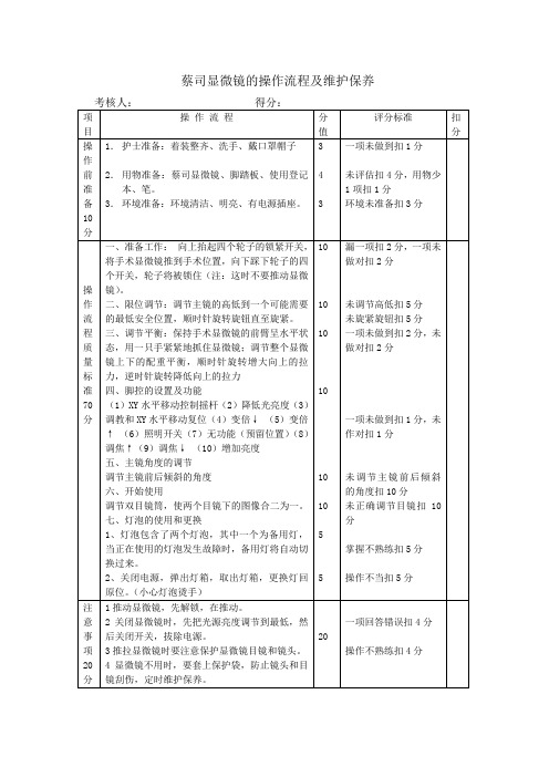
蔡司显微镜的操作流程及维护保养考核人:得分:项目操作流程分值评分标准扣分操作前准备10分1.护士准备:着装整齐、洗手、戴口罩帽子2.用物准备:蔡司显微镜、脚踏板、使用登记本、笔。
3.环境准备:环境清洁、明亮、有电源插座。
343一项未做到扣1分未评估扣4分,用物少1项扣1分环境未准备扣3分操作流程质量标准70分一、准备工作:向上抬起四个轮子的锁紧开关,将手术显微镜推到手术位置,向下踩下轮子的四个开关,轮子将被锁住(注:这时不要推动显微镜)。
二、限位调节:调节主镜的高低到一个可能需要的最低安全位置,顺时针旋转旋钮直至旋紧。
三、调节平衡:保持手术显微镜的前臂呈水平状态,用一只手紧紧地抓住显微镜;调节整个显微镜上下的配重平衡,顺时针旋转增大向上的拉力,逆时针旋转降低向上的拉力四、脚控的设置及功能(1)XY水平移动控制摇杆(2)降低光亮度(3)调教和XY水平移动复位(4)变倍↓(5)变倍↑(6)照明开关(7)无功能(预留位置)(8)调焦↑(9)调焦↓(10)增加亮度五、主镜角度的调节调节主镜前后倾斜的角度六、开始使用调节双目镜筒,使两个目镜下的图像合二为一。
七、灯泡的使用和更换1、灯泡包含了两个灯泡,其中一个为备用灯,当正在使用的灯泡发生故障时,备用灯将自动切换过来。
2、关闭电源,弹出灯箱,取出灯箱,更换灯回原位。
(小心灯泡烫手)10101010101055漏一项扣2分,一项未做对扣2分未调节高低扣5分未旋紧旋钮扣5分一项未做到扣2分,未做对扣2分一项未做到扣1分,未作对扣1分未调节主镜前后倾斜的角度扣10分未正确调节目镜扣10分掌握不熟练扣5分操作不当扣5分注意事项20分1推动显微镜,先解锁,在推动。
2关闭显微镜时,先把光源亮度调节到最低,然后关闭开关,拔除电源。
3推拉显微镜时要注意保护显微镜目镜和镜头。
4显微镜不用时,要套上保护袋,防止镜头和目镜刮伤,定时维护保养。
20一项回答错误扣4分操作不熟练扣4分。
- 1、下载文档前请自行甄别文档内容的完整性,平台不提供额外的编辑、内容补充、找答案等附加服务。
- 2、"仅部分预览"的文档,不可在线预览部分如存在完整性等问题,可反馈申请退款(可完整预览的文档不适用该条件!)。
- 3、如文档侵犯您的权益,请联系客服反馈,我们会尽快为您处理(人工客服工作时间:9:00-18:30)。
显微镜使用说明
显微镜的清洁与保管
无论是显微观察还是显微照相,检查并保持光路系统的清洁是特别重要的。
A ,可能需要清除尘埃的部位:物镜,目镜等(其他 位置非ZEISS 工程师不允许拆卸清洗)。
清洁时先用一个吹风球或软毛刷去掉附着在表面的灰尘和其他异物,然后再做擦拭。
B ,擦拭各镜头时有以下注意事项:
1 擦拭液选用乙醚酒精混合液(乙醚:无水酒精= 7:3)
2 擦拭时一定要采用专用镜头纸或长纤维的脱脂棉签。
3 蘸取擦镜液后用力甩一下,保证擦拭棉签处于半干状态(擦镜液不能多,以保证挥发快速)
擦拭工具和方法如下图
显微镜使用前的注意事项:
1安装完成后,确认显微镜的高度,建议购买可调节高度的坐椅,使得观察者和显微镜高度保持一致.
2放置显微镜的桌子下方,应尽量为空置的位置,方便将腿放入.显微镜应尽量向前摆放,这样观察时腰和颈椎可以保持垂直状态,降低因为长时间观察导致的身体疲劳,并可以有效降低腰和颈椎疾病的发生概率.
3使用者应当明白基本的光学原理,知道如何调节科勒照明,孔径光阑,相差,DIC,荧光等
4使用者应会使用照相软件,知道如何调节至最佳状态.
使用时的注意事项
1 低档显微镜开关时卤素灯电压一般应调至最低。
2 转换物镜时,应旋转物镜架,不要用手直接转物镜。
3 荧光光源汞灯由于使用寿命短(大约200小时) ,为保证尽可能的延长使用时间和
安全,汞灯电源开 /关之间的隔时间必须大于三十分钟。
4 显微镜的各光学部件应调节到位(如, DIC 插件,荧光滤片,物镜等),以免影
响显微镜的正常工作。
如果不到位 ,那么观察时通常会看到月牙形阴影 .
5 使用油镜时应使用专业用油,不要用其它介质(如香柏油),以免损伤镜头。
每
次使用完油镜后,需将油镜擦拭干净(物镜及玻片)。
(正置显微镜,油滴在样品上。
倒置显微镜,油直接滴在物镜上。
)
6 不要用手直接触摸光学部件的表面(如物镜,荧光模块,目镜等),以免留下手指
印在上面,影响观察效果。
7 在拆卸全自动显微镜的聚光镜,荧光滤片盒(倒置显微镜)等部件时,应在断电的
情况下进行操作。
8 因为各款显微镜的有效工作距离/特性等各有不同,使用前应充分了解所用仪器
的特性及观测范围,以免因不恰当的操作,对显微镜及其配件造成损害。
9 调焦首先应该是让物镜向远离样品的方向调节,这样可以充分保证物镜的安全
提示:1,观测样品时(特别是金相样品),切不可将物镜撞及样品,以免将物镜镜头压碎。
2,移动显微镜时,应做到小心轻放,以免因震动而对光学部件及光路造成损害,影响显微镜的使用。
(建议移动显微镜时,先将各光学部件拆除,移位
后,再重新安装。
)。