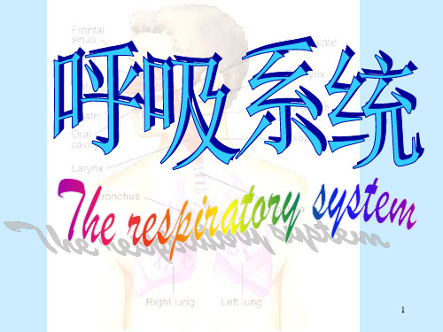
呼吸系统_英文版ppt课件
合集下载
呼吸系统(英文版) PPT-

The nasopharynx is the fist division, and it is nearest to the nasal cavities. It contains the adenoids, which are masses of lymphatic tissue. The adenoids (also known as the pharyngeal tonsils) are more prominent in children, and if enlarged, they can obstruct air passageways.
9
New words
咽
nasopharynx 鼻咽 oropharynx 口咽 laryngopharynx喉咽 pharyngeal 咽的 hypopharyngeal 下咽的
throat 咽喉 adenoid 腺样体
adeno-
adenoma
adenocarcinoma
adenovirus
Responsibilities of respiratory system
Respiration = exchange of gases between body and air
Provides oxygen to body cells for energy Removes carbon dioxide from body cells
tonsil 扁桃体
pharyngeal ~
palatine ~
palatine 腭的 larynx 喉 esophagus 食道 vocal cord 声带 vibrate震动 deterrent 妨碍物 flap 皮瓣 epiglottis 会厌 10
人体解剖学呼吸系统PPT课件
61
3.胸膜下界:在锁骨中线与第8肋相交,在腋中线与第10 肋相交,在肩胛线与11肋相交。
62
二、纵 隔(mediastinum) 1、概念:两侧纵隔胸膜之间的所有器官
和组织的总称。
63
2、分部:上纵隔: 下纵隔:前纵隔、中纵隔、后纵隔
64
学习要点
1、呼吸系统的组成,上下呼吸道的分界。 2、喉的位置、软骨的组成、喉腔的结构特点。 3、气管的位置、左右支气管的形态结构特点。 4、肺的位置、形态、左右肺的形态结构特点。 5、肺的组织结构特点;气血屏障。 6、胸膜与胸膜腔的概念、肋膈隐窝。
25
第三节 肺 (lungs)
• 一、肺的位置和形态
• 1、位置:位于胸腔内,左、右两肺分居
•
纵隔的两侧,膈的上方。
26
• 2、形态:
• 一尖:肺尖
• 一底:肺底
• 三面:膈面
•
胸肋面
•
纵隔面:
•
肺门
•
肺根
•
27
纵隔面:肺门、肺根
28
左右肺的形态区别: 左肺狭而长,前缘有心切迹。以斜裂分上、下两叶。 右肺宽而短,以斜裂、水平裂分上中、下三叶。
•
易患上颌窦炎。
•
蝶窦:前组、中组开口于中鼻道。
•
后组开口于上鼻道。
•
筛窦:开口于蝶筛隐窝。
7
鼻旁窦:鼻腔周围的含气空腔。
8
鼻旁窦
开口部位
蝶窦
蝶筛隐窝
筛窦 后组 上鼻道
前中组 中鼻道
额窦
中鼻道
上颌窦
中鼻道
9
二、喉(larynx)
• 喉的位置: • 位于颈前部的正中,平对第4—6颈椎,上通咽 • 腔,下接气管。
呼吸系统-英文版ppt课件

可编辑课件PPT
6
Diaphragm located below the lungs, attaching to the lower ribs, sternum and lumbar spine and forming the base of the thoracic cavity, is the major muscle of respiration. It is a large, dome-shaped muscle that contracts rhythmically and continually, and most of the time, involuntarily. Upon inhalation, the diaphragm contracts and flattens and the chest cavity enlarges. This contraction creates a vacuum, which pulls air into the lungs. Upon exhalation, the diaphragm relaxes and returns to its domelike shape, and air is forced out of the lungs.
可编辑课件PPT
5
When you breathe, the air: enters the body through the nose or the mouth travels down the throat through the larynx (voice box) and trachea (windpipe) goes into the lungs through tubes called main-stem bronchi one main-stem bronchus leads to the right lung and one to the left lung in the lungs, the main-stem bronchi divide into smaller bronchi and then into even smaller tubes called bronchioles bronchioles end in tiny air sacs called alveoli
呼吸系统的英语ppt课件

Breaking is a complex process
It involves the contract and relaxation of muscles, the movement of air through the passages, and the exchange of gases at the alveoli
04
Trachea and Bronchi
The structure of the tracea and bronchi
要点一
Trachea
要点二
Bronchi
The Trachea, also known as the windpipe, is a cartilaginous tube that extends from the Larynx to the bifurcation at the level of the fourth tropical vertebra It serves as a passage for air to reach the lungs The trajectory is composed of C-shaped rings of cartilage and is covered by mucous membrane
Tonsillitis
Tonsillitis is an inflation of the Tonsils (lands at the back of the stroke) that can cause pain, fever, and diversity switching It is often caused by viruses but can also be bacterial
呼吸系统疾病英文PPT课件
Based on anatomy or X-ray manifestation
❖ Bronchopneumonia ❖ Lobar or Lobular Pneumonia ❖ Interstitial Pneumonia
Based on etiology
❖ Bacterial pneumonia ❖ Viral Pneumonia ❖ Mycoplasma Pneumonia ❖ Chlamydia Pneumonia
Classification of Respiratory Infections
According to the level of the respiratory tree most involved:
❖ Upper respiratory tract infection
❖ Lower respiratory tract infection
❖ Pneumonia remains the most common cause of morbidity in China.
Question
How to classify pneumonia in clinic?
Classification
❖ Anatomy ❖ Pathogens ❖ Severity ❖ Duration ❖ Onset site
What are the signs and symptoms of pneumonia?
The clinical signs and symptoms of pneumonia depend primarily on the age of the patient, the causative organism, and the severity of the disease.
呼吸系统PPT课件:hypoxia

◆ Circulatory hypoxia(循环性缺氧)
Circulatory hypoxia refers to inadequate blood flow leading to inadequate oxygenation of the tissues.
由于组织血流量,使组织供
氧量所引起的缺氧。
O2 in blood
1.5%
physically dissolved
bound to hemoglobin
98.5%
Normal value
PaO2: 100 mmHg ( 13.3kPa ) PvO2: 40 mmHg ( 5.3kPa )
Acting factor ◣Partial pressure of inspired oxygen
100ml血液中Hb所能结合0 ml%
Acting factor
Hb quantity and quality
3. oxygen content, CO2
The total oxygen content of blood includes oxygen that is bound to haemoglobin and physically dissolved in plasma.
◣Etiology
1.Decreased PO2 in inspired air :
plateau
2.External respiratory dysfunction:COPD
3.Venous-to-arterial shunts:
congenital heart disease
◣Characteristics of blood O2
◣Etiology
《呼吸系统疾病》PPT课件
A、肺泡壁:毛细血管受压,充血消退。 B、肺泡腔:大量纤维素和中性粒细胞,纤维素丝 穿过肺泡间孔与相邻肺泡中的纤维素网相连
h
35
(3)灰色肝样变期(5-6天) ① 形成:变态反应达到高峰并逐渐减弱 ② 镜下:
A、肺泡壁:毛细血管受压,充血消退。 B、肺泡腔:大量纤维素和中性粒细胞,纤维素丝 穿过肺泡间孔与相邻肺泡中的纤维素网相连
③ 肉眼:病变肺叶肿胀、暗红色,切面可挤出 泡沫状血性浆液。
h
27
lobar pneumonia(大叶性肺炎)
h
28
④ 临床病理联系:
A、毒血症:寒战高热、外周血白细胞计数升高。 B、呼吸道症状:咳嗽、咳痰。 C、渗出液中可检出肺炎链球菌。 D、X线:片状模糊阴影。
h
29
(2)红色肝样变期 (3-4天) ① 形成:变态反应增强,血管扩张、通透性增高 更加明显,纤维蛋白原渗出。 ② 镜下: A、肺泡壁:毛细血管扩张充血。 B、肺泡腔:大量红细胞、一定量的纤维素
h
14
第一节 第二节 第三节 第四节 第五节 第六节
肺炎 慢性阻塞性肺病 肺尘埃沉着症 慢性肺源性心脏病 呼吸窘迫综合征 肺癌
h
15
n 1
Pneumonia 肺炎
h
16
概述:
➢ 指肺的急性渗出性炎症。
分类依据
病因 性质 病变部位 范围
h
17
一、细菌性肺炎
(一)大叶性肺炎(lobar pneumonia)
通过肺泡间孔蔓延
h
22
2、病因和发病机制
(1)病因: 肺炎链球菌 (2)诱因: 呼吸道防御功能减弱 (3)发病机制:
细菌侵入肺泡内繁殖
Ⅰ型变态反应
h
35
(3)灰色肝样变期(5-6天) ① 形成:变态反应达到高峰并逐渐减弱 ② 镜下:
A、肺泡壁:毛细血管受压,充血消退。 B、肺泡腔:大量纤维素和中性粒细胞,纤维素丝 穿过肺泡间孔与相邻肺泡中的纤维素网相连
③ 肉眼:病变肺叶肿胀、暗红色,切面可挤出 泡沫状血性浆液。
h
27
lobar pneumonia(大叶性肺炎)
h
28
④ 临床病理联系:
A、毒血症:寒战高热、外周血白细胞计数升高。 B、呼吸道症状:咳嗽、咳痰。 C、渗出液中可检出肺炎链球菌。 D、X线:片状模糊阴影。
h
29
(2)红色肝样变期 (3-4天) ① 形成:变态反应增强,血管扩张、通透性增高 更加明显,纤维蛋白原渗出。 ② 镜下: A、肺泡壁:毛细血管扩张充血。 B、肺泡腔:大量红细胞、一定量的纤维素
h
14
第一节 第二节 第三节 第四节 第五节 第六节
肺炎 慢性阻塞性肺病 肺尘埃沉着症 慢性肺源性心脏病 呼吸窘迫综合征 肺癌
h
15
n 1
Pneumonia 肺炎
h
16
概述:
➢ 指肺的急性渗出性炎症。
分类依据
病因 性质 病变部位 范围
h
17
一、细菌性肺炎
(一)大叶性肺炎(lobar pneumonia)
通过肺泡间孔蔓延
h
22
2、病因和发病机制
(1)病因: 肺炎链球菌 (2)诱因: 呼吸道防御功能减弱 (3)发病机制:
细菌侵入肺泡内繁殖
Ⅰ型变态反应
respiratorysystem呼吸系统ppt课件讲解学习
❖Alveolus, totally about 250 to 300 million in lungs of an adult, is supplied by a terminal pulmonary arteriole, which has a diameter of about 35 um and which gives rise to about 1000 capillaries per alveolus. The capillaries are 7 to 10 um in diameter. The distance between the alveolar surface and the capillaries is only 0.050.1 um. The pulmonary capillaries drain into the pulmonary veins and from there into the left atrium. The lung also receives blood through the bronchial arteries from the aorta.
❖ The trachea divides into right and left main bronchi and these in turn divide into lobar bronchi (upper, middle, and lower on the right, and upper and lower on the left). The airways continue to divide into terminal bronchioles, respiratory bronchioles, alveolar ducts and alveolar sacs.
呼吸系统(中英文)PPT课件
呼吸困难 labored breathing (hypoventilation) 右心衰 right-sided heart failure (cor pulmonale)
Treatment
不能根治 控制症状
No cure relieving
symptoms
防止并发症 preventing complications
小细支气管炎
病理学 Pathology
NMU博学至精 明德至善
Clinical features
支气管粘膜炎症、粘液分泌旺盛
咳痰
支气管痉挛,渗出物阻塞
喘
病理学 Pathology
NMU博学至精 明德至善
晚期表现 Late stage menifestation
血氧饱和度低 insufficient oxygenation of blood (hypoxemia)
肺间质、肺泡间隔 :cap. , f, Mφ
病理学 Pathology
NMU博学至精 明德至善 Histology of the Airways
Components Functions
Bronchi are distinguished from bronchioles primarily by the presence of cartilage in their walls. Bronchioles also lack submucosal glands.
Mucosa
Submucosa
Muscles
Cartilage 病理学 Pathology
NMU博学至精 明德至善
Epithelium
Pseudostratified ciliated columnar cells Mucous (goblet) cells
组织学与胚胎学呼吸系统ppt课件
肺静脉
支气管静脉
47
参与多种物质的代谢和转化
肺血管内皮细胞具有很强的多种酶活性
例:合成和降解前列腺素;
合成和分泌心房肽(内皮细胞、血管平滑 肌细胞、II肺泡细胞): A.排钠利尿;
B.扩张肺动脉及支气管;
质的分泌。
C.增加肺泡表面活性物
血管紧张素(AT)-І的转化:AT-I AT-II
5-羟色胺的合成与清除等
48
呼吸系统中与净化空气有关的结 构有哪些?并叙述其主要结构特 点。
49
呼吸系统
(respiratory system)
1
重点: 1.肺导气部的管壁变化规律。 2.肺泡的光镜、超微结构。
2
呼吸系统中与净化空气有关的 结构有哪些?并叙述其主要结 构特点。
3
4
鼻粘膜 前庭部:鼻翼腔面 呼吸部:下、中鼻甲、
鼻道、 鼻中隔中下部 嗅 部:鼻中隔上部、 上鼻甲、 鼻腔顶部
5
上 皮:未角化的复层扁平上皮; 固有层:细密结缔组织、毛囊
(无立毛肌)、皮脂腺等。
6
上皮:假复层纤毛柱状上皮; 固有层:疏松结缔组织、混合腺、 静脉丛和淋巴组织等。
7
8
浆液性嗅腺
施万细胞
假复层柱状上皮
9
10
11
larynx
室襞 喉室
声 襞:膜部(上皮为复扁,固有层为疏松 结缔组织和富含弹性纤维的致密结缔组织)。
软骨部(同室壁和喉室相似)。
12
软骨部 膜部
13
14
15
纤毛细胞 杯状细胞 基细胞 刷细胞 内分泌细胞 (小颗粒细胞)
16
呼吸道上皮内成群的神经内分泌细胞: 细胞内含5-羟色胺、蛙皮素、降钙素、脑 啡肽等
- 1、下载文档前请自行甄别文档内容的完整性,平台不提供额外的编辑、内容补充、找答案等附加服务。
- 2、"仅部分预览"的文档,不可在线预览部分如存在完整性等问题,可反馈申请退款(可完整预览的文档不适用该条件!)。
- 3、如文档侵犯您的权益,请联系客服反馈,我们会尽快为您处理(人工客服工作时间:9:00-18:30)。
Bronchi The trachea divides into two tubes called bronchi, one entering the left and one entering the right lung. Bronchi branch into smaller and smaller tubes known as bronchioles. Bronchioles terminate in grape-like sac clusters known as alveoli. Alveoli are surrounded by a network of thin-walled capillaries.
Larynx: This is also known as the voice box as it is where sound is generated.It contains the vocal cords. It also helps protect the trachea by producing a strong cough reflex if any solid objects pass the epiglottis.
average adult lung.
.
4
The lungs take in oxygen, which all cells throughout the body need to live and carry out their normal functions. The lungs also get rid of carbon dioxide, a waste product of the body's cells. The lungs are a pair of cone-shaped organs made up of spongy, pinkish-gray tissue. They take up most of the space in the chest, or the thorax (the part of the body between the base of the neck and diaphragm). The lungs are separated from each other by the mediastinum, an area that contains the following: heart and its large vessels trachea (windpipe) esophagus thymus lymph nodes The right lung has three sections, called lobes. The left lung has two lobes.
.
2
The Upper Respiratory Tracts
Mouth, nose & nasal cavity: The function of this part of the system is to warm, filter and moisten the incoming air.
Pharynx: Here the throat divides into the trachea (wind pipe) and esophagus (food pipe). There is also a small flap of cartilage called the epiglottis which prevents food from entering the trachea.
Bronchioles: Tertiary bronchi continue to divide and become bronchioles, very narrow tubes. There is no cartilage within the bronchioles and they lead to alveolar sacs.
and lungs
Function
Transports air into the lungs and facilitates the diffusion of oxygen into the blood stream. It also receives waste carbon dioxide from the blood and exhales it.
.
3
The Lower Respiratory Tracts Trachea Muscular cartilaginous tract that is a continuation of the larynx; it divides into two main bronchi, each of which ends in a lung, and allows air to pass. The inner membrane of the trachea is covered in tiny hairs called cilia, which catch particles of dust which we can then remove through coughing.
Alveoli: Individual hollow cavities contained within alveolar sacs (or ducts).
Alveoli have very thin walls which permit the exchange of gases oxygen and
carbon dioxide. They are surrounded by a network of capillaries, into which the
inspired gases pass. There are approximately 3 million alveoli within an. Nhomakorabea1
The respiratory system can be divided into two parts: The upper respiratory tracts:mouth, nose & nasal cavity,pharynx and larynx The lower respiratory tracts:trachea,bronchi,bronchioles,alveoli,diaphragm