Superior disintegrating properties of calcium cross-linked Cassia fistula gum
去除丙叉保护

New sialyl Lewis x mimic containing an a -substitutedb 3-amino acid spacerSilvana Pedatella,a,*Mauro De Nisco,a Beat Ernst,b Annalisa Guaragna,aBeatrice Wagner,b Robert J.Woods c and Giovanni Palumbo aa Dipartimento di Chimica Organica e Biochimica,Universita`di Napoli Federico II,Via Cynthia,4I-80126Napoli,Italy bInstitute of Molecular Pharmacy,Pharmacenter,University of Basel,Klingelberstrasse,50CH-4056Basel,Switzerland cComplex Carbohydrate Research Center,University of Georgia,315Riverbend Road,Athens,GA 30602,USAReceived 11June 2007;received in revised form 20August 2007;accepted 2October 2007Available online 7October 2007Abstract—A highly convergent and efficient synthesis of a new sialyl Lewis x (sLe x )mimic,which was predicted by computational studies to fulfil the spacial requirements for a selectin antagonist,has been developed.With a b 2,3-amino acid residue L -galactose (bioisostere of the L -fucose moiety present in the natural sLe x )and succinate are linked,leading to a mimic of sLe x that contains all the required pharmacophores,namely the 3-and 4-hydroxy group of L -fucose,the 4-and 6-hydroxy group of D -galactose and the carboxylic acid of N -acetylneuraminic acid.The key step of the synthesis involves a tandem reaction consisting of a N-deprotection and a suitable O !N intramolecular acyl migration reaction which is promoted by cerium ammonium nitrate (CAN).Finally,the new sialyl Lewis x mimic was biologically evaluated in a competitive binding assay.Ó2007Elsevier Ltd.All rights reserved.Keywords:sLe x ;Selectin antagonist;b 3-Amino acid;Glycomimetic1.IntroductionSelectins 1are involved in the orderly migration of leuco-cytes from blood vessels to the sites of inflammation.2Although extravasation of leucocytes represents an essential defense mechanism against inflammatory stim-uli,excessive infiltration of leucocytes into the surround-ing tissue can cause acute or chronic reactions as observed in reperfusion injuries,stroke,psoriasis,rheu-matoid arthritis,or respiratory diseases.2An early step in this inflammatory cascade is mediated by selectin/carbo-hydrate interactions.Data from both selectin knock out mice 3a–c and LAD type 2patients,3d clearly demon-strate that the selectin–carbohydrate interaction is a pre-requisite for the inflammatory cascade to take place.Since the tetrasaccharide sLe x (1,Fig.1)is the carbo-hydrate epitope recognized by E-selectin,4it became the lead structure for the design of selectin antagonists.5The search for novel selectin antagonist with enhanced adhesion and improved pharmacokinetic properties 5has led to numerous classes of antagonists.In the initial con-tributions,6,7the structure–activity relationship was eluci-dated,revealing the essential pharmacophores which are the carboxylic acid function of N -acetylneuraminic acid,the 3-and 4-hydroxyl group of L -fucose and the 4-and 6-hydroxyl group of D -galactose (highlighted in Fig.1).8In addition,it has been shown that the D -Glc-NAc moiety is not involved in binding.Its principal func-tion is that of a rigid spacer to accommodate the appropriate spacial orientation of L -fucose and D -galact-ose.5For the design of simplified sLe x antagonists 5a dual strategy was pursued by (1)eliminating monosaccharide moieties by non-carbohydrate linkers,and (2)substitut-ing metabolically labile O-glycosidic bonds.Considering the glycoaminoacid mimics reported by Wong,9which exhibit remarkable pharmacological0008-6215/$-see front matter Ó2007Elsevier Ltd.All rights reserved.doi:10.1016/j.carres.2007.10.001*Corresponding author.Tel.:+39081674118;fax:+39081674119;e-mail:pedatell@unina.itAvailable online at Carbohydrate Research 343(2008)31–38activity,we report herein the synthesis and the biological evaluation of the scaffold 2,a member of a new family of antagonists based on a dihydroxylated b -amino acid as a substitute of galactose.This scaffold is decorated with a suitably modified L -fucose and a carboxylic acid moiety mimicking the native N -acetylneuraminic acid.The corresponding target molecule 2(Fig.1)therefore contains (2S ,3S )-2-hydroxy-b 3-serine which is coupled with the free amino group of 6-amino-6-deoxy-L -galact-ose (L -Fuc replacement)and,at the other end,with a succinic acid (D -NeuNAc replacement).An important factor for affinity is the spacial orientation of the phar-macophores in the bioactive conformation.5d,e This is expected to be realized with the rigid diamide core.2.Results and discussionA conformational analysis of 2by molecular dynamic studies indicated that the low energy conformer-2pre-sented in Figure 1exhibits distances between the carbox-ylate and C-1and C-2of L -galactose (9.2and 9.1A˚,respectively),which are similar to the one found in the bioactive conformation 10of sLe x .Conformer-2is only less stable by 5.29kJ/mol compared to its lowest energy conformation and is characterized by differing flexibili-ties of the dihedral angles h ,u and x (Fig.2).Whereas h and u show considerable flexibility over the 10,000ps period of the MD simulation,x is rather stiff,leading to a partial preorganization of 2in the bioactive conformation.The synthesis started with the preparation of 6-amino-6-deoxy-L -galactose derivative 4from commercially available L -galactose according to a known procedure 11(Scheme 1).According to this procedure,the acetona-tion to achieve 1,2:3,4-di-O -isopropyliden-L -galactose(3),using CuSO 4/H 2SO 4in acetone,gave only poor yields (55%).Using an improved procedure 12(polystyryl diphenyl phosphine–iodine complex,prepared in situ,in anhy-drous acetone,at rt under nitrogen)3was obtained in 95%yield.Tosylation of the primary hydroxy group fol-lowed by substitution with sodium azide and catalytic hydrogenation (Pd/H 2)afforded the free amine 4in 69%overall yield from L -galactose.(2S ,3S )-2-Hydroxy-b 3-serine methyl ester derivative 7,was obtained diastereoselectively 13from commercially available O -benzyl-L -serine via enolate formation (by KHMDS)of the corresponding fully protected b 3-serine 5and the coupling of the enolate with racemic 2-[(4-methylphenyl)sulfonyl]-3-phenyloxaziridine (6)(Scheme 2).The hydrolysis of methyl ester 7afforded 8in excellent yield without any traces of racemization.Then,the 2-hydroxy-b 3-amino acid 8was coupled with the previously prepared 6-amino-L -galactose deriv-ative 4by treatment with DCC and HOBT in CH 2Cl 2,yielding 9in good yield (75%)(Scheme 3).The last step of the synthesis was the introduction of a carboxylate side chain at the N-terminus of 9.The first attempt to remove the 4-methoxybenzyl ether protection (PMB)with cerium ammonium nitrate (CAN)14was accompanied by the formation of several byproducts due to the cleavage of C-2–C-3bond of the b -aminoac-idic moiety.To avoid a possible interference of the free hydroxyl group with CAN,compound 9was first acetyl-ated and then treated with CAN.A single product was formed,which turned out not to be the desired N-depro-tected derivative,but the product of a subsequent O !N intramolecular acetyl migration.15When this O !N intramolecular acyl migration was applied to ester 10,which was obtained byesterificationFigure 1.Identical spacial orientation of the pharmacophores in sLe x (1)and the glycoaminoacid mimic 2.32S.Pedatella et al./Carbohydrate Research 343(2008)31–38of 9with 3-carbomethoxypropionyl chloride,product 11could be isolated in excellent 90%yield (Scheme 3).Finally,the removal of the protecting groups by hydrogenolysis on Pd/C in an ultrasound bath (!12),followed by alkaline hydrolysis (!13),and acidic hydrolysis (TFA/H 2O)of the isopropylidene groups (Scheme 3)yielded 2as an anomeric mixture (a /b =1:2).3.Biological evaluationVarious formats of cell-free competitive binding assays under static conditions have been reported.16–18We used microtiter plates coated with human recombinant E-selectin/IgG or P-selectin/IgG,which were incubated with 2and commercially available sLe x/biotin-polyacry-Figure 2.The plots A,B,and C show the fluctuations of the h ,u ,and x torsional angles in compound 2monitored during the 10,000ps MD simulation in a box of TIP3P water molecules;A,B:the h and u dihedral angles are highly variable within the MD simulation time frame;C:the x torsion angle remains practically unchanged during the MD simulation.S.Pedatella et al./Carbohydrate Research 343(2008)31–3833late.After unbound ligand and polymer were washed from the plate,streptavidin/horseradish peroxidase con-jugate was added to enable colorimetric determination of binding.18b As a reference compound,sialyl Lewis x with an IC50value of0.9mM for E-selectin and P3mM for P-selectin was used.With2,a moderate inhibition of the interaction of sLe x/E-selectin (IC50P6mM)could be detected.However,the interac-tion of P-selectin with the sLe x-polyacrylate polymer could not be inhibited with2(see Table1).4.ConclusionThe new sLe x mimic2was designed and synthesized starting from an L-galactose derivative4and orthogo-nally protected(2S,3S)-2-hydroxy-b3-serine8in six steps.Our synthesis features the use of cerium ammo-nium nitrate(CAN)to perform a useful O!N intra-molecular acyl migration reaction on the succinoyl ester10.However,biological testing revealed that this sLe x mimic shows only low affinity of E-selectin(IC50=6mM)and no activity(IC50>10mM)forP-selectin,respectively.Since the spatially correct arrangement of the phar-macophores can be realized in2,the low affinity can alsobe due to the considerable conformationalflexibility ofthe dihedral angles h and u(Fig.2A and B)leading tosubstantial cost of entropy upon binding.5.Experimental5.1.General methodsTriphenyl phosphine polymer-bound was purchasedfrom Fluka Chemical Co.All moisture-sensitive reac-tions were performed under a nitrogen atmosphere usingoven-dried glassware.Solvents were dried over standarddrying agents and freshly distilled prior to use.Reac-tions were monitored by TLC(precoated silica gel plateF254,Merck).Column chromatography:Merck Kiesel-gel60(70–230mesh);flash chromatography:MerckKieselgel60(230–400mesh).Optical rotations weremeasured at25±2°C in the stated solvent.1H and 13C NMR spectra were recorded on NMR spectro-meters operating at400or500MHz and50,100or125MHz,respectively.Wherever necessary,two-dimen-Table1.Biological evaluation dataTest compound E-Selectin(mM)P-Selectin(mM)Sialyl Lewis x(1)0.9P3Glycoaminoacid(2)P6>1034S.Pedatella et al./Carbohydrate Research343(2008)31–38sional1H–1H COSY experiments were carried out for complete signal bustion analyses were performed using CHNS analyzer.5.2.1,2:3,4-Di-O-isopropylidene-a-L-galactopyranose(3) To a magnetically stirred suspension of dry polystyryl diphenyl phosphine(1.12g,%3.34phosphine units)in anhydrous acetone(10mL)at rt,a solution of I2 (0.85g,3.34mmol)in the same solvent(30mL)was added dropwise in the dark and under dry nitrogen atmosphere.After15min,solid L-galactopyranose (0.33g,1.67mmol)was added in one portion to the sus-pension.TLC monitoring(CHCl3/CH3OH,9:1)showed that the starting sugar was completely consumed within 30min.The reaction mixture was thenfiltered through a glass sinter funnel and washed with acetone.The solvent was removed under reduced pressure and the solid resi-due recrystallized from CHCl3/hexane(1:2)to give thefinal product3(0.41g,95%yield);½a 25D À59.5(c1.5,CHCl3),lit.19½a 25D À55.0(c3.6,CHCl3).1H and13CNMR spectral data matched that reported.125.3.6-Amino-6-deoxy-1,2:3,4-di-O-isopropylidene-a-L-galactopyranose(4)The title compound was prepared following May’s pro-cedure11starting from3.5.4.(2S,3S)-4-(Benzyloxy)-3-[di(4-methoxybenzyl)-amino]-2-hydroxybutanoic acid(8)To a stirring solution of7,prepared as already re-ported,13(1.04g, 2.00mmol)in MeOH(13mL)was added dropwise aq NaOH(1.0M solution in H2O, 4.0mL)at0°C.The reaction,allowed to warm to rt, was stirred for6.5h and then it was diluted with EtOAc (100mL).The organic phase was treated with a solution of HCl(3M,2·100mL),washed with brine(50mL) and dried over Na2SO4.The solvent was removed under reduced pressure and the residue was purified byflash chromatography(CHCl3/MeOH,9:1)to afford8(0.90g,92%)as a pale yellow oil.½a 25D +44.0(c0.9,CHCl3);1H NMR(500MHz,CDCl3):d 3.45(ddd, 1H,J3,211.0,J3,4a9.6,J3,4b3.4Hz,H-3),3.82(s,6H, 2·OCH3), 3.84(d,1H,J2,311.0Hz,H-2), 3.89(d, 2H,J a,b12.0Hz,2·CH a PMB),3.98(dd,1H,J4a,4b 11.3,J4a,39.6Hz,H a-4),4.10(dd,1H,J4b,4a11.3,J4b,33.4Hz,H b-4),4.22(d,2H,J b,a12.0Hz,2·CH b PMB),4.59(d,1H,J a,b11.7Hz,CH a Ph),4.73(d,1H,J b,a 11.7Hz,CH b Ph), 6.89(d,4H,J ortho8.8Hz,ArH), 7.25(d,4H,J ortho8.8Hz,ArH),7.38–7.48(m,5H, PhH);13C NMR(125MHz,CDCl3)d54.2,55.1,60.1, 64.1,66.2,73.9,114.6,128.1,128.6,131.6,136.9, 160.2,175.0.Anal.Calcd for C27H31NO6:C,69.66;H, 6.71;N,3.01.Found:C,69.42;H,6.69;N,3.03.5.5.N1-(1,2:3,4-Di-O-isopropylidene-6-deoxy-a-L-galac-topyranos-6-yl)-(2S,3S)-4-(benzyloxy)-3-[di(4-methoxy-benzyl)amino]-2-hydroxybutanamide(9)HOBT(0.35g,2.58mmol)was added to a stirred solu-tion of b2,3-amino acid8(0.60g,1.29mmol)in anhy-drous CH2Cl2(10mL)at rt.After30min,to the resulting mixture a solution of compound4(0.30g, 1.17mmol)and DCC(0.40g,1.42mmol)in the same solvent(10mL)was added dropwise over5min at 0°C.The solution was allowed to warm slowly to rt and,after15h,most of the solvent was evaporated under reduced pressure and replaced by EtOAc.The precipitate wasfiltered and the solution was washed with saturated NaHCO3solution,brine until neutral, then dried(Na2SO4),and concentrated under reduced pressure.Chromatography of the crude residue on silica gel(hexane/EtOAc,1:1)afforded the oily coupling prod-uct9(0.62g,75%).½a 25D+8.5(c0.9,CHCl3);1H NMR (500MHz,CDCl3)d1.31,1.32,1.43,1.45(4s,12H, 4·CH3), 3.24(ddd,1H,J60a;60b13:9;J60a;508:1; J60a;NH4:4Hz,H a-60), 3.36–3.43(m,1H,H-3), 3.51 (ddd,1H,J60b;60a13:9;J60b;506:8;J60b;NH4:9Hz,H b-60), 3.67(d,2H,J a,b13.2Hz,2·CH a PMB),3.73(d,2H, J b,a13.2Hz,2·CH b PMB), 3.79(s,6H,2·OCH3),3.80–3.85(m,1H,H-50),3.92(dd,1H,J4a,4b10.7,J4a,34.4Hz,H a-4), 3.95–4.08(m,3H,H-40,H-2,H b-4), 4.27(dd,1H,J20;104:9;J20;301:9Hz,H-20), 4.50(dd, 1H,J30;407:8;J30;201:9Hz,H-30), 4.55(d,1H,J a,b 11.7Hz,CH a Ph),4.58(d,1H,J b,a11.7Hz,CH b Ph),5.48(d,1H,J10;204:9Hz,H-10),6.84(d,4H, J ortho8.3Hz,ArH),7.18(d,4H,J ortho8.3Hz,ArH), 7.28–7.40(m,5H,PhH),7.50–7.58(m,1H,NH);13C NMR(125MHz,CDCl3)d22.9,24.7,25.2,26.2,39.7, 54.7,55.5,59.5,66.3,68.4,69.6,70.7,71.0,71.7,73.7, 96.5,108.9,109.6,114.1,127.9,128.7,130.6,131.1, 138.1,159.0,173.8.Anal.Calcd for C39H50N2O10:C, 66.27;H,7.13;N,3.96.Found:C,66.50;H,7.11;N, 3.93.5.6.1-{[(1S,2S)-3-(Benzyloxy)-2-[di(4-methoxybenzyl)-amino]-1-[(1,2:3,4-di-O-isopropylidene-6-deoxy-a-L-galactopyranos-6-yl)carbamoyl]propyl}4-methyl succi-nate(10)3-Carbomethoxy propionyl chloride(0.17g,1.16mmol) and anhydrous pyridine(94l L,1.16mmol)were added to a solution of compound9in anhydrous CH2Cl2 (11mL)at0°C.The stirring solution was allowed to warm slowly to rt and after4h most of the solvent was evaporated under reduced pressure and replaced by EtOAc.The organic phase was washed with brine (2·30mL),dried(Na2SO4),and concentrated under re-duced pressure.The residue oil was purified by silica gel chromatography(hexane/EtOAc,8:2!6:4)to give theoily product10(0.56g,90%).½a 25D+18.0(c0.8,CHCl3);S.Pedatella et al./Carbohydrate Research343(2008)31–38351H NMR(500MHz,CDCl3)d1.29,1.32,1.43,1.45(4s, 12H,4·CH3),2.58–2.68(m,4H,CH2–CH2),3.05(ddd, 1H,J60a;60b13:9;J60a;508:8;J60a;NH3:5Hz,H a-60),3.51–3.56(m,1H,H-2), 3.60(d,2H,J a,b13.1Hz, 2·CH a PMB),3.65(d,2H,J b,a13.1Hz,2·CH b PMB), 3.66(s,3H,CH3OCO),3.67–3.80(m,10H,H b-60,H-50, 2·OCH3,2·H-3),4.13(dd,1H,J40;307:8;J40;501:6Hz, H-40),4.27(dd,1H,J20;104:9;J20;302:5Hz,H-20),4.44 (s,2H,CH2Ph), 4.56(dd,1H,J30;407:8;J30;202:5Hz, H-30),5.46(d,1H,J10;204:9Hz,H-10),5.51(d,1H,J1,2 4.3Hz,H-1),6.54(dd,1H,J NH;60b8:0;J NH;60a3:5Hz, NH),6.82(d,4H,J ortho8.7Hz,ArH),7.27(d,4H,J ortho 8.7Hz,ArH),7.30–7.39(m,5H,PhH);13C NMR (100MHz,CDCl3)d24.8,25.4,26.1,26.4,29.2, 29.6,39.6,52.2,54.4,55.6,58.3,67.2,67.8,71.0, 71.2,71.9,72.9,73.3,96.6,109.2,109.8,114.0, 127.9,128.1,128.7,130.5,132.1,138.7,159.0, 169.4,171.1,173.1.Anal.Calcd for C44H56N2O13:C, 64.38;H,6.88;N,3.41.Found:C,64.19;H,6.85;N, 3.43.5.7.Methyl3-{[(1S,2S)-1-[(benzyloxy)methyl]-2-hydroxy-2-([1,2:3,4-di-O-isopropylidene-6-deoxy-a-L-galactopyranos-6-yl]carbamoyl)ethyl]carbamoyl}pro-panoate(11)A solution of CAN(4.9mL,2.9mmol)in H2O was added to a stirring solution of compound10(0.48g, 0.58mmol)in MeCN at0°C.The reaction was kept to0°C for1h and then was allowed to warm to rt.After 18h,the reaction mixture was quenched by the addition of a saturated NaHCO3solution(50mL)and extracted with CHCl3.The organic layer was washed with brine until neutral,dried(Na2SO4),and concentrated under reduced pressure.The residue,after chromatography on silica gel(CHCl3/MeOH,95:5),gave the oily product11(0.20g,90%).½a 25D À11.0(c0.6,CHCl3);1H NMR(500MHz,CDCl3)d1.31,1.33,1.45,1.47(4s,12H, 4·CH3),2.50(t,2H,J1,26.8Hz,CH2–CH2),2.65(t, 2H,J2,1 6.8Hz,CH2–CH2), 3.28(ddd,1H, J60a;60b13:9;J60a;508:8;J60a;NH4:0Hz,H a-60),3.61–3.68 (m,5H,H b-60,CH a PMB,CH3OCO), 3.84–3.90 (m,2H,H-50,CH b PMB), 4.16(dd,1H, J40;307:9;J40;501:8Hz,H-40),4.23(br s,1H,H-1),4.30 (dd,1H,J20;104:9;J20;302:4Hz,H-20), 4.35–4.40(m, 1H,H-2), 4.48(d,1H,J a,b11.7Hz,CH a Ph), 4.52 (d,1H,J b,a11.7Hz,CH b Ph), 4.59(dd,1H, J30;407:9;J30;202:4Hz,H-30),5.51(d,1H,J10;204:9Hz, H-10),6.31(br d,1H,J NH,16.8Hz,NH serine),7.22–7.26(m,1H,NH galactosamine),7.30–7.40(m,5H,PhH); 13C NMR(100MHz,CDCl3)d24.8,25.3,26.3,26.4, 29.6,31.2,40.0,52.2,53.7,66.6,69.7,70.9,71.2,72.1, 73.5,96.7,109.1,109.9,128.2,128.4,128.9,137.8, 171.9,173.3,173.6.Anal.Calcd for C28H40N2O11:C, 57.92;H,6.94;N,4.82.Found:C,58.14;H,6.91;N, 4.84.5.8.Methyl3-{[(1S,2S)-2-hydroxy-1-(hydroxymethyl)-2-([1,2:3,4-di-O-isopropylidene-6-deoxy-a-L-galactopyr-anos-6-yl]carbamoyl)ethyl]carbamoyl}propanoate(12)A solution of compound11(0.20g,0.35mmol)in MeOH(4mL)was added to a stirring suspension of 5%palladium on carbon(0.07g)in the same solvent (5mL)and then was hydrogenated(1atm)at40°C. Theflask was immersed in an ultrasound cleaning bath filled with water and sonicated for22h.Then the sus-pension wasfiltered through CeliteÒand the solid washed twice with MeOH(2·5mL).The organic phase was evaporated down under reduced pressure to afford the oily product12(0.16g,93%).½a 25D+0.4(c0.5, CHCl3);1H NMR(500MHz,CDCl3)d 1.33, 1.36, 1.47,1.49(4s,12H,4·CH3),2.51–2.57(m,2H,CH2–CH2), 2.63–2.69(m,2H,CH2–CH2), 3.37(ddd,1H, J60a;60b13:4;J60a;508:3;J60a;NH4:4Hz,H a-60),3.65–3.72 (m,4H,H b-60,CH3OCO), 3.74(dd,1H,J Ha,Hb 11.9,J Ha,1 4.9Hz,CH a OH), 3.87(ddd,1H, J50;60a8:3;J50;60b3:9;J50;401:9Hz,H-50),4.05(dd,1H, J Hb,Ha11.9,J Hb,11.5Hz,CH b OH),4.15–4.18(m,1H, H-1),4.19(dd,1H,J40;307:8;J40;501:9Hz,H-40),4.27–4.30(m,1H,H-2),4.31(dd,1H,J20;104:9;J20;302:4Hz, H-20),4.61(dd,1H,J30;407:8;J30;202:4Hz,H-30),5.20 (br s,1H,OH),5.50(d,1H,J10;204:9Hz,H-10),5.90 (br s,1H,OH),6.77(d,1H,J NH,16.3Hz,NH serine), 7.54–7.64(m,1H,NH galactosamine);13C NMR (50MHz,CDCl3)d22.7,23.2,24.3,27.4,28.0,28.7, 38.1,50.2,54.0,60.5,64.4,68.8,69.1,70.1,75.5,94.6, 107.1,107.9,171.6,173.2,173.4.Anal.Calcd for C21H34N2O11:C,51.42;H,6.99;N,5.71.Found:C, 51.28;H,6.96;N,5.73.5.9.3-{[(1S,2S)-2-Hydroxy-1-(hydroxymethyl)-2-([1,2:3,4-di-O-isopropylidene-6-deoxy-a-L-galactopyr-anos-6-yl]carbamoyl)ethyl]carbamoyl}propanoic acid(13) One molar solution of aq NaOH(0.78mL,0.78mmol) was added to a stirring solution of compound12 (0.13g,0.26mmol)in MeOH(13mL)at0°C.The reac-tion mixture was allowed to warm slowly to rt and after 3h(TLC monitoring;CHCl3/CH3OH,8:2)was quenched with some drops of acetic acid until neutral. The precipitate wasfiltered and the organic phase was evaporated under reduced pressure to afford the oilyproduct13(0.11g,91%).½a 25D+9.0(c0.8,CH3OH); 1H NMR(400MHz,CD3OD)d1.33,1.36,1.43,1.47 (4s,12H,4·CH3), 2.40–2.52(m,4H,CH2–CH2), 3.35(dd,1H,J60a;60b14:0;J60a;508:1Hz,H a-60), 3.48 (dd,1H,J60b;60a14:0;J60b;504:4Hz,H b-60), 3.61–3.66 (m,2H,CH2OH),3.94–3.99(m,1H,H-50),4.20(br d,1H,J2,1 3.7Hz,H-2), 4.24–4.31(m,2H,H-40, H-1),4.34(dd,1H,J20;105:0;J20;302:5Hz,H-20),4.63 (dd,1H,J30;408:0;J30;202:5Hz,H-30), 5.48(d,1H, J10;205:0Hz,H-10);13C NMR(125MHz,CD3OD)36S.Pedatella et al./Carbohydrate Research343(2008)31–38d23.2,24.5,25.1,26.3,34.0,34.6,40.7,55.3,61.1,67.4,71.9,72.2,72.9,73.2,97.8,109.9,110.5,174.7,176.3,181.0.Anal.Calcd for C20H32N2O11:C,50.42;H,6.77;N,5.88.Found:C,50.28;H,6.79;N,5.89.5.10.3-{[(1S,2S)-2-Hydroxy-1-(hydroxymethyl)-2-([6-deoxy-L-galactopyranos-6-yl]carbamoyl)ethyl]carbam-oyl}propanoic acid(2)A solution of glycoaminoacid13(0.11g,0.23mmol)inTFA/H2O(9:1,5mL)was stirred at rt.After3h,thereaction solvent was evaporated under reduced pressureto afford a crude that was purified by Sephadex G-10column leading to the mimic2as a mixture of b and aanomers in2:1ratio(0.067g,80%).1H NMR(500MHz,D2O)d 2.33–2.41(m,4H,CH2–CH2),3.30–3.38(m,2.66H,H-20b,2·H-60b,2·H-60a),3.52(dd,0.66H,J30;2010:0;J30;403:3Hz,H-30b), 3.53–3.60 (m,2H,CH2OH), 3.63(br dd,0.66H,J50;60a7:4;J50;60b5:0Hz,H-50b), 3.68(dd,0.34H,J20;3010:3; J20;103:6Hz,H-20a), 3.72(br dd,0.34H,J30;2010:3; J30;403:1Hz,H-30a), 3.76(br d,0.66H,J40;303:3Hz, H-40b),3.82(br d,0.34H,J40;303:1Hz,H-40a),3.96–4.02(m,0.34H,H-50a),4.11–4.23(m,2H,H-1,H-2),4.43(d,0.66H,J10;207:8Hz,H-10b),5.12(d,0.34H, J10;203:6Hz,H-10a);13C NMR(125MHz,D2O)d 31.5,32.5,41.7(C-60b),41.8(C-60a),55.7(C-2),61.8(CH2OH),70.5(C-20a,C-50a),71.3(C-30a),71.4(C-40b),71.8(C-40a),73.6(C-1),74.1(C-20b),74.9(C-30b,C-50b),94.6(C-10a),98.8(C-10b).Anal.Calcd forC14H24N2O11:C,42.43;H,6.10;N,7.07.Found:C,42.30;H,6.09;N,7.09.putational methodsMolecular dynamics simulations were performed usingthe AMBER7.0suite programs20using the SANDER mod-ule.The simulated structure2was built up by module XLEAP of AMBER using the AMBER forcefield PARM99.21 The GLYCAM2000parameters22,23were implemented forthe oligosaccharide simulations.The charges were takenfrom ab initio calculations performed by GAUSSIAN98molecular modelling package,24using the Hartree–Fockmethod with6-31G*basic set.The MD simulation of2were performed with a time step of1fs in a cubic box ofwater8.0A˚to the side containing1102TIP3P watermolecules.25After heating and equilibration for50psthe simulations were performed for10ns,under periodicboundary conditions,at constant pressure(1atm),andconstant temperature(300K).Energy minimizationsand MD simulations were performed with a dielectricconstant of unity,and a cut-offvalue for non-bondedinteractions of8A˚.The1–4electrostatic and van derWaals interactions were scaled by the standard values(SCEE=1.2,SCNB=2.0).Post-processing of the trajectories(torsional,distance and energy analysis)was performed using the CARNAL module of AMBER7.0package.AcknowledgementsS.P.is grateful to Professor Steve Hanessian,who intro-duced her to the fascinating sLe x researchfield.We thank Oto Miedico for collecting valuable results while performing his master thesis work and Professor Romu-aldo Caputo for useful discussions.We are grateful to Dr Cristina de Castro for her help in the purification protocol of thefinal product.1H and13C NMR spectra were performed at Centro Interdipartimentale di Met-odologie Chimico-Fisiche(CIMCF),Universita‘di Na-poli Federico II.Varian Inova500MHz spectrometer is the property of Naples Laboratory of Consorzio Inter-universitario Nazionale La Chimica per l’Ambiente (INCA).References1.Kansas,G.S.Blood1996,88,3259–3287.2.(a)Lobb,R.R.In Adhesion.Its Role in InflammatoryDisease;Harlan,J.M.,Liu,D.Y.,Eds.;Freeman,W.H.: New York,1992,Chapter1,pp1–18;(b)Paulson,J.C.In Adhesion.It’s Role in Inflammatory Disease;Harlan,J.M., Liu, D.Y.,Eds.;Freeman,W.H.:New York,1992, Chapter2,pp19–42;(c)Varki,A.Proc.Natl.Acad.Sci.U.S.A.1994,91,7390–7397.3.(a)Tedder,T.F.;Steeber,D.A.;Chen,A.;Engel,P.FASEB J.1995,9,866–873;(b)McEver,R.;Moore,K.;Cummings,R.D.J.Biol.Chem.1995,270,11025–11028;(c)Lasky,L.Annu.Rev.Biochem.1995,64,113–139;(d)von Adrian,U.H.;Berger, E.M.;Ramezani,L.;Chambers,J.D.;Ochs,H.D.;Harlan,J.M.;Paulson,J.C.;Etzioni,A.;Arfors,K.-E.J.Clin.Invest.1993,91,2893–2897.4.(a)Phillips,M.;Nudelman,E.;Gaeta,F.C.;Perez,M.;Singhal,A.K.;Hakomori,S.-L.;Paulson,J.C.Science 1990,250,1130–1132;(b)Walz,G.;Aruffo,A.;Kolanus, W.;Bevilacqua,M.;Seed,B.Science1990,250,1132–1135;(c)Berg,E.L.;Robinson,M.K.;Mansson,O.;Butcher,E.C.;Magnani,J.L.J.Biol.Chem.1991,266, 14869–14872.5.For reviews see:(a)Ernst,B.;Kolb,H.C.;Schwardt,O.In The Organic Chemistry of Sugars;Levy,D.E.,Fu¨gedi, P.,Eds.;Taylor&Francis Group,2006;pp803–861;(b) Kaila,N.;Thomas,B.E.,IV Med.Res.Rev.2002,22, 566–601;(c)Simanek,E.E.;McGarvey,G.J.;Jablonow-ski,J.A.;Wong,C.-H.Chem.Rev.1998,98,833–862;(d) Kolb,H.C.;Ernst,B.Chem.Eur.J.1997,3,1571–1578;(e)Kolb,H.C.;Ernst,B.Pure Appl.Chem.1997,69,1879–1884;(f)Bertozzi,C.R.Chem.Biol.1995,2,703–708;(g)Musser,J.H.;Anderson,M.B.;Levy,D.E.Curr.Pharm.Des.1995,1,221–232;(h)Giannis,A.Angew.Chem.,Int.Ed.Engl.1994,33,178–180.6.Brandley, B.K.;Kiso,M.;Abbas,S.;Nikrad,P.;Srivasatava,O.;Foxall, C.;Oda,Y.;Hasegawa, A.Glycobiology1993,3,633–641.S.Pedatella et al./Carbohydrate Research343(2008)31–38377.Ramphal,J.Y.;Zheng,Z.-L.;Perez,C.;Walker,L.E.;DeFrees,S.A.;Gaeta,F.C.A.J.Med.Chem.1994,37, 3459–3463,and literatures cited herein.8.(a)Stahl,W.;Sprengard,U.;Kretschmar,G.;Kunz,H.Angew.Chem.,Int.Ed.Engl.1994,33,2096–2098;(b) Somers,W.S.;Tang,J.;Shaw,G.D.;Camphausen,R.T.Cell2000,103,467–479.9.Cappi,M.W.;Moree,W.J.;Qiao,L.;Marron,T.G.;Weitz-Schmidt,G.;Wong, C.-H.Bioorg.Med.Chem.1997,5,283–296.10.(a)Scheffler,K.;Ernst,B.;Katopodis,A.;Magnani,J.L.;Wang,W.T.;Weisemann,R.;Peters,T.Angew.Chem., Int.Ed.Engl.1995,34,1841–1844;(b)Scheffler,K.;Brisson,J.-R.;Weisemann,R.;Magnani,J.L.;Wong,W.T.;Ernst,B.;Peters,T.J.Biomol.NMR1997,9,423–436.11.May,J.A.,Jr.;Sartorelli,J.J.Med.Chem.1979,22,971–976.12.Pedatella,S.;Guaragna,A.;D’Alonzo,D.;De Nisco,M.;Palumbo,G.Synthesis2006,2,305–308.13.Caputo,R.;Cecere,G.;Guaragna, A.;Palumbo,G.;Pedatella,.Chem.2002,17,3050–3054.14.Reetz,M.T.;Ro¨hrig,D.Angew.Chem.,Int.Ed.Engl.1989,28,1706–1709.15.Sohma,Y.;Hayashi,Y.;Skwarczynski,M.;Hamada,Y.;Sasaki,M.;Kimura,T.;Kiso,Y.Biopolymers(Peptide Sci.)2004,76,344–356.16.Rao,B.N.;Anderson,M.B.;Musser,J.H.;Gilbert,J.H.;Schaefer,M.E.;Foxall,C.;Brandley,B.K.J.Biol.Chem.1994,269,19663–19666.17.(a)Foxall,C.;Watson,S.R.;Dowbenko,D.;Fennie,C.;Lasky,L.A.;Kiso,M.;Hasegawa,A.;Asa,D.;Brandley,B.K.J.Cell.Biol.1992,117,895–902;(b)Jacob,G.S.;Kirmaier,C.;Abbas,S.Z.;Howard,S.C.;Steininger,C.N.;Welply,J.K.;Scudder,P.Biochemistry1995,34, 1210–1217;(c)Galustian,C.;Childs,R.A.;Yuen,C.-T.;Hasegawa,A.;Kiso,M.;Lubineau,A.;Shaw,G.;Feizi,T.Biochemistry1997,36,5260–5266;(d)Marinier, A.;Martel, A.;Banville,J.;Bachand, C.;Remillard,R.;Lapointe,P.;Turmel,B.;Menard,M.;Harte,W.E.,Jr.;Wright,J.J.K.;Todderud,G.;Tramposch,K.M.;Bajorath,J.;Hollenbaugh,D.;Aruffo,A.J.Med.Chem.1997,40,3234–3237.18.(a)Weitz-Schmidt,G.;Stokmaier,D.;Scheel,G.;Nifan-t’ev,N.E.;Tuzikov,A.B.;Bovin,N.V.Anal.Biochem.1996,238,184–190;(b)Thoma,G.;Magnani,J.L.;O¨hrlein,R.;Ernst,B.;Schwarzenbach,F.;Duthaler,R.O.J.Am.Chem.Soc.1997,119,7414–7415.19.Schmidt,O.T.Meth.Carbohydr.Chem.1969,1140–1142.20.Case,D.A.;Pearlman,D.A.;Caldwell,J.W.;Cheatham,T. E.,III;Wang,J.;Ross,W.S.;Simmerling, C.L.;Darden,T.A.;Merz,K.M.;Stanton,R.V.;Cheng,A.L.;Vincent,J.J.;Crowley,M.;Tsui,V.;Gohlke,H.;Radmer, R.J.;Duan,Y.;Pitera,J.;Massova,I.;Seibel,G.L.;Singh,U.C.;Weiner,P.K.;Kollman,P.A.AMBER7;University of California:San Francisco,2002.21.Cornell,W.D.;Cieplak,P.;Bayly,C.I.;Gould,I.R.;Merz,K.M.;Ferguson,D.M.;Spellmeyer,D.C.;Fox, T.;Caldwell,J.W.;Kollman,P.A.J.Am.Chem.Soc.1995,117,5179–5197.22.Kirschner,K.N.;Woods,R.J.Proc.Natl.Acad.Sci.U.S.A.2001,98,10541–10545.23.Woods,R.J.;Dwek,R.A.;Edge,C.J.;Fraser-Reid,B.J.Phys.Chem.1995,99,3832–3846.24.Frisch,M.J.;Trucks,G.W.;Schlegel,H.B.;Scuseria,G.E.;Robb,M.A.;Cheeseman,J.R.;Zakrzewski,V.G.;Montgomery,J.A.,Jr.;Stratmann,R.E.;Burant,J.C.;Dapprich,S.;Millam,J.M.;Daniels,A.D.;Kudin,K.N.;Strain,M.C.;Farkas,O.;Tomasi,J.;Barone,V.;Cossi, M.;Cammi,R.;Mennucci,B.;Pomelli,C.;Adamo,C.;Clifford,S.;Ochterski,J.;Petersson,G.A.;Ayala,P.Y.;Cui,Q.;Morokuma,K.;Malick,D.K.;Rabuck,A.D.;Raghavachari,K.;Foresman,J.B.;Cioslowski,J.;Ortiz, J.V.;Stefanov,B.B.;Liu,G.;Liashenko,A.;Piskorz,P.;Komaromi,I.;Gomperts,R.;Martin,R.L.;Fox,D.J.;Keith,T.;Al-Laham,M.A.;Peng,C.Y.;Nanayakkara,A.;Gonzalez, C.;Challacombe,M.;Gill,P.M.W.;Johnson, B.;Chen,W.;Wong,M.W.;Andres,J.L.;Head-Gordon,M.;Replogle,E.S.;Pople,J.A.GAUSSIAN 98,9ed.;Gaussian,1998.25.Jorgensen,W.L.;Chandrasekhar,J.;Madura,J. D.;Impey,R.W.;Klein,M.L.J.Chem.Phys.1983,79,926–935.38S.Pedatella et al./Carbohydrate Research343(2008)31–38。
美国专利到期的药物精选

美国专利到期的药物精选一、精神、神经系统药类1、郁复伸中文通用名:文拉法辛;别名:维拉法辛。
英文通用名:venlafaxine ;英文商品名:Effexor。
药品简介:文拉法辛是中枢神经系统药物,用于治疗精神失常、躁狂抑郁症和抑郁症。
由惠氏公司开发并于1994 年4 月首次在美国上市,此后相继在加拿大、丹麦、英国、意大利、澳大利亚等国上市。
文拉法辛具有临床疗效好、安全性高和治疗成本较低等特点,其2002 年全球销售约为21 亿美元,位列药品销售400 强的第21 位。
美国专利名称:2-苯基-2-(1-羟基环炔基或1-羟基环烷基-2-烯基) 乙胺衍生物(专利号:US4535186)专利权人:American Home Prod注:该专利到期日:2007 年12 月13 日(该专利申请日:1983 年10 月26 日;原来到期日:2002 年12 月13日;后被批准延长5 年) 。
该专利因获得美国儿科药市场独占,到期日延至2008 年6 月13 日。
同族专利: US4535186[1985208213 ]2、罗匹尼罗英文通用名:Ropinirole ;英文商品名:ReQuip 。
药品简介:罗匹尼罗于1997 年首次被批准用于帕金森病,是一种类似多巴胺的多巴胺激动剂,与第一代多巴胺激动剂不同的是其没有麦角林结构。
因为多巴胺激动剂较少引起运动不良反应,2001 年7 月,新的帕金森病治疗指南建议用多巴胺激动剂如葛兰素- 史克公司的罗匹尼罗(ropinirole ,ReQuip ○R ) 代替左旋多巴作为疾病早期的初始一线治疗用药。
2005 年美国食品药品管理局(FDA) 批准罗匹尼罗用于治疗中度到重度的多动腿综合征(RLS) 。
美国专利名称:42氨烷基22 (3H)2吲哚酮类化合物(专利号:US4452808 )专利权人: Smithkline Beckman Corp该专利到期日:2007 年12 月7 日(该专利申请日:1982 年12 月7 日;原来到期日:2002 年12 月7 日;后被批准延长5 年) 。
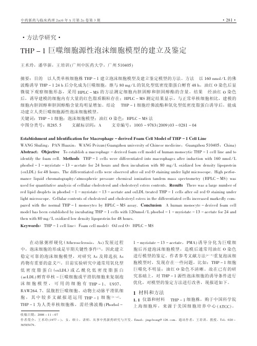
Ind. Eng. Chem. Res.2006; 45(14); 5066
Adsorption of Divalent Cadmium(Cd(II))from Aqueous Solutions onto Chitosan-Coated Perlite BeadsShameem Hasan,Abburi Krishnaiah,Tushar K.Ghosh,*and Dabir S.ViswanathNuclear Science and Engineering Institute,Uni V ersity of Missouri,Columbia,Missouri65211Veera M.Boddu and Edgar D.SmithUS Army Engineering Research and De V elopment Center(ERDC),Construction Engineering ResearchLaboratories,En V ironmental Process Branch(CN-E),Champaign,Illinois61826Chitosan-coated perlite beads were prepared in the laboratory via the phase inversion of a liquid slurry ofchitosan dissolved in oxalic acid and perlite to an alkaline bath for better exposure of amine groups(NH2).The NH2groups in chitosan are considered active sites for the adsorption of heavy metals.The beads werecharacterized by scanning electron microscopy(SEM)and energy-dispersive X-ray spectroscopy(EDS)microanalysis,which revealed their porous nature.The chitosan content of the beads was32%,as determinedusing a thermogravimetric method.The adsorption of Cd2+from an aqueous solutions on chitosan-coatedperlite beads was studied under both equilibrium and dynamic conditions in the concentration range of100-5000ppm.The pH of the solution was varied over a range of2-8.The adsorption of Cd2+on chitosan wasdetermined to be pH-dependent,and the maximum adsorption capacity of chitosan-coated perlite beads wasdetermined to be178.6mg/g of bead at298K when the Cd(II)concentration was5000mg/L and the pH ofthe solution was6.0.On a chitosan basis,the capacity was558mg/g of chitosan.The XPS data suggests thatcadmium was mainly adsorbed as Cd2+and was attached to the NH2group.The adsorption data could befitted to a two-site Langmuir adsorption isotherm.The data obtained at various temperatures provided a singlecharacteristic curve when correlated according to a modified Polanyi’s potential theory.The heat of adsorptiondata calculated at various loadings suggests that the adsorption was exothermic in nature.It was noted thata0.1N solution of HCl could remove the adsorbed cadmium from the beads,but a bed volume of approximatelythree times the bed volume of treated solution was required to completely remove Cd(II)from the beads.However,one bed volume of0.5M ethylenediamine tetra acetate(EDTA)solution can remove all of theadsorbed cadmium after the bed became saturated with Cd(II)during dynamic study with a solution containing100mg/L of cadmium.The diffusion coefficient of Cd(II)onto chitosan-coated beads was calculated fromthe breakthrough curve,using Rosen’s model,and was determined to be8.0×10-13m2/s.IntroductionCadmium plating is used for fasteners and other very-tight-tolerance parts,because of the dual qualities of lubricity at minimal thickness and superior sacrificial corrosion protection to many chemicals at high temperatures.Long-term exposure to cadmium can cause damage to the kidneys,liver,bone,and blood.Therefore,cadmium should be removed from waste streams before discharge into the environment.The removal of cadmium from wastewater is generally accomplished by precipitation with a hydroxide,carbonate,or sulfide compound.Also,several adsorbents s including activated carbon,recycled iron,novel organo-ceramic,hydrous cerium oxide,and low-cost adsorbents such as rice husk,date pits,fly ash,aerobic granular sludge,sewage sludge,tunable biopoly-mers,alginated coated-loofa sponge disks,surfactant-modified zeolites,porous poly(methacrylate)beads,red mud,goethite, perlite,and duolite s have been used for cadmium removal.1-17 The cadmium adsorption capacities of various adsorbents are summarized in Table1.Several researchers18-22have investigated chitosan as an adsorbent for removal of heavy metals,including cadmium from aqueous streams.Chitosan is a natural biopolymer,it is hydrophilic,and it has the ability to form complexes with metals.It is also a nontoxic,biodegradable and biocompatible material. According to Rorrer et al.,23chitosan flake or powder swells and crumbles,making it unsuitable for use in an adsorption column.Chitosan also has a tendency to agglomerate or form a gel in aqueous media.Although the amine and hydroxyl groups in chitosan are mainly responsible for adsorption of metal ions,these active binding sites are not readily available for sorption when it is in a gel or in its natural form.24Guibal et al.25noted that the maximum uptake of chitosan flakes was approximately half of that obtained with chitosan beads for molybdate.The adsorption capacity can be enhanced by spreading chitosan on physical supports that can increase the accessibility of the metal binding sites.Bodmeier et al.26noted that freeze-drying of chitosan gel produced particles with a high internal surface area,which boosted the metal binding capacity. Several attempts have been made to modify the structure of chitosan chemically,and its performance has been evaluated through the adsorption of heavy-metal ions from aqueous solutions.The amine and hydroxyl groups in chitosan allow a variety of chemical modifications.27Kawamura et al.28prepared a porous polyaminated chitosan chelating resin by introducing poly(ethylene amine)onto the cross-linked chitosan beads.The resultant beads showed high capacity and high selectivity for the adsorption of metal ions including cadmium.Hsien and Rorrer29investigated the adsorp-tion of cadmium on porous magnetic chitosan beads.They found*To whom correspondence should be addressed.Tel.:1-573-882-9736.Fax:1-573-884-4801.E-mail address:ghoshT@.5066Ind.Eng.Chem.Res.2006,45,5066-507710.1021/ie0402620CCC:$33.50©2006American Chemical SocietyPublished on Web06/15/2006that the adsorption capacities for1-and3-mm beads were518 and188mg Cd/g of adsorbent,respectively.They also investigated the effects of acylation and cross-linking of chitosan using gutaraldehyde.30They later noted that,although the cross-linked chitosan beads became more resistant to acid,the capacity of cross-linked chitosan for cadmium decreased significantly, compared to non-cross-linked beads.The adsorption of metal ions on gallium(III)-templated oxine type of chemically modified chitosan was reported by Inoue et al.31Chitosan cross-linked with crown ethers32,33showed improved adsorption capacity for cadmium and high selectivity for Ag(I)or Cd(II)in the presence of Pb(II)and Cr(III).Becker et al.34studied the adsorption characteristics of cadmium,along with other metals,on di-aldehyde or tetracarboxylic acid cross-linked chitosan.Although several attempts have been made to enhance the adsorption capacity of chitosan for cadmium and other metal ions through the cross-linking of chitosan,using various chemicals,the sorption capacity for metal ions decreased after the cross-linking of chitosan.Hasan et al.24noted that,by dispersing chitosan on an inert substrate(perlite)enhanced its adsorption capacity for Cr(VI).It is assumed that the active group,such as NH2,became more readily available.In this work,chitosan was dispersed on an inert substance to expose more-active sites for adsorption.Chitosan was coated on perlite,and the coated adsorbent was prepared as spherical beads.It is expected that the swelling and gel formation of chitosan can be reduced in this way,thus allowing regeneration and repeated use of the chitosan adsorbent in a column.The adsorbent was evaluated for cadmium by obtaining equilibrium adsorption data at different temperatures and pH.We also explored the regeneration of the adsorbent using dilute acid and the ethylenediamine tetra acetate(EDTA)solutions. Experimental SectionMaterials.Perlite(grade YM27)was donated by Silbrico Corporation(Hodgkins,IL).Chitosan and cadmium chloride were obtained from Aldrich Chemical Corporation(Milwaukee, WI).The chitosan used in this study was75%-85%deacety-lated and had a molecular weight of∼190000-310000,as determined from the viscosity data by Aldrich Chemical Corporation.All chemicals used in this study were of analytical grade.Oxalic acid,EDTA,and sodium hydroxide were pur-chased from Fisher Scientific Co.(Fairlawn,NJ).A stock solution containing5000mg/L of Cd(II)was prepared in distilled,deionized water using CdCl2.The working solutions of various Cd(II)concentrations were obtained by diluting the stock solution with distilled water.Preparation and Characterization of Chitosan-Coated Perlite Beads.Perlite powder(35mesh)was first soaked with 0.2M oxalic acid for4h.It then was washed with distilled water and dried in an oven for12h.Sixty grams of acid-washed perlite was mixed with30g of chitosan flakes in a beaker that contained1L of0.2M oxalic acid.The mixture was stirred for 4h while heating at313-323K(40-50°C)to obtain a homogeneous mixture.The spherical beads of chitosan,coated on perlite,were prepared via dropwise addition of the mixture into a0.7M NaOH precipitation bath.The beads were washed with deionized water to a neutral pH and freeze-dried for subsequent use.A detailed description of the bead preparation method has been discussed by Hasan et al.24The final diameter of the freeze-dried beads was∼2mm. The Brunauer-Emmett-Teller(BET)surface area of the beads was determined to be25m2/g,compared to3m2/g for the perlite particles.Most of the surface area of the beads is expected to be internal,because the external area is extremely small.The surface morphology of pure perlite changed significantly, following coating with chitosan,as indicated by scanning electron microscopy(SEM)micrographs of the beads.The SEM micrographs of the cross section of a bead also revealed the porous structure of the beads(Figure1a).A transmission electron microscopy(TEM)micrograph(Figure1b)of the beads showed that individual perlite particles were not necessarily coated with chitosan;rather,a group of particles were lumped together and coated by the chitosan film.Energy-dispersive X-ray spectrometry(EDS)microanalysis of the chitosan beads before and after their exposure to the cadmium solution confirmed the presence of Cd(II)in the interior of the beads. The EDS microanalysis shown in Figure2a exhibited peaks for aluminum and silicon,which are two major constituents of perlite.A strong peak at∼3.5keV for Cd on beads that were exposed to Cd(II)can be observed in Figure2b.In the EDS spectrum(Figure2b),a small peak for chlorine was also observed.The chlorine peak may have resulted either from the adsorption of chlorine on chitosan from the solution or fromTable1.Adsorption Capacity of Various Adsorbents for Cadmiumadsorbent pH adsorption capacity(mg/g)reference chitosan flakes 6.09.9Bassi et al.20magnetic chitosan beads1mm size 6.5518Rorrer et al.253mm size 6.5188Rorrer et al.25 non-cross-linked chitosan beads169Hsien et al.26N-acylated chitosan beads216Hsien et al.27cross-linked chitosan crown ethers 2.0-6.532.2Peng et al.29chitosan aryl crown ethers 6.039.3Yang et al.23glutaraldehyde cross-linked chitosan 3.0134.9Becker et al.31rice husk7.00.007Khalid et al.5perlite 6.6-6.70.64Mathialagan and Viraghavan16 diamine grafted chitosan crown ethers28.1Yang et al.30activated carbon 5.0 3.4An et al.21novel organo-ceramic 5.0212.8Gomez-Salazar et al.370.56modified corncorbs 4.89.0Vaughan et al.7Duolite GT-73resin 4.8105.7Vaughan et al.7Amberlite IRC-718resin 4.8258.6Vaughan et al.7Amberlite-200resin 4.8224.8Vaughan et al.7alginate-coated loofa sponge disks 5.088Iqbal and Edyvean11porous poly(methyl methacrylate)beads 6.024.2Denizli et al.13chitin 6.015.0Benguella and Benaissa22chitosan-coated perlite 5.0178.6present workInd.Eng.Chem.Res.,Vol.45,No.14,20065067the solution trapped inside the pores.However,it may be noted that some of the amine groups can undergo protonation in acidic solution,forming NH 3+,which is capable of adsorbing anions such as chlorine.Thermogravimetric analysis (TGA)of the beads indicated that the chitosan content of the bead was ∼32%.Chitosan started to decompose at ∼473K and was completely burned out at773K.The change of mass,as a function of temperature,is shown in Figure 3.Experimental ProcedureEquilibrium batch adsorption studies were performed by exposing the beads to aqueous solutions that contained different concentrations of Cd(II)ions in 125-mL Erlenmeyer flasks to a predetermined temperature.Approximately 0.25g of beads was added to 50mL of solution.The pH of the solutions was adjusted by adding either 0.1N sulfuric acid or 0.1M sodium hydroxide.The flasks were placed in a constant temperature shaker bath for a specific time period.Following the exposure of the beads to Cd(II),the solutions were filtered and the filtrates were analyzed for Cd(II)via atomic absorption spectrometry (AAS).The adsorption isotherm at a particular temperature was obtained by varying the initial concentration of Cd ions.The amount of Cd(II)adsorbed per unit mass of adsorbent (Q e )was calculated using the following equation:Results and DiscussionEffect of Surface Charge and pH on the Adsorption of Cd(II).The surface charge of the bead was determined via a standard potentiometric titration method in the presence of a symmetric electrolyte (sodium nitrate).The magnitude and sign of the surface charge was measured with respect to the point of zero charge (PZC).The pH at which the net surface charge of the solid is zero at all electrolyte concentrations is called the PZC.The pH of the PZC for a given surface is dependent on the relative basic and acidic properties of the solid 35and allows estimation of the net uptake of H +and OH -ions from the solution.The surface charge of the beads in the presence of 0.1M NaNO 3was determined in the following manner.36Four flasks,each containing 2g of chitosan-coated perlite beads,were exposed to 100mL of 0.1M NaNO 3solutions.The flasks were placed in a shaker for 24h at 125rpm.Samples from one of the flasks were titrated directly with 0.1M HNO 3,and samples from another flask were titrated with 0.1M NaOH.After the addition of the acid or base,the pH of the solution was recorded,after it was allowed to equilibrate.Titration was conducted over a pH range of 3-11.The beads were separated from the solutions of the remaining two flasks.The supernatants were titrated in a similar manner,but in the absence of beads.The net titration curve was obtained by subtracting the titration curve of the supernatant that was obtained without the presence of beads from the titration curve obtained with the beads.IntheFigure 1.(a)Scanning electron microscopy (SEM)micrograph of the cross section of the chitosan-coated perlite beads.(b)Transmission electron microscopy (TEM)micrograph of chitosan-coated perlite beads.The black spots represent perliteparticles.Figure 2.(a)Energy-dispersive X-ray spectrometry (EDS)microanalysis of chitosan-coated perlite beads,showing the presence of silicon and aluminum (platinum and silver were from the sputter coating of the sample for electrical contact).(b)EDS microanalysis of chitosan-coated perlite beads following exposure to CdCl 2.Figure 3.Thermogravimetric analysis (TGA)of chitosan-coated perlite beads:(-‚-‚-)freeze-dried beads and (s )oven-dried beads.Q e )(C i -C e )VM(1)5068Ind.Eng.Chem.Res.,Vol.45,No.14,2006absence of specific chemical interaction between the electrolytes and the surface of the bead,the net titration curves usually meet at a point that is defined as the pH PZC.Similar experiments were conducted with a0.05M NaNO3solution.No difference between the two titration curves obtained using two different ion strengths was observed.The surface charge was calculated from the following equation:37[H+]and[OH-]were calculated from the pH of the solution, after appropriate correction was made,using the activity coefficient.The activity coefficient was calculated using the Davis equation:38The results are shown in Figure4.The PZC value of the chitosan-coated perlite bead was determined to be8.5,which was similar to that reported by Jha et al.19for chitosan flake. However,Udaybhaskar et al.39reported a PZC value in the range of6.2-6.8for pure chitosan.The surface charge of chitosan-coated perlite bead was almost zero in the pH range of6-8.5. It may be noted that the p K a value of perlite was determined to be∼7.The protonation of the beads sharply increased at the pH range of3-4.5,making the surface positive.At pH<3.5, the difference between the initial pH and the pH after the equilibration time was not significant,suggesting complete protonation of chitosan.At higher pH(4.5-8.5),the surface charge of the bead slowly decreased,indicating slow protonation of chitosan on the bead.The PZC value of8.5and the behavior of surface charge of the bead could have been due to the modification of chitosan when coated on perlite,which makes it amphoteric in nature.Alumina and silica in perlite may have formed a bond with chitosan,according to the reaction shown in reactions5and6. At different ionic strengths,the surface charge of the bead was almost identical and the pH of the bead suspension did not increase when an electrolyte salt was added.The point to note from this figure is that PZC shifted toward6.5in the presence of Cd ions.As shown in Figure8(given later in this paper), CdCl2could hydrolyze to Cd(OH)2,releasing HCl,which would lower the solution pH.Also,H+ions are released during the adsorption of Cd2+ions by OH groups.As explained later,both the NH2and OH groups are expected to participate in the adsorption process.Their combined effect reduced the PZC value.To adsorb a metal ion on an adsorbent from a solution,it should form an ion in the solution.The types of ions formed in the solution and the degree of ionization are dependent on the solution pH.In the case of chitosan,the main functional group responsible for metal ion adsorption is the amine group(-NH2). Depending on the solution pH,these amine groups can undergo protonation to NH3+or(NH2-H3O)+,and the extent of protonation will be dependent on the solution pH.Therefore, the surface charge on the bead will determine the type of bond formed between the metal ion and the adsorbent surface. The effect of pH on the adsorption of Cd(II)by chitosan-coated perlite beads was studied by varying the pH of the solution over a range of2-8.The pH of the cadmium solutions was first adjusted over a range of2-8using either0.1N H2-SO4or0.1M NaOH and then chitosan-coated perlite beads were added.As the adsorption progressed,the pH of the solution increased slowly.No attempt was made to maintain a constant pH of the solution during the course of the experiment.The amount of cadmium uptake at the equilibrium solution concen-tration is shown for different initial pHs of the solution in Table 2,along with the final pH of the solution.The uptake of Cd(II) by chitosan beads increased as the pH increased from2to8. Although a maximum uptake was noted at a pH of8,as the pH of the solution increased to>7,cadmium started to precipitateσ0(C/m2))(Ca-Cb+[OH-]-[H+])FSa(2)logγi)-0.5109( I I+ I-0.3I)(3)I)0.5∑iCizi2(4)Figure4.Surface charge of chitosan-coated perlite beads in the presenceof CdCl2:([)0.1M NaNO3,(0)0.05M NaNO3,and(2)cadmiumsolution.Table2.Cadmium Uptake at Equilibrium at Various Solution pHat298Kconcentration at equilibriumof the liquid phase(mmol/L)uptake by thesolid phase(mmol/g)final pH ofthe solutionInitial pH of the Solution:20.3030.0285 2.10.5890.0607 2.11.6070.12502.43.4820.1964 2.56.9640.3214 2.6Initial pH of the Solution:4.50.0890.036 4.50.4020.098 4.61.2500.196 4.63.0360.286 5.06.6960.424 4.9Initial pH of the Solution:60.2250.080 6.20.4020.143 6.21.1600.268 6.32.6790.375 6.56.1600.536 6.6Initial pH of the Solution:80.0890.0898.00.3200.1618.11.1600.3218.22.7700.4468.35.9400.5808.6Ind.Eng.Chem.Res.,Vol.45,No.14,20065069out from the solution.Therefore,experiments were not con-ducted at pH >8.0.The increased capacity at pH >7may be a combination of both adsorption and precipitation on the surface.It is concluded that the beads had a maximum adsorption capacity at a pH of ∼6,if the precipitated amount is not considered in the calculation.The amine group of the chitosan has a lone pair of electrons from nitrogen,which primarily act as an active site for the formation of chitosan -metal-ion complex.As mentioned previously,at lower pH values,the amine group of chitosan undergoes protonation,forming NH 3+,which leads to an increased electrostatic attraction between NH 3+and the sorbate anion.Cadmium in an aqueous solution is hydrolyzed with the formation of various species,depending on the solution pH.Moreover,Cd 2+,which is the main hydrolyzed cadmium species in the pH range of 5-7appear in the form of Cd(OH)+,Cd(OH)20,and Cd(OH)3-.Among them,Cd 2+is the predominant species in the solution within this pH range.The fraction of negatively charged hydrolysis products in the solution increases as pH increases.Various hydrolysis reactions are given by Reed and Matsumoto 40and Baes and Mesmer.41Chitosan can form chelates with cadmium ions (Cd 2+)with the release of H +ions.A chelate formation may require the involvement of two or more complexing groups from the molecule.The Cd ion may seek two or more amine groups from chitosan to form the complex.This should normally reduce the pH of the solution.Kaminiski and Modrzejewska 42suggested that the increase in pH may be attributed to the exchange of released H +ions between the surface of the bead and the solution.In the case of chitosan,the protonation of NH 2groups occurs at a rather low pH range.The fact that the pH of the solution increased as the adsorption progressed suggests that Cd(II)formed a covalent bond with the NH 2group.The two NH 2groups could come from two different glucosamine residues of the same molecule,or from two different molecules of chitosan.Jha et al.19compared the stability constants for ammonia and amino complexes with those for chloro complexes of cadmium and noted that the formation of covalent bond with amine nitrogen is the more-preferred reaction.It was noted that the present adsorbent can adsorb 4.98mmol Cd/g of chitosan at 298K when the Cd(II)concentration was 5000mg/L and the pH of the solution was 6.0.The NH 2groups are the main active sites for cadmium adsorption.As can be seen from Figure 8(presented later in this paper),two NH 2groups will benecessary for the adsorption of one Cd ion,because the concentration of NH 2on chitosan is ∼6.9mmol/g.The maximum capacity for cadmium should be ∼3mmol/g.Because the maximum capacity obtained experimentally is 4.98mmol/g,other sites such as CH 2OH,OH,or O groups are also involved in adsorbing cadmium.At pH <2.0,NH 3+starts to hinder the adsorption on the beads,resulting in a further decrease in the capacity.As the pH increases,the deprotonation of amine groups may occur,resulting in a decrease of competition between proton and metal species for surface sites of the bead.21Most of the adsorbents investigated by previous reseahers 16,18,19,36,41did not adsorb any Cd(II)at pH <4,except the present chitosan-coated perlite.It may be noted from Figure 5that both pure perlite and chitosan did not adsorb any cadmium at pH <4.The chitosan-coated perlite beads showed two distinct regions in the pH curve.It seems that,between pH 2and 4.5,one site (mainly NH 2)was most active and at pH >4.5,another site (OH groups)became active for the adsorption of cadmium.Perlite may have provided more-energetic active sites for the adsorption and prevented protonation of amine groups and thus enhanced the adsorption from a more-acidic solution,compared to other adsorbents.Note that pure perlite had an almost-negligible adsorption capacity for Cd(II),as shown in Figure 9(presented later in this paper).Mathialagan and Viraghavan 16also observed a similar capacity for Cd(II)on perlite.The XPS analysis of beads before and after the adsorption of Cd(II)was used to gain a better understanding of adsorption sites onto which Cd(II)was adsorbed.The XPS data were obtained using a KRATOS model AXIS 165XPS spectrometer with nonmonochromatic Mg X-rays (h ν)1253.6eV),which were used as the excitation source at a power of 240W.The spectrometer was equipped with an eight-channel hemispherical detector,and the pass energy of 5-160eV was usedduringFigure 5.Effect of pH on cadmium uptake by pure perlite (initial concentration of 1mg/L;data from ref 16),pure chitosan (initial concentra-tion of 2mg/L;data from ref 19),and chitosan-coated perlite beads (initial concentration of 100mg/L;data from thiswork).Figure 6.X-ray photoelectron spectroscopy (XPS)survey scans for chitosan flakes (top spectrum)and chitosan-coated perlite beads (bottom spectrum).The inset in top spectrum indicates the C 1s position in the chitosan flake,whereas the inset in bottom spectrum indicates the C 1s position in chitosan-coated perlite beads.5070Ind.Eng.Chem.Res.,Vol.45,No.14,2006the analysis of samples.Each sample was exposed to X-rays for the same period of time and intensity.The XPS system was calibrated using peaks of UO 2(4f 7/2),whose binding energy was 379.2eV.A 0°probe angle was used for analysis of the samples.Figures 6a and 6b show the peak positions of carbon,oxygen,and nitrogen present in chitosan flakes and chitosan-coated perlite beads,respectively.The C 1s peak observed at 284.3eV (with a full width at maximum height (fwmh)at 3.27)showed two peaks on deconvolution:one for C -N at 284.3eV and the other one for C -C at 283eV.In chitosan-coated perlite beads,the C 1s peak was observed at 283.5eV,compared to 284.3eV for chitosan flakes.Chemical shifts are considered significant when they exceeded 0.5eV.44The fwhm for all peaks from chitosan-coated perlite beads was observed to be wider than that of chitosan flakes.This indicates that functional groups of chitosan (-NH 2,-OH)may have formed a complex through cross-linking with a constituent of perlite during the coating process,as shown in reactions 5and 6.One of the objectives for coating chitosan on an inert substrate was to expose more NH 2groups on the surface,which are considered active binding sites for chitosan.The nitrogen concentration,as determined from the N 1s peak on chitosan-coated perlite beads,was almost twice that calculated for chitosan flakes.It may be noted that perlite has no nitrogen-containing groups;therefore,the nitrogen in that sample came entirely from chitosan.The high nitrogen content on the bead,as shown in the spectra,was due to the better dispersion of chitosan on the perlite surface.Table 2shows the surface elemental analysis,as determined from the peak area,after correcting for the experimentally determined sensitivity factor ((5%).To understand the binding of Cd(II)to the active sites on chitosan,beads exposed to 100mL of a 1000mg/L cadmium solution at pH 5was used for analysis via XPS.After 24h of exposure,the beads are removed from the solution and dried at room temperature.The dried beads were then analyzed by XPS.A portion of the beads was kept in desiccators and was again analyzed by XPS after approximately one month.The results are shown in Table 3.Note,from the XPS spectrum,that the C 1s and O 1s peaks shifted by 0.5eV,whereas the N 1s peak shifted by 1.78eV.These shifts are a result of the chemical interaction of Cd(II)with the functional groups in chitosan.The larger shift on the N 1s peak is an indication that Cd(II)was strongly bound to the NH 2groups on chitosan.As shown in Figure 7,chitosan beads that were analyzed immediately after 24h of exposure showed predominant bands for Cd 3d,O 1s,and C 1s peaks,whereas N 1s,Cd 3p,Cd 4d,and Auger electrons had smaller bands.For cadmium,the most intense peaks were observed for Cd 3d 5/2at ∼405eV and Cd 3d 3/2at 404eV,which suggests that most of the adsorbed cadmium was on the surface of chitosan.The XPS analysis of beads that were removed from the solution and dried but were analyzed after one month showed a low-intensity peak by theside of the main peak of Cd 3d,which indicates the adsorption of hydrolysis products of cadmium,such as Cd(OH)2.The ratio of Cd/O and Cd/N decreased significantly (particularly the Cd/N ratio).This decrease may be attributed to further transport of cadmium by diffusion onto the inner core.Based on the XPS analysis and the adsorption data at various pH,the adsorption mechanism that was hypothesized is shown in Figure 8.Equilibrium Adsorption Results.As mentioned in the previous section,chitosan-coated beads provided the best capacity for Cd(II)ions at pH 6.0without any precipitation of Cd(II)from the solution.Therefore,the equilibrium studies at various temperatures were performed at this pH.The equilibrium adsorption data at pH 6.0and in the temperature range of 293-313K for Cd(II)are shown in Figure 9.Type I isotherms were obtained at all temperatures.Although the Langmuir equation provided a reasonable fit to the equilibrium adsorption data,the pH dependence could not be correlated using this model.The pH dependence of the metal adsorption can be explained by the competitive adsorption of metal ions and H +ions as follows:where q m is the maximum adsorption amount of metal ions (expressed in terms of mol/g),andIn Figure 5,the pH-dependent cadmium adsorption curve ofTable 3.Absolute Binding Energy (BE)for the Elements Present in the Beads Obtained from X-ray Photoelectron Spectroscopy (XPS)AnalysisCNOCd 3d sample aBE (eV)atomic weight(%)BE (eV)atomic weight(%)BE (eV)atomic weight(%)BE (eV)atomic weight(%)Cd/NCd/O1283.067.25397.5 5.10530.527.072283.557.61397.5 3.91530.528.113283.562.95398.0 3.45530.519.9404 6.07 1.7590.3054284.054.31398.03.80530.531.34404 1.130.2970.036aSample legend:1,chitosan flake;2,chitosan-coated perlite (CP)beads;3,CP beads that were separated from the solution after 24h of exposure to cadmium solution,dried,and analyzed immediately after drying;and 4,CP beads that were separated from the solution after 24h of exposure to cadmium solution and dried,and then the dried beads were kept in a desiccator and analyzed after 1month.Figure 7.Survey scan of chitosan-coated perlite beads exposed to CdCl 2.Inset shows a Cd 3d spectrum of beads exposed to CdCl 2.(Beads were exposed to the cadmium solution for 24h,and XPS analysis on dried beads was conducted immediately.)-SH T -S +H +(K H )(7)-S +M T -SM(K M ,where M is a metal ion)(8)q )q m R K m [M]1+R K m [M](9)R )K H K H +[H +]Z (10)Ind.Eng.Chem.Res.,Vol.45,No.14,20065071。
卡泊芬净醋酸酯

卡泊芬净醋酸酯
张凡;王强
【期刊名称】《临床药物治疗杂志》
【年(卷),期】2006(4)1
【摘要】卡泊芬净醋酸酯(caspofungin acetate)是由Glarea lozoyensis的发酵产物合成得到的半合成肽化合物(棘白素类)。
卡泊芬净是新一类抗真菌药物(葡萄糖合成酶抑制剂)的第一个上市产品.通过抑制真菌细胞壁的重要组成成分B(1,3)-D-葡萄糖的合成而发挥抗真菌作用。
【总页数】6页(P39-44)
【作者】张凡;王强
【作者单位】中国医学科学院中国协和医科大学北京协和医院;中国医学科学院中国协和医科大学北京协和医院
【正文语种】中文
【中图分类】R978.5
【相关文献】
1.正相高效液相色谱法同时测定奶粉中维生素A醋酸酯、维生素A棕榈酸酯、维生素E醋酸酯及α-生育酚 [J], 刘信嘉;林森煜;李秀英;何敏恒;张瑞瑞
2.卡泊芬净联合伊曲康唑在中性粒细胞缺乏伴发热患者中的疗效观察 [J], 徐伟峰;王良妥
3.卡泊芬净预防和治疗侵袭性真菌病的快速卫生技术评估 [J], 李超;张田;李婷;裴艺芳;谭玲
4.伏立康唑与卡泊芬净治疗肝衰竭并发侵袭性肺感染的效果比较 [J], 张聪;戴钱姣;朱潇冉;潘颖璐;戴霞红
5.卡泊芬净初级预防造血干细胞移植患者侵袭性真菌病的疗效及安全性 [J], 姚明康;张晶晶;黄玉;黄倩;王海燕;杨春燕;贾路;吕琳琳;李颖;王健;宋东晓;张颢
因版权原因,仅展示原文概要,查看原文内容请购买。
THP_1巨噬细胞源性泡沫细胞模型的建立及鉴定
稳定可靠的泡沫细胞模型,对研究 =A 及筛选抗 =A 药物有重要的意义 B ! C 。目前实验研究中通常用氧化型 低 密 度 脂 蛋 白 ’ 8DEFE ) 或 乙 酰 化 低 密 度 脂 蛋 白 ’ ,7EFE ) 孵育单核 % 巨噬细胞或平滑肌细胞来复制泡 沫 细 胞 模 型 , 可 用 的 细 胞 有 9GH % #、 I(:&、 J=K!5<6 &、鼠腹腔巨噬细胞、动物主动脉平滑肌细 胞 , 其 中 较 多 文 献 报 道 运 用 9GH % # 细 胞 B : % < C 。 9GH % # 为人类单核细胞株,需经佛波酯 ’ H?8@L8. %
UCV
"N X 丙二醇冲洗 N 7’*,再用 6FI 洗三次,置显微镜 下观察并摄像。 !" ( *+,- . /0 测定胆固醇的条件 ’1 ) !" (" ! 色谱条件 色谱柱: \8^.$/’+ ,B" 柱 R !> B 77 ] BN: 77, @ !7 S ; 流 动 相 : 甲 醇 M 水 R ;: _ B: S ; 流 速::> N 7K Z 7’*;柱温:室温;进样量:B: !K。 !" (" # 质 谱 条 件 离 子 源 为 大 气 压 化 学 电 离 源 R O6,A S ;放电电流 N !O;气化温度 @N: W ;离子传输 毛细管温度 !N: W ;鞘气 R ?! S 流速 @: %$‘’($%$8 1*’(/; 辅助气 R ?! S 流速 N %$‘’($%$8 1*’(/;正离子方式检测;扫 描方式为选择性离子检测 R IA< S ;检测离子:胆固醇, U < a \ V a ,7 Z b @C;,豆甾醇, U < a \ V a ,7 Z b @;N。 !" 2 标准曲线的制备 取胆固醇对照品适量,精密称 定,加无水乙醇制成每毫升含 @:: !2 的储备溶液。 分别精密量取 N,B:,!:,@:,D:,N:,B:: !K 胆固 醇储备液至 B: 7K 量瓶中,各加 B:: !K 浓度为 !:: !2 Z 7K 的豆甾醇内标储备液,用甲醇稀释至刻度, 摇 匀 , 得 质 量 浓 度 为 :> B@"N, :> !H;, :> NN", :> "@H,B> BBC, B> @;N, !> H;: !2 Z 7K 的胆固醇对照 品系列溶液,将上述对照品系列溶液分别注入液质联 用仪,进样量为 B: !K,按前述色谱、质谱条件记录 图谱。以胆固醇的质量浓度为横坐标 R ! S ,其峰面积 与内标峰面积的比值为纵坐标 R " S ,绘制标准曲线。 !" 1 细胞内胆固醇的提取 ’1 ) 细胞接种于 C 孔培养板 内,培养结束后,吸去上清液,冰冻 6FI 洗三次, 用细胞刮刀刮取细胞,加 B 7K 冰冻 :> ; X 生理盐水 重悬细胞,反复冻融三次,冰浴中超声破碎 N 7’*, 得细胞裂解液,分装,每份 !:: !K, M D: W 贮存, 用于测定总胆固醇和游离胆固醇。 总胆固醇的提取:取 BC: !K 上述细胞裂解液,加 @:: !K BN X cG\ 乙醇溶液,N: W 水解 ! -,加正己烷 M 异丙醇 R D _ B S N:: !K 涡漩 @: /, D W 下 @C:: $ Z 7’* 离心 N 7’*,取上层。继用正己烷 M 异丙醇 R D _ B S 如 上法抽提两次,合并三次抽提的有机相,减压真空干 燥,加 !: !K 内标储备液,用甲醇并定容至 ! 7K, 摇匀,作为总胆固醇供试品溶液。 游离胆固醇的提取:取 BC: !K 上述细胞裂解液, 加 @:: !K 乙醇,加正己烷 M 异丙醇 R D _ B S N:: !K 涡漩 @: /,D W 下 @C:: $ Z 7’* 离心 N 7’*,取上层。继用正 己烷 M 异丙醇 R D _ B S 如上法抽提两次,合并三次抽提 的有机相,减压真空干燥,加 !: !K 内标储备溶解并 定容至 ! 7K,摇匀,作为游离胆固醇供试品溶液。 !" 3 *+,- . /0 分析细胞内胆固醇与胆固醇酯的含 量 将正常组和模型组细胞分别用上述方法制得样品
维卡格雷 Vicagrel
Overcoming Clopidogrel Resistance:Discovery of Vicagrel as a Highly Potent and Orally Bioavailable Antiplatelet AgentJiaqi Shan,†,‡Boyu Zhang,†,‡Yaoqiu Zhu,§Bo Jiao,∥Weiyi Zheng,⊥Xiaowei Qi,#Yanchun Gong,†,‡Fang Yuan,#Fusheng Lv,#and Hongbin Sun *,†,‡†State Key Laboratory of Natural Medicines and ‡Center of Drug Discovery,College of Pharmacy,China Pharmaceutical University,24Tongjia Xiang,Nanjing 210009,China §MetabQuest Research and Consulting,1720Oak Avenue,Suite 403,Evanston,Illinois 60201,United States ∥Department of Pharmacology,School of Pharmacy,Shandong University,44Wenhua West Road,Jinan 250012,China ⊥Concord Pharmatech Co.,Ltd.,9Xinglong Road,Nanjing 211800,China #Jiangsu Vcare PharmaTech Co.,Ltd.,15Wanshou Road,Nanjing 211800,China*Supporting Information metabolism studies,since its potency was comparable to that of Preliminary pharmacokinetic study results showed that the bioavailability than that generated from clopidogrel,implying a much lower In summary,9a (vicagrel)holds great promise as a more potent advantages over clopidogrel:(1)no drug resistance for CYP2C19much lower effective dose;(3)faster onset of action.Clopidogrel (1)(Figure 1)is an oral antiplatelet agent used to prevent blood clots in coronary artery disease,peripheral vascular disease,and cerebrovascular disease.Currently,dual treatment with aspirin and clopidogrel is the cornerstone of antiplatelet therapy for patients with acute coronary syndrome (ACS)and prevention of thrombotic events after percutaneous coronary intervention (PCI)with stenting.1The drug works byirreversibly inhibiting P2Y12receptor,a subtype of adenosine diphosphate (ADP)receptors on the platelet membrane.It is a prodrug requiring two-step metabolic conversion by the cytochrome P450(CYP)system to generate clopidogrel active metabolite (AM,5)via clopidogrel thiolactone (4a )(Scheme 1).2However,nonresponsiveness or poor responsiveness to clopidogrel therapy occurs in up to 30%of Caucasian patients (∼2%are poor metabolizers (PMs),∼26%are intermediate metabolizers (IMs)),3especially in PMs carrying CYP2C192*loss-of-function polymorphism,leading to lower levels of the active metabolite of clopidogrel,less inhibition of platelets,and a 1-to 5-fold higher risk for death,myocardial infarction,and stroke in comparison with noncarriers.4,5This refers to the concept of clopidogrel resistance (CR).In 2010,the FDA put a blackbox warning on clopidogrel to make patients and healthcare professionals aware that CYP2C19PMs are at high risk of treatment failure.Currently,clinical practice to overcome CR includes (1)the adjunctive use of cilostazol,(2)increasing the dose of clopidogrel,(3)the use of new antiplatelet agents,such as prasugrel (2)and ticagrelor (3)(Figure 1).6However,the above approaches may increase the risk of major and fatal bleedings and reduce patient compliance.7Taken together,there are still unmet medical needs for the treatment of ACS and its complications,especiallyReceived:January 10,2012Published:March 19,2012Figure 1.Structures of P2Y12receptor antagonists.the clinical management of CR.Novel antiplatelet agents with fast onset of action,less interindividual variability,and low risk of bleeding are urgently needed.Given the fact that clopidogrel is among the most widely prescribed drugs worldwide and its long-term safety profile hasbeen established after14years of clinical use,new drug discovery based on this old drug would be highly desirable.As Nobel laureate James Black said,“The most fruitful basis for the discovery of a new drug is to start with an old drug.”We envisioned that,like the metabolic pattern of prasugrel,ester prodrugs9might be readily converted to clopidogrel thiolactone(4a)by esterase-mediated hydrolysis and sub-sequently to clopidogrel AM(5)through only one CYP-dependent step(Scheme1).In this regard,9could be ideal drug candidates for overcoming CR without increasing the risk of bleeding and other side effects associated with the new antiplatelet agents.Herein,we report the identification of a series of2-hydroxytetrahydrothienopyridine derivatives as novel antiplatelet agents.We also describe the detailed structure−activity relationships(SARs)of this series of compounds.Preliminary efficacy,toxicity,and pharmacokinetic studies led to the discovery of vicagrel(9a)as a highly potent and orally bioavailable antiplatelet agent.■RESULTS AND DISCUSSIONChemistry.Optically pure(R)-methyl2-(2-substitued-phenyl)-2-[(4-nitrobenzenesulfonyl)oxy]acetates7a−c were prepared via reaction of mandelic acid and2-substituted mandelic acids with4-nitrobenzenesulfonyl chloride in the presence of triethylamine at low temperature(Scheme2).N-Alkylation of5,6,7,7a-tetrahydrothieno[3,2-c]pyridin-2(4H)-one hydrochloride(8)with7a−c in the presence of potassium bicarbonate afforded thiolactones4a−c.Reaction of4a−c with acyl anhydrides,acyl chlorides,chlorocarbonic acid esters,or N-substituted carbamic chlorides in the presence of triethylamine or sodium hydride gave optically active2-hydroxytetrahydro-thienopyridine derivatives9a−v with(S)-configuration.The enantiomer of9a((R)-9a)and the racemic mixture of9a ((R,S)-9a)were prepared starting from optically pure(S)-methyl2-(2-chlorophenyl)-2-[(4-nitrobenzenesulfonyl)oxy]-acetate and racemic methyl2-(2-chlorophenyl)-2-[(4-nitrobenzenesulfonyl)oxy]acetate,respectively.The optical purity(%ee)of compounds9a−v and(R)-9a was determinedby chiral HPLC.Inhibition of ADP-Induced Platelet Aggregation inRats and SAR Analysis.Twenty-five2-hydroxytetrahydro-thienopyridine derivatives were evaluated for their inhibitoryeffect on ADP-induced platelet aggregation in rats at dose of3mg/kg.Clopidogrel(1)and prasugrel(2)were used as positivecontrols.ADP-induced platelet aggregation was determined byBorn’s method.8The assay results are summarized in Table1.In our preliminary tests,although clopidogrel showed strongpotency at a dose of10mg/kg(data not shown),it was almostinactive at a dose of3mg/kg.Prasugrel was the most potentone among this set of test pound9a exhibitedvery strong inhibitory effect on platelet aggregation and wasonly slightly less potent than prasugrel.2-Chloro substitutionseemed to be critical for the potency(e.g.,9a−c,l−o),sinceboth2-hydrogen and2-fluoro substitution resulted insignificant loss of potency(e.g.,9u,9v).2-Hydroxy estermoiety of the tetrahydrothienopyridine ring also had asignificant impact on potency.Interestingly,when the size of2-hydroxy alkyl esters increased,the potency decreasedaccordingly(e.g.,potency in the order9a>9b>9c>9d>9e),suggesting that the hindrance at the2-ester groups mightreduce their hydrolysis rates and thus result in lowerconcentrations of clopidogrel AM.The same tendency wasalso observed with carbonic acid esters(e.g.,potency,9m>9n>9p>9q).It was notable that the potency of carbonic acidester9m was similar to that of alkyl ester9a,indicating thatcarbonic acid esters could also be converted to clopidogrel AMas well as alkyl esters.Not surprisingly,clopidogrel thiolactone(4a)was a potent antiplatelet agent,albeit less potent than its Scheme1.Metabolic Activation of Clopidogrel and ProdrugDesignScheme2.Synthesis of Optically Active2-Hydroxytetrahydrothienopyridine Derivatives9a−v aa Reagents and conditions:(a)4-nitrobenzenesulfonyl chloride,CH2Cl2,Et3N,DMAP(cat.),0°C;(b)KHCO3,CH3CN,rt;(c)(R2CO)2O,CH3CN,Et3N,0°C to rt,or R2COCl,THF,Et3N orNaH.For R1and R2,see Table1.acetic ester 9a .Aromatic acid esters (e.g.,9f ,9g ,9h ,9i )were not as potent as alkyl esters,except for nicotinate ester 9l which exhibited strong potency.Substituted carbamate esters were inactive in this assay (e.g.,9s ,9t ).Preliminary tests had shown that 9a seemed to be the most promising drug candidate among this series of compounds,and thus,its enantiomer ((R )-9a )and racemic mixture ((R ,S )-9a )were synthesized and biologically evaluated in order to investigate the effect of configuration on potency.In contrast to 9a ,(R )-9a was almost inactive,while (R ,S )-9a had very weak activity.On the basis of the above results,9a was selected for further in vitro and in vivo metabolism studies.In Vitro Metabolic Activation Studies on Clopidogrel and 9a in Rat Liver Microsomes (RLMs).Clopidogrel (1)could be metabolically activated to form its active metabolite (AM,5)via the thiolactone intermediate (4a )under in vitro incubation conditions with liver microsomes 9or cDNA-expressed P450isozymes 10(Figure 2).The active metabolite (5)is chemically labile probably because of the reactive thiol function.Although it was reported that 5might be stable at 4°C for 24h in quenched incubation mixtures,10for theconvenience of LC −MS/MS-based qualitative and quantitative analyses as well as further purification for NMR studies,5was often derivatized in the incubation mixture with a variety of reagents that were reactive toward the thiol function including glutathione (GSH),acrylonitrile,3′-methoxyphenacyl bromide,N -ethylmaleimide,and dimedone.10,11In this study,glutathione was used to derivatize 5to form the active metabolite −glutathione disulfide adducts (AMGS,Figure 2).To test the metabolic activation of 9a and potential formation of the active metabolite (5),rat liver microsomal incubation was conducted for 9a in parallel with clopidogrel in the presence of NADPH and glutathione.After quenching and centrifugation,the supernatant was analyzed by LC −MS/MS.Incubation of clopidogrel with rat liver microsomes in the presence of GSH leads to NADPH-dependent formation of metabolites (Figure 3A)that exhibited a molecular ion at m /z of 661and a product ion spectrum that is in complete agreement with the formation of the clopidogrel AMGS disulfide adducts.LC −MS/MS analysis of the supernatant resulting from incubation of 9a with rat liver microsomes in the presence of GSH reveals NADPH-dependent formation of glutathione adducts at the same LCTable 1.Inhibitory Effect of 2-Hydroxytetrahydrothienopyridine Derivatives on ADP-Induced Platelet Aggregation in Rats at a Dose of 3mg/kgacompd R 1R 2%ee d platelet aggregation (%)1b99.073.7±5.22c 32.1±9.3**4a 98.148.6±14.6**9a Cl methyl 99.134.6±13.5**9b Cl ethyl 96.546.7±15.4**9c Cl n -propyl 96.352.8±7.7**9d Cl tert -butyl 99.163.4±16.2*9e Cl tert -amyl 99.570.0±23.09f Cl phenyl93.560.1±11.9**9g Cl 4-NO 2-phenyl 10075.6±7.09h Cl 4-MeO-phenyl 96.982.6±9.19i Cl 2-AcO-phenyl 96.077.9±4.99j Cl benzyl 93.553.8±10.4**9k Cl styryl98.784.7±8.79l Cl pyridine-3-yl 97.745.1±9.0**9m Cl methoxy 97.038.6±9.1**9n Cl ethoxy 97.353.0±11.6**9o Cl isopropoxy 97.550.2±9.3**9p Cl isobutoxy 95.565.6±23.29q Cl benzyloxy93.772.2±10.59r Cl phenoxymethyl 89.075.4±4.59s Cl dimethylamino 95.567.7±17.79t Cl pyrrolidine-1-yl 95.780.9±9.29u H methyl 72.081.1±6.09v F methyl86.962.9±12.8*(R )-9a 98.768.4±19.6(R ,S )-9a 63.7±8.0*vehicle75.7±7.7aAssay details are described in Experimental Section.Aggregation data refer to ex vivo measurements 2h after oral administration.(∗)P <0.05.(∗∗)P <0.01vs vehicle.Data are the mean ±SD,n =10.b Clopidogrel hydrogen sulfate was used as a positive control.c Prasugrel free base was used as a positive control.d ee refers to enantiomeric excess.Figure2.Proposed pathways of active metabolite and active metabolite−glutathione adduct formation via in vitro metabolic activation of clopidogrel and9a upon incubation with RLM in the presence of GSH and NADPH.Figure3.LC−MS-selective ion monitoring chromatogram(SIM,M+H+=661)of active metabolite−glutathione(AMGS−1/2/3/4)adducts in RLM incubation of clopidogrel(A)or9a(B)for30min.retention time as the AMGS adducts formed in clopidogrel incubation (Figure 3,B).The molecular ion (MS)spectrum (Figure 4,top)and product ion (MS2)spectrum (Figure 4,bottom)of the glutathione adducts detected in rat liver microsomal incubation of 9a were almost identical to those of the AMGS adducts in clopidogrel incubation (data not shown).The MS spectra of the glutathione adducts show the typical isotopic pattern that is in accordance with the monochloro-containing nature of the AMGS adducts.Fragmentation analysis (Figure 4,inserted)based on the exclusive product ions resulting from collision-induced dissociation (CID)of the molecular ion [M +H]+of m /z 661(MS2spectrum)confirms the formation of the AMGS adducts with the presence of both moieties of 9a and glutathione as well as the disulfide bond formation.The metabolic activation of 9a or clopidogrel confers on the structure of the active metabolite two additional stereochemical sites:one chiral center adjacent to the thio function and one geometric center of the ethylenic double bond,which provide the basis for the formation of four diastereomers.12Chromatographic separation resolved the four diastereomeric AMGS adducts formed from 9a into four peaks with retention times of 9.41min (AMGS −1),9.84min (AMGS −2),10.02min (AMGS −3),and 10.63min (AMGS −4)(Figure 3B).On the basis of the LC −MS/MS studies of in vitro rat liver microsomal incubation of 9a (see Supporting Information)paralleled to clopidogrel,the metabolic activation pathways of 9a are proposed in Figure 2.As shown in the first step,the acetate ester function of 9a undergoes hydrolysis probably by esterases presented in rat liver microsomes and gets deacetylated.The formed 2-hydroxytetrahydrothienopyr-idine (10)is a tautomer of thiolactone 4a ,which is the metabolic intermediate resulting from the first oxidative activation of clopidogrel (1).The metabolic activation pathways of clopidogrel and 9a converge after the first steps through this 2-hydroxytetrahydrothienopyridine −thiolactone tautomerization and share the subsequent pathways including the second step of NAPDH-dependent oxidative ring-opening of 4a that eventually lead to active metabolite formation (Figure 2).Pharmacokinetic Parameters of Clopidogrel Thiolac-tone after Oral Administration of Clopidogrel or 9a to Rats.The dose of clopidogrel or 9a at 24μmol kg −1was orally administered to SD male rats.Blood was collected at 0h (before dosing)and 0.25,0.5,1,2,4,6,8,24h postdose.The dose of clopidogrel thiolactone at 8μmol kg −1was intravenous administration to SD male rats to determine the conversion rate or bioavailability of clopidogrel or 9a to clopidogrel thiolactone.Blood was collected at 0h (before dosing)and 0.083,0.167,0.5,1,2,4,6,8,24h postdose.After sample cleanup,the plasma samples were subjected to LC −MS/MS analysis to determine the plasma concentrations of clopidogrel thiolactone (Figure 5).After oral dosing of clopidogrel,the C max ,T max ,t 1/2,and AUC 0−∞of clopidogrel thiolactone in plasma were 6.93±3.36μg L −1,0.583±0.382h,2.48±0.466h,and 32.2±10.9μg ·h L −1,respectively.After oral dosing of 9a ,the C max ,T max ,t 1/2,and AUC 0−∞of clopidogrel thiolactone in plasma were 67.2±42.3μg L −1,1.17±0.764h,2.19±1.68h,and 211±119μg ·h L −1,respectively.The aforementioned data proved that after oral administration,9a could be readily converted into clopidogrel thiolactone,and bioavailabilityofFigure 4.Averaged molecular ion (MS,top)spectrum and product ion (MS2,bottom)spectrum of the glutathione (AMGS)adducts detected in rat liver microsomal incubation of 9a in the presence of NADPH and glutathione.The inset is proposed fragmentation pathways of the molecular ion [M +H]+of AMGS at m /z of 661.clopidogrel thiolactone generated from 9a was 6-fold higher than that generated from clopidogrel at the same dose.Single-Dose Toxicity of 9a.Single-dose toxicity studies on 9a were performed in GLP laboratories.No mouse (n =10males,10females)died after receiving 5000mg/kg 9a by oral gavage in the 14-day observation period.Liver injury was observed,and no adverse effects were observed in other organs.After oral administration of 9a to beagle dogs,the no-observed-adverse-effect level (NOAEL)was 300mg/kg and the maximal tolerance dose (MTD)was higher than 2000mg/kg.The above data indicate that 9a has very low acute toxicity.■CONCLUSIONSAlthough in 2010,the FDA issued a “boxed warning ”of reduced effectiveness of clopidogrel in patients carrying CYP2C19loss-of-function alleles,there is still debate on whether there is a significant association between CYP2C19genotype and cardiovascular events in patients receiving clopidogrel.5,13,14Nevertheless,it seems no doubt that CYP2C19loss-of-function alleles lead to clopidogrel resistance.In this regard,the cardiovascular risk associated with clopidogrel resistance would be definitely an unnegligible factor,especially for severe ACS patients with PCI,since platelet-mediated thrombotic events might be amplified in the presence of both plaque disruption and interventional procedures.15On the other hand,most of the study population involved in the large-scale clinical trials on clopidogrel were Caucasians among whom the percent of CYP2C19poor metabolizers is only about 2%.By contrast,the percent of CYP2C19poor metabolizers in East Asians is much higher at 15−23%.16That is,the meta-analysis results 13,14based on those clinical trials may not truly reflect the risk associated with clopidogrel resistance,especially for East Asian rge-scale clinical trials addressing clopidogrel resistance are warranted to define the risk of major adverse cardiovascular outcomes among CYP2C19poor metabolizers treated with clopidogrel.In the present study,prodrug design based on clopidogrel thiolactone metabolite was mainly aimed at overcoming clopidogrel resistance.Therefore,252-hydroxytetrahydrothie-nopyridine derivatives were synthesized and evaluated for their inhibitory effect on ADP-induced platelet aggregation in rats.The animal study results showed that some compounds (e.g.,9a ,9b ,9j ,9l ,9m ,9n ,9o )exhibited potent activity as antiplatelet pound 9a was selected for further studies,since its potency was comparable with that of prasugreland was much higher than that of clopidogrel.In vitro metabolism studies revealed that,like clopidogrel,9a could be readily converted to clopidogrel active metabolite by rat liver microsomes.Preliminary pharmacokinetic study results showed that the bioavailability of clopidogrel thiolactone generated from 9a was 6-fold higher than that generated from clopidogrel,implying a much lower clinically effective dose and thus lower dose-related toxicity for 9a in comparison with clopidogrel.In summary,9a holds great promise as a more potent and a safer antiplatelet agent that might have the following advantages over clopidogrel:(1)no drug resistance for CYP2C19poor metabolizers;(2)lower dose-related toxicity due to a much lower effective dose;(3)faster onset of action due to its metabolic activation mechanism.From our point of view,the bleeding risk of 9a should be manageable,since under a proper dosing range,the efficacy of 9a would be equal to that of clopidogrel,and thus,in theory,the bleeding risk of 9a should not be higher than that of clopidogrel.Further preclinical trials on 9a (vicagrel)are currently being conducted in our laboratories to prove the above predictions.■EXPERIMENTAL SECTIONChemistry.Materials and General Methods.All commercially available solvents and reagents were used without further purification.Melting points were determined with a Bu c hi capillary apparatus and were not corrected.1H and 13C NMR spectra were recorded on an ACF *300Q Bruker or ACF *500Q Bruker spectrometer in CDCl 3,with Me 4Si as the internal reference,or in DMSO-d 6.Low-and high-resolution mass spectra (LRMS and HRMS)were recorded in electron impact mode.Reactions were monitored by TLC on silica gel 60F254plates (Qingdao Ocean Chemical Company,China).Column chromatography was carried out on silica gel (200−300mesh,Qingdao Ocean Chemical Company,China).The purity of all final compounds was determined to be ≥95%by analytical HPLC (equipment:Agilent 1100system with a VWD G1314A UV detector;column,Chiralpak IC,4.6mm ×250mm).Detailed analytical HPLC conditions for each final compound are described in the following section.(R )-M e t h y l 2-(2-C h l o r o p h e n y l )-2-(4-nitrophenylsulfonyloxy)acetate (7a).To a stirred mixture of (R )-2-hydroxy-2-(2-chlorophenyl)acetate (98.4g,0.49mol,99.0%ee)and Et 3N (91mL,0.65mol)in CH 2Cl 2(500mL)at 0°C was slowly added a solution of 4-nitrobenzenesulfonyl chloride (120g,0.54mol)in CH 2Cl 2(500mL).After being stirred for 4h at the same temperature,the mixture was quenched with water.The organic layer was separated,dried over anhydrous sodium sulfate,and concentrated under reduced pressure.The residue was recrystallized in MeOH to afford the title compound as a light yellow solid (154.5g,82.0%yield),98.1%ee (Chiral HPLC analytical conditions:Chiralpak IC,4.6mm ×250mm,eluting with 50%n -hexane +50%i -PrOH,flow rate 0.5mL/min,oven temperature 25°C,detection UV 220nm).1H NMR (300MHz,CDCl 3):δ8.30(d,2H,J =8.9Hz),8.07(d,2H,J =8.9Hz),7.39−7.21(m,4H),6.39(s,1H),3.57(s,3H).ESI-MS m /z 408.0[M +Na]+.(S )-Methyl 2-(2-Chlorophenyl)-2-(2-oxo-7,7a-dihydrothieno-[3,2-c ]pyridin-5(2H ,4H ,6H )-yl)acetate (4a).To a solution of (R )-methyl 2-(2-chlorophenyl)-2-(4-nitrophenylsulfonyloxy)acetate (58.1g,0.15mol)in CH 3CN (500mL)were added 5,6,7,7a-tetrahydrothieno[3,2-c ]pyridin-2(4H )-one hydrochloride (32.3g,0.17mol)and potassium bicarbonate (37.8g,0.38mol).After being stirred at room temperature for 26h under N 2atmosphere,the mixture was filtered and the liquid was concentrated under reduced pressure.The residue was purified by column chromatography to give a yellow oil,which was recrystallized in EtOH to afford the title compound as a white solid (35.4g,70%yield),mp 146−148°C,98.1%ee (determined after conversion to 9a ).[α]D19+114.0°(c 0.5,MeOH).1H NMR (300MHz,CDCl 3):δ7.54−7.51(m,1H),7.44−Figure 5.Plasma concentrations of clopidogrel thiolactone after oral administration of clopidogrel or 9a to SD rats at a dose of 24μmol kg −1.7.41(m,1H),7.32−7.26(m,2H),6.02(s,1H),4.91(s,1H),4.16 (m,1H),3.92(m,1H),3.73(s,3H),3.25(d,1H,J=12.3Hz),3.03 (d,1H,J=12.7Hz),2.65−2.59(m,1H),2.37−2.31(m,1H),1.93−1.79(m,1H).13C NMR(75MHz,CDCl3):δ198.4,170.7,167.1, 134.8,132.8,130.0,129.7,127.1,126.7,67.2,52.2,51.5,51.0,49.6, 33.8.ESI-MS m/z338.1[M+H]+.HRMS calcd for C16H17NO3SCl [M+H]+m/z338.0618,found338.0626.(S)-Methyl2-(2-Acetoxy-6,7-dihydrothieno[3,2-c]pyridin-5(4H)-yl)-2-(2-chlorophenyl)acetate(9a).To a stirred mixture of 4a(6.5g,19mmol,97.5%ee)and Et3N(5.4mL,38.5mmol)in CH3CN(100mL)at0°C was slowly added acetic anhydride(3.6mL, 38.5mmol).The mixture was stirred for2h at room temperature and quenched with water.Then AcOEt(100mL)was added to the mixture and the organic layer was washed with saturated sodium bicarbonate solution,brine and dried over anhydrous sodium sulfate. The organic layer was concentrated under reduced pressure and the residue was purified by column chromatography to afford a light yellow oil,which was recrystallized in EtOH to afford the title compound as a white solid(6.8g,93.2%yield),mp73−75°C,99.1% ee(Chiral HPLC analytical conditions:Chiralpak IC,4.6mm×250 mm,eluting with92%n-hexane+8%THF+0.1%Et2NH,flow rate 0.5mL/min,oven temperature25°C,detection UV254nm).[α]D23 +45.00°(c1.0,MeOH).1H NMR(300MHz,CDCl3):δ7.70−7.24 (m,4H),6.26(s,1H),4.92(s,1H),3.72(s,3H),3.60(ABq,2H,J =14.2Hz),2.90(s,2H),2.79−2.65(m,2H),2.26(s,3H).13C NMR (75MHz,CDCl3):δ170.7,167.2,149.1,134.2,133.3,129.4,129.3, 128.9,128.8,126.6,125.3,111.5,67.3,51.6,49.8,47.6,24.5,20.2.ESI-MS m/z380.0[M+H]+.HRMS calcd for C18H19NO4SCl[M+H]+ m/z380.0723,found380.0737.(S)-5-(1-(2-Chlorophenyl)-2-methoxy-2-oxoethyl)-4,5,6,7-tetrahydrothieno[3,2-c]pyridin-2-yl Propionate(9b).Following a procedure similar to that described for the preparation of9a except that an equivalent amount of propionic andydride was used in place of acetic anhydride,the title compound was obtained as a colorless oil in 66.0%yield,96.5%ee(Chiral HPLC analytical conditions:Chiralpak IC,4.6mm×250mm,eluting with90%n-hexane+10%i-PrOH+ 0.1%Et2NH,flow rate0.5mL/min,oven temperature25°C, detection UV254nm).[α]D20+36.00°(c0.50,MeOH).1H NMR(300 MHz,CDCl3):δ7.69−7.26(m,4H),6.26(s,1H),4.91(s,1H), 3.72(s,3H),3.59(ABq,2H,J=14.2Hz),2.88−2.87(m,2H), 2.78−2.76(m,2H),2.55(q,2H,J=7.7Hz),1.23(t,3H,J=7.4 Hz).13C NMR(75MHz,CDCl3):δ171.2,149.8,123.7,130.0,129.8, 129.5,129.1,127.2,125.6,111.7,106.2,67.8,52.2,50.3,48.2,27.4, 25.0,21.1,8.8.ESI-MS m/z394.1[M+H]+.HRMS calcd for C19H21NO4SCl[M+H]+m/z394.0883,found394.0880.(S)-5-(1-(2-Chlorophenyl)-2-methoxy-2-oxoethyl)-4,5,6,7-tetrahydrothieno[3,2-c]pyridin-2-yl Butyrate(9c).Following a procedure similar to that described for the preparation of9a except that an equivalent amount of butyric anhydride was used in place of acetic anhydride,the title compound was obtained in41.0%yield, 96.3%ee(Chiral HPLC analytical conditions:same as those for9b). [α]D20+32.00°(c0.50,MeOH).1H NMR(300MHz,CDCl3):δ7.69−7.24(m,4H),6.25(s,1H),4.90(s,1H),3.72(s,3H),3.58(ABq,2 H,J=14.3Hz),2.89−2.86(m,2H),2.78−2.76(m,2H),2.52−2.47 (m,2H),1.74(q,2H,J=5.2Hz),1.00(t,3H,J=5.2Hz).13C NMR(75MHz,CDCl3):δ171.2,170.4,149.7,134.7,133.8,130.0, 129.8,129.4,129.2,127.1,125.7,111.8,67.9,52.1,50.3,48.2,35.8, 25.0,18.2,13.5.ESI-MS m/z408.1[M+H]+.HRMS calcd for C20H23NO4SCl[M+H]+m/z408.1035,found408.1036.(S)-5-(1-(2-Chlorophenyl)-2-methoxy-2-oxoethyl)-4,5,6,7-tetrahydrothieno[3,2-c]pyridin-2-yl Pivalate(9d).To a solution of4a(337.5mg,1.0mmol,97.5%ee)in THF(15mL)was added Et3N(836μL,5.9mmol).After being stirred for10min,the mixture was cooled to0°C and pivaloyl chloride(738μL,6.1mmol)was added.After being stirred at25°C for4h,the mixture was poured into saturated sodium bicarbonate solution(60mL)and extracted with EtOAc(30mL).The organic layer was washed with brine,dried over anhydrous sodium sulfate,and concentrated under vacuum.The residue was purified by column chromatography to give the title compound in85%yield,99.1%ee(Chiral HPLC analytical conditions:same as those for9a).[α]D20+38.00°(c0.50,MeOH).1H NMR(300 MHz,CDCl3):δ7.69−7.23(m,4H),6.26(s,1H),4.90(s,1H),3.72 (s,3H),3.59(ABq,2H,J=14.1Hz),2.88−2.87(m,2H),2.79−2.77 (m,2H),1.30(s,9H).13C NMR(75MHz,CDCl3):δ175.1,171.2, 150.0,134.6,133.7,129.9,129.7,129.3,129.0,127.1,125.5,111.3, 67.7,52.0,50.2,48.1,39.0,26.9,24.9.ESI-MS m/z422.2[M+H]+. HRMS calcd for C21H25NO4SCl[M+H]+m/z422.1198,found 422.1193.(S)-5-(1-(2-Chlorophenyl)-2-methoxy-2-oxoethyl)-4,5,6,7-tetrahydrothieno[3,2-c]pyridin-2-yl2,2-Dimethylbutanoate (9e).Following a procedure similar to that described for the preparation of9d except that an equivalent amount of2,2-dimethylbutanoyl chloride was used in place of pivaloyl chloride,the title compound was obtained as a white solid in74.9%yield,mp98−100°C,99.5%ee(Chiral HPLC analytical conditions:same as those for9a).[α]D20+36.00°(c0.50,MeOH).1H NMR(300MHz,CDCl3):δ7.69−7.22(m,4H),6.25(s,1H),4.90(s,1H),3.71(s,3H),3.59 (ABq,2H,J=14.3Hz),2.88−2.87(m,2H),2.78−2.76(m,2H), 1.64(q,2H,J=7.3Hz),1.26(s,6H),0.88(t,3H,J=7.4Hz).13C NMR(75MHz,CDCl3):δ174.7,171.2,150.0,134.7,133.7,129.9, 129.7,129.4,129.1,127.1,125.5,111.4,67.8,52.1,50.3,48.1,43.1, 33.3,24.9,24.4,9.2.ESI-MS m/z436.2[M+H]+.HRMS calcd for C22H27NO4SCl[M+H]+,m/z436.1352,found436.1349.(S)-5-(1-(2-Chlorophenyl)-2-methoxy-2-oxoethyl)-4,5,6,7-tetrahydrothieno[3,2-c]pyridin-2-yl Benzoate(9f).Following a procedure similar to that described for the preparation of9a except that an equivalent amount of benzoic anhydride was used in place of acetic anhydride,the title compound was obtained as a white solid in 52.0%yield,mp84−86°C,93.5%ee(Chiral HPLC analytical conditions:same as those for9b).[α]D20+34.00°(c0.50,MeOH).1H NMR(300MHz,CDCl3):δ8.17−7.26(m,9H),6.42(s,1H),4.95 (s,1H),3.73(s,3H),3.68−3.57(m,2H),2.93−2.82(m,4H).13C NMR(75MHz,CDCl3)δ163.5,149.9,134.7,133.9,130.2,130.0, 129.8,129.5,128.6,128.5,127.2,125.9,112.1,67.8,52.2,50.4,48.2, 25.0.ESI-MS m/z442.1[M+H]+.HRMS calcd for C23H21NO4SCl [M+H]+m/z442.0891,found442.0880.(S)-5-(1-(2-Chlorophenyl)-2-methoxy-2-oxoethyl)-4,5,6,7-tetrahydrothieno[3,2-c]pyridin-2-yl4-Nitrobenzoate(9g).Fol-lowing a procedure similar to that described for the preparation of9d except that an equivalent amount of4-nitrobenzoyl chloride was used in place of pivaloyl chloride,the title compound was obtained as a yellow solid in26.0%yield,mp100−102°C,100%ee(Chiral HPLC analytical conditions:Chiralpak IC,4.6mm×250mm,eluting with 50%n-hexane+50%i-PrOH+0.1%Et2NH,flow rate0.5mL/min, oven temperature25°C,detection UV254nm).[α]D20+30.00°(c 0.50,MeOH).1H NMR(300MHz,CDCl3):δ8.18(s,4H),7.70−7.26(m,4H),6.47(s,1H),4.95(s,1H),3.73(s,3H),3.70−3.57 (m,2H),2.95−2.91(m,2H),2.84−2.82(m,2H).13C NMR(75 MHz,CDCl3):δ171.3,161.7,151.0,149.2,134.8,133.9,133.6,131.3, 130.0,129.8,129.5,127.2,126.6,123.8,112.6,67.8,67.3,52.2,51.6, 51.1,50.3,49.7,48.1,29.7,25.1.ESI-MS m/z487.0[M+H]+.HRMS calcd for C23H20N2O6SCl[M+H]+m/z487.0736,found487.0731. (S)-5-(1-(2-Chlorophenyl)-2-methoxy-2-oxoethyl)-4,5,6,7-tetrahydrothieno[3,2-c]pyridin-2-yl4-Methoxybenzoate(9h). Following a procedure similar to that described for the preparation of 9d except that an equivalent amount of4-methoxybenzoyl chloride was used in place of pivaloyl chloride,the title compound was obtained as a yellow oil in30.0%yield,96.9%ee(Chiral HPLC analytical conditions:same as those for9g).[α]D20+26.00°(c0.50,MeOH).1H NMR(300MHz,CDCl3):δ8.09−8.06(m,2H),7.68−7.26(m,4H),6.95−6.92(m,2H),6.38(s,1H),4.92(s,1H),3.85(s,5H),3.71−3.59(m,5H),2.90−2.79(m,4H).13C NMR(75MHz, CDCl3):δ171.2,164.5,163.1,149.9,134.6,133.7,132.7,132.3,129.9, 129.7,129.4,127.1,125.7,121.2,120.6,114.1,113.6,111.8,67.8,55.4, 52.0,50.3,48.1,25.0.ESI-MS m/z472.2[M+H]+,494.2[M+Na]+. HRMS calcd for C24H23NO5SCl[M+H]+m/z472.0993,found 472.0985.(S)-5-(1-(2-Chlorophenyl)-2-methoxy-2-oxoethyl)-4,5,6,7-tetrahydrothieno[3,2-c]pyridin-2-yl2-Acetoxybenzoate(9i). Following a procedure similar to that described for the preparation。
Synthetic approaches to the 2002__ new drugs
Keywords: Synthesis, New Drug, New Chemical Entities, Medicine, Therapeutic Agents. INTRODUCTION Dozens of new drugs are registered and launched every year around the world. Although thousands of drugs have been marketed historically, the structure similarity among some drugs is obvious and even more so for drugs targeting in the same gene family. Furthermore, it has been demonstrated that molecules which share the same or similar chemical template can be further modified for different therapeutic indications against the similar gene family. Therefore, medicinal chemists, being aware of these new drug structures, can strike and adopt ideas for their own innovations. In addition, preparation of these drug molecules has been studied extensively to make it concise due to the cost of goods consideration and to ensure environmentfriendliness. Having such robust and reliable synthetic methods in hand to access these core structures will steer synthetic efforts more effectively toward the most promising compounds and help focus the optimization toward other challenging properties such as ADME. In 2002 alone, 33 NCEs including biological drugs, and two diagnostic agents reached the market [1-5]. This review article will focus on the syntheses of the 28 new drugs marketed last year (Figure 1), but excludes new indications for known drugs, new combinations and new formulations. The syntheses of these new drugs were published sporadically in different journals and patents. It is our intention to compile the syntheses of new drugs yearly into an annual review for the readers’ advantage. The synthetic routes cited here represent the most scalable methods according to the best of the authors’ knowledge and appear in alphabetical order by generic name. Adefovir Dipivoxil (HepseraTM) Adefovir dipivoxil (1), discovered by Gilead, became the first nucleoside analogue to gain FDA approval for the treatment of chronic hepatitis B infection [6]. Adefovir works by blocking viral replication [6]. The synthesis [7,8] of adefovir dipivoxil (1) involves a four-step process [9,10] as depicted in Scheme 1. Adenine (29) was condensed with ethylene carbonate (30) in hot DMF to afford intermediate 9*Address correspondence to these authors at the Pfizer, Groton, CT 06340, USA; Tel: 1-860-7153552; E-mail: jin_li@; kevin_k_liu@ 1389-5575/04 $45.00+.00
防治糖尿病血管并发症药物筛选——山茱萸有效部位及有效成分的作用和作用机理分析
防治糖尿病血管并发症药物筛选——山茱萸有效部位及有效成分的作用和作用机理分析__■■_I作者简历姓名:许惠琴性别:女出生年月:1961年3月籍贯:江苏省常熟市民族:汉学习经历:1981.9~1985.7 南京药学院(现中国药科大学)药学专业(药理专门化)获学士学位1986.10~1986.12中山医科大学临床药理进修班结业1992.9~1993.7 南京中医药大学中药学硕士研究生课程班结业1995.9~1996.7 南京中医药大学英语口语班结业1998.9~至今南京中医药大学中药药理专业博士研究生工作经历:1985.7~1986.7南京中医学院中药系药理教研室见习助教1986.7~1992.4南京中医学院中药系药理教研室助教1992.5~1998.5南京中医药大学中药学院药理教研室讲师1998.6~至今南京中医药大学药学院药理教研室副教授、教研室副主任、实验室主任在读期间从事教学及科研等情况教学承担本科生《药理学》、《中药药理学》、《药代动力学》和《药理实验方法学》4门课程和硕博研究生《药理实验方法学》、《中药药理研究进展》的理论和实验课的教学任务,同时指导和协助指导本科生、硕士和博士研究生毕业论文的实验和撰写工作。
科研主持省级科研课题1项,主要负责厅局级以上科研课题1 5项。
2000年获得省级科技进步二等奖、三等奖各1项,省中管局科技进步一等奖2项、三等奖1项。
已通过省级鉴定的有3项,省中管局鉴定的有1项。
主要内容有:l、主持省科委自然科学基金项目山茱萸抗糖尿病血管病变的物质基础及作用机理分析2、省教委科研项目明党参有效成分及炮制机理研究,2000年获省科技进步二等奖3、省教委科研项目化瘀祛瘀、降气平喘法治疗哮喘发作期的研究,2000年获省科技进步三等奖4、省中管局科研项目马宝生药成分与药理的基础研究,2000年获省中管局科技进步一等奖5、省中管局科研项目中药山茱萸抗厥脱有效成分的筛选及其作用机理的研究,2000年获省中管局科技进步一等奖6、省教委科研项目养心复脉口服液治疗早搏的J晦床和实验研究,>2000年获省中管局科技进步三等奖7、省中医管理局中医药、中西医结合科研项目垂盆草的化学、药理及其注射液制备工艺和质量标准研究,2000年通过省科技厅鉴定,评定结果为居国内领先水平8、省计划生育委员会科研项目戕麟口服液催经止孕临床及实验研究,2000年通过省中管局鉴定,评定结果为居国内先进水平9、省社会发展基金项目清化瘀热、滋阴补气法治疗非胰岛素依赖性糖尿病微血管并发症研究,2001年通过省科技厅鉴定,评定结果为居国内领先水平10、省社会发展基金项目中药复方降糖清胶囊治疗非胰岛素依赖型糖尿病的研究,2001年通过省科技厅鉴定,评定结果为居国内先进水平iI、国家中医管局青年基金项目穴贴定喘膏的制备及其透皮吸收机理研究12、省自然科学基金项目LⅡ莱萸抗休克的物质基础及作用机理研究13、省社会发展基金项目脉安颗粒治疗早搏的临床和实验研究14、省教委科研项目中药山茱萸抗厥有效成分的筛选及作用机理研究15、省中医管理局科研项目癌痛平胶囊治疗癌性疼痛的研究16、省中医管理局科研项目二至丸复方研究论文及论著在省级以上fU物公丌发表学术研究沦文12篇,其中第一作者5篇。
- 1、下载文档前请自行甄别文档内容的完整性,平台不提供额外的编辑、内容补充、找答案等附加服务。
- 2、"仅部分预览"的文档,不可在线预览部分如存在完整性等问题,可反馈申请退款(可完整预览的文档不适用该条件!)。
- 3、如文档侵犯您的权益,请联系客服反馈,我们会尽快为您处理(人工客服工作时间:9:00-18:30)。
Ideally, superdisintegrants should not only produce stronger tablets but also, disintegrate the tablet in the oral cavity in less than 30 s. In addition, directly compressible superdisintegrant(s) are preferred probably due to the reason that direct compression method is inexpensive, most convenient and produces tablets of sufficient mechanical integrity without the use of complicated unit operations (Terashita & Imamura, 2002). However, the disintegration of fast dissolving tablets (FDTs) prepared by direct compression method is often compromised while improving the tensile strength of tablets. Superdisintegrants like croscarmellose sodium, crospovidone and sodium starch glycolate can disintegrate the tablets faster. However, they are of limited use when tablets are prepared with crushing strength of more than 4 kg (Fukami, Ozawa, Yonemochi, Yoshihashi, & Terada, 2005). Also, microcrystalline cellulose (Avicel-PH101 & PH102) or dicalcium phosphate added in FDTs for enhancing their disintegration often causes unpleasant feeling of grittiness in mouth. 理想的情况下,分解性能优越的物质不仅能产生更强 的药剂片,也能在口腔中,30秒内瓦解片剂。此外,直接可压缩的分解性能优越的物质是首选的原因可能是由于直接压缩 方法是廉价的,便捷的,并且生产片剂 有足够的机械完整性,无需使用复杂的 单元操作 (Terashita & Imamura, 2002). 。然而, 由直接压缩方式而准备的快速溶解的药片(FDTs)往往缺乏抵抗性,同时也提高了片剂的拉伸强度。分解性能优越的物质,如交联羧甲基 钠, 交联聚维酮和钠淀粉乙醇酸可以使得 片剂分解得更快。然而,在破碎强度超过4公斤时,他们的使用是有限的(Fukami, Ozawa,Yonemochi, Yoshihashi, & Terada, 2005)。此外,微晶纤维素(Avicel-PH101 & PH102)或为提高分解速度而添加了FDTs的磷酸氢钙往往会导致嘴里不愉快的砂性的感觉。
Faster wetting of the superdisintegrating system accompanied with rapid swelling was considered to be the most vital requirement for exhibiting effective superdisintegration. The findings also suggested that increase in mechanical strength (i.e. increase in compression) led to increase in wetting time that resulted in increase in disintegration time and hence, loss of superdisinte-gration potential (Goel, Kaur, Tiwary, & Rana, 2010). On this basis attempts made at developing a combination of wetting agent (like amino acids) and swelling agents (like carmellose, alginates, etc.) for preparing FDTs with sufficient mechanical strength by direct compression have not been found suitable for water soluble drugs like ondansetron (Goel, Vora, & Rana, 2008; Vora & Rana, 2008). Hence, there is a need to develop an alternative sweet tasting disintegrating system for preparing FDTs for water soluble drugs having sufficient mechanical strength and quick disintegration that is independent of tablet crushing strength (TCS). 分解性能优越系统的加快湿润,以及伴随着的快速膨胀 被认为分解性能优越的有效展示。该项发现也暗示在机械强度的增加(即在压缩方面的加强)可能导致湿润(wetting)时间的增加,从而导致分解的时间增加,因此,导致 superdisinte-gration 的可能性减少(Goel, Kaur, Tiwary, & Rana, 2010). 基于此基础,为了通过直接压缩使得FDTs拥有充足的机械强度,尝试着开发结合润湿剂(如氨基酸)和膨胀剂(像carmellose,藻酸盐等代理),而目前还没发现该尝试适合于水溶性药物,如盐酸昂丹司琼 (HCl)(Goel, Vora, & Rana, 2008; Vora & Rana, 2008).
因此,有必要为 FDTs 开发一个替代甜味分解系统,因为水溶性药物有充足的机械强度和快速分解,并不受压力强度(TCS).的影响。
Gum obtained from the seeds of Cassia fistula comprises - (1→4) linked d-mannopyranose units with random distribution of -(1→6) linked d-galactopyranose units as side chain having mannose:galactose ratio of 3.0 (Shrivastava & Kapoor, 2005). Carboxymethylation as well as carbamoylethylation of Cassia gum is reported to improve cold water solubility, improve viscosity and increase microbial resistance as compared to native gum (Sharma, Kumar, & Soni, 2003; Soni & Sharma, 2000). Therefore, an attempt was made to incorporate calcium or sodium salts of carboxymethylated or carbamoylethylated C. fistula gum as superdisintegrant in the formulation development of FDTs.
从槐瘘 (Cassia fistula )的种子的牙床(Gum)包括(1→4)与随机分布的d-mannopyranose单位对-(1→6)与d-galactopyranose单位作为侧链甘露:半乳糖的比例为3(Shrivastava & Kapoor, 2005).羧甲基化 以及Cassia gum 的carbamoylethylation,据报道,用于提高冷水溶解度,提高粘度和增加微生物的耐药性相比本地胶(Sharma, Kumar, & Soni, 2003; Soni & Sharma, 2000). 。因此,企图在 制定fdts的发展中,将钠钙盐的制备或carbamoylethylated角瘘胶作为一种超崩解剂(superdisintegrant)。 Metoclopramide hydrochloride (MET) is an antiemetic agent. It is used for the prophylaxis of vomiting associated with cisplatin or other chemotherapeutic agents. The drug is highly soluble in water and is rapidly absorbed after oral administration (Ganza-Gonzalez, Anguiano-Igea, Otero-Espinar, & Blanco Mendez, 1999; Reynolds, 1993). It has a short biological half life and is usually administered in a dose of 10–15 mg four times a day to maintain effective concentration throughout the day. Rapid action of MET is not achievable by administering it in conventional tablet form. This is because the persistent vomiting resulted in loss of hydrochloric acid, alkalosis