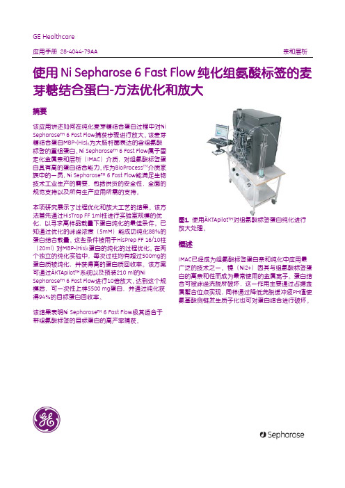
His Mag Sepharose(GE公司)
MBP-his融合蛋白纯化及放大

图5. 采用Axichrom 50柱对Ni Sepharose™ 6 Fast Flow纯化MBP-(His)6的工艺进行十倍放大。样品: 2125 ml含5500 mg MBP-(His)6的大肠杆菌提取物。 通过280 nm吸收对纯化进行监测。
图6. 在ExcelGel™ Gradient 8-18胶上进行非还原 SDS-PAGE分析。收集经HisPrep FF 16/10柱和 Axichrom 50柱中Ni Sepharose 6 Fast Flow纯化的洗 脱峰成分并合并(见图4和5)。纯化中使用的样品: 表达MBP-(His)6的大肠杆菌提取物。每个泳道上样量 一致。胶通过Coomassie™考马斯亮蓝进行染色。
层析柱和层析纯化系统
本项研究采用分别预填充有1 ml的HisTrap FF介质 和20 ml HisPrep FF 16/10介质的Ni Sepharose™ 6 Fast Flow柱。室温下选用带10 mm UV-cell的 ÄKTAexplorer™ 100层析系统。在中试规模的试验
中,将Ni Sepharose™ 6 Fast Flow填入AxiChrom 50 柱(内径为50 mm)后,采用ÄKTApilot™层析系统 在室温下操作。Ni Sepharose™ 6 Fast Flow用去离子 水搅匀后载入AxiChrom 50柱,以60 cm/h(19.6 ml/min)线性流速填充。填充床进一步通过轴向挤 压压缩15%至床高10.7cm,柱体积210ml。
材料与方法
除非特别说明,否则所有的设备和层析介质均来自 GE Healthcare(乌普萨拉,瑞典),并且所有使用 的化学物质均为分析级。
原材料的准备
本项研究中目标蛋白MBP-(His)6为大肠杆菌表达的 组氨酸标签重组麦芽糖结合蛋白,其分子量为 44000,等电点(PI)为4.4。
层析介质的品质影响因素——基质材料

层析介质的品质影响因素——基质材料导论我们在纯化生物大分子的时候,层析技术是最普遍和通用的纯化方法之一,层析介质又是层析技术的核心,那么层析介质的哪些因素会影响其品质了,最终会影响我们纯化工艺过程及纯化结果呢?今天首先介绍介质基质材料对其品质的影响。
层析介质常用的基质材料有无机化合物组成硅胶,也有有机化合物组成的聚苯乙烯,还有纯化生物大分子最常用的多糖聚合物(琼脂糖,葡聚糖,纤维素)等。
(1) 硅胶系列的介质,其刚性大,但不耐碱,多用于制备正反向层析介质,在小分子化合物提取分离,特别是植物提取行业广泛应用,也多用于分析型色谱柱介质的制备。
(2) 聚苯乙烯系列填料,刚性较强,层析过程传质速度高,但是其亲水性差疏水性强,生物相容性差,多用于多肽,生物碱等物质的分离提纯,也有少部分用于生物大分子纯化。
(3) 多糖系列层析介质,目前有琼脂糖微球,葡聚糖微球,琼脂糖交联葡聚糖微球,琼脂糖交联纤维素微球等为基质制备而成的各种层析介质,广泛用于疫苗,抗体,蛋白,血液制品,生长因子,酶的分离纯化,其生物相容性良好。
如Focudex G-25广泛用于生物大分子的脱盐,是葡聚糖交流纤维素制备而成的凝胶过滤介质,而Focurose 4FF广告用于疫苗的分离纯化,是交联琼脂糖制备而成的凝胶过滤介质等。
交联琼脂糖制备而成的介质应用最为广泛,如GE公司的Sepharose FF系列介质及汇研生物Focurose FF系列介质,在离子交换介质,亲和介质,疏水介质制备上应用最为广泛。
葡聚糖交联纤维微球介质,如GE公司Sephadex系列介质及汇研生物Focudex系列介质,在生物大分子脱盐及小分子多肽分离纯化中广泛应用。
葡聚糖交联琼脂糖微球介质,如GE公司的Sepharose XL及汇研生物的Focurose XL 系列介质,其即可避免介质和蛋白结合的空间位阻,又提高了可利用配基的密度,从而大幅度提高了介质的载量,比FF基质的填料有明显的载量优势,也广泛用于生物大分子的分离纯化。
人呼吸道卡他莫拉菌快速检测胶体金试纸的研制

人呼吸道卡他莫拉菌快速检测胶体金试纸的研制黄莹琪;张秋;陶冶;胡征;张改平;张华山【摘要】旨在研制一种快速、简便检测呼吸道感染病人呼吸道中卡他莫拉菌的试纸,并建立其检测方法.经生物信息学分析找到UspA1胞外结构域单一线性表位UspA1 Line,偶联血蓝蛋白KLH形成复合蛋白UspA1Line-KLH,并利用已构建的表达UspA1抗原的工程菌,表达纯化重组蛋白UspA1-His,免疫新西兰大白兔制备多克隆抗体,取血清后利用Protein A亲和层析和多肽亲和层析两步纯化抗体,得到纯度高,特异性强的多克隆抗体.采用柠檬酸三钠还原法制备40 nm胶体金颗粒,与多克隆抗体结合,形成双抗体夹心,达到快速检测卡他莫拉菌的目的.结果显示,建立的检测方法可在10 min内完成对样本的检测,特异性检出样本中卡他莫拉菌,与其他常见的呼吸道病原菌无交叉反应,试纸条在24℃保存具有良好的重复性和稳定性.成功研制了可快速检测卡他莫拉菌的胶体金试纸条.【期刊名称】《生物技术通报》【年(卷),期】2018(034)009【总页数】6页(P184-189)【关键词】卡他莫拉菌;单一线性表位;UspA1;免疫层析法【作者】黄莹琪;张秋;陶冶;胡征;张改平;张华山【作者单位】湖北工业大学生物工程与食品学院,武汉430068;湖北工业大学生物工程与食品学院,武汉430068;湖北工业大学生物工程与食品学院,武汉430068;湖北工业大学生物工程与食品学院,武汉430068;发酵工程教育部重点实验室(湖北工业大学),武汉430068;工业发酵湖北省协同创新中心,武汉430068;河南农业大学,郑州450002;湖北工业大学生物工程与食品学院,武汉430068;发酵工程教育部重点实验室(湖北工业大学),武汉430068;工业发酵湖北省协同创新中心,武汉430068【正文语种】中文卡他莫拉菌(Moraxella catarrhalis,M. catarrhalis)属革兰氏阴性、兼性厌氧双球菌,是慢性肺病患者中引起下呼吸道感染的重要病原体[1]。
包涵体的纯化和复性情况总结

板块一、大肠杆菌(这里讨论的大肠杆菌为非分泌到培养基中的重组蛋白,是否有重组蛋白分泌到培养基中的工程菌我没有见过。
)一、关于菌体的量大肠杆菌表达的基因工程蛋白是纯化人员最方便获得的原料,对纯化工艺开发来说几乎没有原料方面的限制。
常看到有战友用个几毫升的菌液去做纯化,对此我十分不解,同样要做,为什么不多做点呢?很少的菌体会给纯化带来一些难以估计的问题,工艺的重复性和放大往往出现问题。
因此,要做个好工艺就多发酵表达一些菌体吧。
我做纯化时,初始工艺摸索用的菌体量一般为10g左右。
二、关于是否包涵体表达包涵体的定义我就不讲了。
我要讲的是,一个基因工程蛋白是否是包涵体表达的说法本身就不完全准确。
至于包涵体在电镜下的晶体颗粒表现等等对我们纯化来说毫无意义,我相信做纯化工艺的人没有谁去看这个电镜,也不关心。
我们判断的依据只是SDS-PAGE,目的蛋白在破菌沉淀中,我们就认为是包涵体表达,但这是一个似是而非的结论。
看着没问题,实际上是有毛病的。
关键在于你用的是什么破菌缓冲液!有些蛋白在用缓冲液A破菌时是在破菌沉淀中,而用缓冲液B破菌时却在破菌上清中。
缓冲液A和B的差别可能只是pH上相差1-2个单位。
那么它是包涵体表达还是可溶上清表达呢?说这个问题主要在于有些战友往往非常在意他的目标蛋白是否包涵体表达。
甚至还有包涵体表达就用专门的包涵体蛋白纯化方法等等。
我们应该关心的是目标蛋白在什么缓冲体系下是可溶的,在什么缓冲体系下是不溶的!不要让包涵体这个概念给你误导。
三、关于表达量我们常常在发表的文章上看到,我这个工程菌的目标蛋白的表达量达到菌体总蛋白的30%、50%等等。
我要说都是文章的作者在忽悠。
不知道他们是如何定量的,用的最多的大概就是SDS-PAGE的扫描分析吧。
且不说一个SDS-PAGE不能表现出所有的菌体蛋白,电泳的染色方法、染色脱色强度、照片的曝光强度、扫描分析时的条带选择等等无不对这个百分比影响巨大。
在公司的QC部门做的对此应该最有体验,20%的条带要它变成30%又有何难?我的观点是对待表达量的描述不可定量,只能定性。
HiTrap DEAE FF, 1 ml and 5 ml

GE HealthcareInstructions 71-5017-51 AG HiTrap ion exchange columns HiTrap DEAE FF,1 ml and 5 ml HiTrap CM FF, 1 ml and 5 ml HiTrap Q FF, 1 ml and 5 mlHiTrap SP FF, 1 ml and 5 ml HiTrap ANX FF (high sub), 1 ml and 5 mlHiTrap™ SP FF, HiTrap CM FF, HiTrap Q FF, HiTrap DEAE FF, and HiTrap ANX FF (high sub) are prepacked 1 ml and 5 ml, ready to use cation and anion exchange HiTrap columns for method scouting, group separations, sample concentration and sample clean-up of charged biomolecules. The special design of the column, together with a modern chromatography media, provides fast, reproducible, and easy separations in a convenient format.The columns can be operated with a syringe, peristaltic pump or liquid chromatography system such as ÄKTA™ design.Connectorkit1Union 1/16” female/M6 male is also needed.2Union M6 female/1/16” male is also needed.Code No. Product Type of Medium No. supplied17-5157-01 HiTrap SP FF Strong cation exchanger 5 × 5 ml 17-5056-01 HiTrap CM FF Weak cation exchanger 5 × 1 ml 17-5155-01 HiTrap CM FF Weak cation exchanger 5 × 5 ml 17-5053-01 HiTrap Q FF Strong anion exchanger 5 × 1 ml 17-5156-01 HiTrap Q FF Strong anion exchanger 5 × 5 ml 17-5055-01 HiTrap DEAE FF Weak anion exchanger 5 × 1 ml 17-5154-01 HiTrap DEAE FFWeak anion exchanger5 × 5 ml 17-5162-01 HiTrap ANX FF (high sub) Weak anion exchanger 5 × 1 ml 17-5163-01 HiTrap ANX FF (high sub) Weak anion exchanger5 × 5 mlConnectors suppliedUsageNo. supplied1/16” male/luer female Connection of syringe to top ofHiTrap column 1Tubing connector flangeless/M6 female Connection of tubing (e.g. Peristaltic Pump P1) to bottom of HiTrap column 1 1Tubing connector flangeless/M6 male Connection of tubing (e.g. Peristaltic Pump P1) to top of HiTrap column 2 1Union 1/16” female/M6 male Connection to original FPLC™ System through bottom of HiTrap column 1Union M6 female/1/16” maleConnection to original FPLC System through top of HiTrap column1Stop plug female, 1/16” Sealing bottom of HiTrap column2, 5 or 7Table of contents1.Description (4)2.General considerations (7)3.Operation (13)4.Purification (13)5.Optimizing starting conditions (15)6.Further optimization (16)7.Determination of binding capacity (19)8.Scaling up (20)9.Storage (20)10.Intended use (22)11.Ordering information (22)1DescriptionColumnThe columns are 1 ml and 5 ml columns made of polypropylene, which is biocompatible and non-interactive with biomolecules. The top and bottom frits are manufactured from porous polyethylene. It is delivered with a stopper on the inlet and snap-off end on the outlet.The separation can be easily achieved using a syringe together with the supplied luer connector, a peristaltic pump, or in a chromatography system such as ÄKTA design.The column cannot be opened or refilled.Note:To prevent leakage it is essential to ensure that the connector is tight.Characteristics of HiTrap column are listed in Table 1.Table 1. Characteristics of HiTrap column.Media PropertiesQ Sepharose™ Fast Flow, DEAE Sepharose Fast Flow, SP Sepharose Fast Flow, and CM Sepharose Fast Flow are based on a robust, 6% highly cross-linked beaded agarose matrix with excellent flow properties and high loading capacities.ANX Sepharose 4 Fast Flow (high sub) is based on 4% highly cross-linked beaded agarose. This results in a medium with higher porosity, which is particularly useful for the purification of high molecular mass proteins.Characteristics of the ion exchangers are listed in Table 2.Column volumes 1 ml or 5 mlColumn dimensions 0.7 × 2.5 cm (1 ml) or 1.6 × 2.5 cm (5 ml)Maximum flow rates HiTrap 1 ml: 4 ml/min, HiTrap 5 ml: 20 ml/min Recommended flow ratesHiTrap 1 ml: 1ml/min, HiTrap 5 ml: 5 ml/minMaximum back pressure 0.3 MPa, 3 barChemical stabilityAll commonly used aqueous buffers, 1 MNaOH, 8 M urea, 6 M guanidine hydrochloride, 70% ethanolTable 2. Characteristics of Sepharose Fast Flow ion exchangersCation exchangers SP Sepharose Fast Flow CM Sepharose Fast FlowBead structure 6% highly cross-linkedagarose 6% highly cross-linked agaroseBead size 45–165 µm 45–165 µm Type of gel Strong cation Weak cation Charged group -CH2CH2CH2SO3- -O-CH2COO-Total ionic capacity 0.18–0.25 mmol H+/ml medium 0.09–0.13 mmol H+/ ml mediumDynamic binding capacity170 mg Ribonuclease A/ml medium50 mg Ribonuclease A/ml mediumpH stability:Short term23–14 2–14Working4–13 6–10Long term34–13 4–13Storagetemperature4°C to 30°C 4°C to 30°CStorage buffer 20% ethanol,0.2 M sodium acetate20 % ethanolChemical stability All commonly used aqueous buffers, 1 M NaOH, 8 M urea,6 M guanidine hydrochloride, 70% ethanolAvoid Oxidizing agents, cationic detergents and buffers.See Table3 and Figure 2.1Determination of dynamic binding capacity: DEAE Sepharose Fast Flow, Q Sepharose Fast Flow, SP Sepharose Fast Flow and CM Sepharose Fast Flow: Samples were applied at 75 cm/h until 50% breakthrough. Columns: 0.5 x 5 cm. Buffers: 0.05 M Tris, (+ 2 M NaCl in the elution buffer), pH 7.5 (Q and DEAE), 0.1 M acetate, (+ 2 M NaCl in the elution buffer), pH 5.0 (SP and CM). ANX Sepharose 4 Fast Flow (high sub): Sample was applied at 300 cm/h until 10% breakthrough. Column: 1.6 × 13 cm. Buffer: 0.05 M Tris, (+ 1 M NaCl in the elution buffer), pH 7.5.2 Refers to the pH interval for short term such as regeneration and cleaning.3 Refers to the pH interval where the medium is stable over a long period of time without adverse effects on its subsequent chromatographic performance.Note:The active end of the charged group is the same for DEAE Sepharose Fast Flow and ANX Sepharose Fast Flow (high sub), the difference is the length of the carbon chain of the charged group. DEAE Sepharose Fast Flow has a diethylaminoethyl-group bound to the agarose whilst ANX Sepharose 4 Fast Flow has a diethylaminopropyl-group attached.Anion exchangers Q Sepharose Fast Flow DEAE SepharoseFast Flow ANX Sepharose 4 Fast Flow (hig sub)Bead structure 6% highly cross-linked agarose 6% highly cross-linked agarose 4% highly cross-linked agarose Bead size 45–165 µm 45–165 µm 45–165 µm Type of gel Strong anion Weak anion Weak anion Charged group -N +(CH 3)3 -N +(C 2H 5)2H -N +(C 2H 5)2HTotal ioniccapacity 0.18–0.25 mmol Cl -/ml medium 0.11–0.16 mmol Cl -/ml medium 0.13–0.17 mmol Cl -/ml medium Dynamic binding capacity 1 120 mg HSA/ml medium 110 mg HSA/ml medium 43 mg BSA/ml medium pH stability:Short term 2 1–14 1–14 2–14Working 2–12 2–9 3–10Long term 3 2–12 2–13 3–10Storagetemperature 4°C to 30°C 4°C to 30°C 4°C to 30°C Storage buffer 20% ethanol20% ethanol20% ethanolChemical stability All commonly used aqueous buffers, 1 M NaOH, 8 M urea, 6 M guanidine hydrochloride, 70% ethanol AvoidOxidizing agents, anionic detergents and buffers.See Table 4 and Figure 3.2General considerationsSelection of ion exchangerIon exchange chromatography is based on the binding of charged sample molecules to oppositely charged groups attached to an insoluble matrix.Substances are bound to ion exchangers when they carry a net charge opposite to that of the ion exchanger. This binding is electrostatic and reversible.The pH value at which a biomolecule carries no net charge is called the isoelectric point (pI). When exposed to a pH below its pI, the biomolecule will carry a positive net charge and will bind to a cation exchanger (SP and CM). At pH’s above its pI the biomolecule will carry a negative net charge and will bind to an anion exchanger (Q, DEAE and ANX) (Fig 1).If the sample components are most stable below their pI’s, a cation exchanger should be used. If they are most stable above their pI’s, an anion exchanger is used. If stability is high over a wide pH range on both side of the pI, either type of ion exchanger can be used.Weak ion exchangers have a limited pH working range (Table 3).Information on the pI and how the net charge on the molecule varies with pH gives valuable information regarding the choice of starting conditions. Electrophoretic titration curves enable the determination of the charge/pH relationship for the molecules present across the pH range of interest.Fig 1. The net charge of a protein as a function of pH.Attached to cation exchangersAttached to anion exchangersI s o e l e c t r i c p o i n tN e t c h a n g e o f p r o t e i nSelection of buffer pH and ionic strengthBuffer pH and ionic strength are critical for the binding and elution of material (both target substances and contaminants) in ionexchange chromatography. Selection of appropriate pH and ionic strength for the start and elution buffers allows the use of threepossible separation strategies.Strategy 1. Binding and elution of all sample componentsBinding is achieved by choosing a start buffer with a low pH for SP Sepharose Fast Flow, and CM Sepharose Fast Flow, or high pH for Q Sepharose Fast Flow, DEAE Sepharose Fast Flow and ANXSepharose 4 Fast Flow (high sub). The ionic strength should be kept as low as possible to allow all components to bind to the ionexchanger (< 5 mS/cm).This results in a concentration of the target substance and acomplete picture of the total sample. The drawback of this strategy is that the binding capacity of the ion exchanger for the targetsubstance depends on the amount of contaminants in the sample.Strongly binding contaminants can also displace bound targetprotein if a large volume of sample is loaded.Note:Starting conditions are subject to the stability of the sample components.Strategy 2. Enrichment of target proteinThis is achieved by choosing a start buffer with a pH optimized to allow maximal binding of target protein, and as high as possible an ionic strength to supress binding of sample contaminants.This strategy results in a concentration of the target substances.Strategy 3. Binding of sample contaminantsThis is achieved by choosing a start buffer with a pH and an ionic strength that promotes the binding of some or all contaminantsbut allows the target substance to pass through the column.The drawback of this approach is that the target substance is not concentrated and the amount of sample that can be applied to the ion exchanger depends on the amount of contaminants in thesample.Start bufferThe concentration of buffer required to give effective pH controlvaries with the buffer system. A list of suitable buffers andsuggested starting concentrations is shown in Tables 3 and 4,Figures 2 and 3. In the majority of cases a concentration of at least10 mM is required to ensure adequate buffering capacity. The ionicstrength of the buffer should be kept low (< 5 mS/cm) so as not to interfere with sample binding. Salts also play a role in stabilizingprotein structures in solution and it is important the ionic strength should not be so low that protein denaturation or precipitationoccurs.The buffering ion should carry the same charge as the ionexchange group and should have a pKa within 0.5 pH units of the pH used in the separation. Buffering ions of opposite charge may take part in the ion exchange process and cause localdisturbances in pH.Table 3. Buffers for cation exchange chromatography 1Ref: Handbook of chemistry and physics, 83rd edition, CRC, 2002-2003.pH interval Substance Conc. (mM) Counter-ion pKa (25°C)1+2.6–3.6 Methyl malonic acid 20Na + or Li + 3.072.6–3.6 Citric acid 20 Na + 3.133.3–4.3 Lactic acid 50 Na + 3.863.3–4.3 Formic acid 50 Na + or Li + 3.753.7–4.7;5.1–6.1Succinic acid50 Na + 4.21;5.644.3–5.3 Acetic acid 50 Na + or Li + 4.755.2–6.2 Methyl malonic acid 50Na + or Li + 5.765.6–6.6 MES 50 Na + or Li + 6.276.7–7.7 Phosphate 50 Na + 7.207.0–8.0 HEPES 50 Na + or Li + 7.567.8–8.8 BICINE50Na + 8.33Table 4. Buffers for anion exchange chromatography 1Ref: Handbook of chemistry and physics, 83rd edition, CRC, 2002–2003.pH interval Substance Conc. (mM) Counter-ionpKa (25°C)1-4.8–5.8 Piperazine 20 Cl - or HCOO - 5.335.5–6.5 L-Histidine 20 Cl - 6.046.0–7.0 bis-Tris 20 Cl - 6.486.2–7.2; 8.6–9.6bis-Tris propane20Cl -6.65;9.107.3–8.3 Triethanolamine 20 Cl - or CH 3COO - 7.767.6–8.6 Tris 20Cl - 8.078.0–9.0 N-Methyl-diethanolamine 20 SO 42- 8.528.0–9.0 N-Methyl-diethanolamine 50 Cl - or CH 3COO - 8.528.4–9.4 Diethanolamine 20 at pH 8.4 50 at pH 8.8Cl - 8.888.4–9.4 Propane 1,3-Diamino 20Cl - 8.889.0–10.0Ethanolamine 20Cl - 9.509.2–10.2Piperazine20 Cl - 9.7310.0–11.0 Propane 1,3-Diamino 20Cl - 10.5510.6–11.6 Piperidine20Cl - 11.12chromatography.Fig 3. Recommended buffer substances for anion exchange chromatography.Starting pHCation exchangers (SP, CM): At least 1 pH unit below the pI ofsubstance to be bound.Anion exchangers (Q, DEAE, ANX): At least 1 pH unit above the pI of substance to be bound.3OperationThe columns can be operated by a syringe, a peristaltic pump or a chromatography system.Buffer preparationWater and chemicals used for buffer preparation should be of high purity. It is recommended to filter the buffers by passing themthrough a 0.45 µm filter before use. See Tables 3 and 4, Figures 2 and 3 for recommended buffers.Sample preparationThe sample should be adjusted to the composition of the startbuffer by buffer exchange using HiTrap Desalting, HiPrep™ 26/10 Desalting or PD-10 columns. The sample should be filtered througha 0.45 µm filter or centrifuged immediately before it is applied tothe column (See Table5).4Purification1Fill the syringe or pump tubing with start buffer (low ionic strength). Remove the stopper and connect the column to thesyringe (with the provided connector), or pump tubing, ”drop todrop” to avoid introducing air into the column.2Remove the snap-off end at the column outlet.3Wash out the preservatives with 5 column volumes of start buffer, at 1 ml/min for HiTrap 1 ml and 5 ml/min for HiTrap 5 ml.4Wash with 5 column volumes of elution buffer (start buffer with1 M NaCl).5Finally equilibrate with 5–10 column volumes of start buffer. 6Apply the sample at 1 ml/min for HiTrap 1 ml and 5 ml/min forHiTrap 5 ml using a syringe fitted to the luer connector or by pumping it onto the column.7Wash with at least 5 column volumes of start buffer or until no material appears in the effluent.8Elute with 5–10 column volumes of elution buffer, see Choice of gradient type.9The purified eluted fractions can be desalted using a HiTrap Desalting, HiPrep 26/10 Desalting or a PD-10 column ifnecessary.10After completed elution, regenerate the column by washing with 5 column volumes of regeneration buffer (start buffer with1 M NaCl) followed by 5–10 columns volumes of start buffer. Thecolumn is now ready for a new sample.For a first experiment the following conditions are recommended:Flow rates: 1 ml/min using HiTrap 1 ml column5 ml/min using HiTrap 5 ml columnStart buffer: See Tables 3 and 4, Figures 2 and 3Elution buffer: Start buffer + 1 M NaClGradient volume: 20 ml5Optimizing starting conditions If the composition of the sample is unknown, a simple screeningtest using a syringe or pump can be performed to optimize starting pH and ionic strength.1Set up a series of buffers with different pH’s, in the range 4–8 (SP, CM) or 5–9 (Q, DEAE, ANX), with 0.5–1 pH unit intervalsbetween each buffer. Make one series with 1 M NaCl included inthe buffers (elution buffer) and the other without NaCl (startbuffer).2Equilibrate the column with start buffer, see Purification.3Adjust the sample to the chosen start buffer, see Sample preparation.4Apply a constant known amount of the sample at 1 ml/min using HiTrap 1 ml column and at 5 ml/min using HiTrap 5 mlcolumn. Collect the eluate.5Wash with at least 5 column volumes of start buffer or until no material appears in the effluent. Collect the eluate.6Elute bound material with elution buffer. 3–5 column volumes are usually sufficient but other volumes may be requireddependent on the exact experimental conditions. Collect theeluate.7Analyse all eluates (by activity assay for example) and determine the purity and the amount bound to the column.8Perform steps 2–7 for the next buffer pH.9Decide which pH should be used for the selected purification strategy.10To decide on starting ionic strength conditions, a similar screening is done, but the buffer pH is held constant and thesalt concentration is varied in the interval 0–0.5 M, withintervals of 0.05–0.1 M salt between each buffer.6Further optimizationThe recommendations given above will give a sound basis fordeveloping an efficient purification step. Details of how flow rate, sample loading, particle size and elution scheme may be optimized to meet the special needs can be found in the handbook, IonExchange Chromatography & Chromatofocusing, Principles andMethods, Code No. 11-0004-21.A wide range of ion exchange chromatography media forpurification of biomolecules at all scales is available. See Ordering information, /protein-purification orcontact your local representative.Choice of gradient type1Stepwise gradients are easy to produce and require minimal equipment. Eluted peaks are very sharp and elution volumesminimal. However, care must be exercised in the design of thesteps and the interpretation of results for substances eluted bya sharp change in pH or small differences in ionic strength.Peaks tend to have sharp fronts and pronounced tailing sincethey frequently contain more than one component.2Continuous salt gradients are the most frequently used type of elution. Many types of gradient forming systems are available.Two buffers of differing ionic strength, the start and elutionbuffer (start buffer + 1 M NaCl or higher buffer saltconcentration), are mixed together and if the volume ratio ischanged linearly, the ionic strength changes linearly.Note:Another, but less common, method to desorb boundmaterial is to increase (SP and CM) or decrease (Q, DEAEand ANX) the pH of the eluent.Continuous pH gradients are difficult to produce atconstant ionic strength, since simultaneous changes inionic strength, although small, also occur (bufferingcapacities are pH dependent).In the case of pH gradients using weak ion exchangers (CM,DEAE and ANX) the buffer may have to titrate the ionexchanger and there will be a short period ofre-equilibration before the new pH is reached.Elution with stepwise ionic strength gradientsStepwise elution is the sequential use of the same buffer atdifferent ionic strengths. It is technically simple and fast, and issuitable for syringe operation. It is often used for sampleconcentration and sample clean-up. Stepwise elution gives small peak volumes and the resolution depends on the difference inelution power between each step.1Choose starting conditions as outlined under Optimizing starting conditions.2Equilibrate the column, see Purification.3Adjust the sample to the chosen starting pH and ionic strength, see Sample preparation.4Apply the sample at 1 ml/min using HiTrap 1 ml column and at5 ml/min using HiTrap 5 ml column. Collect eluate.5Wash with at least 5 column volumes of start buffer or until no material appears in the effluent. Collect eluate.6Elute with the first step ionic strength buffer. The volumes required for stepwise elution depend on the operatingconditions. However, 3–5 column volumes are usuallysufficient. Collect eluate.7Elute with next ionic strength buffer. Collect eluate.8After completed elution, regenerate the column by washing with 5 column volumes of regeneration buffer (start buffer with1 M NaCl) followed by 5–10 volumes of start buffer. The columnis now ready for a new sample.Elution with continuous ionic strength gradients Continuous salt gradient elution is the most frequently used type of elution in ion exchange chromatography. It is very reproducible and leads to improved resolution, since zone sharpening occursduring elution. Continuous gradients can be prepared in different ways, depending on available equipment.• A peristaltic pump and a gradient mixer e.g. pump P-1, gradient mixer GM-1.• A one pump system, e.g. ÄKTAprime™ plus.• A two pump system, e.g. ÄKTA design.1Choose starting conditions as outlined under Optimizing starting conditions.2Equilibrate the column, see Purification.3Adjust the sample to the chosen starting pH and ionic strength, see Sample preparation.4Apply the sample at 1 ml/min using HiTrap 1 ml column and at5 ml/min using HiTrap 5 ml column. Collect eluate.5Wash with at least 5 column volumes of start buffer or until no material appears in the effluent.6Start the gradient elution. A gradient volume of 10–20 column volumes and an increase in ionic strength to 0.5 M NaCl isusually sufficient.7Regenerate the column by washing with 5 column volumes of start buffer with 1 M NaCl followed by 5–10 column volumes ofstart buffer. The column is now ready for a new sample.7Determination of binding capacityThe amount of sample which can be applied to a column depends on the capacity of the column and the degree of resolutionrequired. The capacity is dependent on the sample composition, chosen starting conditions of pH and ionic strength and the flow rate at which the separation is done. The influence of flow rate and pH on the capacity for some model proteins are shown in Figure 4.Samples were applied until 5% of the start material appeared in the eluent. The column was then washed with 10 ml 20 mM Tris-HCl, pH 8.2 or 9.0 before elution with elution buffer, 20 mM Tris-HCl, 1 M NaCl, pH 8.2 or 9.0.Fig 4. Binding capacity of human IgG, HSA and human transferrin at different pH’s on HiTrap Q HP, 1 ml.1Equilibrate the column, see Purification.2Adjust the sample to the chosen starting pH and ionic strength, see Sample preparation.3Determine the concentration of the specific proteins by UV, SDS-PAGE, ELISA or other appropriate techniques.flow rate, ml/minm g p r o t e i n e l u t e dlgG, pH 8.2HSA, pH 8.2Transferrin, pH 8.2lgG, pH 9.04Apply the sample solution to the column with a pump or a syringe, at a flow rate equal to the flow rate to be used in thepurification method. Collect fractions and continue sampleapplication until the column is saturated.5Wash the column with 5–10 column volumes of start buffer or until no material appears in effluent.6Elute bound proteins with 3–5 column volumes of elution buffer (start buffer with 1 M NaCl) and collect eluate.7Analyse fractions and eluates from steps 4 and 6 for the specific protein and determine the breakthrough profile(sample concentration as a function of the amount of sampleapplied). The dynamic capacity is the amount that can beapplied without any significant breakthrough. The totalcapacity for the specific protein is determined from step 6.8Scaling upFor quick scale-up of purification, two or three HiTrap ionexchange columns of the same type can be connected in series.For further scale-up Q Sepharose Fast Flow, SP Sepharose FastFlow, CM Sepharose Fast Flow and DEAE Sepharose Fast Flow are available as prepacked HiPrep 16/10 columns or as lab packs. See ordering information.9StorageHiTrap Q FF, HiTrap DEAE FF, HiTrap ANX FF (high sub) andHiTrap CM FF: Rinse with water then wash with 5 column volumes 20% ethanol at 1 ml/min (HiTrap 1 ml column) or at 5 ml/min(HiTrap 5 ml column) to prevent microbial growth.HiTrap SP FF: Rinse with water then wash with 5 column volumes 20% ethanol containing 0.2 M sodium acetate at 1 ml/min (HiTrap1 ml column) or at 5 ml/min (HiTrap 5 ml column).Seal the column with the supplied stoppers. The recommendedstorage temperature is 4ºC to 30ºC.Table 5. Prepacked columns for desalting and buffer exchange10Intended useHiTrap DEAE FF, HiTrap CM FF, HiTrap Q FF, HiTrap SP FF, and HiTrap ANX FF are intended for research use only, and shall not be used in any clinical or in vitro procedures for diagnostic purposes.11Ordering informationProduct No. supplied Code no.HiTrap IEX Selection Kit 7 × 1 ml 17-6002-33HiTrap Q FF 5 × 1 ml 17-5053-015 × 5 ml 17-5156-01HiTrap SP FF 5 × 1 ml 17-5054-015 × 5 ml 17-5157-01HiTrap DEAE FF 5 × 1 ml 17-5055-015 × 5 ml 17-5154-01HiTrap CM FF 5 × 1 ml 17-5056-015 × 5 ml 17-5155-01HiTrap Q XL 5 × 1 ml 17-5158-015 × 5 ml 17-5159-01HiTrap SP XL 5 × 1 ml 17-5160-015 × 5 ml 17-5161-01HiTrap ANX FF (high sub) 5 × 1 ml 17-5162-015 × 5 ml 17-5163-01HiTrap Desalting 5 × 5 ml 17-1408-01100 × 5 ml 211-0003-2917-0510-10ml Q Sepharose Fast Flow1 25300 ml 17-1510-01 SP Sepharose Fast Flow125 ml 17-0729-10300 ml 17-0729-01ml 17-0709-10 DEAE Sepharose Fast Flow1 25500 ml 17-0709-01 CM Sepharose Fast Flow125 ml 17-0719-10500 ml 17-0719-01 ANX Sepharose 4 Fast Flow (high sub)1 25 ml 17-1287-10500 ml17-1287-01 HiPrep DEAE FF 16/10 1 × 20 ml28-9365-41HiPrep CM FF 16/10 1 × 20 ml28-9365-42HiPrep SP FF 16/10 1 × 20 ml28-9365-441Process scale quantities are available. Please contact your local representative.2Special package. Delivered on specific customer order.1One connector included in each HiTrap package.2Two, five, or seven stop plugs female included in HiTrap packages depending on the product.3One fingertight stop plug is connected to the top of each HiTrap column at delivery.HiPrep Q FF 16/101 × 20 ml 28-9365-43HiPrep 26/10 Desalting 1 × 53 ml 17-5087-014 × 53 ml 17-5087-02PD-10 Desalting column30 17-0851-01AccessoriesNo. SuppliedCode No.1/16” male/luer female 1 218-1112-51Tubing connector flangeless/M6 female 1 2 18-1003-68Tubing connector flangeless/M6 male 1 2 18-1017-98Union 1/16” female/M6 male 1 6 18-1112-57Union M6 female /1/16” male 1 5 18-3858-01Union luerlock female/M6 female 218-1027-12HiTrap/HiPrep, 1/16” male connector for ÄKTA design8 28-4010-81Stop plug female, 1/16”2 5 11-0004-64Fingertight stop plug, 1/16”3 511-0003-55LiteratureCode No.Ion Exchange Chromatography & Chromatofocusing Handbook, Principles and Methods11-0004-21Ion Exchange Chromatography Columns and Media, Selection Guide 18-1127-31HiTrap Column Guide18-1129-81ProductNo. supplied Code no.For local office contact information, visit: /contactGE Healthcare Bio-Sciences ABBjörkgatan 30751 84 UppsalaSweden/hitrap/protein-purification GE Healthcare Europe GmbH Munzinger Strasse 5,D-79111 Freiburg,GermanyGE Healthcare UK Ltd Amersham PlaceLittle Chalfont Buckinghamshire, HP7 9NAUKGE Healthcare Bio-Sciences Corp 800 Centennial AvenueP.O. Box 1327Piscataway, NJ 08855-1327 USAGE Healthcare Bio-Sciences KK Sanken Bldg.3-25-1, Hyakunincho Shinjuku-ku, Tokyo 169-0073 JapanGE, imagination at work and GE monogram are trademarks of General Electric Company. HiTrap, Sepharose, FPLC, ÄKTAprime, ÄKTA, Drop Design, Sephadex, HiPrep, MiniTrap andMidiTrap are trademarks of GE Healthcare companies.© 1999-2009 General Electric Company – All rights reserved.First published Dec. 1999.All goods and services are sold subject to the terms and conditions of sale of the company within GE Healthcare which supplies them. A copy of these terms and conditions is available on request. Contact your local GE Healthcare representative for the most current information.71-5017-51 AG 03/2009imagination at work。
- 1、下载文档前请自行甄别文档内容的完整性,平台不提供额外的编辑、内容补充、找答案等附加服务。
- 2、"仅部分预览"的文档,不可在线预览部分如存在完整性等问题,可反馈申请退款(可完整预览的文档不适用该条件!)。
- 3、如文档侵犯您的权益,请联系客服反馈,我们会尽快为您处理(人工客服工作时间:9:00-18:30)。
GE HealthcareInstructions 28-9775-37 ACMag SepharoseHis Mag Sepharose™ NiHis Mag Sepharose Ni is available in the following pack sizes (instructions for use included in all pack sizes):
•His Mag Sepharose Ni, 5% medium slurry, 2 × 1 ml•His Mag Sepharose Ni, 5% medium slurry, 5 × 1 ml•His Mag Sepharose Ni, 5% medium slurry, 10 × 1 mlNote:1 ml medium slurry is sufficient for 5 reactions according to the recommended protocol. 1 ml of 5% (v/v) medium slurry contains 50 µl magnetic beads.
PurposeHis Mag Sepharose Ni products are magnetic beads designed for simple small-scale purification of histidine-tagged proteins. The magnetic beads are suitable for purification of a single sample or multiple samples in parallel for example in screening experiments.
Intended useHis Mag Sepharose Ni is intended for research only, and should not be used in any clinical or in vitro procedures for diagnostic purposes.
CAUTION: The product contains nickel, Ni2+, which is potentially allergenic. Always use normal protection devices like gloves and safety glasses when handling His Mag Sepharose Ni.228-9775-37 AC
Table of contents1Principle....................................................................................32Advice on handling.............................................................43Operation.................................................................................64Purification protocol for high capacity......................75Purification protocol for high purity............................86Optimization of parameters...........................................97Characteristics......................................................................98Compatibility.......................................................................109Ordering information......................................................1128-9775-37 AC 3
1PrincipleHis Mag Sepharose Ni is an affinity chromatography medium with high affinity for histidine-tagged proteins from various sources. The medium consists of magnetic beads based on Sepharose coupled with IMAC ligand and immobilized with nickel ions.
Purification of histidine-tagged proteins on IMAC media is a balance between capacity and purity, modulated by the concentration of imidazole in the sample and in the binding/wash buffer. To simplify the purification procedure two purification protocols are included in this instruction, one with focus on high capacity and the other one with focus on high purity.
His Mag Sepharose Ni provides flexible purification allowing a wide range of sample volumes and easy scale-up by varying the bead quantity and is suitable for purification of multiple samples in parallel.
Mag Sepharose products can be used together with Eppendorf microcentrifuge tubes and a magnetic rack, for example MagRack 6 (see Section 2). The magnetic beads are easily separated from the liquid phase during the different steps in the purification protocol. His Buffer Kit can be used to facilitate buffer preparation. 428-9775-37 AC
2Advice on handlingNote:His Mag Sepharose Ni is intended for single use only.General magnetic separation stepIt is recommended to use 1.5 ml Eppendorf tubes and MagRack 6 in the included protocol (see Section4 and Section5).
1 Remove the magnet before adding liquid.
2Insert the magnet before removing liquid. When using volumes above 1.5 ml, e.g. 50 ml, a magnetic pickpen can be used for collecting the magnetic beads. Another alternative is to spin down the beads by using a swing-out centrifuge.
Dispensing the medium slurry•Prior to dispensing the medium slurry, make sure it is homogeneous by vortexing the vial thoroughly.
•When the medium slurry is resuspended, immediately pipette the required amount of medium slurry into the desired tube.
•Repeat the resuspension step between each pipetting from the medium slurry vial.28-9775-37 AC 5
Handling of liquid•Use the magnetic rack with the magnet in place for each liquid removal step.
•Before application of liquid, remove the magnet from the magnetic rack.
•After addition of liquid, allow resuspension of the beads by vortexing or manual inversion of the tube. When processing multiple samples, manual inversion of the magnetic rack is recommended.
•If needed, a pipette can be used to remove liquid from the lid of the vial.
Incubation•During incubation, make sure the magnetic beads are well resuspended and kept in solution by end-over-end mixing or by using a benchtop shaker.
