抵御新发_突发病毒性传染病的新思_省略_和针对宿主的广谱抗病毒药物的研发_秦晓峰
艾滋病疫苗研究的现状与未来

艾滋病疫苗研究的现状与未来艾滋病,是由人类免疫缺陷病毒(HIV)感染引起的一种病毒性疾病,以免疫功能失调、感染性疾病和肿瘤等多种病理表现为特征。
截至目前,全球已有超过7700万人感染HIV,其中超过3600万人已死亡。
虽然通过抗病毒治疗,可以延缓疾病进展,增加患者的生存率,但是没有可靠的治愈方法,只能选择终身使用抗病毒药物,而大量使用抗病毒药物又有助于病毒耐药性的产生。
因此,寻求一种安全有效的艾滋病疫苗是防控HIV传播的重要措施。
目前,艾滋病疫苗的研究方向主要集中于3个方面:预防疫苗、治疗疫苗和保护性疫苗。
其中研究最充分的是预防疫苗,也是大多数人所熟知的“艾滋病疫苗”。
那么,这些疫苗现在的研究进展和未来的发展方向是什么呢?在目前的研究中,最被关注的是HIV病毒外壳蛋白(Env)诱导的中和抗体(NAbs),因为这些抗体能够抵御感染来自不同传播途径的HIV变种。
科学家们在设计疫苗时,通过对Env蛋白进行修改和组合,将其引入宿主体内,诱导机体产生特定的NAbs。
目前,多项临床试验表明,这类涉及Env蛋白诱导NAbs的预防疫苗在人体免疫反应、安全性等方面的表现都令人期待,但是迄今为止,还没有一种疫苗获得了足够的保护效果。
与此同时,研究人员也在不断探索其他疫苗方案,包括合成肽、DNA免疫、向量疫苗等。
其中,治疗疫苗被认为是未来的研究方向之一。
治疗疫苗是将HIV患者自身的病毒群体中经过筛选的免疫原发挥作用,增强机体免疫力,起到治疗作用。
目前,这种治疗疫苗尚处于早期实验室或动物实验阶段,但仍有望成为治疗艾滋病的一个重要手段。
除此之外,研究人员还在不断寻求增加艾滋病疫苗保护效果的新方法,比如联合使用不同的疫苗方案、改变给药途径、加入上调机体免疫反应的佐剂等。
这些方法都在实验室中进行验证,或已经进入临床实验阶段。
虽然在艾滋病疫苗的研究中仍面临许多困难,但是科学家们依然坚持着不断探索的步伐,一方面希望能够解决艾滋病流行的问题,另一方面也对HIV研究产生了新的启示,推动了对免疫学和发展物理学的研究。
PRRS
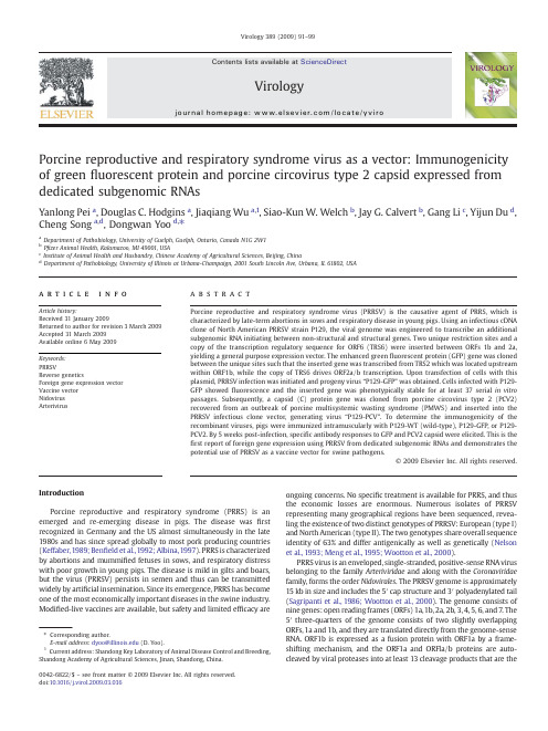
Porcine reproductive and respiratory syndrome virus as a vector:Immunogenicity of green fluorescent protein and porcine circovirus type 2capsid expressed from dedicated subgenomic RNAsYanlong Pei a ,Douglas C.Hodgins a ,Jiaqiang Wu a ,1,Siao-Kun W.Welch b ,Jay G.Calvert b ,Gang Li c ,Yijun Du d ,Cheng Song a ,d ,Dongwan Yoo d ,⁎aDepartment of Pathobiology,University of Guelph,Guelph,Ontario,Canada N1G 2W1bP fizer Animal Health,Kalamazoo,MI 49001,USA cInstitute of Animal Health and Husbandry,Chinese Academy of Agricultural Sciences,Beijing,China dDepartment of Pathobiology,University of Illinois at Urbana-Champaign,2001South Lincoln Ave,Urbana,IL 61802,USAa b s t r a c ta r t i c l e i n f o Article history:Received 31January 2009Returned to author for revision 3March 2009Accepted 31March 2009Available online 6May 2009Keywords:PRRSVReverse geneticsForeign gene expression vector Vaccine vector Nidovirus ArterivirusPorcine reproductive and respiratory syndrome virus (PRRSV)is the causative agent of PRRS,which is characterized by late-term abortions in sows and respiratory disease in young ing an infectious cDNA clone of North American PRRSV strain P129,the viral genome was engineered to transcribe an additional subgenomic RNA initiating between non-structural and structural genes.Two unique restriction sites and a copy of the transcription regulatory sequence for ORF6(TRS6)were inserted between ORFs 1b and 2a,yielding a general purpose expression vector.The enhanced green fluorescent protein (GFP)gene was cloned between the unique sites such that the inserted gene was transcribed from TRS2which was located upstream within ORF1b,while the copy of TRS6drives ORF2a/b transcription.Upon transfection of cells with this plasmid,PRRSV infection was initiated and progeny virus “P129-GFP ”was obtained.Cells infected with P129-GFP showed fluorescence and the inserted gene was phenotypically stable for at least 37serial in vitro passages.Subsequently,a capsid (C)protein gene was cloned from porcine circovirus type 2(PCV2)recovered from an outbreak of porcine multisystemic wasting syndrome (PMWS)and inserted into the PRRSV infectious clone vector,generating virus “P129-PCV ”.To determine the immunogenicity of the recombinant viruses,pigs were immunized intramuscularly with P129-WT (wild-type),P129-GFP,or P129-PCV2.By 5weeks post-infection,speci fic antibody responses to GFP and PCV2capsid were elicited.This is the first report of foreign gene expression using PRRSV from dedicated subgenomic RNAs and demonstrates the potential use of PRRSV as a vaccine vector for swine pathogens.©2009Elsevier Inc.All rights reserved.IntroductionPorcine reproductive and respiratory syndrome (PRRS)is an emerged and re-emerging disease in pigs.The disease was first recognized in Germany and the US almost simultaneously in the late 1980s and has since spread globally to most pork producing countries (Keffaber,1989;Ben field et al.,1992;Albina,1997).PRRS is characterized by abortions and mummi fied fetuses in sows,and respiratory distress with poor growth in young pigs.The disease is mild in gilts and boars,but the virus (PRRSV)persists in semen and thus can be transmitted widely by arti ficial insemination.Since its emergence,PRRS has become one of the most economically important diseases in the swine industry.Modi fied-live vaccines are available,but safety and limited ef ficacy areongoing concerns.No speci fic treatment is available for PRRS,and thus the economic losses are enormous.Numerous isolates of PRRSV representing many geographical regions have been sequenced,revea-ling the existence of two distinct genotypes of PRRSV:European (type I)and North American (type II).The two genotypes share overall sequence identity of 63%and differ antigenically as well as genetically (Nelson et al.,1993;Meng et al.,1995;Wootton et al.,2000).PRRS virus is an enveloped,single-stranded,positive-sense RNA virus belonging to the family Arteriviridae and along with the Coronaviridae family,forms the order Nidovirales .The PRRSV genome is approximately 15kb in size and includes the 5′cap structure and 3′polyadenylated tail (Sagripanti et al.,1986;Wootton et al.,2000).The genome consists of nine genes:open reading frames (ORFs)1a,1b,2a,2b,3,4,5,6,and 7.The 5′three-quarters of the genome consists of two slightly overlapping ORFs,1a and 1b,and they are translated directly from the genome-sense RNA.ORF1b is expressed as a fusion protein with ORF1a by a frame-shifting mechanism,and the ORF1a and ORFla/b proteins are auto-cleaved by viral proteases into at least 13cleavage products that are theVirology 389(2009)91–99⁎Corresponding author.E-mail address:dyoo@ (D.Yoo).1Current address:Shandong Key Laboratory of Animal Disease Control and Breeding,Shandong Academy of Agricultural Sciences,Jinan,Shandong,China.0042-6822/$–see front matter ©2009Elsevier Inc.All rights reserved.doi:10.1016/j.virol.2009.03.036Contents lists available at ScienceDirectVirologyj o u r n a l ho m e p a g e :w w w.e l s ev i e r.c o m /l o c a t e /y v i r onon-structural proteins Nsp1α,Nsp1β,and Nsp2through Nsp12.The Nsps are thought to be involved in genome replication and subgenome transcription(van Dinten et al.,1999;van Marle et al.,1999).The viral structural proteins are encoded by ORFs2a,2b,and3through7which are located downstream of ORFs1a and1b.These genes are expressed as a3′-coterminal nested set of subgenomic(sg)RNAs(de Vries et al., 1990).The5′untranslated leader sequences of the sgRNAs are derived from the5′end of the viral genome and fused to the body segments of the sgRNAs at conserved hexanucleotide motifs[5′UCAAC(U/C)3′] located immediate upstream of every transcription unit(de Vries et al., 1990;den Boon et al.,1996).The conserved hexanucleotide motif and poorly conservedflanking sequences form secondary structures in the sgRNAs that make up the transcriptional regulatory sequences(TRS)that are necessary for sgRNA formation(Pasternak et al.,2000).With the exception of sgRNA7,sgRNAs are structurally polycistronic but,with the exception of sgRNA2a/b,functionally monocistronic as only the5′most proximal gene of each sgRNA is translated.ORFs2a,2b,and3through7 code for GP2(glycoprotein2),E(envelope),GP3,GP4,GP5,M (membrane)and N(nucleocapsid)proteins,respectively,and they make up virion particles(Meulenberg et al.,1995).The recent development of infectious clones for PRRSV has allowed specific alterations of viral genomes and generation of mutant viruses(Yoo et al.,2004).However,manipulation of arterivirus genomes is complicated by the condensed organization of the viral genome.In the case of the European genotype of PRRSV,each gene overlaps slightly except for ORFs 1b and2a(Meulenberg et al.,1993),although the overlap of genes seems less important for virus replication and growth for equine arteritis virus (de Vries et al.,2000).For the North American genotype,structuralgenes Fig.1.Genomic organization of P129-WT,P129-GFP,and P129-PCV2.(A)Genomic organization and sequence of the region surrounding the ORF1b/ORF2junction of the unmodified “wild-type”P129strain of PRRSV(P129-WT).The boxed sequence indicates the core hexanucleotide of TRS2.Amino acids indicate translated sequences of polyprotein1a/b(ORF1b),the GP2protein(ORF2a),and the E protein(ORF2b).The initiation codons are indicated in bold,and the E protein sequence is italicized.Numbers in parenthesis indicate genomic sequence positions.Stars indicate translation stops.(B)P129-GFP.Unique AflII and Mlu I sites and a copy of TRS6were introduced into the non-coding region between ORF1b and ORF2a,creating expression vector pCMV-S-P129-1bMCS2.The GFP gene was amplified by PCR and inserted between the AflII and Mlu I restriction sites.(C)P129-PCV2.The PCV2 capsid gene was amplified by PCR and cloned between the AflII and Mlu I restriction sites.92Y.Pei et al./Virology389(2009)91–99also overlap with the exceptions of ORFs 1b and 2a,and ORFs 4and 5(Nelsen et al.,1999;den Boon et al.,1991;Snijder and Meulenberg,1998).Thus,the entire genome of the North American type PRRSV possesses only 4short non-coding regions:191nucleotides of 5′untranslated region (UTR),1nucleotide between ORF1b and ORF2a,10nucleotides between ORF4and ORF5,and 151nucleotides of 3′UTR upstream of the polyadenylation tail.The 5′and 3′UTRs contain genome replication and transcription signals,and therefore offer limited sites for gene insertion and manipulation.The presence of overlapping genes hampers mutational analysis of the N-and C-termini of the structural proteins and also makes it dif ficult to insert heterologous genes into the viral genome.In the present study,we used a genomic cDNA clone of PRRSV (Lee et al.,2005)to generate a vector for foreign gene expression from a dedicated subgenomic transcription unit inserted in the region between the structural and non-structural genes.The vector was used to express GFP in vitro and in vivo .The inserted gene was tolerated by the virus,stable for at least 37passages in cell culture,and induced antibodies to GFP in young pigs.Using this approach,we also generated a recombinant PRRSV expressing the capsid protein gene of porcine circovirus type 2(PCV2).PCV2is a small DNA virus in the Circoviridae family,with a genome ofonly 1.76kb.PCV2ORF1is essential for viral DNA replication (Mankertz et al.,1998;Fenaux et al.,2000),while ORF2encodes the capsid protein containing type-speci fic epitopes that are believed to be important for virus neutralization (Nawagitgul et al.,2000;Fenaux et al.,2004).Accumulating evidence suggests a major role for PCV2in postweaning multisystemic wasting syndrome (PMWS)and porcine dermatitis and nephropathy syndrome (PDNS)(Hasslung et al.,2005).These syndromes cause serious economic impacts in the swine industry today (Chae 2005).We showed the recombinant PRRSV P129-PCV2induced an anti-PCV2antibody response in immunized pigs in the presence of maternal antibodies to PCV2.Our vector construction may be applicable not only for PRRSV,but also for other members of the families Arteriviridae and Coronaviridae .ResultsDevelopment of PRRSV as an expression vector for GFPTo explore the possibility of developing PRRSV as a gene expression vector,the GFP gene was inserted between the stop codon ofORF1bFig.2.(A)Restriction patterns of P129-WT (lane 1),P129-GFP (lane 2),and P129-PCV2(lane 3)genomic clones generated by Sma I digestion.Fragments of 590bp,4736bp and 10,779bp are expected from all three clones.The fourth fragment varies in size depending on the gene inserted at the non-structural and structural gene junction.Relative to the P129-WT fragment (2787bp,lane 1),the GFP-containing fragment from P129-GFP is 766bp larger (3553bp,lane 2)and the PCV2capsid-containing fragment from P129-PCV2is 754bp larger (3541bp,lane 3).(B –E)Recovery of recombinant PRRSV foci from full-length genomic clones.(F)Integration of the GFP gene in P129-GFP viral genome.Genomic RNA was extracted from P129-GFP virus and digested with DNase I prior to reverse transcription.Without reverse transcription,no products were ampli fied from P129-GFP using ORF7-speci fic PCR primers P129-7F and P129-7R (lane 1)or primers P129-1bF and P129-2aR that span the insertion site (lane 2).With reverse transcription,primers P129-7F and P129-7R ampli fied the ORF 7fragment (534bp)from both P129-WT (lane 3)and P129-GFP (lane 4).Using P129-1bF and P129-2aR,a product of 766bp larger (lane 6)than the product from P129-WT (lane 5)was ampli fied from P129-GFP.(G)Integration of the PCV2C gene in PRRSV.Genomic RNA from P129-WT (lanes 9,10,12)and P129-PCV2(lanes 7,8,and 11)was ampli fied using primers P129-1bF and pared to P129-WT (lane 9)a product that is 754bp larger was ampli fied from P129-PCV2(lane 7).Using PCV2C gene speci fic primers PCV2-F and PCV2-R,the expected 497bp product was generated from P129-PCV2(lane 8)but not from P129-WT (lane 10).Using PRRSV ORF4-speci fic primers P129-4F and P129-4R,the expected 567bp product was ampli fied from both P129-PCV2and P129-WT (lanes 11and 12,respectively).93Y.Pei et al./Virology 389(2009)91–99and the start codon of ORF2a (Fig.1A).This non-coding region is extremely short,comprising only one adenosine nucleotide.The TRS associated with ORFs 2a and 2b (TRS2;TGAACC)is positioned 26nucleotides upstream from the start of ORF2a and is embedded in ORF1b.Upon insertion of GFP to the region,TRS2will drive transcription of the GFP gene instead of ORFs 2a and 2b.Thus,a synthetic TRS (TTAACC)with flanking sequences derived from TRS6was introduced 22nucleotides downstream of GFP and 17nucleotides upstream from the ORF2a start (Fig.1B).TRS6was chosen because RNA secondary structure suggested that it was shorter than other PRRSV TRS elements and because the distance between the copy of TRS6driving ORF2a/b and the authentic TRS6ensured that potential intramolecular homologous RNA recombination would result in a non-viable (ORF2-5deleted)virus.The PRRSV genomic clone containing GFP was designated P129-GFP,and the insertion was con firmed by restriction patterns (Fig.2A,lane 2)and sequencing.MARC-145cells were transfected with P129-GFP and the production of virus was monitored daily for development of cytopathic effect (CPE)(Figs.2B,C,D).CPE was observed 4days post-transfection and the development of CPE was one day slower than for P129-WT.After three consecutive passages,P129-GFP virus was re-examined for GFP sequence integration in the viral genome (Fig.2F).While ORF7ampli fication products were identical in size for P129-WT (lane 3)and P129-GFP (lane 4),1bF and 2aR primers produced a larger size product from P129-GFP (lane 6)than from P129-WT (lane 5).The larger product was the expected size for correct insertion of GFP gene intothe viral genome.Replication of P129-GFP was slightly slower than that of P129-WT at passages 1to 3.However,plaques were comparable in size and morphology for P129-WT and P129-GFP,and the titers at passage 3were 5×10⁵plaque forming units (PFU)/ml and 1×10⁵PFU/ml,respectively.Fluorescence was evident in P129-GFP-infected cells (Fig.3A),demonstrating the expression of GFP during infection.To examine the genetic stability of recombinant PRRSV,P129-GFP was passaged 37times in MARC-145cells and GFP expression was monitored by fluorescence microscopy.Individual plaques of 20formed by P129-GFP were randomly chosen and examined for fluorescence.The selected plaques were all positive for fluorescence (Table 1),and sequencing of the viral RNA con firmed stability of the insert (data not shown).This data showed the genetic stability of P129-GFP after serial passages in cell culture and the stable expression of GFP.It also demonstrates that the region between ORF1b and ORF2a is a suitable site for foreign gene insertion for PRRSV.This was the first demonstration of the use of a nidovirus as an expression vector wherein the foreign gene is inserted in the region between the non-structural and structural protein codingsequences.Fig.3.Expression of GFP or PCV2capsid protein by P129-GFP and P129-PCV2in MARC-145cells.(A)Live cells infected with P129-GFP passage 3;(B)live cells infected with P129-GFP passage 37;(C)uninfected cells;(D)P129-PCV2passage 3fixed and stained with PCV2-speci fic antibody conjugated with FITC at 48h post-infection;(E and F)P129-PCV2infected cells fixed and co-stained with PCV2-speci fic antibody conjugated with FITC (E)or GP4protein-speci fic monoclonal antibody 169(F).Table 1Stability of GFP expression in P129-GFP virus.Virus Titer (PFU/ml)GFP expressing plaques Passage 38.3×10e620positive/20plaques Passage 372.4×10e720positive/20plaques94Y.Pei et al./Virology 389(2009)91–99Construction of PRRSV expressing PCV2capsidPCV2is associated with porcine multisystemic wasting disease (PMWS,now termed PCVAD [PCV-associated disease])and porcine dermatitis and nephropathy syndrome (PDNS).PCV2is transmitted by the oro-nasal route and shed in the bronchial secretions and feces,thus the transmission route is similar to that of PRRSV.The capsid (C)protein is the major antigen able to elicit protective immunity against PCV2.Thus,using the PRRSV vector system described above,an additional recombinant PRRSV was constructed to carry the PCV2C gene.A 702bp C gene was cloned by PCR from a lung tissue positive for PCV2.Sequencing of the C gene showed 99–100%amino acid identity to published sequences available in the GenBank database.The P129-PCV2clone was constructed by inserting the C gene into PRRSV in the same way as the GFP gene insertion for P129-GFP (Fig.1C).The insertion of the C gene was con firmed by restriction digestion pattern (Fig.2A,lane 3)and sequencing.The P129-PCV2recombinant virus was recovered from MARC-145cells by transfection (Fig.2E),and the insertion of the C gene in the viral genome was con firmed by RT-PCR of the viral RNA (Fig.2G).The titer of passage 3virus was 2×10⁵PFU/ml and the plaque morphology was indistinguishable from P129-WT.Infection of cells with P129-PCV2and staining with PCV2-speci fic antibody produced distinct fluorescence (Fig.3E),which shows the expression of the C protein by P129-PCV2.The capsid protein of PCV2is arginine-rich and normally shuttles into the nucleus during PCV2replication (Liu et al.,2001),and similarly,the PRRSV N has also been shown to localize in the nucleus and nucleolus (Lee et al.,2006;Pei et al.,2008).Thus,in cells infected with P129-PCV2,the synthesis of legitimate PCV2capsid should be evident by the translocation of capsid into the nucleus.Thus,the C protein expression by P129-PCV2was con firmed by co-staining of virus-infected cells with PRRSV N protein-speci fic antibody and PCV2-speci fic antiserum (Fig.4).While the PRRSV N protein was found in the both cytoplasm and the nucleolus as usual (panel A),the PCV2capsid protein was speci fically localized to the nucleus and nucleolus (panel C)in the same cell,clearly demonstrating the expression of PCV2capsid protein by the recombinant PRRSVP129-PCV2.Fig.4.Co-expression of the PRRSV N protein (green)and the PCV2capsid protein (red)during infection of P129-PCV2.MARC-145cells were infected with P129-PCV2and stained 24h post-infection with PRRSV N-speci fic MAb SDOW-17(A and B)or PCV2-speci fic pig serum (C and D).Arrows indicate the PRRSV N protein in the nucleolus (panel A)in addition to the cytoplasm and the PCV2capsid protein in the nucleus and nucleolus (panel C).Panel E shows the merge of A and C.Panel F shows the merge of B and D.Table 2PCR primers and their genomic Sequence (5′–3′)aGenomic position b PurposePCV2-F cacggatattgtagtcctggt 1093–1114PCV2PCR test PCV2-R ccgcaccttcggatatactgtc1565–1586PCV2PCR testPCV2-orf2F gatgcttaagatgacgtatccaaggtggcg 1715–1734PCV2ORF2ampli fication PCV2-orf2R gtacacgcgtcattaagggttaagtcccccc 1031–1050PCV2ORF2ampli ficationP129-F1F aacagaagagttgtcgggtccac11,699–11,721P12911,783–12,055,ampli fication P129-F1R gctttcacgcgtccccacttaagttcaattcaggcctaaagttggttca 12,031–12,055Introduction of A flII and Mlu IP129-F2F gcgacgcgt gttccgtggcaacccctttaaccagagtttcagcggaaga atgaaatggggtctatacaaagcctcttcgaca 12,056–12,089P12912,056–12,697,ampli fication and introduction of Mlu I and TRS6P129-F2R aacagaacggcacgatacaccacaaa 13,819–13,844P12912,056–12,679,ampli fication P129-7F tcatccgattgcggcaaatg 14,724–14,743P129ORF7ampli fication P129-7R agaatgccagcccatca 15,242–15,258P129ORF7ampli fication P129-4F gtttcacctagaatggctg 13,213–13,231ORF4ampli fication P129-4R ccccaacatacttgaacattc 13,750–13,770ORF4ampli ficationP129-1bF ggtgaggactgggaggattac 11,921–11,941ORF1b –ORF2a,region ampli fication P129-2aRcagtacgtagcattggaacc12,758–12,777ORF1b –ORF2a,region ampli ficationa Restriction sites are underlined.TRS6and surrounding sequences are indicated in bold.bGenomic positions for PCV2primers were based on GenBank accession AF027217.Genomic position for P129primers were based on GenBank accession AF494042.95Y.Pei et al./Virology 389(2009)91–99Infection of pigs and antibody responses to GFP and PCV2capsid protein To determine antibody responses to GFP and the PCV2capsid protein,pigs were immunized with the recombinant PRRS viruses.Fifteen PRRSV-free pigs at 4weeks of age were randomly allotted to 3groups of 5pigs each.Animals were immunized twice on days 0and 21by intramuscular injection of 5×10⁵PFU per animal with either P129-WT,P129-GFP,or P129-PCV2.Following inoculation,the animalswere maintained for 5weeks for clinical observation and serum collection.Clinical signs of PRRS were minimal and comparable in all 3groups (data not shown).Mild clinical signs were not unexpected,since the infectious cDNA clone used in these studies was not attenuated.Tonsil samples were collected at necropsy on day 35and assessed by RT-PCR for the presence of PRRSV ORF7using primers P129-7F and P129-7R (Table 2)as well as the GFP and PCV2capsid inserts using primers P129-1bF and P129-2aR.All 15pigs were positive for ORF7,indicating infection and persistence of PRRSV in the tonsils.PCR products from the ORF1b/ORF2a junction were not detected in these tonsil samples,possibly due to the much lower molar ratio of ORF1b-containing RNA template relative to ORF7-containing RNA in infected cells.Alternatively,it is possible that the GFP and PCV2capsid genes might have been unstable in vivo and lost in the inoculated pigs over time.To examine antibody responses in these pigs,ELISAs were conducted for PRRSV,GFP,and PCV2C protein.All pigs produced good levels of antibodies to PRRSV (Fig.5A),and the antibody titers were comparable among groups.P129-GFP elicited speci fic antibodies to GFP,whereas pigs immunized with P129-PCV2or P129-WT were negative for GFP (Fig.5B).The GFP antibodies increased following first immunization,and the second immunization at day 21boosted the antibody response somewhat (Fig.5B).Similarly,antibodies for PCV2C protein were detected in pigs immunized with P129-PCV2(Fig.5C).In these pigs,however,PCV2antibodies were detected at day 0in all 3treatment groups and tended to wane over time.These antibodies likely represent maternal antibodies taken up in colostrum shortly after farrowing at the farm of origin and prior to experimental infection.However,an increase in anti-PCV2antibodies was observed at 28days in the P129-PCV2group only (Fig.5C)due to boosting effects from the second immunization at day 21.In contrast,anti-PCV2antibodies in the other two treatment groups waned gradually from day 0until at least until day 35.To further determine the speci fic antibody responses to GFP and the PCV2C protein,Western blots were conducted using sera from these pigs.Serum from the P129-GFP group was reactive with GFP (Fig.6A),consistent with the ELISA data (Fig.5B).Similarly,serum from the P129-PCV2group showed a strong reaction with C pro-tein (Fig.6B).Weaker reactions were observed using sera from the P129-WT and P129-GFP groups,consistent with the presence of maternalantibodies.Fig.5.ELISA showing induction of speci fic antibodies in sera from pigs inoculated with P129-GFP,P129-PCV2,or P129-WT viruses.(A)antibodies to PRRSV N protein;(B)antibodies to GFP;(C)antibodies to PCV2Cprotein.Fig. 6.Western blots showing induction of speci fic antibodies in sera from pigs inoculated with P129-GFP,P129-PCV2,or P129-WT at day 0or day 35post-inoculation.Blots contain GFP protein (A)or PCV2virions (B).Arrows indicate positions of GFP and PCV2capsid protein.C denotes positive control (anti-GFP monoclonal antibody).M indicates molecular weight markers.The virus used to infect pigs that contributed to the serum pools is indicated above each lane.96Y.Pei et al./Virology 389(2009)91–99DiscussionThe primary target cell of PRRSV is the alveolar macrophage,and pigs are the only animal species known to be susceptible to PRRSV infection.Therefore,development of PRRSV as a vaccine vector would be useful for the delivery of porcine pathogen genes to the respiratory tract of the pig.Arterivirus genomes are organized in a complex way. Most genes overlap one another in different reading frames,making it difficult to engineer the genome for foreign gene insertion.An early approach involved engineering the3′terminal region of the N gene (Groot Bramel-Verheije et al.,2000).Modification of the3′terminal sequence of N gene was possible and caused only minimal effects on virus replication and growth.However,no more than7amino acids were inserted.More recently,Nsp2,a large product of proteolytic cleavage of the ORF1a and ORF1a/b polyproteins,was found to be remarkably heterogeneous in sequence,with several hypervariable regions.Therefore,the GFP gene was inserted in-frame into or between the hypervariable regions to create nsp2-GFP fusion proteins (Fang et al.,2006;Kim et al.,2007).This approach was successful and allowed for the production of recombinant viruses.However,in all cases the inserted GFP gene was not phenotypically stable and lost greenfluorescence after several passages in cell culture(Fang et al., 2006;Kim et al.,2007;Han et al.,2007).In the present study,we inserted GFP and the PCV2capsid genes into the short region separating the non-structural protein genes from the structural protein genes.In contrast to the nsp2-GFP fusion proteins described above,we expressed GFP as a separate transcription unit resulting in an additional sgRNA.This approach has the advantage of eliminating the need to alter the coding sequence of a viral gene,and also minimizes effects on expression and post-translational modification of viral gene products.As a result,our recombinant virus was stable for at least37cell culture passages without loss of the gene or the greenfluorescent phenotype.PRRSV is known to induce immune suppressive effects in pigs (Charerntantanakul et al.,2006)and can persist up to6months in infected pigs(Wills et al.,1997).For these reasons,co-infection of PRRSV with other pathogen such as PCV2can result in much more severe clinical outcome than either agent alone(Harms et al.,2001; Kim et al.,2003).Therefore,a dual-purpose vaccine capable of protecting pigs against both PRRS and PCV2would be advantageous. Our study demonstrates the potential of PRRSV as a viral vaccine vector.Prior to the purchase of animals for infection studies using P129-PCV2,all pigs were screened for PCV2by PCR.PCR is the gold standard for detection of PCV2,whereas antibody screening is not routinely conducted in diagnostic laboratories due to cross-reactivity between PCV2and PCV1which is ubiquitous and widely distributed in thefield (Magar et al.,2000).Although all pigs entering the present study tested negative for the presence of PCV2by PCR,antibodies were detected in sera collected on day0.These antibodies were most likely of maternal origin,since they decreased in concentration over the duration of the experiment in pigs receiving P129-WT or P129-GFP. Serological studies show that maternal antibodies for PCV2decay during thefirst2months of life(Rodríguez et al.,2002;Larochelle et al.,2003).Western blots and ELISA gave comparable results in this regard.In the current study,antibodies to PCV2only increased after day21of the study and did so only in pigs receiving P129-PCV2, suggesting that the increases were most likely specific responses to P129-PCV2vaccination.At the termination of the study(day35post-infection),tonsil samples in all3groups were positive for the PRRSV N gene by RT-PCR.The presence of the inserted GFP and PCV2genes in these same samples could not be confirmed using primersflanking the region of the gene insertion.No products were amplified,even from pigs infected with the P129-WT virus.Failure to amplify the ORF1b/ORF2a junction is likely the result of template RNA concentra-tions below the level of detection of the PCR assay,and is consistent with the observation that the copy number of ORF1b(present only on genomic RNA)is much lower than the copy number of ORF7(present on all sgRNAs as well as genomic RNA).In conclusion,a PRRSV gene expression vector was generated, capable of expressing a foreign gene from an additional transcription unit located in the region between the non-structural and structural genes of the virus.The recombinant PRRSVs expressing the GFP or PCV2capsid genes were generated and shown to replicate well in cell culture.The addition of766nt(GFP)or754nt(PCV2capsid)of foreign genetic material,representing approximately5%of the PRRSV genome,was tolerated with no evidence of compensatory deletions or rearrangements elsewhere in the genome.Pigs inoculated with these recombinant PRRSVs produced foreign gene specific antibodies.Our study demonstrates the potential of PRRSV to function as a vector for development of multivalent vaccines against swine diseases.Materials and methodsCells and virusesMARC-145African green monkey kidney cells(Kim et al.,1993) were maintained as previously described(Lee et al.,2003).Dulac porcine kidney cells,kindly provided by L.Babiuk(Vaccine and Infectious Disease Organization,SK,Canada),were grown in Modified Eagle's Medium(MEM)supplemented with5%fetal bovine serum (FBS;Gibco BRL),penicillin(100U/ml),and streptomycin(50μg/ml). Cells were maintained at37°C with5%CO2.Stocks of recombinant viruses derived from infectious clones were prepared by passaging three times on MARC-145cells.Titers of PRRSV were determined by standard plaque assays on MARC-145cells using6-well plates(35mm diameter)in duplicate.Plaques were stained with0.01%neutral red. For isolation of PCV2,lung tissues were obtained from a PCR-positive pig(Ontario18099)submitted to Animal Health Laboratory of the University of Guelph(Guelph,ON,Canada).The tissues were homogenized in PBS and thefiltrate was used to infect Dulac cells. At3days post-infection,cells were stained using a porcine circovirus hyperimmune serum(VMRD,Pullman,WA,USA)to confirm infection. On day4post-infection,cells were harvested and freeze–thawed three times.Cell debris was removed by centrifugation at5000×g, and the supernatant was stored at−80°C until use.Construction of a PRRSV expression vector and recombinant PRRSVsFor construction of the PRRSV expression vector,the regions flanking the ORF1b/ORF2a junction were amplified by PCR using the shuttle plasmid p2-7D4(containing genomic positions11,504to 15,395)as template.Two DNA products corresponding to positions 11,783to12,055and12,056to12,697were amplified.The primer set P129F1-F(containing an Eco47III site)and P129F1-R(containing AflII and Mlu I sites)was used for the upstream product(Table2).The primers set P129F2-F(containing Mlu I site and TRS6)and P129F2-R (containing a Bsr GI site)was used to amplify the downstream product(Table2).The twoflanking products were digested with Eco 47III–Mlu I and Mlu I–Bsr GI,respectively,and included in a three-way ligation with Eco47III–Bsr GI-digested full-length genomic cDNA clone pCMV-S-P129(Lee et al.,2003).The resulting construct pCMV-S-P129-1bMCS2contained a complete PRRSV genome with unique AflII and Mlu I sites and a copy of TRS6inserted between ORF1b and ORF2a.Transfection of MARC-145cells with this construct produced viable virus that replicated normally(data not shown).For insertion of foreign genes,pCMV-S-P129-1bMCS2was digested with AflII–Mlu I and ligated to either the GFP gene or the PCV2capsid gene into which AflII and Mlu I sites were introduced during PCR (Fig.1).Recombinant genomic clones were screened by Sma I digestion(Fig.2),and selected clones were sequenced to confirm the presence of insertions.The PCV2capsid protein gene was cloned97Y.Pei et al./Virology389(2009)91–99。
治愈HIV突破病毒根除或长效控制?专家解读研究成果

治愈HIV突破病毒根除或长效控制?专家解读研究成果最新讨论成果:治愈HIV病毒根除或长效掌握?近年来,HIV治疗方案不断更新,已经从之前单一的药物治疗渐渐进展为复合抗病毒治疗,但由于HIV病毒自复制速度快,耐药性高等特点,照旧存在治愈难度大的问题。
近日,有学者通过使用高通量HLA 技术与CRISPR基因编辑技术的联用,实现了人造T细胞对HIV毒株的清除,这一成果为治愈HIV病毒供应了新的思路。
本文将从技术、成果、应用前景、措施四个方面分别对最新讨论成果进行阐述。
技术突破:高通量HLA与CRISPR技术联用近日,哈佛高校讨论团队发布了一篇题为“基于高通量HLA和CRISPR 基因编辑的T细胞治疗HIV感染”的文章。
该团队正确鉴定了携带T 细胞免疫相关分子HLA-B57的患者,设计了利用CRISPR技术使人工T 细胞有效识别并删除HIV毒株的试验方案,并取得了初步胜利。
这一发觉不仅使得HLA分型技术得到了有效应用,同时也为基因编辑技术在治疗HIV病毒方面的应用供应了新的思路。
成果展现:利用人造T细胞进行治疗该试验结果意味着,利用基因编辑技术和相关免疫技术,人类有望通过无需使用药物,直接调动人体免疫系统来直接消退HIV病毒。
这一成果为治愈HIV病毒打下了坚实的技术基础。
虽然现实中,要让每一位携带HIV的患者都有机会进行此项人造T细胞技术的治疗仍面临不少问题,但这样的尝试与成果已经开头向我们展现着更加清楚的治愈方向。
应用前景:开创治愈HIV虽然目前这项技术还处于早期试验阶段,但是不得不说,它已经向我们呈现了治愈HIV病毒的前瞻阶段。
一旦此项技术胜利应用于临床实践中,对于携带HIV的广阔患者来说,将意味着从今告辞长期抗病毒治疗,向着健康彻底而不是表面上的稳定,从今长期的生活方向前行。
这也为治愈其他病毒感染带来了更大的启发和启发性意味。
措施展望:多元化策略综合应用光靠一项技术或一门技术手段是无法实现HIV病毒治愈的。
AIDS病毒的研究进展与治疗

AIDS病毒的研究进展与治疗在现代医学发展的进程中,AIDS(获得性免疫缺陷综合症)是一个备受瞩目的疾病。
自从该疾病首次被发现以来,科学家们一直在努力寻找治疗方法,以帮助那些受到病毒感染的患者。
本文将探讨AIDS病毒的研究进展和治疗,以及未来可能存在的治愈方案。
病毒背景首先,让我们看一下AIDS病毒背景。
这种病毒的全称是人体免疫缺陷病毒。
它是由HIV-1和HIV-2两种慢病毒引起的。
HIV-1是导致全球AIDS病例的主要原因,而HIV-2则主要在非洲出现。
这些病毒在人体中攻击免疫细胞,导致患者免疫系统的逐渐完全瘫痪。
目前还没有可用的疫苗或治疗方法,而快速传播的程度也使该病成为全球性的流行病。
病毒研究进展自AIDS病毒爆发以来,科学家们一直在研究该病毒及其传播方式。
他们还尝试解析AIDS病毒的遗传组成,以期了解这种病毒是如何在人体内攻击免疫系统的。
这种研究给我们带来了很多见解,例如:- 研究揭示了AIDS病毒是怎样侵害免疫系统的。
这种病毒会感染T細胞,它们是免疫系统的关键部分。
在未来的感染中,病毒会破坏和杀死这些细胞,并在身体内繁殖自己,从而进一步削弱免疫系统的功能。
- 学者已经解析了这种病毒基因的不同区域,以期了解它们的具体作用。
这些信息有助于开发更好的试剂,以检测患者是否感染了病毒,以及研制新的药物。
- 研究还揭示了AIDS病毒是如何在人体内传播的。
大多数人感染HIV是通过人体的体液直接接触,如性接触,及与感病人共用针头和输血。
幸运的是,这些传播途径现在是可以被控制的。
治疗方法虽然目前无法治愈AIDS病毒,但能够使用许多药物来防止病毒从繁殖和传播。
这些药物配方会变化不定,以及其他药品的可能出现,为病人提供更良好的治疗方案。
以下是目前常用的AIDS 药物:- 抗逆转录病毒药物(ART)是最常用的治疗方法之一。
这些药物可以防止病毒从繁殖和传播。
它们被称为逆转录酶抑制剂或核苷类似物,以他们的主要制造型式来命名。
抗艾滋病药物的研究进展(北京大学药学院)

N
N
H 2N N N HO O
O
DAPP
❖ F-ddA 很有效,但不良反应严重,甚至有病 人死亡,已终止了临床试验。
NH2
N
N
NN HO O
F
F -d d A
NNRTI的研究进展
❖NNRTIS的作用机理
作用部位:HIV-1RT疏水腔 (亲脂性强)与其底物 作用部位1nm处。
作用方式:NNRTI进入“疏水腔”后与其表面的 活 性AA形成稳定的复合物。
第二代NRTIS
❖ Tenofovir(替诺福韦)为单磷酸核苷类药物, 越过了体内单磷酸化 (体内活化最困难步骤)。
❖ 前药形式:将磷酸核苷做成某种前药形式,以 提高体内的吸收,是提高药物生物利用度的一 种有效形式。
❖ 较成功例子:
NH2
O
N
N
iprO CO CH 2O O N N
O
PO
iprO CO CH 2O
扎西他宾(Zaicitabine, ddC) 1992. 6 Roche
司他夫定(Stavudine, d4T) 1994. 6 Bristol Myers Squibb
拉米夫定(Lamivudine, 3TC) 1995. 11 Glaxo Smith Kline
双汰芝(AZT + 3TC)
1997. 9 Glaxo Smith Kline
S
CH3 H O
OH H
N
Ritonvir
Neifinavir
Combination Therapy
❖ 主要问题:抗药性 ❖ 组合疗法 (鸡尾酒疗法) —三种药物,占总处方的
76.8%。
1 PI + 2 NRTI + 0 NNRTI 0 PI + 2 NRTI + 1 NNRTI 0 PI + 3 NRTI + 0 NNRTI 2 PI + 2 NRTI + 0 NNRTI 0 PI + 2 NRTI + 0 NNRTI 其他
211132684_猪PCSK9单克隆抗体的制备及其在PRRSV感染过程中的应用
·研究论文·Chinese Journal of Animal Infectious Diseases中国动物传染病学报摘 要:实验室前期shot-gun 质谱结果表明PCSK9在PAM 空细胞和PRRSV 感染组PAM 细胞间存在差异表达。
为了深入研究PCSK9与PRRSV 间的相互作用机制,本实验在体外合成猪PCSK9基因并克隆至原核表达载体pET-28a 中,构建重组表达质粒pET-28a-PCSK9。
利用该原核表达系统获得了高效表达的重组蛋白PCSK9(约75 kDa ),进一步通过镍柱亲和层析技术纯化重组蛋白PCSK9作为免疫原,皮下免疫5-6周龄的BALB/c 小鼠(100 ng/只),获得了1株产生PCSK9特异性单克隆抗体的杂交瘤细胞株(2A2B12)。
应用此单克隆抗体对猪繁殖与呼吸综合征病毒(PRRSV )感染PAM 细胞后的内源性PCSK9变化情况进行了检测。
Western blot 和IFA 结果表明本实验制备的PCSK9单克隆抗体具有良好的特异性,抗体针对的表位位于PCSK9的C 端结构域(463-705 aa );此外,Western blot 结果表明PRRSV 感染可抑制PCSK9蛋白的表达,过表达PCSK9对PRRSV 的复制有明显的抑制作用。
本研究中研制的抗体可为今后猪源PCSK9与PRRSV 的相互作用研究提供工具支持。
关键词:PCSK9;单克隆抗体;PRRSV 中图分类号: S858.28文献标志码: A文章编号:1674-6422(2023)01-0106-07Preparation of a Monoclonal Antibody to Porcine PCSK9 and its Applicationduring PRRSV InfectionZHANG Yujiao 1, LIU Changlong 1,2, JIANG Yifeng 1,2, YU Lingxue 1,2, ZHOU Yanjun 1,2, ZHAO Kuan 1,LI Liwei 1,2, CAO Yunlei 1, TONG Guangzhi 1,2, GAO Fei 1,2(1. Shanghai Veterinary Research Institute, CAAS, Shanghai 200241, China; 2. Jiangsu Co-Innovation Center for the Prevention andControl of Important Animal Infectious Disease and Zoonoses, Yangzhou University, Yangzhou 225009, China)收稿日期:2020-09-23基金项目:国家自然科学基金(31670158);国家重点研发计划政府间国际科技创新合作重点专项(2016YFE0112500)作者简介:张玉娇,女,博士研究生,预防兽医学专业通信作者:高飞,E-mail:***************.cn猪PCSK9单克隆抗体的制备及其在PRRSV 感染过程中的应用张玉娇1,刘长龙1,2,姜一峰1,2,虞凌雪1,2,周艳君1,2,赵 款1,李丽薇1,2,曹云雷1,童光志1,2,高 飞1,2(1.中国农业科学院上海兽医研究所,上海200241,2.扬州大学 江苏省动物重要疫病与人兽共患病防控协同创新中心,扬州225009)2023,31(1):106-112Abstract: Previous studies of shot-gun mass spectrometry have demonstrated that proprotein convertase subtilisin/kexin type 9(PCSK9)is differently expressed in PAM cells with or without Porcine reproductive and respiratory syndrome virus (PRRSV) infection. In order to further explore the interaction mechanism between PCSK9 and PRRSV , pig-originated PCSK9 gene was synthesizedin vitro and cloned into prokaryotic expression vector pET-28A followed by generating recombinant expression plasmid PET-28a-PCSK9. PCSK9 protein of about 75 kDa was expressed as inclusion body at a high level in the prokaryotic expression system and purifi ed by nickel column affinity chromatography. The purified PCSK9 protein was used to subcutaneously immunize BALB/C mice at 5-6 week-age (100 ng/mouse) and then a hybridoma cell line secreting monoclonal antibody (MAb) 2A2B12 against PCSK9 was obtained· 107 ·张玉娇等:猪PCSK9单克隆抗体的制备及其在PRRSV 感染过程中的应用第31卷第1期前蛋白转化酶枯草溶菌素9(p r o p r o t e i n convertase subtilisin kexin 9, PCSK9)又称为神经凋亡调节转化酶(neural apoptosis-regulated convertase 1, NARC-1),属于前蛋白转化酶(Proprotein Convertases, PC)家族[1],这些蛋白酶参与酶原、激素、生长因子、细胞因子、粘附分子以及受体等分泌蛋白的激活、灭活、细胞定位过程[2-3]。