2016EANM实践指南:放射性核素骨显像检查
放射性核素骨显像对骨转移癌诊断的应用进展
放射性核素骨显像对骨转移癌诊断的应用进展摘要:放射性核素骨显像如今已经成为临床上对骨转移癌作出诊断的常规方法之一而愈来愈受到临床医生的重视。
作为一项诊断技术,放射性核素骨显像在早期诊断、合理治疗及预后方面均有重要的价值,其方法简便,一次显像就能为检测骨转移癌提供灵敏的信息,但其较突出的缺点是特异性较CT、MRI低。
正鉴于此,我们参考了数十篇国内外相关的文献,想就放射性核素骨显像对骨转移癌诊断的原理及其应用方面作一个简要地阐释,以寄对临床医生有一定的参考价值。
主题词放射性核素骨显像诊断骨转移癌应用进展APPLIED PROGRESS OF RADIONUCLIDE BONE IMAGING IN DIAGNOSIS OF METASTATIC OSTEOMAABSTRACTRadionuclide bone imaging nowadays has become one of formal methods in the clinical diagnosis of metastatic osteoma, which has been attached much more importance to by the clinical doctors. As a diagnosis technique, radionuclide bone imaging has a very important value in the early diagnosis、reasonable treatment and evaluement of prognosis, which is a simple method and provides very sensitive information in detecting the metasatic osteoma by imaging all over body once. however, more distinctive shortcomings of it is the lower sensitivity than CT and MRI.. As a result of it, we have referred to tens of related articles at home and aboard. As far as the fundermental theory and application of radionuclide bone imaging in metastatic osteoma are concerned, a simple explanation is illustrated so as to have a certain referring value for the clinical doctors.KEY WORDS:RADIONUCLIDE BONE IMAGING DIAGNOSIS METASTATIC OSTEOMA APPLIED PROGRESS放射性核素骨显像是一种无创伤和灵敏度高的骨病诊断方法,尤其是对临床疑有骨转移癌的患者,更能够早期诊断和一次完成全身扫描,这具有与其他检查方法无法比拟的优点。
(医学课件)核医学骨显像
放射性核素在体内分布与骨骼的 形态和代谢状态密切相关,通过 显像设备可获得骨骼的形态和功 能信息。
发展历程与现状
发展历程
核医学骨显像技术经历了从早期的X 线骨显像到现代的SPECT/CT、 PET/CT等技术的发展过程。
现状
目前,SPECT/CT和PET/CT等技术已 广泛应用于临床,为骨骼疾病的诊断 和治疗提供了重要手段。
06
核医学骨显像的优缺点及未来 发展趋势
核医学骨显像的优点与局限性分析
优点 高灵敏度:核医学骨显像能够检测到早期、微小的骨病变,提高诊断的准确性。
全身性检查:可对全身骨骼进行一次性检查,有助于发现多发病灶。
核医学骨显像的优点与局限性分析
• 定量分析:通过半定量或定量分析,可评估病变的严重程 度和病程进展。
骨小梁结构发生改变,如骨质 疏松、骨坏死等,对诊断骨骼
疾病具有重要价值。
骨皮质异常
骨皮质发生改变,如骨折、骨 肿瘤等,对诊断骨骼疾病具有
重要价值。
关节间隙异常
关节间隙发生改变,如关节炎 、关节损伤等,对诊断关节疾
病具有重要价值。
鉴别诊断与临床意义
与其他影像学检查比较
核医学骨显像可以与其他影像学检查 (如X线、CT、MRI等)进行比较, 提高诊断的准确性和可靠性。
03
优化检查流程:通过改进检查流程和操作规范,降低检查时间和辐射 剂量,提高患者舒适度。
04
加强政策支持:政府应加大对核医学骨显像等高端医学影像技术的政 策支持力度,推动其在基层医疗机构的应用。
THANKS
谢谢您的观看
则骨等。
骨小梁结构
清晰显示骨小梁的分布 和密度,反映骨小梁的
生理状态。
(医学课件)核医学骨显像

核医学骨显像可以早期发现骨肿 瘤、炎症等病变,而X线检查可能
难以发现。
与CT检查比较
核医学骨显像可以显示骨骼的血液 供应和功能状态,而CT检查主要 显示骨骼的形态结构。
与MRI检查比较
核医学骨显像可以显示骨骼的病变 范围和程度,而MRI检查对软组织 的显示更为敏感。
04
核医学骨显像在临床诊断中的 应用
核医学骨显像的优缺点分析
• 无辐射:与X线相比,核医学骨显像检查过程中患者接受 的辐射量较少。
核医学骨显像的优缺点分析
01
缺点
02
03
04
价格昂贵:核医学骨显像检查 费用较高,限制了其在临床的
广泛应用。
有一定放射性:尽管辐射量较 少,但仍然存在一定的放射性
,需要特殊防护。
对设备和人员要求高:核医学 骨显像需要专业的设备和人员 ,限制了其在基层医院的应用
原理
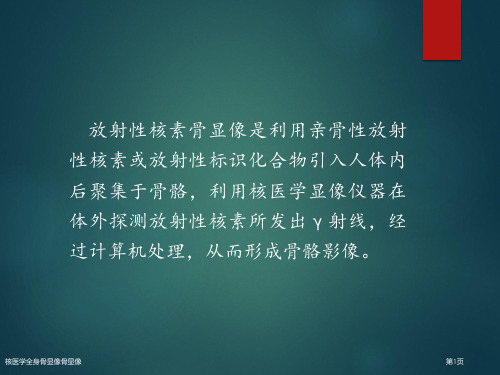
放射性核素标记的化合物被骨骼 吸收后,在体外通过γ闪烁照相机 等设备进行显像,从而反映骨骼 的代谢和形态变化。
发展历程与现状
发展历程
核医学骨显像技术自20世纪50年代 开始发展,经历了多代技术更新和改 进,目前已经广泛应用于临床。
现状
随着技术的不断进步,核医学骨显像 在准确性、敏感性和特异性等方面得 到了显著提高,为临床提供了更加准 确、可靠的诊断依据。
骨质疏松症评估
骨质疏松症诊断
核医学骨显像能够评估骨质疏松症的程度和范围,通过观察骨骼中放射性核素的分布情况,可以判断 骨密度和骨质量。
骨质疏松症治疗评估
核医学骨显像可以监测骨质疏松症治疗的效果,通过比较治疗前后的放射性核素分布情况,评估治疗 效果和调整治疗方案。
05
全身骨显像检查流程及处理

全身骨显像检查流程及处理下载温馨提示:该文档是我店铺精心编制而成,希望大家下载以后,能够帮助大家解决实际的问题。
文档下载后可定制随意修改,请根据实际需要进行相应的调整和使用,谢谢!并且,本店铺为大家提供各种各样类型的实用资料,如教育随笔、日记赏析、句子摘抄、古诗大全、经典美文、话题作文、工作总结、词语解析、文案摘录、其他资料等等,如想了解不同资料格式和写法,敬请关注!Download tips: This document is carefully compiled by the editor. I hope that after you download them, they can help yousolve practical problems. The document can be customized and modified after downloading, please adjust and use it according to actual needs, thank you!In addition, our shop provides you with various types of practical materials, such as educational essays, diary appreciation, sentence excerpts, ancient poems, classic articles, topic composition, work summary, word parsing, copy excerpts,other materials and so on, want to know different data formats and writing methods, please pay attention!全身骨显像检查是一种非常常见的医学影像学检查方法,通过对整个身体骨骼进行放射性同位素示踪,用以检测骨骼结构及功能异常。
放射性核素骨扫描解读

定期体检:监测辐射暴露情况,及时采取防护措施
放射性核素骨扫描的未来发展
结合PET技术:提高代谢活性评估,实现多模态融合诊断
结合超声技术:提高血流动力学评估,减少操作风险
结合MRI技术:提高软组织对比度,减少伪影
结合CT技术:提高定位准确性,减少辐射剂量
技术进步:提高图像质量和分辨率,降低辐射剂量
成像原理:放射性核素在骨骼中的分布和代谢情况,反映骨骼的健康状况
伽马相机:检测放射性核素的伽马射线,形成图像
伽马相机:用于检测放射性核素发出的伽马射线
放射性核素注射器:用于将放射性核素注入患者体内
计算机:用于处理和存储扫描数据
显示器:用于显示扫描图像和结果
注射放射性核素:将放射性核素注射到患者体内,使其在骨骼中聚集。
放射性核素骨扫描在肿瘤诊断中的实际应用案例
放射性核素骨扫描在肿瘤诊断中的优势
放射性核素骨扫描可以评估骨折愈合情况
通过扫描结果,医生可以判断骨折是否愈合
扫描结果还可以帮助医生确定是否需要进行进一步的治疗
放射性核素骨扫描是一种无创、无痛的检查方法,对患者来说更加安全舒适
放射性核素骨扫描的原理
骨关节炎的临床表现和诊断难点
放射性核素骨扫描在骨关节炎诊断中的优势
放射性核素骨扫描在骨关节炎诊断中的实际应用案例
骨转移瘤的诊断:通过骨扫描可以发现骨转移瘤的部位和范围
骨感染性疾病的诊断:骨扫描可以帮助医生判断骨感染性疾病的部位和程度
骨代谢性疾病的诊断:骨扫描可以评估骨代谢性疾病的严重程度和治疗效果
骨肿瘤的诊断:骨扫描可以帮助医生判断骨肿瘤的部位、大小和恶性程度
放射性核素骨扫描的解读方法
骨扫描图像的特点:黑白对比,清晰度高
放射性核素骨显像

2024/1/25
1
contents
目录
2024/1/25
• 放射性核素骨显像概述 • 放射性核素骨显像技术 • 放射性核素骨显像解读 • 放射性核素骨显像在临床应用 • 放射性核素骨显像优势与局限性 • 放射性核素骨显像未来展望
2
01
放射性核素骨显像概述
2024/1/25
3
定义与原理
20
局限性讨论
1 2
特异性较低
放射性核素骨显像虽然灵敏度高,但特异性相对 较低,可能出现假阳性或假阴性结果。
分辨率有限
由于放射性核素的物理特性,骨显像的分辨率相 对较低,可能无法清晰显示细小的骨骼结构。
3
受干扰因素影响
某些药物、治疗或生理状态可能影响放射性核素 的分布和代谢,从而影响骨显像的准确性。
定义
放射性核素骨显像是一种利用放射性核素标记的骨骼寻求剂,通过注射入体内 后,在骨骼中聚集并发出γ射线,进而通过外部探测器进行成像的技术。
原理
骨骼寻求剂在体内分布后,与骨骼中的羟基磷灰石晶体发生特异性结合,从而 在骨骼中聚集。通过γ射线探测器接收到的信号,经过计算机处理,可以生成骨 骼的二维或三维图像。
14
04
放射性核素骨显像在临床应用
2024/1/25
15
恶性肿瘤骨转移评估
早期发现骨转移
放射性核素骨显像具有高灵敏度 ,能够在X线平片发现骨转移前数
周或数月内检测到骨破坏累及的 范围。
2前后的骨显像图,可 以评估治疗效果,如肿瘤缩小、骨 转移灶减少等。
预测预后
2024/1/25
4
发展历程及现状
01
02
03
早期阶段
核医学全身骨显像骨显像
移性骨肿瘤最常见影像特征:
多发病灶 放射性异常浓聚 随机分布
核医学全身骨显像骨显像
第42页
临床应用-转移性骨肿瘤
二.转移性骨肿瘤核素显像表现*
2. 转移性骨肿瘤影像表现
单发性/孤立性放射性增高 放射性缺损区(“冷区”) 弥漫性放射性增高(superscan):超级骨显像 无异常放射性分布(假阴性) 闪烁现象 软组织显影(肿瘤摄取显像剂)
显像方法
七.SPECT/CT融合显像 将SPECT功效图像和CT解剖图像空间位置/坐标配准
后进行叠加, 使融合影像取得增量互补信息。融合显像 优势是同机采集、 定位准确,显著改进了对骨骼病变检 出率及判别诊疗能力,降低了骨显像诊疗骨转移假阳性, 提升了诊疗特异性。
核医学全身骨显像骨显像
第13页
核医学全身骨显像骨显像
核医学全身骨显像骨显像
前列腺癌
第46页
临床应用-转移性骨肿瘤
核医学全身骨显像骨显像
乳腺癌
肺癌
第47页
临床应用-转移性骨肿瘤
超级骨显像
核医学全身骨显像骨显像
前列腺癌
胃癌
第48页
临床应用-转移性骨肿瘤
核医学全身骨显像骨显像
乳腺癌随访,3个月后
第49页
临床应用-转移性骨肿瘤
第36页
超级骨显像(super scan)
核医学全身骨显像骨显像
第37页
核医学全身骨显像骨显像
第38页
核医学全身骨显像骨显像
metastatic calcification
第39页
骨显像适应症
判别原因不明骨痛,排除骨肿瘤; 判断原发骨肿瘤受累范围,了解有没有远端骨转移; 判断X线片难以确定应力性骨折及细微骨折; 诊疗各种代谢性骨病及骨关节疾病; 骨组织病理活检定位; 评价骨病治疗后疗效; 判别陈旧性或新近发生压缩性椎体骨折; 股骨头缺血坏死诊疗及预后判断; 诊疗骨髓炎,尤其是X线片阴性,但临床高度怀疑情况。
核医学全身骨显像骨显像

汇报人: 日期:
目录
• 核医学全身骨显像骨显像概述 • 检查前准备 • 检查过程 • 检查结果解读 • 检查后处理 • 核医学全身骨显像骨显像展望
01
核医学全身骨显像骨显像概述
定义与原理
定义
核医学全身骨显像是一种利用放射性核素示踪技术来检测全身骨骼系统结构和 功能的医学影像方法。
鉴别诊断需要考虑多种因素
需要对病变部位进行鉴别诊断,需要考虑多种因素,如年龄、性别、职业等。
结合其他影像学检查
为了提高诊断的准确性和可靠性,需要结合其他影像学检查方法,如CT、MRI等。
05
检查后处理
放射性废物的处理
分类收集
将使用过的显像剂、清洁 用品等放射性废物进行分 类收集,避免与普通垃圾 混放。
。
对专业人员的培训与教育
提高专业素养
加强对核医学专业人员的培训和教育 ,提高他们的专业知识和技能水平, 以确保准确、安全地进行诊断和治疗 。
跨学科合作
鼓励核医学专业人员与其他医学领域 的专家进行合作和交流,共同推动核 医学技术在临床实践中的应用和发展 。
THANKS
谢谢您的观看
骨髓炎、骨结核等疾病。
骨皮质不连续
骨皮质出现缺损或凹陷,提示 可能存在骨折、骨肿瘤等病变 。
骨髓腔异常
骨髓腔出现增宽或狭窄,提示 可能存在骨髓炎、骨结核等病 变。
骨纹理模糊
骨纹理出现模糊或中断现象, 提示可能存在骨质疏松、骨关
节炎等病变。
诊断分析与鉴别诊断
基于病变部位和影像表现进行诊断
根据全身骨显像的结果,结合患者的临床表现和其他检查结果,对病变部位进行诊断。
02
检查前准备
- 1、下载文档前请自行甄别文档内容的完整性,平台不提供额外的编辑、内容补充、找答案等附加服务。
- 2、"仅部分预览"的文档,不可在线预览部分如存在完整性等问题,可反馈申请退款(可完整预览的文档不适用该条件!)。
- 3、如文档侵犯您的权益,请联系客服反馈,我们会尽快为您处理(人工客服工作时间:9:00-18:30)。
The EANM practice guidelines for bone scintigraphyT.Van den Wyngaert1,2&K.Strobel3&W.U.Kampen4&T.Kuwert5&W.van der Bruggen6&H.K.Mohan7&G.Gnanasegaran8&R.Delgado-Bolton9&W.A.Weber10&M.Beheshti11&ngsteger11&F.Giammarile12&F.M.Mottaghy13,14&F.Paycha15&On behalf of the EANM Bone&Joint Committee and the Oncology Committee.Received:2May2016/Accepted:3May2016#The Author(s)2016.This article is published with open access at AbstractPurpose The radionuclide bone scan is the cornerstone ofskeletal nuclear medicine imaging.Bone scintigraphy is ahighly sensitive diagnostic nuclear medicine imaging tech-nique that uses a radiotracer to evaluate the distribution ofactive bone formation in the skeleton related to malignantand benign disease,as well as physiological processes.Methods The European Association of Nuclear Medicine(EANM)has written and approved these guidelines to pro-mote the use of nuclear medicine procedures of high quality.Conclusion The present guidelines offer assistance to nuclearmedicine practitioners in optimizing the diagnostic procedureand interpreting bone scintigraphy.These guidelines describethe protocols that are currently accepted and used routinely,but do not include all existing procedures.They shouldtherefore not be taken as exclusive of other nuclear medicinemodalities that can be used to obtain comparable results.It isimportant to remember that the resources and facilities avail-able for patient care may vary.Keywords Bone scintigraphy.Bone scan.Bone SPECT/CT.Bone diseasePreambleThe aim of this document is to provide general informationabout bone scintigraphy.This guidelines describe current rou-tine clinical procedures but should not be interpreted as* F.Paychafrederic.paycha@aphp.fr1Department of Nuclear Medicine,Antwerp University Hospital,Wilrijkstraat10,2650Edegem,Belgium2Faculty of Medicine and Health Sciences,University of Antwerp,Universiteitsplein1,2610Wilrijk,Belgium3Department of Radiology and Nuclear Medicine,Lucerne CantonalHospital,Lucerne,Switzerland4Nuclear Medicine Spitalerhof,Spitalerstraße8,20095Hamburg,Germany5Clinic of Nuclear Medicine,Friedrich-Alexander-UniversityErlangen-Nürnberg,Erlangen,Germany6Department of Radiology and Nuclear Medicine,SlingelandHospital,Doetinchem,The Netherlands7Department of Nuclear Medicine,Royal Free London NHSFoundation Trust,London,UK8Department of Nuclear Medicine,Guy’s and St Thomas’NHSFoundation Trust,London,UK9Department of Diagnostic Imaging(Radiology)and NuclearMedicine,San Pedro Hospital and Centre for Biomedical Research ofLa Rioja(CIBIR),University of La Rioja,Logroño,La Rioja,Spain10Department of Radiology,Memorial Sloan Kettering Center,NewYork,NY,USA11PET-CT Center Linz,Department of Nuclear Medicine andEndocrinology,St Vincent’s Hospital,Seilerstaette4,4020Linz,Austria12Department of Nuclear Medicine,Centre Hospitalier Universitaire deLyon,Lyon,France13Department of Nuclear Medicine,University Hospital Aachen,RWTH Aachen University,Aachen,Germany14Department of Nuclear Medicine,Maastricht University MedicalCenter(MUMC+),Maastricht,The Netherlands15Department of Nuclear Medicine,Hôpital Lariboisière,AssistancePublique-Hôpitaux de Paris,2rue Ambroise Paré,75010Paris,Franceexcluding alternative procedures also employed to obtain equivalent data.It is important to remember that the resources and facilities available for patient care may vary from one country to another and from one medical institution to another. This document has been prepared primarily for nuclear medi-cine physicians,physicists and technicians with the intention of offering assistance in optimizing procedural protocols and di-agnostic information that can currently be obtained from bone scintigraphy.The guidelines were written by the EANM Bone &Joint and Oncology Committees and then reviewed and approved by all EANM committees and the Board,as well as by the National Societies represented within the EANM.The EANM wrote and approved the guidelines to promote the use of high-quality nuclear medicine procedures.These guidelines are intended to assist practitioners in providing ap-propriate nuclear medicine care for patients.They are not in-flexible rules or requirements for practice and are not intended, nor should they be used,to establish a legal standard of care. For these reasons and those set out below,the EANM cautions against the use of these guidelines in litigation in which the clinical decisions of a practitioner are called into question.The ultimate judgment regarding the propriety of any specific procedure or course of action must be made by medical profes-sionals taking into account the unique circumstances of each case.Thus,an approach that differs from the guidelines does not necessarily imply that the approach was below the standard of care.To the contrary,a conscientious practitioner may respon-sibly adopt a course of action different from that set out in the guidelines when,in the reasonable judgment of the practitioner, such course of action is indicated by the condition of the patient, the limitations of available resources,or advances in knowledge or technology subsequent to publication of the guidelines.The practice of medicine involves not only the science,but also the art of dealing with the prevention,diagnosis,allevia-tion,and treatment of disease.The variety and complexity of human conditions make it impossible at times to identify the most appropriate diagnosis or to predict with certainty a par-ticular response to treatment.Therefore,it should be recog-nized that adherence to these guidelines will not assure an accurate diagnosis or a successful outcome.All that should be expected is that the practitioner will follow a reasonable course of action based on current knowledge,available re-sources,and the needs of the patient to deliver effective and safe medical care.The sole purpose of these guidelines is to assist practitioners in achieving this objective. Introduction and goalsThe radionuclide bone scan is the cornerstone of skeletal nu-clear medicine imaging.Bone scintigraphy is a highly sensitive diagnostic nuclear medicine imaging technique that uses a ra-diotracer to evaluate the distribution of active bone formation in the skeleton related to malignant and benign diseases,as well as physiological processes.Phosphate analogues can be labelled with technetium-99m(99m Tc)and are used for bone imaging because of their high uptake in the skeleton and rapid clearance from soft tissues after intravenous injection.Tracer accumula-tion occurs in proportion to local blood flow and bone remod-elling activity(dependent on osteoblast/osteoclast activity),and unbound tracer is rapidly cleared from surrounding soft tissues.Most pathological bone conditions,whether of infectious, traumatic,neoplastic or other origin,are associated with an increase in vascularization and local bone remodelling.This accompanying bone reaction is reflected on bone scans as a focus of increased radioactive tracer uptake.Bone scintigra-phy is a sensitive technique that can detect significant meta-bolic changes very early,often several weeks or even months before they become apparent on conventional radiological im-ages.In addition,the technique provides an overview of the entire skeleton at a relatively modest radiation exposure.While MRI has been shown to be more sensitive than pla-nar bone scans in detecting skeletal metastases in vertebral bodies,bone SPECT has been found to have comparable di-agnostic sensitivity for vertebral body metastases and a higher diagnostic sensitivity for metastases located in the pedicles [1].Furthermore,the majority of studies that have compared bone scanning with MRI did not use a reliable gold standard and typically included various solid tumours and even lym-phomas which usually produce osteolytic lesions with poor osteoblastic bone reaction so that they are not detectable by bone scintigraphy.Hence,there is no reliable evidence that bone scintigraphy is generally less sensitive than whole-body MRI in all solid cancers.Multimodality SPECT/CT of-fers the unique opportunity to correlate scintigraphic findings with anatomical images and introduces novel algorithms to further enhance SPECT image quality based on the CT data (e.g.correction for attenuation and scatter).This results in improved correlation of areas with physiological variants or abnormal tracer accumulation with anatomical landmarks[2]. However,this has increased the complexity of this technique, increasing the need for standardization and practice guidelines in order to maximize the diagnostic yield of the examination.The corresponding guidelines from the Society of Nuclear Medicine and Molecular Imaging(SNMMI),the Dutch Society of Nuclear Medicine(NVNG),and the French Society of Nuclear Medicine and Molecular Imaging (SFMN),as well as the existing EANM bone scintigraphy procedures guideline for tumour imaging were consulted while preparing this consensus document[3–6].For specific applications of bone scintigraphy in selected indications or populations,the reader is also referred to the EANM guide-lines for paediatric bone scanning,and the EANM and SNMMI practice guidelines for sodium18F-fluoride PET/CT bone scans[7–9].Other excellent reference works are also recommended[10].Eur J Nucl Med MolImaging The goal of these guidelines is to provide an educational tool designed to assist the nuclear medicine practitioner in appropriately recommending,performing,interpreting,and reporting the results of bone scintigraphy.Definitions1.Planar whole-body images in the anterior and posteriorprojections of the axial and appendicular skeleton.If nec-essary,additional localized or spot views can be obtained.2.Focal planar images limited to a specific portion of theskeleton.3.Single photon emission tomography(SPECT,also knownas SPET)allows the visualization of the three-dimensional distribution of the radiopharmaceutical in the skeleton.4.SPECT/CT images consist of a SPECT acquisition com-bined with CT using an integrated CT scanner.5.Multiphase bone scan produces planar images of the vas-cular inflow and the soft tissue phase,and delayed phase images of the radiopharmaceutical over a given area of the skeleton.The vascular inflow images are acquired during intravenous injection.The study of the soft tissue distri-bution of the radiopharmaceutical in the region of interest is performed within the first5–10min after injection.Finally,delayed whole-body,focal views,and/or tomo-graphic images are usually acquired between2and4h after injection of the radiopharmaceutical.In some pa-tients,it may be useful to acquire late-phase images up to24h after tracer administration[11].6.Quantification is the process of calculating the osseousradioactivity concentration expressed as standardized up-take values(SUV).Common clinical indications for bone scanThe indications for bone scintigraphy are numerous and can generally be classified into three distinct clinical scenarios:(1) when a specific bone disease is present or suspected,(2)to explore unexplained symptoms,and(3)for the metabolic as-sessment prior to the start of therapy.While the diagnostic sensitivity of bone scintigraphy is very high,the low specific-ity often requires further investigation with other imaging mo-dalities(e.g.plain radiography,CT or MRI)or nuclear medi-cine studies(e.g.FDG PET/CT).For this reason,anatomical imaging and bone scintigraphy should be considered as com-plementary methods,each of which cannot be replaced by the other.Conversely,bone scintigraphy is not indicated in a number of specific conditions due to limitations of the technique in a specific disease context or lack of clinical impact of imaging results.However,ultimately it must be the nuclear medicine physician who evaluates and decides on the indication in each particular case and on the specific protocol that should be applied.Both indications and nonrecommended indications are outlined in the following sections.Bone disease present or suspected1.Oncology&Solid tumours with high affinity for bone,includingprostate,breast,lung and renal cancer[12–17] &Malignant haematological conditions limited to bone,including Hodgkin’s disease and non-Hodgkin’s lym-phoma[18]&Bone tumours and bone dysplasia,including osteosar-coma,osteoid osteoma,osteoblastoma,fibrous dys-plasia,giant cell tumour and osteopoikilosis[19] &Soft tissue sarcomas,including rhabdomyosarcoma &Paraneoplastic syndromes,including hypertrophicpulmonary osteoarthropathy,algodystrophy,polymyalgia rheumatica,poly(dermato)myositis andosteomalacia&Assessment of bone remodelling prior to radionuclide therapy(223Ra-Cl2,89Sr-Cl2,153Sm-EDTMP,186Re-HEDP)Whole-body FDG PET and PET/CT are other im-aging modalities that enable detection of the primarytumour and metastases by visualization of the in-creased glucose consumption of malignant tissue.The majority of studies comparing FDG PET with18F-fluoride PET or SPECT with99m Tc-labelledbisphosphonates have shown a higher diagnostic sen-sitivity of FDG for osteolytic metastases and a higherdiagnostic sensitivity of bone-affine radiotracers fordetecting osteoblastic metastases[20–24].Primary tumours of bone are relatively rare in adults,whereas bone metastases of other cancer enti-ties(e.g.breast,prostate,lung,renal cancer,etc.)arevery frequent.In prostate cancer,the assessment ofbone scans is increasingly standardized by calculationof the bone scan index and reporting of progressionaccording to PCWG-2criteria[25,26].2.Rheumatology&Chronic inflammatory arthritis,including rheumatoid arthritis,spondyloarthropathies and related disorders(ankylosing spondylitis,psoriatic arthritis,Reiter’s ar-thritis,SAPHO syndrome[synovitis,acne,pustulosis,hyperostosis,osteitis],chronic recurrent multifocalosteomyelitis)and sacroiliitis[27–29]Eur J Nucl Med MolImaging&Osteoarthritis of the lumbar facet joints,hip, femorotibial and femoropatellar osteoarthritis,rhizarthrosis and tarsal osteoarthritis[30,31] &Enthesopathies,including plantar fasciitis,Achilles tendinitis and bursitis&(Avascular)osteonecrosis,which is most frequently located at the femoral head,femoral condyle and tib-ial plateau&Osteonecrosis of the jaw(ONJ)[32]&Complex regional pain syndrome type I of the hand, hip,knee and foot&Tietze’s syndrome(costochondritis)&Polymyositis&Paget’s disease&Langerhans cell histiocytosis(LCH):single system LCH and multisystem LCH with bone involvement &Non-Langerhans cell diseases,such as Erdheim–Chester disease,Schnitzler syndrome,and RosaïDorfman disease&Other rare osteoarticular diseases,such as sarcoidosis with bone involvement,mastocytosis,Behçet’s dis-ease,and familial Mediterranean fever3.Bone and joint infections[33]&Osteomyelitis(acute,subacute or chronic,of bacteri-al,mycobacterial or fungal origin)&Septic arthritis&Spondylodiscitis or spondylitis&Septic loosening or mechanical complication of inter-nal fixation(long bones or spine)or arthroplasty(hip,knee,ankle or shoulder)&Malignant(necrotizing)external otitisThe reader is also referred to other relevant EANM consensus documents and guidelines[34,35].4.Orthopaedics,sports and traumatology[36,37]&Periostitis,including shin splints and thigh splints[38]&Enthesopathies,including plantar fasciitis,Achilles tendinitis and bursitis&Spondylolisthesis(acute or subacute)&Radiological occult stress-related fractures(e.g.scaphoid,tarsals)or nonspecific symptoms[39] &Insufficiency fractures,including osteoporotic verte-bral or occult fractures,sacral fractures,femoral heador neck fractures,tibial plateau fractures,tarsal andmetatarsal fractures&Septic loosening,mechanical complication,and syno-vitis of internal fixation(long bones or spine)or pros-thesis(hip,knee,ankle,or shoulder)&Pseudoarthrosis(delayed union,non-union)&Periarticular heterotopic ossification&Viability of bone graft5.Metabolic bone disease[40–42]&Hyperparathyroidism(primary and secondary)&Osteomalacia&Renal osteodystrophy&Rare skeletal manifestations of endocrine disorders, including hyperthyroidism and acromegaly &Vitamin D deficiency6.Paediatrics&Osteochondritis of the hip(Legg-Calvé-Perthes disease)&Transient synovitis of the hip&Osteoid osteoma&Battered child syndrome&Mandibular condylar hyperplasia&Bone infarction(sickle cell disease,thalassaemia) Exploration of unexplained symptoms1.Subacute or chronic musculoskeletal or bone pain withnormal clinical examination and radiographs&Arthralgia,monoarthritis,oligoarthritis,polyarthritis, localized or multifocal bone pain and backache 2.Further exploration of abnormal biochemical(e.g.phos-phate or calcium metabolism)or radiological findings 3.Fever of unknown origin:exclusion of osteomyelitisMetabolic assessment prior to initiation of therapy1.Evaluation of the activity of arthropathies and to confirmactive synovitis prior to radiation synovectomy or before infiltration of facet joints with corticosteroids2.Evaluation of osteoblast activity in case of Paget’s diseasebefore initiating treatment with bisphosphonates3.Assessment of benign or malignant vertebral compressionfracture prior to vertebroplasty or kyphoplasty Treatment monitoringQuantitative bone SPECT/CT is a novel technique with po-tentially useful applications in treatment response monitoring in bone[43].However,the exact role in routine clinical prac-tice has yet to be determined.Eur J Nucl Med MolImaging Bone scan not indicatedBone scan may not be the preferred investigation in the fol-lowing conditions1.Bone lesions with known inconsistent scintigraphic find-ings,such as plasmacytoma,multiple myeloma, chordoma,or Ewing’s sarcoma2.Benign bone lesions and incidentalomas when properlycharacterized by radiological imaging,including bone is-land,uncomplicated haemangioma,osteitis condensans ilii,nonossifying fibromas,asymptomatic enchondroma of the long bones,ganglion cyst and asymptomatic Paget’s disease3.Symptomatic degenerative joint disease well character-ized on radiological imaging,properly diagnosed based on the pain syndrome and a well performed clinical examinationEven though bone scintigraphy may in general not be the preferred imaging modality in the conditions listed above,this recommendation should be assessed within the specific clini-cal context of the patient.Specification of the examination procedure Qualifications and responsibilities of personnelAll physicians and personnel involved in performing and reporting bone scintigraphy should be sufficiently qualified and experienced in accordance with applicable laws,and in-dividual responsibilities should be documented in standard operating procedures.RequestThe written or electronic request form should provide suffi-cient information to demonstrate the medical necessity of the examination.This should include current signs and symp-toms,relevant history(skeletal surgery,trauma,or recent ra-diation or chemotherapy),and the specific reason for the ex-amination or provisional diagnosis.Outpatients should also bring the results of all other relevant examinations that have already been performed(laboratory,radiological,scintigraph-ic,or other).Patient preparationWhen making an appointment,the patient should receive information on how the examination is performed and its estimated duration.Patients should be informed that they may eat and drink and of the need to report a pregnancy,any delay in menstrual cycle,or active breastfeeding. Information leaflets and/or displays should be available in the waiting area of the nuclear medicine service,and all information should preferably be accessible through the website of the institution.Prior to tracer injection,the nuclear medicine physician or technologist must explain the purpose of the examination,the expected benefits,and answer any questions.The patient is informed as to how the examination is to be performed(e.g. multiple planar acquisitions,additional SPECT/CT,etc.),tak-ing into account the specific clinical problem.Relevant infor-mation that may assist in interpretation of imaging findings are checked with the patient,including:1.History of fractures,trauma,osteomyelitis,cellulitis,oe-dema,arthritis,neoplasms,metabolic bone disease,or limitation of function2.Current symptoms and physical findings3.Results of prior bone scintigraphy or other imaging stud-ies such as conventional radiography,CT and MRI(it is strongly recommended that hard copies or computer files of previous examinations be obtained)4.History of recent nuclear medicine studiesboratory results6.History of therapy that might affect the results of bonescintigraphy(see Precautions)7.History and dates of prior orthopaedic surgery(e.g.pres-ence and location of prosthetic implants)8.History of anatomical or functional renal/urinary tractabnormalities9.Contraindications for hydrationAt this time,the physical condition of the patient should be assessed and whether current symptoms(e.g. pain,immobility,etc.)may hamper acquisition of optimal images.In patients with severe pain,an appropriate anal-gesic strategy should be implemented in consultation with the treating/referring physician.In addition,the scanning parameters may be adapted to accommodate the patient (see Protocol/image acquisition).Unless contraindicated,patients should be well hydrated and instructed to drink one or more litres of water during the time between injection and imaging.All patients should be asked to void their bladder frequently during the time between injection and delayed imaging as well as immediately prior to the scan.The patients should drink a large amount of fluids during the24h after radiopharmaceutical administration.In patients on renal replacement therapy,haemodialysis per-formed from15min to5h after injection of the radiopharma-ceutical can successfully decrease blood pool and soft tissue activity to nearly normal levels[44,45].However,careful planning of the examination and consultation with the treating nephrologist are recommended.Eur J Nucl Med MolImagingPrecautionsPregnancy and lactationIn women of childbearing age,it is necessary to verify the absence of pregnancy.In a patient who is known or suspected to be pregnant,a clinical decision is necessary to consider the benefits against the possible harm of carry-ing out any procedure.If medically justified,radiation ex-posure should be delayed until after both pregnancy and breastfeeding.Evaluation by other techniques such as ul-trasonography or MRI is preferred.While interruption in breastfeeding is not essential according to the ICRP,this is on the basis that there is no free pertechnetate in the radio-pharmaceutical[46].Therefore,an interruption of at least 4h during which one meal is discarded is advised to be on the safe side.Possible drug interactionsThe main drugs that may interfere with the quality of scinti-graphic images are:1.Aluminium:reduced skeletal tracer uptake,diffuse hepat-ic tracer uptake,increased renal tracer uptake2.Androgen deprivation therapy for prostate cancer(bicalutamide,oestrogens):increased mammary tracer uptake in case of gynecomastia3.Bone-modifying agents(including bisphosphonates anddenosumab)or agents interfering with osteoblast func-tion(e.g.cabozantinib):reduced skeletal tracer uptake[47]4.Corticosteroids:reduced skeletal tracer uptake,reducedtracer uptake at fracture sites5.Haematopoietic growth factors:increased spinal traceruptake,possible increased tracer uptake in the appendic-ular skeleton6.Iron:increased renal tracer uptake,increased tracer uptakeat site of intramuscular injection,diffuse hepatic tracer uptake7.Methotrexate:diffuse hepatic tracer uptake8.Nephrotoxic chemotherapy:increased renal tracer uptakeand reduced skeletal tracer uptake9.Nifedipine:reduced skeletal tracer uptake RadiopharmaceuticalsPhysical characteristics of the radionuclide99m Tc decays by isomeric transition with a half-life of6.02h to99Tc by emitting one140.5-keV gamma ray per decay.Characteristics of the tracer moleculeThe molecules most commonly used for performing bone scintigraphy are bisphosphonates:methylene diphosphonate (MDP),hydroxymethylene diphosphonate(HMDP)or hydroxyethylene diphosphonate(HDP),and2,3-dicarboxypropane-1,1-diphosphonate(DPD).PharmacokineticsThe injected radiolabelled bisphosphonates adsorb to the sur-face of hydroxyapatite crystals in proportion to local bone vascularization and osteoblastic activity.After intravenous ad-ministration,the plasma clearance of bisphosphonates is biexponential and a function of skeletal uptake and urinary elimination.Four hours after injection,approximately50to 60%of the injected amount is fixed in the skeleton,the un-bound fraction(34%)is excreted in the urine,and only6% remains in the circulation.Tracer elimination through the gas-trointestinal tract is insignificant.Maximum bone accumula-tion is reached1h after tracer injection and remains practically constant up to72h.Radiopharmaceutical preparation and storageThe bisphosphonates listed above are commercially available and supplied in a vial containing the bisphosphonate,a stan-nous reducing agent and other excipients in the form of a powder ready for labelling.Vials containing the sterile nonpyrogenic lyophilisate should be stored at4–8°C or room temperature,as required by the manufacturer.These kits can be used until the expiry date of the batch(1to2years after the date of manufacture).The radiopharmaceutical is prepared by the addition of the required amount of sodium pertechnetate[99m TcO4−]diluted in sterile physiological saline to the vial in accordance with the manufacturer’s instructions.After labelling,the preparation should be kept at between4°C and8°C or at room temper-ature,depending on the brand used,and remains stable for8h. Because the radiopharmaceutical is susceptible to oxidation, care should be taken to avoid introducing air into the multidose vial during preparation or removal of doses.The radiopharmaceutical should be used within6h of preparation unless allowed otherwise by the manufacturer. Administered activityThe radiopharmaceutical has to be administered by the intra-venous route,e.g.by direct intravenous injection,via an in-dwelling catheter or using a butterfly needle.The activity of the radiopharmaceutical to be administered should be determined after taking into account Directive 2013/59/EURATOM of the Council of the European Union,Eur J Nucl Med MolImaging which guarantees health protection of individuals with respect to the dangers of ionizing radiation in the context of medical exposure.According to this directive,member states are re-quired to bring into force such regulations as may be neces-sary to comply with the directive.One of the criteria is the designation of Diagnostic Reference Levels(DRL)for radio-pharmaceuticals;these are defined as levels of activity for groups of standard-sized patients and for broadly defined types of equipment.These levels are expected not to be exceeded for standard procedures when good and normal practice regarding diagnostic and technical performance is applied.For the reasons discussed above,the activities recom-mended below for99m Tc-bisphosphonate should be consid-ered only a general indication,based on data in the literature and current experience.However,nuclear medicine physi-cians in each country should respect the DRLs and the rules set out in local laws.For bone scintigraphy in adults,the average activity admin-istered by a single intravenous injection should be500MBq (300–740MBq,8–20mCi;Table1).The administered ac-tivity usually ranges between8and10MBq/kg for adults. Lower activities may be used when equipment with higher detector sensitivity or resolution recovery resulting in similar image quality is available.For markedly obese adult patients, the administered activity may be increased to11–13MBq/kg. If the injected activity falls outside these recommended limits for clinical reasons,the deviation should be kept as small as possible.Practitioners could be required to justify administra-tion of activities greater than local national DRLs.The activities administered to children should be a fraction of those administered to adults calculated in relation to body weight according to the factors given by the EANM/SNMMI Paediatric Dosage Harmonization Working Group[9,48].In children,the EANM recommends a baseline activity of 35MBq for99m Tc-bisphosphonates(with a minimum activity of40MBq)that should be adjusted based on the class of the radiopharmaceutical(class B)and the weight of the child (Table1).Protocol/image acquisitionInstrumentationSingle-headed or dual-headed gamma camera equipped with a low-energy,high-resolution parallel-hole collimator is recommended.A low-energy general purpose collimator may alternatively be used for early dynamic and blood pool images.The energy window is centred on the photon energy peak of99m Tc(140keV)and the window width is generally set at15%or20%.An asymmetrical window may be used to improve resolution(e.g.20%with a3%offset)but its use must be supported by a physicist and an appropriately tailored quality assurance programme[49,50].Planar and whole-body acquisitionsFor the vascular phase of the examination,the camera is positioned centred on the area of interest.The dynamic acquisition of30to60images with a duration of1–2s each and a matrix of64×64or128×128pixels is started simultaneously with the intravenous tracer injection.The early and late planar images are focused on one or more region(s).The early images are acquired between1min and10min after intravenous injection of99m Tc-labelled bisphosphonate,with an acquisition time of3min to 5min and a matrix size of128×128or256×256. Delayed images are usually acquired2h to5h after injec-tion of the radiolabelled bisphosphonate using a predefined duration(4–10min)or number of counts,with a matrix size of128×128or256×256.When using a predefined number of counts,at least700,000to1,000,000counts are required for scanning the thoracoabdominal region,250, 000to400,000counts for the large joints and skull,and 150,000to250,000for the distal joints,depending on the field of view(FOV)of the gamma camera.The larger the FOV,the larger the number of total counts required to give similar count densities over equivalent regions of the skel-eton[51].Moreover,the presence of organs(typically the kidneys)with physiologically high count densities may hamper visualization of contiguous structures(typically the spine).A pinhole collimator acquiring high-resolution planar views of a small area can be used to complete the examination, particularly in infants and children.A total count of50,000to 100,000is recommended.Zoom magnification or a converg-ing collimator may also be used to improve resolution.The physician interpreting the image should be notified when a collimator that introduces distortions,such as a pinhole,has been used.Additional projections,such as lateral,oblique, tangential and special views may be obtained if necessary.Table1Applicable dose reference levels for bone scintigraphyChildren Adults3.5kg10kg20kg30kg40kg50kgActivity(MBq)4095170240310375300–740 Effective dose(mSv) 2.0 2.4 2.5 2.6 2.7 2.8 2.9–4.0Eur J Nucl Med MolImaging。