Chapter six Molecular Cloning
Chapter 4 Molecular Clonning Methods
Chapter 4 Molecular Cloning Methods第四章分子克隆方法4.1 基因克隆分子克隆概述•分子克隆(molecular cloning,基因克隆):通过体外重组技术,将一段目的DNA经切割、连接插入适当载体,并导入受体细胞,扩增形成大量子代分子的过程•基因克隆的核心---体外重组(Recombination):人工将一段目的DNA插入一个载体的过程。
孙悟空也会“克隆”一个细菌经过20分钟左右就可一分为二;仙人掌切成几块,每块落地就生根,......凡此种种,都是生物靠自身的一分为二或自身的一小部分的扩大来繁衍后代,这就是无性繁殖。
无性繁殖的英文名称叫“Clone”,音译为“克隆”。
实际上,英文的“Clone”来源于希腊文“Klone”,原意是用“嫩枝”或“插条”繁殖。
时至今日,“克隆”的含义已不仅仅是“无性繁殖”,凡来自一个祖先,无性繁殖出的一群个体,也叫“克隆”。
可以这样说,关于克隆的设想,我国明代的大作家吴承恩已有精彩的描述--孙悟空经常在紧要关头拔一把猴毛变出一大群猴子,就是克隆猴。
4.1.1 限制性内切酶什么是限制性内切酶?限制性核酸内切酶(restriction endonuclease):识别并切割特异的双链DNA序列的一种内切核酸酶。
[别名]Endodeoxyribonuclease[单位定义]在适当的温度下,在1小时内,1μgDNA在50μl体积中,被完全消化所需的限制性内切酶的量。
一般说来,线性DNA比超螺旋DNA更易消化。
限制性内切酶的发现•19世纪60年代末期,Stewart Linn和Werner Arbert从E.coli中发现了限制性核酸内切酶。
这些酶通过切断外源DNA来防止外源DNA,如病毒DNA的入侵,可以说它们“限制”了病毒的宿主范围,且它们的切割位点在外源DNA的内部,因此称限制性核酸内切酶。
•1971 美国人Daniel Nathans 和Hamilton Smith 发展了核酸酶切技术。
分子克隆

3.与反转录相关的PCR扩增
RT-PCR(reverse transcriptase-PCR,RT-PCR): 又称反转录PCR, 是以反转录的cDNA作模板所进行的PCR,
可对基因的表达和序列多态性分析。
RT-PCR
反转录
逆转录酶
AAAnA mRNA
AAAnA
T T TnT
cDNA第一链
DNA聚合酶 cDNA
目的基因的获取-----PCR技术:
定义: PCR技术又称聚合酶链式反应(polymerase chain reaction),是通过模拟体内 DNA 复制的 方式,在体外选择性地将 DNA 某个特殊区域扩 增出来的技术。
Taq DNA多聚酶的发现
Heat-stable polymerase is vital to the ease of the process…
4.PCR反应程序
⑴94~96℃ ⑵94℃ ⑸ 25- ⑶50-60℃ 35个循 ⑷72℃ 环 ⑹72℃ ⑺4-10℃ 30’’-3’ 预变性(使模板DNA充分变性) 30’’ 变性 30’’-1’ 复性(使引物与模板充分退火) n’(按1’扩增1kb计算)延伸 3-7’ 总的延伸(使产物延伸完整) 保存
质粒自身含有复制起始点,与相应的顺式调控元件组成一个复制子(replicon), 能利用细菌的酶系统进行独立的复制及转录。质粒具有多种遗传选择标记, 包括各种抗药基因或营养代谢基因等。
氨苄青霉素抗性(ampicillin resistance,ampr)基因:
此基因编码ß 内酰胺酶,该酶能 水解氨苄青霉素ß —内酰胺环,使之 失效而使细菌产生耐药。
1988年Saiki 等从温泉 ( Hot spring)中分离 的一株水生嗜热杆菌 (thermus aquaticus) 中提取到一种耐热DNA 聚合酶。
Chapter 6 Cloning Vectors for E and eukaryotes
1
Contents
6.1 Cloning vectors based on E.coli plasmids 6.1.1 The nomenclature of plasmid cloning vectors 6.1.2 The useful properties of pBR322 6.1.3 Other typical E.coli plasmid cloning vectors 6.1.5 pMD-19T 6.2 Vectors for yeast and other fungi 6.2.1 Vectors based on the 2 m plasmid 6.2.2 A Yep may insert into yeast chromosomal DNA 6.2.3 Other types of yeast cloning vector 6.2.4 Artificial chromosomes can be used to clone large pieces of DNA in yeast 6.2.5 Vectors for other yeasts and fungi
A map of pBR322
7
6.1.3 The pedigree of pBR322
Fig 6.2 The pedigree of pBR322:(a) the manipulations involved in construction of pBR322, and (b) a summary of the origins of pBR322
2
contents
氯化锂沉淀rna原理

氯化锂沉淀rna原理一、引言RNA(核糖核酸)是生物体内一类重要的核酸分子,具有传递遗传信息、调控基因表达等重要功能。
为了研究RNA的结构和功能,科学家们开展了许多RNA的提取和纯化研究。
其中,氯化锂沉淀RNA是一种常用的方法之一。
二、氯化锂沉淀RNA的原理氯化锂沉淀RNA的原理是利用氯化锂与RNA中的磷酸根结合形成沉淀,从而将RNA分离出来。
具体步骤如下:1. 细胞破碎:将待提取的RNA的细胞破碎,使RNA释放到溶液中。
2. 细胞裂解液处理:加入细胞裂解液,使细胞内的蛋白质、DNA等杂质被裂解。
3. 加入氯化锂溶液:加入氯化锂溶液,使细胞裂解液中的RNA与氯化锂结合形成沉淀。
4. 沉淀处理:通过离心将沉淀与上清液分离开来。
5. 沉淀洗涤:利用乙醇洗涤沉淀,去除杂质。
6. RNA溶解:将沉淀中的RNA用适当的溶液进行溶解,得到纯化的RNA溶液。
三、实验操作1. 提取RNA的样品准备:准备待提取RNA的样品,可以是细菌、真核生物细胞等。
2. 细胞破碎:使用细胞破碎液将细胞破碎,使细胞内的RNA释放到溶液中。
3. 细胞裂解液处理:加入细胞裂解液,使细胞内的蛋白质、DNA等杂质被裂解。
4. 加入氯化锂溶液:根据实验所需的RNA浓度,加入适量的氯化锂溶液,使RNA与氯化锂结合形成沉淀。
5. 沉淀处理:将混合物进行离心,使沉淀与上清液分离开来。
6. 沉淀洗涤:用75%乙醇洗涤沉淀,去除杂质。
7. RNA溶解:用适当的溶液(如RNase-free水)将沉淀中的RNA 溶解,得到纯化的RNA溶液。
四、注意事项1. 实验操作时应注意严格遵守无菌操作规范,以保证RNA的纯度和完整性。
2. 实验过程中应避免RNase的污染,如需使用工具、试剂等,应先进行RNase去污处理。
3. 实验室中的仪器和试剂应保持干净,以避免杂质对RNA提取的干扰。
4. 操作过程中应注意个人安全,如需使用有毒试剂,应戴好防护手套和口罩。
五、总结氯化锂沉淀RNA是一种常用的RNA提取和纯化方法,其原理是利用氯化锂与RNA中的磷酸根结合形成沉淀。
分子克隆英文版

分子克隆英文版Molecular CloningMolecular cloning is a fundamental technique in modern molecular biology and biotechnology, allowing for the isolation, amplification, and manipulation of specific DNA sequences. This process involves the transfer of a DNA fragment from one organism to a self-replicating genetic element, such as a plasmid or a virus, which can then be introduced into a host cell. The host cell, typically a bacterium or a eukaryotic cell, then replicates the inserted DNA, producing multiple copies of the desired genetic material.The primary motivation behind molecular cloning is the need to obtain large quantities of a specific DNA sequence for various applications, including genetic research, disease diagnosis, and the production of recombinant proteins. By cloning a gene of interest, researchers can generate an unlimited supply of the target DNA, enabling further analysis, modification, and utilization.The process of molecular cloning can be divided into several key steps. The first step involves the isolation of the desired DNA fragment, which can be obtained from a variety of sources, such asgenomic DNA, complementary DNA (cDNA), or synthetic DNA. This DNA fragment is then inserted into a suitable vector, such as a plasmid or a viral genome, using specialized enzymes called restriction endonucleases and DNA ligase.The resulting recombinant DNA molecule is then introduced into a host cell, typically a bacterial cell like Escherichia coli, through a process called transformation. The transformed host cells are then cultured, and the cells containing the desired recombinant DNA are selected and amplified. This amplification process allows for the production of large quantities of the target DNA sequence.One of the most important applications of molecular cloning is the production of recombinant proteins. By cloning a gene encoding a specific protein, researchers can express and purify the protein in a host cell, such as a bacterium or a eukaryotic cell line. This technique has been instrumental in the development of numerous therapeutic proteins, including insulin, growth hormones, and monoclonal antibodies, which have revolutionized the field of medicine.Molecular cloning is also essential for the study of gene function and regulation. By cloning a gene and introducing it into a host cell, researchers can investigate the expression, localization, and interactions of the encoded protein, as well as the regulatory mechanisms that control its activity. This knowledge is crucial forunderstanding the complex biological processes that underlie human health and disease.In addition to its applications in research, molecular cloning has also found widespread use in the field of genetic engineering. By modifying the genetic material of organisms, scientists can create genetically modified organisms (GMOs) with desirable traits, such as increased crop yield, improved nutritional value, or resistance to pests and diseases. This technology has significant implications for agriculture, environmental conservation, and the development of new therapeutic strategies.Despite its many benefits, molecular cloning is not without its challenges and ethical considerations. The potential for misuse or unintended consequences of genetic manipulation has sparked ongoing debates and the need for robust regulatory frameworks to ensure the responsible and ethical use of this powerful technology.In conclusion, molecular cloning is a fundamental technique in modern molecular biology and biotechnology, enabling the isolation, amplification, and manipulation of specific DNA sequences. This technology has revolutionized numerous fields, from genetic research and medicine to agriculture and environmental conservation. As the field of molecular biology continues to evolve, the applications and implications of molecular cloning willundoubtedly continue to expand, posing both exciting opportunities and important ethical considerations for the future.。
Molecular Cloning and Characterization
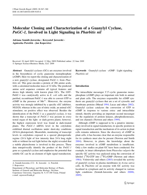
Molecular Cloning and Characterization of a Guanylyl Cyclase,PnGC-1,Involved in Light Signaling in Pharbitis nilAdriana Szmidt-Jaworska ÆKrzysztof Jaworski ÆAgnieszka Pawełek ÆJan KopcewiczReceived:20April 2009/Accepted:12May 2009/Published online:22June 2009ÓSpringer Science+Business Media,LLC 2009Abstract Guanylyl cyclases (GCs)are enzymes involved in the biosynthesis of cyclic guanosine monophosphate (cGMP).Here we report the cloning and characterization of a new guanylyl cyclase,designated PnGC-1,from Phar-bitis nil .This gene encodes a protein of 286amino acids,with a calculated molecular mass of 32kDa.The predicted amino acid sequence contains all typical features and shows high identity with known plant GCs.The GST-PnGC-1was catalytically active in E.coli cells and the purified,recombinant PnGC-1was able to convert GTP to cGMP in the presence of Mn 2?.Moreover,the enzyme activity was strongly inhibited by a specific sGC inhibitor,NS2028,whereas in the case of nitric oxide,an animal sGC stimulator,no positive effect was observed.Besides the identification of the PnGC-1as a guanylyl cyclase,it was shown that a transcript of PnGC-1was present in every tested organ of the light-or dark-grown plants;however,the highest expression level was found in dark-treated plants.The PnGC-1mRNA level in the cotyledons exhibited diurnal oscillations under short-day conditions (8/16-h photoperiod).Meanwhile,monitoring of transcript levels in cotyledons exposed to a special photoperiodic regime (24h light of low intensity then 24h long night with or without far-red light before the night)revealed that a stabile phytochrome is involved in this process.These data unequivocally identify the product of the PnGC-1gene as a guanylyl cyclase and emphasize the potential that soluble GC can be an element of light signal transduction.Keywords Guanylyl cyclase ÁcGMP ÁLight signaling ÁPharbitis nilIntroductionThe intracellular messenger 30,50-cyclic guanosine mono-phosphate (cGMP)plays an important role both in animal and plant cells.The enzymes responsible for cGMP syn-thesis are guanylyl cyclases that are a set of cytosolic and membrane proteins (Murad 1994;Lucas and others 2002).Guanylyl cyclase catalyzes the conversion of GTP to cGMP in response to various extra-and intracellular stimuli,thereby providing an important second messenger for the regulation of protein kinases,phosphodiesterases,and ion channels (Newton and others 1999).Although cGMP is supposed to be a putative interme-diary involved in signal transduction,its specific position in signal transduction and the mechanism of its action in plant cells remains unknown.Since the discovery of cGMP in plant cells,it has become clear that an enzyme responsible for its synthesis must also be present (Newton and Smith 2004).However,in plants,the knowledge about the enzymes involved in cGMP metabolism is insufficient.Only a few studies on plant GC have been conducted.For example,in intact chloroplasts from Phaseolus vulgaris the specific GC activity was analyzed using the conversion of labeled [32P]-GTP into [32P]-cGMP (Newton and others 1984).Volotovsky and others (2003)revealed the activity of guanylyl cyclase in plasma membranes in Avena sativa cells.In Pharbitis nil an enzyme with GC activity was localized in cytoplasm and its activity changed in various light conditions (Szmidt-Jaworska and others 2008b ,2009).A.Szmidt-Jaworska (&)ÁK.Jaworski ÁA.Pawełek ÁJ.KopcewiczDepartment of Physiology and Molecular Biology of Plants,Institute of General and Molecular Biology,NicolausCopernicus University,Gagarina St.9,87-100Torun,Poland e-mail:asjawors@umk.plJ Plant Growth Regul (2009)28:367–380DOI 10.1007/s00344-009-9105-8Recently,important advances using molecular and genetic approaches have been made in plant GC research.A small gene family of GCs encoding different isoforms of the enzyme has been identified.There are three reports of cloning of putative cyclase from higher plants(Ludidi and Gehring2003;Kwezi and others2007;Yuan and others 2008).A motif search of the Arabidopsis genome based on conserved and functionally assigned amino acids in the catalytic domain of GCs returned one candidate(AtGC-1) that contained the glycine-rich motif typical for GCs. When AtGC-1was expressed in E.coli,the cell extract contained2.5times more cGMP than the control one.The amino acid sequence of protein indicates that it is soluble-type cyclase,unaffected by NO(Ludidi and Gehring2003). Using the same strategy,Kwezi and others(2007)cloned and expressed a recombinant protein(AtBRI1-GC).This brassinosteroid receptor harbors the putative catalytic domain and can convert GTP to cGMP in vitro.It seems that AtBRI1-GC may belong to a novel class of cyclases that contains a GC and cytosolic kinase domain.Recently, a guanylyl cyclase-like gene from Zea mays has been cloned and characterized(Yuan and others2008).Multiple copies of this gene have been mapped and it was shown that gene expression is associated with Fusarium grami-nearum resistance.Cyclic GMP as a second messenger plays an essential role in many important cellular processes.In animal and plant cells,transient changes in cGMP level in the cytosol have been observed during growth,development,and under stress conditions(Cousson2001;Maathuis2006). Significant changes in cGMP levels have been reported in response to light treatment,in phytochrome-dependent gene expression required for chloroplast development and anthocyanin biosynthesis(Bowler and others1994),gib-berellic acid-dependent a-amylase synthesis(Penson and others1996),and photoperiodicflower induction(Szmidt-Jaworska and others2004,2008a).It is known that the endogenous level of cGMP can be modulated by either controlling the activity of GCs,of cyclic nucleotide phosphodiesterase,or of both.Moreover, cGMP formation may be regulated also indirectly at the transcriptional and post-transcriptional levels(Jiang and Stojilkovic2006).The mechanisms by which plant GC can be regulated are important questions which touch upon molecular cloning of the GC gene as well as analysis of the expression of GC coding gene(s),especially since expres-sion of the plant GC was not tested by any of the constructs reported in the earlier works.Studies focusing on such a problem and identification of the GC gene in P.nil are the subject of this report.Moreover,the biochemical charac-terization of the recombinant protein is described and confirms that the analyzed enzyme belongs to a class of plant GCs.Materials and MethodsPlant Material and Light TreatmentsThe investigations were conducted on5-day-old seedlings of morning glory(Pharbitis nil L.Chois),the Japanese variety Violet(Marutane Seed Co.,Kyoto,Japan).Seeds were soaked in concentrated sulfuric acid for50min and then washed in running tap water for3h.They were left in distilled water at25°C overnight.The swollen seeds were planted on a mixture of vermiculite and sand(2:1w/w)and grown at25°C in various light conditions.For expression analysis in vegetative organs,plants were grown in darkness or under continuous light(130l mol m-2s-1,white lightfluorescent tubes,Polam)for5days. The cotyledons,hypocotyls,and roots were picked,imme-diately frozen in liquid nitrogen,and stored at-80°C.For expression analysis in various light/dark regimes in variant I,plants were growing in continuous light for5days.A portion of5-day-old plants was left in such conditions. The rest of the plants were exposed to a16-h-long darkness, but for some of them the long night was disrupted by a 5-min-long pulse of red light(R,1.5l mol m-2s-1,fluo-rescent tubes TLD15R/18W,Philips)at the8th hour or R followed by a10-min-long pulse of far-red light(FR, 0.1l mol m-2s-1,narrow bandfilter FR=730±2,half-bandwidth=9nm).In variant II,plants were grown for 4days in darkness and then cultivated for24h in low-intensity white light(40l mol m-2s-1,cool whitefluo-rescent tubes,Philips).A portion of these plants was left in such conditions.The rest were moved to darkness for24h (long inductive night).Some of the seedlings were irradiated with a10-min-long pulse of FR light at the end of the24-h white-light period or FR light followed by R light.Afterward,for both variants,cotyledons were harvested every hour,immediately frozen in liquid nitrogen,and stored at-80°C.Molecular Cloning of PnGC-1Molecular cloning of PnGC-1consisted of three steps: PnGC-1fragment production by PCR,30RACE(rapid amplification of cDNA ends),and full-length PnGC-1 cDNA amplification.PnGC-1fragment production by PCR.Total RNA was isolated from green cotyledons of P.nil seedlings and then the mRNA was purified using an Oligotex mRNA Mini Kit (Qiagen).Thefirst-strand cDNA for RT-PCR was synthe-sized with RevertAid M-MuLV RT(Fermentas)following the manufacturer’s instructions.Degenerate oligonucleo-tide primers50-TGG GA(C/T)TGC GG(C/T)CT(C/T) GCT-30and50-ACA TAT(C/T)AC(A/G)AC ATA(A/ G)TG(A/C/T)CC-30,corresponding to the conservedamino acid sequences WDCGLA and GHYVVIC,respec-tively,were designed for two conserved regions located in the regulatory domain of previously cloned putative plant GCs.The database accession numbers are as follows: Arabidopsis thaliana(AY118140,AAM51559),Hordeum vulgare(ABD18447),Zea mays(DQ372067),and Triticum aestivum(DQ372070).PCR was performed with50ng of cDNA and100ng of degenerate primers.PCR parameters consisted of50s at95°C for denaturing,50s at53°C for annealing,and50s at74°C for extension for35cycles,and afinal extension step of7min at74°C.PCR products were separated on a 1.3%(w/v)agarose gel,eluted,and sequenced.30RACE of PnGC-1.30-RACE-ready cDNA synthesis was performed with the BD SMART RACE cDNA Amplification Kit(BD Biosciences Clontech).The mRNA from total green cotyledons’RNA was purified and then reverse-transcribed with the30-RACE CDS Primer A [50-AAG CAG TGG TAT CAA CGC AGA GTA C(T)30VN-30,N=A,C,G,or T and V=A,G,or C].30 RACE was performed with the Advantage2Polymerase Mix(Clontech).The PCR reaction was performed by using a GSP(gene-specific primer)1(50-TTG AGG AAT TGG CAC AGT CCT GCT CT-30)and Universal Primer A Mix (UPM;Long:50-CTA ATA CGA CTC ACT ATA GGG CAA GCA GTG GTA TCA ACG CAG AGT-30;Short: 50-CTA ATA CGA CTC ACT ATA GGG C-30)under the following conditions:94°C for30s,68°C for30s,and72°C for2min for35cycles.The PCR product was purified and cloned into a pTZ57R vector(Fermentas)for sequencing.Full-length PnGC-1cDNA amplification.An alignment of sequences obtained from degenerate GSP1primers and a search of the NCBI EST database was used to predict a full-length PnGC-1cDNA.The sequence of50cDNA end was found in the dbEST of Ipomoea nil(synonym Phar-bitis nil)8-day-old seedling shoots(gi74391850).Thus, two PCR primers PnGC-1(50-GGT CCC AGT TTT GCA ACT TT-30)and PnGC-2(50-CAA AAT CAG TCA ACC CAG CA-30)were designed for the amplification of full-length cDNA.The mRNA from green cotyledons was purified and reverse transcribed with the SuperScript III Reverse Transcriptase(Invitrogen)to obtain a PCR tem-plate.PCR was performed in a total volume of50l l reaction solution containing5l l109buffer A(minus Mg2?),2.5l l50mM MgCl2,1l l5mmol/l l each of dNTPs,1l l10l mol/l l PnGC-1,1l l10l mol/l l PnGC-2, 1%(v/v)DMSO,2l l cDNA,and1unit Yellow PfuPlus DNA polymerase(Eurx)using the following protocol: 95°C for5min followed by30cycles of95°C for30s, 61°C for30s,and72°C for1min,with afinal extension of 7min at72°C.The nucleotide sequence of PnGC-1 reported here is available in GenBank under the accession number DQ672602.RNA Extraction and Reverse TranscriptionSamples were quick-frozen in liquid nitrogen and ground to powder by mortar and pestle.Total RNA was isolated from various organs(cotyledons,roots,hypocotyls)with the GeneMATRIX Universal RNA Purification Kit(Eurx) according to the manufacturer’s instructions.Prior to reverse transcription,RNA samples were treated with RNase-free DNaseI(Fermentas)and then reverse tran-scribed with the MMLV RT enzyme(Epicentre)in20l l at 37°C for1h.Semiquantitative PCRRT-PCR analysis was performed to analyze the expression of the PnGC-1gene.PCR was conducted at the linearity phase of the exponential reaction for each gene.The gene-specific primer pairs were as follows:for PnGC-1,forward primer:50-TGA ACG TTC GTC TCA ACT GC-30and reverse primer:50-ACC GAC CAA AGC AAA CTC A-30; and for actin4gene(ACT),forward primer:50-GAA TTC GAT ATC CGA AAA GAC TTG TAT GG-30and reverse primer:50-GAA TTC CAT ACT CTG CCT TGG CAA TC-30.Amplifications were performed in a thermocycler programmed for30cycles of30s at94°C,45s at60°C, and45s at72°C.The actin4expression level was used as a quantitative control.Expression and Purification of GST-PnGC-1Fusion ProteinThe full-length PnGC-1cDNA was amplified by PCR.The PCR product was verified by DNA sequencing and intro-duced into the plasmid pTZ57R/T(Fermentas).For expression of the GST-PnGC-1in bacteria,the PnGC-1ORF (open reading frame)was cut from pTZ57R/T and inserted into the pGEX-6P2(GE Healthcare)expression vector at Not1and Sal1restriction sites.The E.coli BL21strain, transformed with the resulting plasmid,was used to produce the GST-tagged protein.The expression of fusion protein was induced by the addition of isopropyl b-D-thiogalacto-side(IPTG)to afinal concentration of1mM and incubation at24°C for3.5h.The bacteria cells were collected by centrifugation,suspended in lysis buffer[50mM Tris–HCl (pH8.0),150mM NaCl,5mM EDTA,0.5%(v/v)NP-40, 1mM PMSF,and0.2mg ml-1lysozyme],and disrupted by sonication.The soluble fraction was separated by centrifu-gation at12,000g for10min at4°C and the GST-tagged proteins were adsorbed onto glutathione–Sepharose4B beads(GE Healthcare).After washing the column with buffer containing50mM Tris–HCl(pH8.0)and150mM NaCl,the GST-PnGC-1complex was either eluted with 10mM glutathione in50mM Tris–HCl(pH8.0)or PnGC-1was released from the fusion protein by proteolytic cleavage of the protein with PreScission protease(GE Healthcare) following the manufacturer’s instructions.For a control expression,the pGEX-6P2vector was used or GST alone was purified as described above.Electrophoresis and Western Blotting AnalysisThe homogeneity and purity of eluted protein fractions were analyzed by12%(v/v)SDS–PAGE and gels were stained with Coomassie Blue.For Western blotting analysis,pro-teins resolved by SDS–PAGE were transferred onto poly-vinylidenefluoride(PVDF)membranes(Hybond-P,GE Healthcare)by the semidry system(BioRad)in25mM Tris,192mM glycine buffer(pH8.3)with20%(v/v) methanol.After blocking in TBS buffer(20mM Tris, 100mM NaCl)containing3%(w/v)nonfat dry milk,the membrane was incubated with polyclonal anti-GST anti-bodies(1:10,000)(GE Healthcare).The Western blots were visualized with horseradish peroxidase-conjugated sec-ondary anti-goat IgG antibodies(1:30,000)(Sigma-Aldrich) and the blots were detected using a chemiluminescence kit (ECL plus)following the manufacturer’s instructions(GE Healthcare).Determination of cGMP and cAMP ConcentrationThe concentration of cGMP or cAMP was measured with a specific[3H]-radioimmunoassay kit(Amersham Pharmacia Biochem).Analyzed samples were added to scintillation vials containing scintillation cocktail and counted in a liquid scintillation counter(Wallac1407).Data are expressed as pmol cGMP g-1fresh tissue or pmol cyclic nucleotide(cGMP or cAMP)min-1mg-1protein.All measurements were performed in duplicates and repeated three times.Guanylyl Cyclase ActivityGuanylyl cyclase activity was determined as described previously by Kamisaki and others(1986)and Szmidt-Jaworska and others(2008b)by estimating the rate of cGMP formation.The analyzed fractions(containing PnGC-1)were assayed for guanylate cyclase activity in a final volume of100l l at30°C for10min with gentle shaking.The incubation buffer contained50mM Tris–HCl (pH7.5),4mM MnCl2,1mM GTP,1mM DTT,and 0.5mM isobutylmethylxanthine.The reaction was termi-nated by the addition of96%(v/v)ethanol.The samples were shaken for5min,incubated on ice for10min,and then centrifuged at13,000g for10min.Supernatants were collected and lyophilized.The concentration of the formed cyclic GMP was determined by radioimmunoassay as described above.The enzyme activity was defined as the amount of cGMP produced by1mg of protein per minute.The effect of temperature on the activity was evaluated with the standard activity assay at pH7.5and different temperatures in the20–37°C range.Kinetic parameters(K m and V max)were determined from rate measurements(1–20min)using GTP concentrations from0.1to3.0mM.The data were processed following the classical Lineweaver-Burk-type plots.All the measure-ments were performed in three replicates.Moreover,ATP (0.1–3mM)was used to analyze the specificity of the identified enzyme.The inhibitor’s and activators’effects were determined by comparing the enzymatic reaction rates obtained when 1mM SNP and1mM NS2028were added in the presence or absence of divalent cations(Mn2?or Mg2?).ResultsMolecular Cloning of PnGC-1The PnGC-1gene was isolated using degenerated PCR primers that were derived from conserved sequences of previously cloned GCs of other plant species.RT-PCR amplification was performed on mRNA from cotyledons.A DNA fragment of the expected size was recovered from the agarose gel,TA-cloned into the plasmid vector,and sequenced.The complete coding sequence of the GC gene, designated as PnGC-1,was obtained using RACE-PCR. The full-length PnGC-1cDNA contains start and stop codons and consists of a861-bp ORF which encodes a286-amino-acid peptide(Fig.1)with a theoretical molecular mass of32.3kDa and a pI of5.7.The full-length sequence of PnGC-1has been placed into GenBank under accession number DQ672602.A search of the protein database using BLASTP revealed that the deduced amino acid sequence of PnGC-1 exhibited homology to other GCs,including S.lycopersi-cum(61%identity),A.thaliana AtGC-1(56%identity),T. aestivum(52%identity),and O.sativa(57%identity) (Fig.2).PnGC-1contains all typical features of other plant GC(Fig.3).It contains a variable catalytic motif in its N-terminal region and highly conserved C-terminal region.The catalytic domain exhibited high conservation of glycines(G,positions32and39)and arginine(R,position 21),all responsible for interaction with the phosphate group in GTP via a water molecule.Moreover,it possesses cysteine(C,position13)responsible for recognition of the guanine.The glutamic acid and/or aspartic acids(E and D, position25and/or51)are believed to bind essential metal ions(Mn2?or Mg2?).The asparagine(N,position33)and lysine(K,position18)are likely responsible for transition-state stabilization.It is possible to find putative myris-toylation sites in both N and C terminals of the predicted GC protein.To assess the properties of PnGC-1,the full-length gene was expressed in E.coli BL21as a glutathione S-trans-ferase (GST)-fusion protein.The PnGC-1was cloned into pGEX-6P2and the recombinant PnGC-1was expressed in E.coli using the GST-fusion system.When the protein expression was induced by addition of IPTG,GST-PnGC-1emerged as a clear main band with a molecular mass of 56kDa,which was not observed in control fractions (empty pGEX vector or pGEX-PnGC-1without IPTG application)(Fig.4a).When a sample of this purified protein was digested with PreScission protease,one main 32-kDa band appeared,corresponding to PnGC-1(Fig.4a).Western blot analysis showed that an anti-GST antibody reacted with the 56-kDa peptide as well as with GST(26kDa)(Fig.4b),demonstrating that the induced protein was GST-PnGC-1.The possibility that the expression of pGEX-PnGC-1forms an active enzyme was analyzed by in vivo and in vitro analyses of enzyme activity.When an equal amount of cells was extracted and assayed for cGMP,a 2.5-fold increase was observed in extracts from transformed,induced bacteria compared with extracts from a nonin-duced cell and the control (empty pGEX vector)(Fig.5).Enzymatic analysis performed with the supernatant also revealed a positive reaction.The supernatant after IPTG treatment had a GC activity of about 0.18pmol min -1mg -1protein.Cyclic GMP,as a product of PnGC-1activity,was undetectable when IPTG was not applied (Fig.6a).To analyze the kinetic properties of purified PnGC-1,cGMP formation was determined in the presence of increasing GTP concentration (Fig.6b).A Lineweaver-Burk plot of the data revealed a K m value of 0.87mM GTP and V max of 78.1pmol min -1mg -1protein,suggesting proper folding of the catalytic domain.Moreover,the possibility that a recombinant protein might act as both an adenylyl and guanylyl cyclase was examined directly.However,in the case of PnGC-1,no associated adenylyl cyclase activity toward ATP was detected (Fig.6b).The effect of temperature on PnGC-1activity was determined within a range of 20–37°C,and the optimum temperature was found to be approximately 30°C (Fig.6c).As was shown in Fig.6d,PnGC-1essentially requires divalent metal ions for its enzymatic activity and Mn 2?isFig.1Nucleotide and deduced amino acid sequences of guanylyl cyclase from Pharbitis nil .Numbering starts at the putative transla-tion initiation codon for nucleotide (left margin).Stop codon is marked by anasteriskFig.2A rooted phylogenetic tree based on the nucleotide sequences illustrates the relationship between the cloned plant GC.The percentage of identical (%Id)and similar (%Sim)residues is presented.The phylogenetic tree was constructed using ClustalW in GenBank.The EMBL database accession numbers are as follows:P.nil (ABG67691),A.thaliana (AAM51559),Z.mays (DQ372067),S.lycopersicum (ABK15531),G.max (ABK15530),H.vulgare (ABD18447),T.aestivum (ABD18449),and O.sativa (ABD18448)the most favorable.Apart from metal ions,the influence of SNP (as a NO donor and animal sGC stimulator)and NS 2028(as a GC inhibitor)on the activity of recombinant PnGC-1was tested.Application of SNP did not alter the enzyme activity,whereas NS 2028caused a significant (4.5-fold)reduction in the production of cGMP.Expression Analysis in Vegetative OrgansTo investigate changes in the PnGC-1expression level,plant organs’specificity was analyzed first.A semiquanti-tative RT-PCR technique was used to monitor the tissue-specific transcript levels of PnGC-1.Total RNAwasFig.3A comparison of the amino acid sequences ofputative guanylyl cyclases from various plants.Black and shaded backgrounds indicate regions of identity and consensus,respectively,between eight GC from various plants.The alignment was generated using the VECTOR program.Dashes represent sequence gaps to allow for maximum alignment.In the catalytic domains the glycine-rich motifs are indicated as r ;the PP i -binding residue is indicated as ;;the guanine-binding residue is indicated as r ;the metal ion-bindingresidues are indicated as d ;the amino acids responsible for transition state stabilization are indicated as H .The arrows indicate the amino acid residues corresponding to the degenerate oligonucleotides.The accession numbers of the GC sequences are indicated in Fig.2isolated from cotyledons,stems,and roots of P.nil seed-lings grown either in continuous white light or treated with darkness.As shown in Fig.7,PnGC-1mRNA wasdetected in all organs,but the highest expression pattern was observed when plants were grown in darkness.In these plants the expression level was higher in hypocotyls and roots than in cotyledons.The transcript levels of PnGC-1were found to be downregulated by light.The most pro-nounced decrease (2-and 2.5-fold)was found in cotyle-dons and hypocotyls,respectively.The amplification of the actin4gene in the corresponding samples was used to determine the availability of relatively intact RNA samples and to normalize the quantification of the target (PnGC-1)transcript.The level of actin4mRNA did not show any statistically significant variation,thereby validating the results obtained.Expression Analysis Under Photoperiodic Conditions To determine the relationship between PnGC-1expression and the light/dark treatment,we followed changes in PnGC-1expression during two different light/dark regimes.To analyze the expression pattern of PnGC-1during short-day conditions (8/16h photoperiod),we compared mRNA populations extracted from cotyledons that either had or had not been exposed to a 16-h-long night.Total RNA was isolated from cotyledons at 1-h intervals,and the levels of mRNA were determined by RT-PCR analysis (Fig.8a).The normalized PnGC-1to actin4transcript ratio changed during the long night.The transcript level reached three peaks at the 3rd,7,and 11th hour after moving plants to the darkness and then fell during the subsequent dark period.In contrast,in the seedlings that were kept in light,the level of GC mRNA remained at a basallevel.Fig.4Purification and characterization of recombinant PnGC-1.The full-length PnGC-1reading frame was expressed in E.coli as a GST-fusion protein.Purification was monitored by SDS-PAGE followed by Coomassie Brillant Blue staining (a )or Western blot analysis with anti-GST antibody (b ).a ne 1is protein molecular mass marker (kDa).Lane 2is the supernatant from noninduced E.coli ne 3is the supernatant from IPTG-induced E.coli ne 4is the recombinant GST-PnGC-1purified using a glutathione affinity ne 5is a PnGC-1after digestion with PreScission ne 6is GST protein.b Western nes 2and 3are supernatants from noninduced (-)and IPTG-induced (?)culture of E.coli transformed with ne 4is a purified ne 5is a recombinant ne 6is a GSTproteinFig.5Content of endogenous cGMP in E.coli cells determined with [3H]cGMP radioimmunoassay system.The control (C)is the concentration of cGMP in the whole-cell E.coli extract containing empty pGEX 6P2after IPTG addition.?and -show the value of cGMP in IPTG-induced or noninduced extracts of E.coli containing the construct (pGEX-PnGC-1).All assays were performed in duplicate and each experiment was repeated three timesIn addition,an examination was undertaken to discover whether light also affects the mRNA level in cotyledons already exposed to the darkness.Seedlings of P.nil exposed to a 16-h-long night were treated with R or FR in the middle of the night.The interruption of the darkness by R reduced the level of PnGC-1mRNA rapidly,and then a renewed increase was observed (Fig.8b).FR light alone did not reduce the PnGC-1mRNA level (data not shown).To determine whether FR could reverse the effect of R,the same set of experiments were performed and R and FR were added in the middle of the 16-h-long night,respec-tively.The effectiveness of R followed by FR was still very high (data not shown);therefore,FR was not able to reverse the effect caused by R.Experiments conducted in traditional conditions for cultivation of P.nil seedlings did not distinguish between the phytochrome types involved.This was the reason why special photoperiodic conditions for cultivation of seed-lings were used (72-h-long darkness,24-h-long white light of low intensity,and 24-h-long night with or without FR before the night).In such conditions,moving plants to the darkness changed the amount of the transcript,reaching a maximal level at 3,7,and 12h of the night and decreasing during the subsequent dark period (Fig.9).When FR treatment preceded the night,the oscillations did not occur.However,when FR and subsequent R were given prior to the 24-h-long night,the PnGC-1expression pattern was restored (data notshown).Fig.6Biochemical properties of recombinant PnGC-1.Values are the means of threereplicates.Bars represent SE.a Guanylyl cyclase activity toward Mn-GTP.C is the enzyme activity in thesupernatant from noninduced and IPTG-induced E.coli cells containing empty pGEX 6P2.S is the enzyme activity in the supernatant from noninduced and IPTG-induced E.coli cells transformed with pGEX-PnGC-1.b Determination ofrecombinant PnGC-1cyclase activity in response toincreasing concentration of substrate (GTP or ATP,in the range of 0.1-3mM).c Effect of temperature on PnGC-1activity.d Comparison of enzyme activity after application of various substances (divalent cations:Mn 2?and Mg 2?;SNP as a NO donor and potentsoluble GC stimulator;NS2028as a potent soluble GC inhibitor)。
how to read Literature
看文献的学问结合看到的一些真知灼见,加上我这次历时4周的prelim(preliminary,准备;预赛;初步措施)考试(精读+粗读,怎么也得两三百篇吧),我也来也谈谈看文献。
其实看文献也是一种学问,因为掌握多少知识并不重要,至少不是最关键的,知道怎样去掌握知识才是最关键的。
千万不要小看这个,这里面可大有文章呢。
看文献最关键的在于要自己明白这篇文献到底说了什么,bottom line是什么,到底哪一点是有新意的(才开始的时候不容易做到)。
看文献的大忌就是读的时候似乎都明白,读完后就不知所云。
在我看来,以下几点:1. 不管是粗读还是精读,读完后,都要自己做个brief的总结,take home message(重点总结)是什么,文章着重是解决什么问题,有什么巧妙的地方。
什么,不知道?打回去重读,看abstract、disscusion、conclusion,一般这种信息不会很多的,也不是要死记硬背的那种,如果你发现你很费劲才能记住,那说明你没抓住重点。
我的经验是:如果看几遍都毫无进展,那说明要么是你的阅读太慢了(-->至少浏览每段的首句,强迫自己集中精力读快,你会发现经过一段时间你是能够做到的),要么是你的背景知识不够(-->追踪参考文献,我粗略估计了一下,一般在2~3篇文章的references list(reference,参考;参照;涉及,提及;参考书目;引用)里面就会出现一篇很好的review(review,回顾;复习;评论呢;检讨;检阅),接下来请看suggestion 2)。
2. 如果是新手才入门的,或是想了解新的领域,最好的办法就是先看大牛们的reviews(如果你不知道谁是大牛,那就在pubmed里面搜这个领域的关键词+review type,或是在同等类型文章后面的list比对,出现频率较高的),看review 的诀窍是少而精。
不求一遍就看懂(如果那样你都可以写review 了),仔细翻查references,如果你能潜心钻研其中的2/3,那么恭喜你,你已经入门了;当然如果有认识的朋友在相关领域的,不妨交流交流,可以省去你走很多弯路的。
(完整版)名词解释分子生物学

(完整版)名词解释分子生物学分子生物学名词解释基因组,Genome,一般的定义是单倍体细胞中的全套染色体为一个基因组,或是单倍体细胞中的全部基因为一个基因组半保留复制(semiconservative replication):一种双链脱氧核糖核酸(DNA)的复制模型,其中亲代双链分离后,每条单链均作为新链合成的模板。
因此,复制完成时将有两个子代DNA分子,每个分子的核苷酸序列均与亲代分子相同,半不连续复制(Semi-ondisctinuousreplication)。
是指DNA复制时,前导链上DNA的合成是连续的,后随链上是不连续的,故称为半不连续复制。
dNTP,deoxy-ribonucleoside triphosphate(脱氧核糖核苷三磷酸)的缩写。
是包括dATP, dGTP, dTTP, dCTP,dUTP等在内的统称,N是指含氮碱基,代表变量指代A、T、G、C、U等中的一种。
在生物DNA、RNA合成中,以及各种PCR(RT-PCR(reverse transcription PCR)、Real-time PCR)中起原料作用。
转座子是一类在细菌的染色体,质粒或噬菌体之间自行移动的遗传成分,是基因组中一段特异的具有转位特性的独立的DNA序列.多顺反子(polycistronicmRNA)在原核细胞中,通常是几种不同的mRNA连在一起,相互之间由一段短的不编码蛋白质的间隔序列所隔开,这种mRNA叫做多顺反子mRNA。
这样的一条mRNA链含有指导合成几种蛋白质的信息。
基因表达:(gene expression)是指生物基因组中结构基因所携带的遗传信息经过转录、翻译等一系列过程,合成特定的蛋白质,进而发挥其特定的生物学功能和生物学效应的全过程外显子(expressed region)是真核生物基因的一部分,它在剪接(Splicing)后仍会被保存下来,并可在蛋白质生物合成过程中被表达为蛋白质。
- 1、下载文档前请自行甄别文档内容的完整性,平台不提供额外的编辑、内容补充、找答案等附加服务。
- 2、"仅部分预览"的文档,不可在线预览部分如存在完整性等问题,可反馈申请退款(可完整预览的文档不适用该条件!)。
- 3、如文档侵犯您的权益,请联系客服反馈,我们会尽快为您处理(人工客服工作时间:9:00-18:30)。
Cha pter six: Molecular Cloning 第六章:分子克隆Introduction 介绍Molecular cloning-not to be confused with cell cloning or whole animal cloning-is an important tool in our endeavour to understand the structure, function and regulation of individual genes and their products.分子克隆-不要与细胞克隆或整个动物的克隆相混淆-是我们的努力了解单个基因的结构,功能和调控以及他们(所编码的)产物的一个重要工具。
An important resource is the creation of a DNA library, which is a complete collection of DNA fragments, each located singly within a cloning vector that is capable of replication when transferred to an appropriate host cell.(分子克隆的)一个重要应用是建立一个DNA文库,这是一个全部的DNA片段的集合,其中每个片段都单独坐落在一个克隆载体中,在被转移到一个适当的宿主细胞后具备复制能力。
Two distinct types of library can be formed, depending on whether the cloned DNA was derived by fragmentation of the genome (genomic library) or was copied from cellular messenger RNA (cDNA library).根据被克隆的DNA来源于基因组碎片(基因组文库)还是从细胞mRNA复制(cDNA库) ,可以形成两种不同类型的文库。
The following basic steps are involved in the molecular cloning of genes.以下基本步骤参与基因的分子克隆。
Choice of DNA DNA的选择The first step involves choosing which DNA to clone, genomic DNA or cDNA?第一步涉及选择那种DNA来克隆,基因组DNA还是cDNA?If the cloned DNA is to be representative of the entire genome of a particular species (ie.如果克隆的DNA是代表一个特定物种的全基因组 (即。
genomic DNA - facilitating genome analysis, the control of gene expression, gene structure and intronic sequences), total DNA must be prepared from somatic cells of that species.基因组DNA——促进基因组分析,控制基因表达、基因结构和内含子序列的测定),那么该物种体细胞的全部DNA都必须制备出来。
Because genomic DNA is identical throughout all somatic cells of an individual, the exact cellular source of material is not important.因为在个体的所有体细胞基因组DNA完全相同的, 该材料的确切的细胞来源并不重要。
However, if the DNA is to be representative only of the coding regions of those genes expressing proteins, complementary DNA (ie.然而,如果DNA是仅仅代表的这些编码蛋白质区域的基因,互补DNA(即。
cDNA - representing the mRNA population currently active in the cell and indicative of proteins being actively synthesized) must first be created.cDNA——代表细胞中此时活跃的mRNA群和正在指导合成蛋白质)必须首先被合成。
cDNA is so-called because it is complementary to cellular mRNA.之所以称为互补DNA是因为它与细胞mRNA互补。
Because many genes are switched off in different cells at different times, the identities and amounts of transcribed mRNA are in constant flux within a cell, varying at different stages of development and in response to the changing needs of the cell.因为在不同的细胞不同的时间,许多基因是关闭的,所以,在发育的不同阶段和响应细胞需求的变化,细胞内被转录的mRNA的特性和数量时刻都是变化的。
Consequently, it is important that the tissue or cell type is carefully chosen with respect to age, function and stage of the cell cycle, prior to cDNA production.因此,鉴于年龄、功能在细胞周期中所处的阶段而选择组织或细胞类型是十分重要的,(甚至)优先于cDNA的合成。
Insertion of the chosen DNA into a vector将所选DNA插入一个载体The cloning of DNA requires the production of large quantities of the DNA of interest.DNA的克隆需要生产大量所感兴趣的DNA。
This is accomplished by taking advantage of the fact that bacteria and bacteriophage replicate their DNA with high fidelity, many times, in a relatively short time-frame.这一过程利用细菌和噬菌体可以在一个在相对较短的时限内,高保真的多次复制他们的DNA这一事实下得以完成。
There are two sorts of cloning vector: viral vectors (known as bacteriophage, or simply, phage) and plasmid vectors.有两种类型的克隆载体:病毒载体(称为噬菌体,或简单,噬菌体)和质粒载体。
Transformation 转化Typically, phage transformation is used when substantial replication of material is required, being up to 200-fold more efficient than plasmid vectors, the latter being used when the final step involves the preparation of pure DNA.通常,大量复制所需的材料时使用转换, 噬菌体载体比质粒载体有效200倍,当最后一步涉及纯DNA的制备时使用后者。
Many contemporary vectors incorporate features from both phage and plasmids and are known as phagemid vectors.许多现在使用的载体具有噬菌体和质粒的双重特性被称为噬菌粒载体。
The animation below encapsulates the processes involved in either plasmid or phage transformation.Play the animation to see a schematic of vector formation using a restriction enzyme.下面的动画包括涉及到质粒或噬菌体转化的过程。
播放动画看到载体使用限制性内切酶进行转化的示意图。
A recombinant vector DNA molecule containing foreign insert DNA is formed. The principles involved in the use of both vector types are conceptually similar.一个重组载体DNA分子上包含外源插入的DNA序列。
这个原理涉及到两种载体的使用在理论上是相似的。
The circular form is cleaved with a restriction nuclease giving a linear vector with cohesive ends.环形(的质粒载体)被限制性内切核酸酶切割成线性的、含有两个粘性末端的载体。
The insert DNA, cleaved by the same or compatible restriction enzyme, is incubated with the restricted vector, causing the two fragments to anneal.Covalent sealing utilizes DNA ligase, which to the 3' hydroxyl of the adjacent nucleotide on the other fragment.插入DNA培养在用限制性内切酶处理过的载体中,导致两个片段退火杂交。