Antigen modification regulates competition of broad and narrow neutralizing HIV antibodies
bvla430101转基因生物安全证书

bvla430101转基因生物安全证书
《转基因生物安全证书》是根据《中华人民共和国生物安全法》及其实施条例
规定,由国家生物安全委员会发布的一项规范性文件,其主要目的是为了保护人类、动物和植物的健康和生态环境,确保转基因生物的安全使用。
该证书的主要内容包括:一是转基因生物的安全性评价,二是转基因生物的安
全管理,三是转基因生物的安全应用,四是转基因生物的安全监测,五是转基因生物的安全报告,六是转基因生物的安全审查,七是转基因生物的安全宣传。
该证书的有效期为三年,期满后需要重新申请。
申请人需要提交转基因生物安
全性评价报告、转基因生物安全管理报告、转基因生物安全应用报告、转基因生物安全监测报告、转基因生物安全报告、转基因生物安全审查报告和转基因生物安全宣传报告等相关材料,并经过审核后方可获得证书。
转基因生物安全证书的发放,旨在确保转基因生物的安全使用,保护人类、动
物和植物的健康和生态环境,防止转基因生物对环境和人类健康造成不良影响。
因此,申请人应当认真履行转基因生物安全证书的相关规定,严格遵守国家有关转基因生物安全的法律、法规和规章,确保转基因生物的安全使用。
抗甲状腺过氧化物酶抗体(Anti-TPO)测定试剂盒(磁微粒化学发光免疫分析法)产品技术要求北方生物

抗甲状腺过氧化物酶抗体(Anti-TPO)测定试剂盒(磁微粒化学发光免疫分析法)校准品和质控品具有批特异性,具体浓度见瓶签。
1.1包装规格100测试/盒,200测试/盒1.2主要组成成分注:1、不同批号试剂盒中各组分不可以互换使用。
2、校准品和质控品具有批特异性,具体浓度见瓶签。
2.1外观试剂盒各组分应齐全、完整、液体无渗漏;磁微粒试剂摇匀后为棕色含固体微粒的均匀悬浊液,无明显凝集;其他液体组分应澄清,无沉淀或絮状物;包装标签应清晰,易识别。
2.2装量各组分装量应不得低于标示体积。
2.3溯源性根据GB/T21415-2008及有关规定,提供试剂盒内校准品的来源、赋值过程以及测量不确定度等内容,溯源至中国食品药品检定研究院提供的国家标准品(编号:150557)。
2.4线性在[5.0,600.0]IU/mL线性区间内,相关系数r应不低于0.9900。
2.5检出限应不高于2.5IU/mL。
2.6准确度以国家标准品作为样本进行检测,其测量结果的相对偏差应在±10.0%范围内。
2.7重复性分别用(30.0±6.0)IU/mL和(200.0±40.0)IU/mL的样本各重复检测10次,其变异系数(CV,%)应不高于10.0%。
2.8质控品的测定值质控品的测定结果均应在规定的质控范围内。
2.9批间差用3个批号的试剂盒分别检测浓度为(30.0±6.0)IU/mL和(200.0±40.0)IU/mL的样本,3个批号试剂盒之间的批间变异系数(CV,%)应不高于15.0%。
2.10稳定性试剂盒在2~8℃保存,有效期为12个月,在有效期结束的前后两个月内,检测试剂盒的线性、检出限、准确度、重复性、质控品的测定值,应符合2.4、2.5、2.6、2.7、2.8相应的规定。
抗环瓜氨酸肽抗体测定试剂盒(胶乳免疫比浊法)产品技术要求zhongshengbeikong

抗环瓜氨酸肽抗体测定试剂盒(胶乳免疫比浊法)适用范围:本试剂用于体外定量测定人血清中抗环瓜氨酸肽抗体的含量。
1.1规格液体双剂型试剂1(R1):60mL×2, 试剂2(R2):20mL×2;试剂1(R1):60mL×1, 试剂2(R2):20mL×1;试剂1(R1):45mL×1, 试剂2(R2):15mL×1;选配校准品(2个浓度):0.5mL×2;选配质控品(2个水平):0.75mL×2。
1.2规格划分说明根据净含量划分规格。
1.3主要组成成分试剂盒由试剂1(R1)液体、试剂2(R2)液体、校准品液体(选配)和质控品液体(选配)组成。
1.3.1 试剂1(R1)液体:甘氨酸缓冲液100mmol/L 1.3.2 试剂2(R2)液体:包被环瓜氨酸肽的胶乳颗粒0.12w/v%1.3.3 校准品:人血清基质抗环瓜氨酸肽抗体定值范围:浓度1:1U/mL~10U/mL;浓度2:70U/mL~120U/mL(每批定值)1.3.4 质控品:人血清基质抗环瓜氨酸肽抗体定值范围:水平1:10U/mL~30U/mL;水平2:30U/mL~70U/mL(每批定值)2.1 外观试剂盒中各组件的外观应满足:a)试剂1(R1)应为无色透明溶液,无杂质、无絮状物,外包装完整无破损;b)试剂2(R2)应为白色乳浊液,无杂质、无絮状物,外包装完整无破损;c) 校准品应为淡黄色、黄色或淡红色透明溶液,无混浊、无未溶解物,外包装完整无破损;d) 质控品应为淡黄色、黄色或褐色透明溶液,无混浊、无未溶解物,外包装完整无破损。
2.2 净含量液体试剂净含量应不少于标示值。
2.3 试剂空白吸光度在波长546nm (530nm-570nm)(光径1cm)处,试剂空白吸光度(A)应≤1.800。
2.4 准确度测定抗环瓜氨酸肽抗体纯品, 回收率应在80%~120%范围内。
人基质质控血清

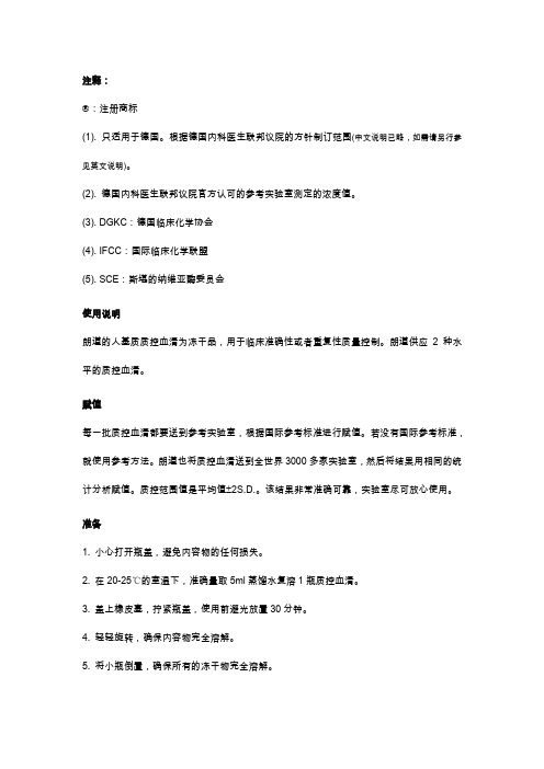
注释:
®:注册商标
(1). 只适用于德国。
根据德国内科医生联邦议院的方针制订范围(中文说明已略,如需请另行参见英文说明)。
(2). 德国内科医生联邦议院官方认可的参考实验室测定的浓度值。
(3). DGKC:德国临床化学协会
(4). IFCC:国际临床化学联盟
(5). SCE:斯堪的纳维亚酶委员会
使用说明
朗道的人基质质控血清为冻干品,用于临床准确性或者重复性质量控制。
朗道供应2种水平的质控血清。
赋值
每一批质控血清都要送到参考实验室,根据国际参考标准进行赋值。
若没有国际参考标准,就使用参考方法。
朗道也将质控血清送到全世界3000多家实验室,然后将结果用相同的统计分析赋值。
质控范围值是平均值±2S.D.。
该结果非常准确可靠,实验室尽可放心使用。
准备
1. 小心打开瓶盖,避免内容物的任何损失。
2. 在20-25℃的室温下,准确量取5ml蒸馏水复溶1瓶质控血清。
3. 盖上橡皮塞,拧紧瓶盖,使用前避光放置30分钟。
4. 轻轻旋转,确保内容物完全溶解。
5. 将小瓶倒置,确保所有的冻干物完全溶解。
6. 勿摇晃小瓶。
复溶后的血清既可以用于手工测试,也可以用于全自动生化分析仪。
该血清只能按照上述步骤复溶。
稳定性
该血清自生产之日起,在4℃下保存可以稳定4年。
效期标在试剂盒的侧面。
该血清一旦复溶,在25℃下可以稳定24小时,在4℃下可以稳定7天,在-20℃至少可以稳定1个月(见受限情况)。
5通道全自动乳化仪

全自动多通道抗原乳化系统,适用于生物科研机构生物公司制备抗体需要免疫动物时,自动将抗原与试剂制成乳剂,然后给动物注射,减轻了科研人员的工作负担,缓解了科研压力。
性能特点:1、自带水浴制冷全程保持低温操作,减少因摩擦发热产生的不良影响;2、自带升降平台可以实现人水分离,优化试验操作;3、高效省时能同时进行5组相同或者不同抗原的乳化操作;4、螺纹口连接不易脱落或崩坏,可以在高温高压清洗消毒后重复使用;5、一次性常规注射器耗材都是一次性常规注射器,干净无菌,经济实惠;6、全程无须人员看守仪器根据压力传感器自动判断乳化效果;7、任意设置推拉次数进行持续往复的混合反应,使乳化更均匀;8、多微孔连接通道能有效提高乳化效率和乳化质量;9、接头可单买乳化接头可以单独购买,满足客户需求。
南京巴傲得生物科技有限公司( Biogot technology, co, Ltd),是专一服务于生命科学研究的专业技术型企业。
是一大批志同道合的资深专业人士共同创办的具有现代化经营管理水平的生物高科技企业,从事生物技术产品的开发及销售,致力于将巴傲得生物打造成中国乃至世界一流的生物技术产品供应商。
巴傲得生物长期备有Bioworld抗体现货8000多种抗体可以在一周左右送到客户的手里。
精湛品质、合理价格、专业服务,是我们为您服务的宗旨。
在细胞信号通路、免疫学、蛋白组学上拥有显著优势,其抗体产品、生长因子等为生命科学科研工作者提供了极大的便利。
客户的满意、微笑和信任是对我们最大的鼓舞与激励,巴傲得生物愿以专业的知识、卓越的服务作为您事业腾飞的基石!The full-automatic multi-channel antigen emulsification system is suitable for automatically making the antigen and reagent into emulsion and then injecting the emulsion into the animal when preparing antibodies in biological research institutions and biological companies, which reduces the workload of researchers and relieves the pressure of scientific research.Performance characteristics:1. Bring your own water bath refrigerationKeep low temperature operation in the whole process to reduce the adverse effects caused by friction heating;2. Bring your own lifting platformIt can realize the separation of human and water and optimize the test operation;3. Efficient and time-saving20 groups of the same or different antigens can be emulsified simultaneously;4. Thread connectionIt is not easy to fall off or collapse, and can be reused after high temperature and high pressure cleaning and disinfection;5. Disposable conventional syringeConsumables are disposable conventional syringes, which are clean, sterile and economical;6. There is no need for personnel to guard the whole processThe instrument automatically judges the emulsification effect according to the pressure sensor;7, arbitrarily set the number of push and pullCarry out continuous reciprocating mixing reaction to make emulsification more uniform;8, microporous connection channelCan effectively improve emulsification efficiency and emulsification quality;9. The joint can be bought separatelyEmulsified joints can be purchased separately to meet customer requirements.Bioworld technology co., Ltd manufactures and supplies antibodies for worldwide distribution. We provide antibodies in bulk quantity and OEM basic services. The experienced scientific and technical professionals make Bioworld Technology a highly innovative, first-class biotechnological company on both manufacturing and marketing. Bioworld strives for a national leader and the world-class company in manufacturing biological products for industry, health, and research. We look forward to serving your research needs and becoming yourlong-term research partner.。
GMP常用英语单词

Abbreviated New drug简化申请的新药Accelerated approval加速批准Adverse effcet副作用Adverse reaction不良反应Agency审理部门ANDA(Abbreviated New drug application)简化新药申请Animal trial动物试验Archival copy存档用副本Batch production records生产批号记录Batch production批量生产CFR (Code of federal regulation )(美)联邦法规Clinical trial临床试验COS/CEP欧洲药典符合性认证Dietary supplement食品补充品DMF(Drug master file)药物主文件Drug substance原料药Generic name非专利名称ICH(International Conference onHarmonization of Technical Requirements for Registration of Pharmaceuticals for Human Use)人用药物注册技术要求国际协调会议IND(Investigation new drug)临床研究申请(指申报阶段,相对于NDA);研究中的新药(指新药开发阶段,相对于新药而言,即临床前研究结束)Informed consent知情同意INN(international nonproprietary name)国际非专有名称Investigator研究人员;调研人员Labeled amount标示量NDA(New drug application)新药申请NF(National formulary)(美)国家药品集NIH(National Institute of Health)(美)国家卫生研究所Panel专家小组preparing and Submitting起草和申报Prescription drug处方药Proprietary name专有名称Regulatory methodology质量管理方法Regulatory methods validation管理用分析方法的验证Regulatory specification质量管理规格标准Review copy审查用副本Sponsor主办者(指负责并着手临床研究者)Standard drug标准药物Strength规格;规格含量(每一剂量所含有效成分的量)Submission申报;递交Treatment IND研究中的新药用于治疗生产工艺相关Acceptance criteria可接受标准air driers手烘箱Airlock Room气闸室analytical methods分析方法anhydrous无水API原料药Assay 含量at rest静态batch size批量Blending Batches混批Blending Room总混间calibrating校正case-by-case具体分析centigrate摄氏度Changing Room更衣室Charge-in进料chemical properties化学性质Clarity,completeness,or PH of solutions溶液的澄明度、溶解完全性及PH值cleaning agents清洗媒介cleaning procedures清洁程序Cleaning Tools Room洁具室Coating Mixture Preparing Room配浆间Commercial scale可配伍性Concentrated Solution Room浓配室consistency of the process工艺的稳定性critical process关键步骤dedicated专用的Documentation System文件系统dosage form剂型electronic form电子格式electronicsignatures电子签名Emergency Door安全门established schedule预先计划Excipient辅料exhaust排气fermentation发酵Granulation颗粒HAVC(Heating ventilation and air conditioning)空调净化系统Heavy metal重金属historical date历史数据Hydrochloric acid盐酸in operation动态incoming materials进厂物料in-house testing内控检测installation qualification(IQ)安装确认intermediate中间体intermal audits self-inspection自检laboratory control record实验室控制记录laboratory information managementsystem(LIMS)实验室信息管理系统local authorities当地药政部门Loss on drying干燥失重Meet the requirement符合要求Melting point熔点Melting range熔程microbiological specifications微生物标准microorganisms微生物Milling磨粉Mix-ups混放modified facilities设施变更molecular formula分子式Non-dedicated equipment非专用设备Operational qualification(OQ)运行确认Out-of-specification不合格Packaging包装Particle size粒度Perform a blank determination作一个空白对照Personnel Hygiene人员卫生pilot scale中试规模potable water饮用水premises设施process parameters工艺参数Process validation工艺验证,过程验证product quality reviews产品质量回顾production batch records批生产记录proposed indication适应症purification纯化performance qualification(PQ)性能确认Process flow diagrams(PFDS)工艺流程图product validation产品验证regulatory inspection evaluation药政检查Related substance有关物质release放行Residual solvents残留溶剂retention periods保留期限Retention samples留样retention time保留时间Retrospective validation回顾性验证Revalidation再验证review and approve审核并批准route of administration给药途径Sanitation环境卫生scale-up reports报产报告serious GMP deficiencies严重GMP缺陷Sip sterilization in place在线灭菌sodium hydroxide氢氧化钠Specific rotation比旋度specifications标准stability date稳定性数据stability monitoring program稳定性监控计划status状态sterile APIs无菌原料药sterilization消毒succ essive batches连续批号supplier供应商technical transfer技术转化total microbial counts微生物总数traceable可追踪的turnover packages验证文件集Validation master plan验证总计划Validation report验证报告常用中译英系统system物料平衡reconciliation批batch or lot批号batch number批生产记录batch records文件document标准操作规程standard operating proceddures (SOP)生产工艺规程master formula工艺用水water for processing纯化水purified water注射用水water for injection状态标志status mark/label中间产品intermediate product理论产量theoretical yield物料material待验quarantine起始原料staring material洁净室(区)clean room(zone)待包品bulk product成品finished product灭菌sterilization控制点control point质量监督quality surveillance生产过程控制in-process control退货returned product拒收rejected交叉污染cross contamination放行released质量要求quality requirement可追溯性traceability计量确认metrologial confirmation人员净化室room for cleaning human body物料净化室room for cleaning material悬浮粒子airborne particles洁净度cleanliness净化cleaning传递箱pass box洁净服clean working garment洁净工作台clean bench静态at-rest动态operational粗效过滤器roughing filter中效过滤器medium efficiency filter高效过滤器hepa filter安装确认instalation qualification(IQ)运行确认operational qualification(OQ)性能确认performance qualification(PQ)工艺验证process validation。
人脂磷壁酸LTA试剂盒使用方法

人脂磷壁酸(LTA)试剂盒使用方法检测范围:96T20pg/ml-480pg/ml使用目的:本试剂盒用于测定人血清、血浆及相关液体样本中脂磷壁酸(LTA)含量。
实验原理本试剂盒应用双抗体夹心法测定标本中人脂磷壁酸(LTA)水平。
用纯化的人脂磷壁酸(LTA)抗体包被微孔板,制成固相抗体,往包被单抗的微孔中依次加入脂磷壁酸(LTA),再与HRP标记的脂磷壁酸(LTA)抗体结合,形成抗体-抗原-酶标抗体复合物,经过彻底洗涤后加底物TMB显色。
TMB在HRP酶的催化下转化成蓝色,并在酸的作用下转化成最终的黄色。
颜色的深浅和样品中的脂磷壁酸(LTA)呈正相关。
用酶标仪在450nm波长下测定吸光度(OD值),通过标准曲线计算样品中人脂磷壁酸(LTA)浓度。
试剂盒组成标本要求1.标本采集后尽早进行提取,提取按相关文献进行,提取后应尽快进行实验。
若不能马上进行试验,可将标本放于-20℃保存,但应避免反复冻融2.不能检测含NaN3的样品,因NaN3抑制辣根过氧化物酶的(HRP)活性。
操作步骤1.标准品的稀释:本试剂盒提供原倍标准品一支,用户可按照下列图表在小试管中进行2.加样:分别设空白孔(空白对照孔不加样品及酶标试剂,其余各步操作相同)、标准孔、待测样品孔。
在酶标包被板上标准品准确加样50μl,待测样品孔中先加样品稀释液40μl,然后再加待测样品10μl(样品最终稀释度为5倍)。
加样将样品加于酶标板孔底部,尽量不触及孔壁,轻轻晃动混匀。
3.温育:用封板膜封板后置37℃温育30分钟。
4.配液:将30倍浓缩洗涤液用蒸馏水30倍稀释后备用5.洗涤:小心揭掉封板膜,弃去液体,甩干,每孔加满洗涤液,静置30秒后弃去,如此重复5次,拍干。
6.加酶:每孔加入酶标试剂50μl,空白孔除外。
7.温育:操作同3。
8.洗涤:操作同5。
9.显色:每孔先加入显色剂A50μl,再加入显色剂B50μl,轻轻震荡混匀,37℃避光显色15分钟.10.终止:每孔加终止液50μl,终止反应(此时蓝色立转黄色)。
促黄体生成素检测试剂注册技术审查指导原则

促黄体生成素检测试剂注册技术审查指导原则促黄体生成素(luteinizing hormone,LH)是一种由前垂体腺紧张素释放激素(gonadotropin-releasing hormone,GnRH)调控的垂体激素,它在调控生殖系统中具有重要的功能。
促黄体生成素检测可以用于监测女性的生理状态,例如排卵、月经周期和疾病的诊断和治疗。
因此,促黄体生成素检测试剂的注册技术审查指导原则(2024年第213号)是监管部门对该类产品进行注册审核时的技术指导原则。
促黄体生成素检测试剂(胶体金免疫层析法)是一种常用的促黄体生成素检测方法,该方法基于抗原抗体反应原理,通过检测样品中的促黄体生成素含量,来确定样品中的生理状态。
因此,对于该类检测方法的注册技术审查,有以下指导原则:1.技术原理和方法说明:注册申请人应明确说明检测方法的原理和步骤,包括样品预处理、准备试剂、反应体系、信号检测等关键步骤,确保方法的准确性和可靠性。
2.试剂的质量控制:注册申请人应提供检测试剂的生产设施和质量控制体系,包括原辅材料的选择和采购、生产工艺的控制和标准化等,确保试剂的质量符合规定标准。
3.检测灵敏度和特异性:注册申请人应提供方法对促黄体生成素的检测灵敏度和特异性的验证数据,以及与其他常用检测方法的比较数据,确保该方法能够准确和特异地检测促黄体生成素。
5.没有干扰物:注册申请人应提供方法对于可能存在的干扰物(如高浓度抗体、其他类似分子)的抗干扰数据,以确保该方法在实际应用中的可靠性和准确性。
6.校准方法和质量控制策略:注册申请人应提供检测试剂的标准品溯源和校准方法,以及合理的质量控制策略,保证检测结果的准确性和可靠性。
7.临床性能验证:注册申请人应提供方法在不同人群样本中的临床性能验证数据,包括不同性别、年龄、健康状况等人群中的测试结果,以评估方法的准确性和可靠性。
8.使用说明和结果解读:注册申请人应提供使用说明书和结果解读指南,包括样品采集和处理方法、结果解读的参考范围和标准等,以确保用户能够正确使用和解读检测试剂。
- 1、下载文档前请自行甄别文档内容的完整性,平台不提供额外的编辑、内容补充、找答案等附加服务。
- 2、"仅部分预览"的文档,不可在线预览部分如存在完整性等问题,可反馈申请退款(可完整预览的文档不适用该条件!)。
- 3、如文档侵犯您的权益,请联系客服反馈,我们会尽快为您处理(人工客服工作时间:9:00-18:30)。
to structural analysis,including other ferritins that could not be solved by x-ray crystallography. During the first4e–/Å2on gold substrates,1to 2Åof in-plane motion remain.These first few electrons are critical,as they potentially contain the most high-resolution information(26,27). Future work will focus on substrate design and image acquisition conditions to reduce the initial motion even more.Along with further improve-ments in electron detector efficiency,this will bring cryo-EM to the physical limits imposed by the homogeneity of the protein preparation, the electron scattering cross sections of the bio-logical specimen(4),and the counting statistics of detecting individual electrons.We anticipate that high-efficiency,stationary particle imaging will allow:(i)measurement and modeling of the progressive damage to the primary and second-ary structure of the molecules,(ii)improved re-finements using dose-fractionation that includes the use of tilt,and(iii)direct modeling and re-finement of molecular structure from particle images.This will enable routine structure deter-mination for many molecules and complexes that are currently too difficult to be practical. REFERENCES AND NOTES1. A.Amunts et al.,Science343,1485–1489(2014).2.M.Liao,E.Cao,D.Julius,Y.Cheng,Nature504,107–112(2013).3.M.Allegretti,ls,G.McMullan,W.Kühlbrandt,J.Vonck,eLife3,e01963–e01963(2014).4.R.Henderson,Q.Rev.Biophys.28,171–193(1995).5.P.B.Rosenthal,R.Henderson,J.Mol.Biol.333,721–745(2003).6.R.Henderson,G.McMullan,Microscopy62,43–50(2013).7. A.Faruqi,R.Henderson,M.Pryddetch,P.Allport,A.Evans,Nucl.Instrum.Methods546,170–175(2005).8.R.Henderson,R.M.Glaeser,Ultramicroscopy16,139–150(1985).9. E.R.Wright,C.V.Iancu,W.F.Tivol,G.J.Jensen,J.Struct.Biol.153,241–252(2006).10.R.M.Glaeser,G.McMullan,A.R.Faruqi,R.Henderson,Ultramicroscopy111,90–100(2011).11. A.F.Brilot et al.,J.Struct.Biol.177,630–637(2012).12.G.McMullan,S.Chen,R.Henderson,A.R.Faruqi,Ultramicroscopy109,1126–1143(2009).13.G.McMullan,A.R.Faruqi,D.Clare,R.Henderson,Ultramicroscopy147,156–163(2014).14.M.G.Campbell et al.,Structure20,1823–1828(2012).15.X.-C.Bai,I.S.Fernandez,G.McMullan,S.H.Scheres,eLife2,e00461(2013).16.X.Li et al.,Nat.Methods10,584–590(2013).17. C.J.Russo,L.A.Passmore,Nat.Methods11,649–652(2014).18.D.Rhinow,W.Kühlbrandt,Ultramicroscopy108,698–705(2008).19.C.Yoshioka,B.Carragher,C.S.Potter,Microsc.Microanal.16,43–53(2010).20.C.J.Russo,L.A.Passmore,J.Struct.Biol.187,112–118(2014).21.S.H.Banyard,D.K.Stammers,P.M.Harrison,Nature271,282–284(1978).22.R.R.Crichton,J.-P.Declercq,Biochim.Biophys.Acta1800,706–718(2010).23.W.H.Massover,Micron24,389–437(1993).24.S.H.W.Scheres,J.Struct.Biol.180,519–530(2012).25.N.de Val,J.-P.Declercq,C.K.Lim,R.R.Crichton,J.Inorg.Biochem.112,77–84(2012).26.W.Baumeister,M.Hahn,J.Seredynski,L.M.Herbertz,Ultramicroscopy1,377–382(1976).27.H.Stark,F.Zemlin,C.Boettcher,Ultramicroscopy63,75–79(1996).ACKNOWLEDGMENTSWe thank R.Henderson for guidance and advice throughout this project;ls and W.Kühlbrandt for the use of the Polara electron microscope at the Max Planck Institute of Biophysics, Frankfurt;I.S.Fernandez and the V.Ramakrishnan lab for the gift of ribosomes;G.McMullan,S.Chen,C.Savva,J.Grimmett,T.Darling,and M.Skehel for technical assistance;our colleagues at the Laboratory of Molecular Biology—especially S.Scheres, G.Murshudov,and R.A.Crowther—for many helpful discussions;and R.A.Crowther,V.Ramakrishnan,and E.Rajendra for a criticalreading of the manuscript.This work was supported by theEuropean Research Council(ERC)under the European Union’sSeventh Framework Programme(FP7/2007-2013)/ERC grantagreement no.261151and MRC grant U105192715.C.J.R.and L.A.P.are inventors on a patent application filed by theMRC on the gold substrates.The coordinates of the refinedapoferritin model are deposited in the Protein Data Bank underaccession code4v1w and the EM density map is depositedSUPPLEMENTARY MATERIALS/content/346/6215/1377/suppl/DC1Materials and MethodsFigs.S1to S10Table S1References(28–40)Movie S14August2014;accepted13November2014 glycoprotein(Env)elicits a polyclonal an-tibody response that targets diverse epi-topes(1,2).Antibodies that display narrowbreadth of neutralization(narrow neutral-izing antibodies;nNAbs)develop during the firstmonths of infection whereas those capable of neu-tralizing heterologous viruses(broadly neutralizingantibodies;bNAbs)develop several years later in~10to30%of HIV-1–positive individuals(3).bNAbsisolated from HIV-1–infected patients are more pro-tective than nNAbs in experimental HIV-1/SHIVinfection(4)and will likely be a key component ofan effective HIV-1vaccine.Even though nNAbs andbNAbs target the same regions of Env(2,5–7),re-combinant Env(rEnv)immunogens are poorly rec-ognized by germline-reverted(gl)bNAbs(glbNAbs)and their corresponding B cell receptors(BCRs)(5,8–21),suggesting that the lack of bNAb gen-eration during immunization may be due toinefficient stimulation of naïve bNAb BCR pro-genitors(17,20).In contrast,little is known aboutgenitors of nNAbs.Understanding why B cellresponses against nNAb epitopes dominate overthose targeted by bNAbs in the context of rEnvimmunization will inform on basic immunolog-ical mechanisms of epitope competition and pro-vide new information relevant to the developmentof an effective HIV-1vaccine.Here we investigated whether glnNAbs fromdistinct clonal lineages that targeted the CD4-binding site(BS)and V3regions of Env(2)alsodisplay minimal rEnv recognition.Amino acid dif-ferences between the mutated and gl sequences ofnNAbs range from2.4to7.3%for the heavy chainsand2.7to5.6%for the light chains for the nNAbCD4-BS antibodies(table S1and fig.S1).In con-trast,prototypic CD4-BS bNAbs,VRC01(33.9%heavy,23%light),NIH45-46(a clonal relative ofVRC01;39.8%heavy,26.1%light),b12(21%heavy,19%light),8ANC131(33%heavy,24%light),andCH103(12.7%heavy,10%light)are more mutated(5,8,16,22).The anti-V3nNAbs are more mutated(11.6to21.6%heavy,9.7to13.8%light)than theanti-CD4-BS nNAbs.In contrast to the anti-CD4-BS glbNAbs,whichdo not bind rEnv(5,8,16,17,20)(table S2),glnNAbsdisplayed broad Env recognition(from51to100%)(table S2).The binding affinities of the glnNAbswere generally weaker than those of the corre-sponding mutated antibodies,owing to increasedoff rates in most cases(fig.S2).Whereas the glVRC01class bNAbs were unable to neutralize any of the 1Seattle Biomedical Research Institute,Seattle,WA98109,USA.2Laboratory of Molecular Immunology,The RockefellerUniversity,New York,NY10065,USA.3Laboratory of HumoralResponse to Pathogens,Department of Immunology,InstitutPasteur and CNRS-URA1961,75015Paris,France.4Departmentof Global Health,University of Washington,Seattle,WA98109,USA.*These authors contributed equally to this work.†Present address:Fred Hutchinson Cancer Research Center,Seattle,WA98109,USA.‡Present address:GlycoVaxyn,Grabenstrasse3,CH-8952Schlieren,Switzerland.§Corresponding author.E-mail:lstamata@viruses tested,three of the five glnNAbs exhibited neutralizing activity against tier1viruses(table S3). Overall,we conclude that the glnNAbs and glbNAbs recognize the CD4-BS on soluble and virion-associated Env differently(23,24).Two of the three anti-V3glnNAbs displayed neutralizing activity against several tier1viruses(table S3).We next investigated whether B cells stably expressing glnNAb and glVRC01-class BCRs(fig. S3)could become activated by(Fig.1A)and in-and C.As previously reported,none of the rEnvstested activated glVRC01-class B cells(17,20);however,they did activate glnNAb B cells tar-geting either the CD4-BS or V3(Fig.1A).Sim-ilarly,glnNAb B cells readily internalized diverserEnvs,whereas glVRC01class B cells did not(Fig.1B).Combined,the above results indicate thatrEnv immunogens can activate naïve nNAb Bcells but not naïve VRC01-class B cells.We recently reported that the disruption ofN460D,and N463D—on the clade C426c rEnv(herein called426c.NLGS.TM)confers binding toand activation of glNIH45-46and glVRC01B cells(17).In contrast,wild-type(WT)426c Env is rec-ognized by only the glnNAbs(table S2).gl1-154,gl1-695,gl1-732,and gl4-341nNAbs inhibitedbinding of glNIH45-46to426c.NLGS.TM(fig.S5A),an indication that the epitopes of the anti-CD4-BS glnNAbs used here overlap those of theVRC01-class glbNAbs.The anti-V3gl2-59mono-germline reverted antibodies to trimeric426c Env gp140variants measured by BLI.NA:binding was undetectable.426c NLGS.TM trimeric gp140426c NLGS.TM trimeric D V1D2D3 Apparent K A(1/M)k on(1/Ms)K off(1/s)Apparent K A(1/M)K on(1/Ms)K off(1/s)2.85×108 2.96×10⁴ 1.04×10⁻⁴ 1.77×10⁸ 1.57×1048.85×10–56.30×1087.48×104 1.19×10⁻⁴ 2.65×1089.71×103 3.66×10–54.12×107 1.61×10⁴ 3.91×10⁻⁴ 3.31×1067.73×1035.88×10–46.77×1079.89×103 1.46×10⁻⁴ 2.24×10⁷ 4.66×10³ 2.08×10⁻⁴NA NA NA NA NA NA4.04×1079.87×103 2.44×10⁻⁴ 3.52×10⁸ 1.40×10⁴ 3.98×10–52.67×108 5.51×10³ 2.06×10⁻⁴ 2.64×1089.07×1033.43×10–5NA NA NA NA NA NA1.19×1011 1.85×10⁵ 1.55×10⁻⁶NA NA NANA NA NA NA NA NAFig.2.Activation by and internalization of Envby glnNAb and glVRC01-class B cells in responseto modified Env immunogens.(A)glNIH45-46or glVRC01B cells were mixed with the indicatedglnNAb B cells.The pooled B cells were then chal-lenged with WT426c(top row),426c.NLGS.TM(mid-dle row),or426c.NLGS.TM.D V1-3(bottom row)at a1m M final concentration.Calcium flux was monitoredconcurrently in both B cell lines with the strategydepicted in fig.S6.Black arrow indicates time of Envaddition.(B)Area under the calcium flux curve de-termined from duplicate experiments in(A).Errorbars represent SD from the mean of10(glVRC01-class)or4replicates(other B cell lines).Green as-terisks and blue stars over the bars indicatesignificant differences(P<0.05)from glNIH45-46and glVRC01B cells,respectively,for each proteinusing a two-tailed Student’s t test.(C)Quantifica-tion of internalization of the indicated Envs byglVRC01class and glnNAb B cells.Error bars rep-resent SD from the mean of two independent ex-periments each performed in duplicate(n=4).Green astrices and blue stars represent significantdifferences(P<0.05)from glNIH45-46and glVRC01,respectively,for each protein using a two-tailedStudent’s t test.Fig.3.B cell activation and antigen uptake of Env by glVRC01-class B cellsin the presence of soluble anti-HIVantibodies.(A)Calcium flux was monitored in glNIH45-46(solid bars)or glVRC01(patterned bars)B cells incubated with 426c.NLGS.TM(white bars)or with426c.NLGS.TM.D V1-3(gray bars)in the presence of equimolar concentrations of the indicated antibodies.Bars repre-sent mean and SD of the area under the calcium flux response curve relative to that of Env in the absence of soluble mAb from three independent experiments. Asterisks represent a significant difference(P<0.05)in the calcium flux re-sponse between426c.NLGS.TM and426c.NLGS.TM.D V1-3using a two-tailed Student’s t test.Env internalization by glNIH45-46(B)or glVRC01(C)B cells incubated with426c.NLGS.TM(white bars)or426c.NLGS.TM.D V1-3(gray bars) in the absence or presence of equimolar concentrations of the indicated nNAbs. Bars represent the mean of two independent experiments.Asterisk denotes significant difference(P<0.05)in fluorescence compared to cells incubated with Env alone using a two-tailed Student’s t test in(B)and(C).Dotted line corresponds to mean426c.NLGS.TM internalization by B cells in the absence of Ab in(B)and(C).in the binding of glNIH45-46(fig.S5A).The gl1-676,gl1-79,and gl2-1261antibodies,which do not bind426c.NLGS.TM(Table1),had no effect on the binding of glNIH45-46(fig.S5A).In germinal centers(GCs),B cells expressing higher-affinity BCRs selectively expand,whereas lower-affinity B cells are eliminated(25).The anti-CD4-BS gl1-154,gl1-695,gl1-732,and gl4-341,and the anti-V3gl2-59bound426c.NLGS.TM with a higher apparent affinity than glNIH45-46(Table1).glVRC01 bound more strongly to426c.NLGS.TM than gl1-154 and gl1-695,but had a slightly lower apparent affinity than the gl4-341and gl1-732mAbs.The gl2-59V3–directed mAb bound more strongly to 426c.NLGS.TM than any of the glCD4-BS Abs,con-sistent with the higher propensity of gl2-59B cells to be activated and take up rEnv(Fig.1).We antic-ipate that upon immunization with the426c.NLGS. TM,naïve2-59–like B cells,or B cells that target immunodominant epitopes on the variable regions of Env(V1,V2and V3),would have an advantage over naïve VRC01-class B cells in becoming acti-vated,taking up antigen,and obtaining T cell help. To investigate the effect that differences in binding affinity have on the relative activation of B cells expressing different anti-Env BCRs,we de-veloped an assay that allows for the concurrent monitoring of B cell activation in distinct B cell populations(fig.S6).As expected,WT426c ac-tivated glnNAb B cells(Fig.2A top row,and Fig. 2B),but not glVRC01-class B cells(Fig.2A top row, and Fig.2B).426c.NLGS.TM activated gl VRC01-class B cells,but with the exception of gl1-154,it activated glnNAb B cells more strongly(Fig.2A, middle row).426c.NLGS.TM activated gl1-695more strongly than glNIH45-46and comparably to glVRC01(Fig.2A,middle row,and Fig.2B). Because426c.NLGS.TM showed the strongest activation of gl2-59B cells,we aimed to abolish ac-tivation of B cells targeting the V1,V2,and V3re-gions of426c.NLGS.TM,by deleting these regions to create426c.NLGS.TM.D V1-3.As expected,426c. NLGS.TM.D V1-3did not bind to gl2-59mAb(Table1) and did not activate gl2-59B cells(Fig.2,A and B).With the exception of glNIH45-46,deletion of the variable loops reduced the apparent binding affinities of the anti-CD4-BS antibodies tested (Table1).Consistent with these changes in bind-ing,426c.NLGS.TM.D V1-3activated glNIH45-46B cells more robustly than426c.NLGS.TM(Fig.2,A and B).The relative activation of all but one anti-CD4-BS glnNAb B cell(gl4-341)by426c.NLGS. TM.D V1-3was less than that observed for glVRC01-class B cells(Fig.2,A and B).gl4-341B cells were activated comparably to glVRC01class B cells, although the activation was reduced relative to that observed with WT426c and426c.NLGS.TM (Fig.2,A and B).At lower concentrations of426c. NLGS.TM.D V1-3,only glVRC01-class and gl4-341 B cells were activated(fig.S7).To represent a more physiological B cell repertoire,we challenged glVRC01-class B cells mixed with an excess of pooled glnNAb,and non-Env reactive B cells (fig.S8).WT426c only activated nNAb B cells, and although426c.NLGS.TM could activate the minority glVRC01-class B cells,a preferential ac-tivation of the nNAb B cell pool was observed.In contrast,426c.NLGS.TM.D V1-3activated glVRC01-class B cells preferentially at higher concentrationsand exclusively at lower concentrations(fig.S8).When the internalization of rEnv by B cells wasquantitated,426c NLGS.TM was preferentiallyinternalized by nNAb B cells,whereas426cNLGS.TM.D V1-3was preferentially internalizedby glVRC01-class and gl4-341B cells(Fig.2C).The differential impact of removing the variableEnv regions on the binding affinities,B cell activa-tion,and antigen internalization for the bNAbsversus nNAbs to the CD4-BS is likely due to differ-ences in the angle of approach to the CD4-BS bythese two types of antibodies(16,22,24,26–29).Additionally,the nNAbs may make contacts withportions of the variable loops.Antibodies can enter GCs and inhibit BCRbinding to an immunogen unless the affinity ofthe BCR for the immunogen is higher than forthe soluble antibody(30).Through this feedbackmechanism,higher-affinity antibodies hinderthe ability of low-affinity,antigen-specific B cellsto receive T cell help and thus select for higher-affinity B cell clones(30).Because the apparentaffinities of glVRC01-class bNAbs for426c.NLGS.TM D V1-3are higher than those for the glnNAbsstudied here(Table1),we expect that such afeedback mechanism will provide a selectiveadvantage to glVRC01-class B cell responses ifthis rEnv is used as an immunogen.Inversely,owing to the higher apparent affinities of theglnNAbs for the426c.NLGS.TM relative to theglVRC01class bNAbs(Table1),we would expectthis immunogen to drive the development ofnNAb responses through the same antibody-feedback mechanism.Indeed,the binding ofanti-CD4-BS nNAbs to426c.NLGS.TM reducedthe activation of glVRC01-class B cells by thatrEnv(Fig.3A and fig.S9).In contrast,activationof glVRC01class B cells by426c.NLGS.TM.D V1-3rEnv was observed even in the presence of thegl1-154,and gl1-695Abs(Fig.3A and fig.S9).Thus,removal of the V1,V2,and V3regions reduces theability of some(but not all)anti-CD4nNAbs toblock rEnv activation of glVRC01class B cells.Similar observations were made when theinternalization of these two Envs by glVRC01class B cells was determined in the absence andpresence of glnNAbs(Fig.3,B and C).glNIH45-46B cells more readily internalized426c.NLGS.TM.D V1-3compared to426c.NLGS.TM,even inthe presence of inhibitory antibodies,with theexception of the gl4-341mAb(Fig.3B,comparewhite and gray bars).Overall,we conclude thatthe presence of anti-CD4-BS glnNAbs can inhibitthe activation of,and the antigen internalizationby,glVRC01-class B cells in response to the426c.NLGS.TM,but that these effects are less pro-nounced when the variable regions V1,V2,andV3are removed from this rEnv.Thus,selectingwhich immunogen is used during the primingphase of immunization(e.g.,426c.NLGS.TM versus426c.NLGS.TM.D V1-3)will be critically importantto the eventual elicitation of anti-CD4-BS bNAbs.In sum,our study provides a mechanistic expla-nation as to why rEnv immunogens elicit narrowrather than broadly neutralizing antibody re-sponses to the CD4-BS of Env.This is primarily dueto the broad Env-recognition properties of glnNAbBCRs,which contrasts with the inefficiency by whichbNAb BCRs recognize rEnv.Notably,we demon-strate that rational immunogen modifications canreduce(and in certain cases eliminate)the activa-tion of naïve B cells that give rise to such nNAbs,while promoting the activation of naïve B cellsthat give rise to glVRC01-class bNAbs,even whenthe epitope of these antibodies substantially over-lap.As such,our results are relevant to efforts toelicit protective antibody responses against notonly HIV-1but potentially other pathogens as well.REFERENCES AND NOTES1.H.Mouquet et al.,PLOS ONE6,e24078(2011).2.J.F.Scheid et al.,Nature458,636–640(2009).3.L.Stamatatos,L.Morris,D.R.Burton,J.R.Mascola,Nat.Med.15,866–870(2009).4. F.Klein et al.,Science341,1199–1204(2013).5.J.F.Scheid et al.,Science333,1633–1637(2011).6.J.E.Robinson et al.,J.Virol.84,3443–3453(2010).7. A.Changela et al.,J.Virol.85,2524–2535(2011).8.S.Hoot et al.,PLOS Pathog.9,e1003106(2013).9.X.Xiao,W.Chen,Y.Feng,D.S.Dimitrov,Viruses1,802–817(2009).10.X.Xiao et al.,mun.390,404–409(2009).11.M.Huber et al.,J.Virol.84,10700–10707(2010).12.H.Mouquet et al.,Proc.Natl.Acad.Sci.U.S.A.109,E3268–E3277(2012).13.D.Sok et al.,PLOS Pathog.9,e1003754(2013).14.M.Bonsignori et al.,J.Virol.85,9998–10009(2011).15.B.J.Ma et al.,PLOS Pathog.7,e1002200(2011).16.T.Zhou et al.,Science329,811–817(2010).17.A.T.McGuire et al.,J.Exp.Med.210,655–663(2013).18.T.Ota et al.,J.Immunol.189,4816–4824(2012).19.K.J.Doores et al.,J.Virol.87,2234–2241(2013).20.J.Jardine et al.,Science340,711–716(2013).21.A.T.McGuire,J.A.Glenn,A.Lippy,L.Stamatatos,R.W.Doms,J.Virol.88,2645–2657(2014).22.H.-X.Liao et al.,Nature496,469–476(2013).23.R.W.Sanders et al.,PLOS Pathog.9,e1003618(2013).24.D.Lyumkis et al.,Science342,1484–1490(2013).25.G.D.Victora,M.C.Nussenzweig,Annu.Rev.Immunol.30,429–457(2012).26.T.Zhou et al.,Nature445,732–737(2007).27.L.Chen et al.,Science326,1123–1127(2009).28.R.Diskin et al.,Science334,1289–1293(2011).29.T.Zhou et al.,Immunity39,245–258(2013).30.Y.Zhang et al.,J.Exp.Med.210,457–464(2013).31.Cells expressing the g1-732BCR did not internalize the clade AQ168a2and Q461e2rEnvs that activated B cells expressingthat BCR.This discrepancy can be attributed to pHrodolabeling of these two rEnvs,which reduced the binding of thegl1-732mAb(fig.S4).ACKNOWLEDGMENTSWe thank M.Nussenzweig for providing the nNAb antibody expressionplasmids,J.Mascola and X.Wu for providing the IgG1glVRC01antibody,D.Rawlings for providing the pRRL expression plasmids,andL.Scharf and P.Bjorkman for technical assistance.The datapresented in this paper are found in the main paper and in thesupplementary materials.Accession numbers for the mutated andgermline-reverted nNAbs used in this study can be found at GenBank:KM595240-KM595271.This work was supported by NIH grants P01AI094419-01and U1919AI109632-01(L.S.),Canadian Institutes ofHealth Research fellowship(A.T.M),and Swiss National ScienceFoundation fellowship PBBSP3_144245and P300P3_151140(A.M.D.).SUPPLEMENTARY MATERIALS/content/346/6215/1380/suppl/DC1Materials and MethodsAuthor ContributionsFigs.S1to S9Tables S1to S3References(32–43)25July2014;accepted13November201410.1126/science.1259206。
