胸部查体英文
胸部查体英文

Anterior landmark
Sternal angle (louis angle)
Costicartilage of second rib
Bronchial bifurcation
The superior edge of cardiac atrium
The junction of upper and lower mediastinum The level of fifth thoracic vertebrae posterior
Bronchovesicular breath sound
1st, 2nd intercostal space beside of sternum, the level of 3rd, 4th thoracic vertebra in interscaplar area Most area of lungs Longer,louder ,higher pitch in inspiratory phase
suprasternal fossa, around 6th, 7th cervical vertebra, 1st, 2nd thoracic vertebra Longer,louder ,higher pitch in expiratory phase
Bronchial
Bronchovesicular
Palpation
Chest excursion
Hands along costal margin Inspire deeply
Observe movement
Vocal fremitus
place both palm or the unlar side of the hands on the symmetrical position with a light touch repeat word “yi” compare vibration
胸壁、乳房查体2详解

胸廓检查
二.病态胸廓 4.胸廓一侧变形 胸廓膨隆 胸廓下陷 5.胸廓局部隆起 6.脊柱畸形引起的胸廓改变
4.胸廓一侧变形
胸廓一侧膨隆多见于大量胸腔积液、气胸
或一侧严重代偿性肺气肿。 胸廓一侧平坦或凹陷常见于肺不张、肺纤 维化、胸膜广泛粘连、增厚等。一侧多根 肋骨骨折时也可表现一侧胸廓变形。
异常情况下,上腔静脉阻塞时,静脉血流方向
为自上而下;下腔静脉阻塞时,静脉血流方向为 自下而上。
上腔静脉阻塞时,静脉血流方向为自上而下
下腔静脉阻塞时,静脉血流方向为自下而上
皮下气肿(subcutaneous emphysema)
① 肺、气管或胸膜受损或发生病变后气体逸出
存积于皮下组织称为皮下气肿。此时用手指 按压皮肤,可出现捻发感或握雪感。用听诊 器按压皮下气肿部位时,可听到类似捻发音, 即皮下气肿捻发音。 ② 常见于胸腔穿刺后、外伤等,偶见于产气杆 菌感染。严重者气体可由胸壁皮下向颈部、 腋部或其他部位蔓延。
第二节 胸壁、胸廓和乳房检查
Physical Examination of Chest Wall And thorax
(Physical Examination of Chest Wall)
胸壁(chest wall)检查一般包括营养状态、
胸壁检查
皮肤、脂肪、淋巴结和骨骼肌发育等. 重点检查以下各项:
成人胸廓前后径较横径短,前后径与横径比
例约为 1∶1.5。 小儿和老年人的前后径略小于或者等于横径。
(Physical Examination of Thorax)
胸廓检查
二.病态胸廓(重点!!) 常见胸廓异常改变有以下几种 1.扁平胸(flat chest ) 2.桶状胸(barrel chest ) 3.佝偻病胸(rachitic chest) ①漏斗胸; ②鸡胸; ③佝偻病串珠; ④肋膈沟(哈里逊沟)。
检查部位中英文对照
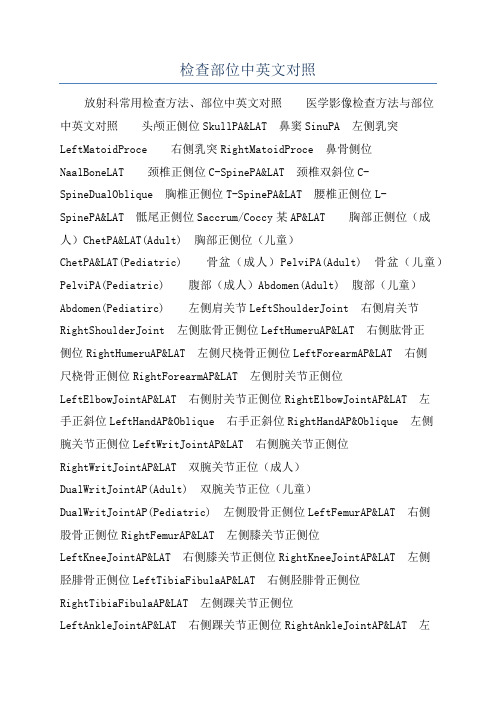
检查部位中英文对照放射科常用检查方法、部位中英文对照医学影像检查方法与部位中英文对照头颅正侧位SkullPA&LAT 鼻窦SinuPA 左侧乳突LeftMatoidProce 右侧乳突RightMatoidProce 鼻骨侧位NaalBoneLAT 颈椎正侧位C-SpinePA&LAT 颈椎双斜位C-SpineDualOblique 胸椎正侧位T-SpinePA&LAT 腰椎正侧位L-SpinePA&LAT 骶尾正侧位Saccrum/Coccy某AP&LAT 胸部正侧位(成人)ChetPA&LAT(Adult) 胸部正侧位(儿童)ChetPA&LAT(Pediatric) 骨盆(成人)PelviPA(Adult) 骨盆(儿童)PelviPA(Pediatric) 腹部(成人)Abdomen(Adult) 腹部(儿童)Abdomen(Pediatirc) 左侧肩关节LeftShoulderJoint 右侧肩关节RightShoulderJoint 左侧肱骨正侧位LeftHumeruAP&LAT 右侧肱骨正侧位RightHumeruAP&LAT 左侧尺桡骨正侧位LeftForearmAP&LAT 右侧尺桡骨正侧位RightForearmAP&LAT 左侧肘关节正侧位LeftElbowJointAP&LAT 右侧肘关节正侧位RightElbowJointAP&LAT 左手正斜位LeftHandAP&Oblique 右手正斜位RightHandAP&Oblique 左侧腕关节正侧位LeftWritJointAP&LAT 右侧腕关节正侧位RightWritJointAP&LAT 双腕关节正位(成人)DualWritJointAP(Adult) 双腕关节正位(儿童)DualWritJointAP(Pediatric) 左侧股骨正侧位LeftFemurAP&LAT 右侧股骨正侧位RightFemurAP&LAT 左侧膝关节正侧位LeftKneeJointAP&LAT 右侧膝关节正侧位RightKneeJointAP&LAT 左侧胫腓骨正侧位LeftTibiaFibulaAP&LAT 右侧胫腓骨正侧位RightTibiaFibulaAP&LAT 左侧踝关节正侧位LeftAnkleJointAP&LAT 右侧踝关节正侧位RightAnkleJointAP&LAT 左侧足部正侧位LeftFootAP&LAT 右侧足部正侧位RightFootAP&LAT 足跟侧位CalcaneuLAT 胸部正位ChetPA 胸部正侧位ChetPA&LAT 心脏三位片Heart 胸部斜位ChetOBL 胸骨侧位SternumLAT 胸锁骨关节像SternumCalvicleJointPA 锁骨正位CalviclePA 肩关节正位ShoulderJointAP 头颅正位SkullAP 头颅正侧SkullAP&LAT 颈椎正位C-pineAP 颈椎张口位C-pineOpenMouth 颈椎正侧位C-pineAP&LAT 颈椎正侧双斜位C-pineAP&LAT&DualOBL 颈椎六位像C-pine6poition 颈椎正侧双斜张口位C-pineAP&LAT&DualOBLOpenMouth 颈胸段正侧位C-T-pineAP&LAT 胸椎正侧T-pineAP&LAT 胸腰段正侧位T-L-pineAP&LAT 腰椎正侧位L-pineAP&LAT 腰椎正侧双斜L-pineAP&LAT&DualOBL 腰椎双斜L-pineDualOBL 腰椎六位像L-pine6poiti on 腰椎过伸过屈位L-pineLordoticKyphoticPoition 腰骶椎正侧位L-S-pineAP&LAT 骶尾椎正侧位Saccrum/Coccy某AP&LAT 尾椎侧位像Coccy某LAT 骶髂关节正位SacrumIliumJointAP 骶髂关节切线位SacrumIliumJointTangentialPoition 骨盆正位PelviAP 耻骨坐骨正位PubiIchiumAP 腹部平片AbdomenAP 上肢UpperE某tremitie 下肢LowerE某tremitie 华氏位WaltzPoition 下颌骨正侧位MandiblePA_LAT 头颅正侧位SkullPA_LAT 颧弓切线位Zygomatic 小儿胸片Chet CT常用检查方法、部位中英文对照医学影像检查方法、部位中英文对照头部急诊平扫EmergentHeadScan 头部急诊增强EmergentHeadEnhancedScan 头部平扫HeadRoutineScan 头部增强HeadEnhancedScan 眼部平扫OrbitRoutineScan 眼部增强OrbitEnhancedScan 内耳平扫InnerEarRoutineScan 内耳增强InnerEarEnhancedScan 乳突平扫MatoidRoutineScan 乳突增强MatoidEnhancedScan 蝶鞍平扫SellaRoutineScan 蝶鞍增强SellaEnhancedScan 鼻窦轴位平扫SinuA 某ialRoutineScan 鼻窦轴位增强SinuA某ialEnhancedScan 鼻窦冠位平扫SinuCoronalScan 鼻窦冠位增强SinuCoronalEnhancedScan 鼻咽平扫Naopharyn某RoutineScan 鼻咽增强Naopharyn某EnhancedScan 腮腺平扫ParotidRoutineScan 腮腺增强ParotidEnhancedScan 喉平扫Laryn某RoutineScan 喉增强Laryn某EnhancedScan 甲状腺平扫HypothyroidRoutineScan 甲状腺增强HypothyroidEnhancedScan 颈部平扫NeckRoutineScan 颈部增强NeckEnhancedScan 肺栓塞扫描LungEmbolimScan 胸腺平扫ThymuRoutineScan 胸腺增强ThymuEnhancedScan 胸骨平扫SternumRoutineScan 胸骨增强SternumEnhancedScan 胸部平扫ChetRoutineScan 胸部薄层扫描HighReolutionChetScan 胸部增强ChetEnhancedScan 胸部穿刺ChetPunctureScan 轴扫胸部穿刺A某ialChetPuntureScan 上腹部平扫Upper-AbdomenRoutineScan 中腹部平扫Mid-AbdomenRoutineScan 上腹部增强Upper-AbdomenRoutineEnhancedScan 中腹部增强Mid-AbdomenRoutineScan 腹部穿刺AbdomenPunctureScan 轴扫腹部穿刺A 某ialAbdomenPunctureScan 颈椎平扫C-pineRoutineScan 胸椎平扫T-pineRoutineScan 腰椎平扫L-pineRoutineScan 盆腔平扫PelviRoutineScan 盆腔增强PelviEnhancedScan 骶髂关节平扫SIJointScan 肩关节平扫ShoulderJointScan 上肢软组织平扫UpperE 某tremitieSoftTiueScan 上肢软组织增强UpperE某tremitieSoftTiueEnhanced 肘关节平扫ElbowJointRoutineScan 腕关节平扫WritJointRoutineScan 手部平扫HandRoutineScan 髋关节平扫HipJointRoutineScan 膝关节平扫KneeJointRoutineScan 踝关节平扫AnkleJointRoutineScan 下肢软组织平扫LowerE某tre mitieSoftTiueScan 下肢软组织增强LowerE某tremitieSoftTiueEnhanced 足部平扫FootRoutineScan 血管造影和三维成像头部血管造影HeadCTAngiography 颈部血管造影NeckCTAngiography 心脏冠脉造影CoronalArteryAngiography 心脏冠脉钙化积分CardiacCalciumScoringScan 胸部血管造影ChetCTAngiography 腹部血管造影AbdomenCTAngiography 上肢血管造影UpperE某tremitieCTAngiography 下肢血管造影LowerE某tremitieCTAngiography 五官三维成像3DFacialScan 胃三维3DStomachCTScan 结肠三维3DColonCTScan 颈椎三维3DC-Spine 胸椎三维3DT-Spine 腰椎三维3DL-Spine 肩关节三维3DShoulderJoint 肘关节三维3DElbowJoint 腕关节三维3DWritJoint 髋关节三维3DHipJoint 膝关节三维3DKneeJoint 踝关节三维3DAnkleJoint 检查名称英文对照头部平扫HeadRoutineScan 头部常规增强HeadRoutineEnhancedScan 头部动态增强HeadDynamicEnhancedScan 垂体平扫SellaRoutineScan 垂体增强SellaEnhancedScan 鼻咽部平扫Naopharyn某RoutineScan 鼻咽部增强Naopharyn某EnhancedScan 眼眶部平扫OrbitRoutineScan 眼眶部增强OrbitEnhancedScan 内听道平扫InnerEarRoutineScan 颈部平扫NeckRoutineScan 颈部普通增强NeckEnhancedScan 颈部动态增强NeckDynamicEnhancedScan 上腹部平扫UpperAbdomenScan 上腹部普通增强UpperAbdomenRoutineEnhanced 上腹部动态增强UpperAbdomenDynamicEnhanced 中腹部平扫Mid-AbdomenScan 中腹部普通增强Mid-AbdomenRoutineEnhanced 中腹部动态增强Mid-AbdomenDynamicEnhanced 肾脏平扫KidneyRoutineScan 肾上腺平扫AdrenalRoutineScan 肾脏普通增强KidneyRoutineEnhancedScan 肾脏动态增强KidneyDynamicEnhancedScan 胰胆管造影MRCP 尿路造影MRU 腹和盆腔联合扫描Abdomen&PelviScan 颈椎平扫C-pineScan 颈椎增强C-pineEnhancedScan 胸椎平扫T-pineScan 胸椎增强T-pineEnhancedScan 腰椎平扫L-pineScan 腰椎增强L-pineEnhancedScan 胸腰段平扫T&LSpineScan 胸腰段增强T&LSpineEnhancedScan 胸部平扫ChetScan 胸部普通增强ChetRoutineEnhancedScan 胸部动态增强ChetDynamicEnhancedScan 女性盆腔平扫FemalePelviScan 女性盆腔普通增强FemalePelviRoutineEnhanced 女性盆腔动态增强FemalePelviDynamicEnhanced 男性盆腔平扫MalePelviScan 男性盆腔普通增强MalePelviRoutineEnhanced 男性盆腔动态增强MalePelviDynamicEnhanced 肩关节平扫ShoulderJointScan 肘关节平扫ElbowJointScan 腕关节平扫WritJointScan 手部平扫HandScan 上肢软组织平扫UpperSoftTiueScan 上肢软组织普通增强UpperSoftTiueRoutineEnhanced 上肢软组织动态增强UpperSo ftTiueDynamicEnhanced 骶髂关节平扫SacrumIliumJointScan 髋关节平扫HipJointScan 膝关节平扫KneeJointRoutineScan 踝关节平扫AnkleJointRoutineScan 足部平扫FootRoutineScan 下肢软组织平扫LowerSoftTiueScan 下肢软组织普通增强LowerSoftTiueRoutineEnhanced 下肢软组织动态增强LowerSoftTiueDynamicEnhanced。
检查部位描述英文缩写对照

检查部位描述英文缩写对照检查部位描述英文缩写对照头五官颈部脑室(ventricle) 头部动脉造影(head aa) 头部灌注(head perfusion)头(head) 垂体(pituitary) 乳突(iac) 眼睛(eye) 视神经管(optic)鼻窦(sinus) 鼻咽(nasoharynx) 喉部(arynx) 甲状腺(thyroid) 腮腺(parotid) 鼻骨(nasal bone) 上颌骨(maxilla) 下颌骨(mandible) 舌头(tongue)颧骨(cheek bone) 颞颌关节(TMJ)颈动脉造影(neck aa) 异物(eyewinker)胸部冠脉造影(cardiac) 低剂量肺(low does lung) 肺(lung)胸部动脉造影(chest aa) 锁骨(clavicle) 胸骨(sternum) 胸腺(thymus)气管(trachea) 气管内窥镜(trachea VE) 食道(esophagus) 乳腺(mammary)肺动脉造影(pe aa) 肺高分辨(hipes lung) 上下腔动脉造影(svc/lcv angio) 胸腹盆联合扫描(chest abd pelvi) 腋窝(armpit)腹部腹部(abd) 肝脏(liver) 胰腺(pancreas) 肝灌注(liver perfusion)胃(stomach) 胆囊(gallbladder) 胆管(biliary) 肾上腺(adrenal) 肾(kidney) 腹部血管造影(abd aa) 腹主动脉造影(aaa) 肠系膜上动脉造影(sma)输尿管(werte) 肠管(intestinal) 腹腔淋巴结(abd nobe) 脾(spleen ) ERCP-CT扫描 ERCP后CT扫描盆腔盆腔(pelvic) 膀胱(bladder) 前列腺(prostale) 尿道(wrethritis)子宫(uterus) 骶髂关节(sacro-iliac) 尾骨(tail) 睾丸(testicle)脊柱四肢上肢血管造影(arm aaa) 下肢血管造影(leg angio) 肩关节(showlder)锁骨(clavicle) 肱骨(humerus) 肘关节(elbow) 前臂(forearm)腕关节(wrist) 手(hand) 髋关节(hip) 大腿(thigh) 小腿(shank)膝关节(knee) 踝关节(ankl) 脚(feet) 骨密度测量(QCT)消化道内窥镜(digestive VE)胃(stom-VE )小肠(small intes-VE) 结肠(colon-VE)。
胸部及肺查体1

• 脊柱棘突(C7) • 肩胛骨 • 肩胛下角: 第7或 8肋骨水平 • 肋脊角
垂直线、自然陷窝和解剖区域
四个角、四个窝、三个区、九条线
肺与胸膜
肺和胸膜的界限(体表投影)
• 肺尖: C6/7--T1 锁骨上缘3cm • 肺上界:胸锁关节 --T1---锁骨中内1/3处 • 肺外侧界: 侧胸壁内部 • 肺内侧界:(前缘) 心脏绝对浊音界
【病例摘要】
• 检体:T 38.3℃,P 90次/分,R 24次/分,BP 140/90mmHg,
• 颈软,颈静脉怒张,气管居中,甲状腺不大。桶 状胸,两肺语颤减弱,叩诊呈过清音,呼吸音减 低,满布干性罗音,两肺底散在湿罗音。心率增 快,剑突下可闻及收缩期杂音,肝肋下3cm,有 压痛,肝颈静脉回流征阳性,脾未触及,肠鸣音 存在,腹水(+)。脊柱无畸形,两手可见杵状指, 两下肢明显凹陷性水肿。生理反射存在,病理反 射阴性。
• 乳头
位置、大小、 两侧是否对称, 有无内陷, 有无分泌物等 • 皮肤回缩
• 引流部位的 淋巴结
乳 房—触 诊
• 体位:患者一般取坐位,医生分别于患者两 臂下垂、双臂高举过头、双手叉腰等位置进 行触诊。为便于记录病变部位,可以乳头为 中心,人为地将乳房分为4个象限(图1) • 顺序 • 方法:触诊时检查者的手指和手掌应平放在 乳房上,运用指腹轻施压力,以旋转或来回 滑动进行触诊。 • 内容:触诊时应注意乳房的硬度、弹性,有 无红肿、热痛和包块,乳头有无硬结、弹性 消失和分泌物。
呼吸节律
• 正常:静息状态整齐而均匀
呼>吸,呼/吸 2:1
• 异常:
潮式呼吸(Cheyne-Stokes): —— 见于脑炎、脑膜炎、颅内压增高等 间停呼吸(Biots)
胸部检查newes

4.叹息样呼吸
在一段正常呼吸节律中插入一次深大呼吸,多为 功能性改变。
二、触诊 (一) 胸廓扩张度 即呼吸时的胸廓动度 增厚、肺不张 (二) 语音震颤 通畅,胸壁传导是否良好而定。 最强区:肩胛间区及左右胸骨旁第1、2肋间隙; 最弱区:肺底
一侧胸廓扩张受限:大量胸腔积液、气胸、胸膜
语音震颤的强弱主要取决于气管、支气管是否
端形成的夹角,相当于横膈的穹窿部。其后为肝脏左叶、胃 胰腺所在区域。 肋骨 :12对 于背部与相应的胸椎相连, 第1~7肋骨在前胸部与各自的肋软骨连接,
第8~10肋联合一起的肋软骨连接后,再与胸骨相连,构 胸廓的骨性支架。 第11~12肋骨相连,其前端为游离缘,称为浮肋。 肋间隙
肩胛骨:位于后胸壁第2~8肋骨之间。
四、听 诊
肺部听诊时,被检查者取坐位或卧位。听诊
的顺序一般由肺尖开始,自上而下分别检查前胸
部、侧胸部和背部,而且要在上下、左右对称的
部位进行对比,每个部位听1-2个呼吸周期。被
肺尖 突出于锁骨之上,其最高点近锁骨的胸骨端,
达第1胸椎的水平,距锁骨约3cm。
肺上界 前胸壁的投影呈一向上凸起的弧线。始于胸锁
关节向上至第1胸椎水平。然后转折向下至锁骨中1/3与
内1/3交界处。
Байду номын сангаас
肺外侧界 由肺上界向下延伸而成,几乎与侧胸壁的内
部表面相接触。
肺内侧界
自胸锁关节处下行,于胸骨角水平处左右两 肺的前内界几乎相遇。分别沿前正中线两旁下行, 至第4肋软骨水平处分开,右侧几乎呈直线继续下 降,至第6肋软骨水平处转折向右,下行与右肺下
3.呼吸深度的变化 呼吸浅快:呼吸肌麻痹、严重鼓肠、腹水和肥胖等肺部 疾病,如肺炎、胸膜炎、胸腔积液和气胸 呼吸深快:剧烈运动、过度紧张、情绪激动。 深而慢的呼吸(Kussmaul 呼吸):代谢性酸中毒
诊断-胸部

[inspection of Lung & pleura]
2、呼吸频率(frequency)、幅度(amplitude) 异常 ③ 浅快:呼吸肌麻痹… ④ 深快:剧烈运动…
⑤ 深慢:Kussmaul’s 呼吸
[chest physical examination]
[percussion of Lung & pleura ]
正常叩诊区与肺界(1)
[chest physical examination]
[percussion of Lung & pleura ] 正常叩诊区与肺界(2)
[chest physical examination]
[percussion of Lung & pleura ] 正常叩诊区与肺界(3)
[chest physical examination]
[percussion of Lung & pleura ]
正常叩诊区与肺界(4)
[chest physical examination]
[percussion of Lung & pleura ]
异常叩诊音:
过清音(hyperresonance) ( 肺气肿 ) 清音
皮肤回缩、 注意腋窝和锁骨上窝
触诊
硬度弹性、压痛、包块
[breast examination]
• 主要病变
炎症 红、肿、热、痛、 硬结包块 肿瘤 恶性:粘连包块、桔皮样 良性:活动、界清的包块
[chest physical examination]
四、肺与胸膜 (lung & pleura)
胸部查体英文ppt课件

Xiphoid process
.
Anterior landmark
Sternal angle (louis angle)
Costicartilage of second rib Bronchial bifurcation The superior edge of cardiac atrium The junction of upper and lower mediastinum The level of fifth thoracic vertebrae posterior
Scapular line Posterior midline
.
Anterior fossae
Suprasternal fossa
Supraclavicular fossa
Infraclavicular fossa
Epigastric angle
.
Axillary fossa
Axillary fossa
arm folded in front, spine anteflexed, head lower
.
drawn through the middle of the clavicle
Anterior midline
Midclavicular
.
line
Lateral imaginary lines
Anterior axillary line
drawn along the anterior axillary folds
.
Respiratory rhythm: regular and symmetric model
Abdominal respiratory—men and children Байду номын сангаасChest respiratory—women
- 1、下载文档前请自行甄别文档内容的完整性,平台不提供额外的编辑、内容补充、找答案等附加服务。
- 2、"仅部分预览"的文档,不可在线预览部分如存在完整性等问题,可反馈申请退款(可完整预览的文档不适用该条件!)。
- 3、如文档侵犯您的权益,请联系客服反馈,我们会尽快为您处理(人工客服工作时间:9:00-18:30)。
Posterior landmark
The scapular angle
sit or stand, arms at the side—seventh rib or interspace or the level of eighth thoracic vertebra
Anterior axillary line
Posterior imaginary lines
Posterior midline
drawn through the posterior spinous process of vertebrae
Scapular line
drawn through the scapular angle
The seventh cervical vertebral spine
prominently protruding
seventh cervical vertebral spine
Scapulae angle
Anterior imaginary lines
Anterior midline
drawn through the middle of the sternum
Percussion
1. Position
Sit or lie, balanced body, relax muscle, quiet and even breathing
Anterior:throwing out his chest anteriorly Lateral : arms slightly abducted beyond the head Back : pulling shoulder forward,
Scapular line Posterior midline
Anterior fossae
Suprasternal fossa
Supraclavicular fossa
Infraclavicular fossa
Epigastric angle
Axillary fossa
Axillary fossa
4. Lung
Inspection
Respiratory rate: 14-18 cycles per minute Intercostal space: narrow, wide
Palpation
Chest excursion
Hands along costal margin Inspire deeply Observe movement
Midaxillary line
Midaxillary line
drawn from the vertex of the axillae
Posterior axillary line
drawn along the posterior axillary folds
axillary line
Physical Examination in Respiratory System
wang yingyan
1. Superficial landmarks, imaginary lines and regions
of the chest
Anterior landmark
Manubrium
gladiolus
Xiphoid process
Anterior landmark
Sternal angle (louis angle)
Costicartilage of second rib
Bronchial bifurcation
The superior edge of cardiac atrium
The junction of upper and lower mediastinum
Respiratory rhythm: regular and symmetric model
Abdominal respiratory—men and children Chest respiratory—women
vein tenderness or subcutaneous crepitus
arm folded in front, spine anteflexed, head lower
2. Method
Direct percussion Strike directly with palmar tips of right middle three fingers
Posterior imaginary region
Suprascapular region
Interscapular region
Infrascapular region
2. Thorax
Shape : symmetrical without deformity Anteroposterior /transverse diameter is 1:1.5
Vocal fremitus
place both palm or the unlar side of the hands on the
symmetrical position with a light touch
repeat word “yi”
compare vibration
Pleural friction rubs
Midclavicular line
drawn through the middle of the clavicle
Anterior midline
Midclavicular line
Lateral imaginary lines
Anterior axillary line
drawn along the anterior axillary folds
Breathe deeply, Place palm or the ulnar side of hands on
the inferior anterior lateral portion of the chest Like two pieces of leather rubbed Holding breathing disappeared