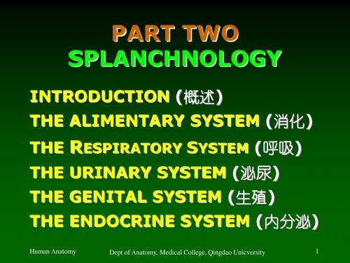
修改chapter7 Digestive system
四年级下英语牛津作文my week

四年级下英语牛津作文my weekHere is an English essay with more than 1000 words, as requested. The title is "My Week" and the content is written entirely in English without any additional punctuation marks.My WeekMonday morning starts off with a bit of a rush as I try to get ready for the school day ahead. I quickly eat a bowl of cereal for breakfast and gather my books and supplies before heading out the door. When I arrive at school, I make my way to my fourth-grade classroom and take my seat. Our English lesson is first on the agenda and we dive right into discussing the latest chapter of our Oxford reading book. I enjoy these discussions as they allow me to practice my English speaking skills. After English, we move on to math where we are working on fractions. I find fractions a bit tricky to grasp but the teacher is patient and offers plenty of examples to help us understand.During our break, I head outside with my friends to run around and play a quick game of tag. It feels good to get some fresh air and physical activity after sitting in the classroom. When the bell rings,we head back inside for science class. Today we are learning about the human body and the different organ systems. I find this topic fascinating and ask lots of questions. Our teacher seems impressed by my curiosity and encourages me to keep exploring this area of study.After a quick lunch, we have our weekly spelling test. I've been practicing my spelling words each night and I'm confident I will do well. Sure enough, I ace the test and feel a sense of pride in my hard work. The last class of the day is art, which is one of my favorite subjects. We are working on sketching landscapes today and I lose myself in the peaceful process of putting pencil to paper.When the final bell rings, I gather my belongings and head home. On the bus ride, I chat with my friends about our plans for the evening. Many of us are getting together to play at the park near our houses.I arrive home, have a snack, and quickly finish my homework so I can join my friends. We spend a couple of hours running around, playing games, and laughing. It's the perfect way to end the school day.Tuesday morning starts off much the same as Monday, with a rushed breakfast and gathering of school supplies. In our English lesson, we begin reading a new chapter of our Oxford book. I'm excited to see what happens next in the story. Math class focuses on decimals today, which I find a bit easier to grasp than fractions. During ourbreak, I practice my soccer skills with a few classmates. I'm trying to make the school soccer team and know I need to keep working on my ball control.Science class covers the respiratory system, which I find fascinating. I ask the teacher lots of questions about how our lungs and diaphragm work together to allow us to breathe. Spelling test goes well again, and I'm pleased with my performance. Art class has us experimenting with watercolor paints today, creating landscapes with soft, blended colors. I really enjoy the calming process and the beautiful results.After school, I head straight to soccer practice. Our coach puts us through some drills to work on dribbling and passing, then we scrimmage for the last part of the session. I feel like I'm improving with each practice and I'm hopeful I'll make the team. When practice ends, I grab a quick dinner before settling in to finish my homework. In the evening, I read for pleasure, losing myself in an adventure story.Wednesday begins with a spelling quiz first thing in the morning. I breeze through it, feeling confident in my preparation. Our English lesson has us analyzing poetry, which is a new challenge for me. I'm intrigued by the way poets use language to convey emotions and paint vivid pictures. In math, we move on to working withpercentages, which I find straightforward compared to some of our previous topics.During the break, I join a group of friends for a quick game of handball. It's a fun way to get some exercise and competition. Science class covers the circulatory system today, building on our previous lessons about the human body. I'm fascinated to learn how the heart pumps blood throughout our bodies to deliver oxygen and nutrients. The art project has us experimenting with pastels, blending colors to create still life drawings. I find the soft, rich textures of the pastels very appealing.After school, I head to the library to work on a research project for social studies. I spend a couple of hours reading through reference books and taking notes. It's tedious work, but I know it will pay off when I put together my final report. In the evening, I enjoy a home-cooked meal with my family and then settle in to play a strategy game on my computer.Thursday dawns and I feel a bit tired from the busy week so far. But I push through my morning routine and make it to school. Our English lesson has us practicing persuasive writing techniques, which I find challenging but interesting. In math, we review what we've learned about percentages and then move on to probability. I enjoy the logic and problem-solving involved in working with probability.During the break, I chat with friends about our weekend plans. Many of us are looking forward to a school field trip on Friday. In science, we learn about the digestive system, which builds on our previous lessons. I'm amazed by how complex and efficient our bodies are in breaking down the food we eat. Spelling test goes well again, andI'm pleased to see my hard work paying off.Art class has us experimenting with printmaking techniques today. I enjoy the process of carving a design into a block and then pressing it onto paper to create a repeating pattern. After school, I head to my piano lesson. I've been practicing diligently and my teacher compliments my progress. In the evening, I video chat with my grandparents who live in another city. It's wonderful to catch up with them and share the highlights of my week.Friday arrives and I'm excited for our class field trip. We pile onto the bus and head to a local nature center, where we'll be learning about different ecosystems. Our guide leads us on a hike through the woods, pointing out various plants and animals. I'm fascinated by all the life thriving in this natural environment. We also get to visit the center's small zoo, where we see animals like owls, snakes, and turtles up close.After the field trip, we return to school for our final classes of theweek. In English, we have a lively discussion about the persuasive writing techniques we practiced earlier in the week. I feel like I'm really starting to understand how to craft a compelling argument. Math class is a review session to prepare us for our upcoming test on percentages and probability. I feel confident in my understanding of these concepts.When the final bell rings, I gather my things and head home, looking forward to the weekend. I spend the afternoon playing outside with neighborhood friends, then enjoy a family dinner. In the evening, I wind down by reading a book and getting to bed early, ready to start a new week of learning and adventure.。
09消化管(1)
Human Anatomy
Dept of Anatomy, Medical College, Qingdao Unicversity
7
E. The Abdominal Regions
(腹部分区)
Rt. hypochon- Epigastric driac region region Lt hypochondriac region
Human Anatomy Dept of Anatomy, Medical College, Qingdao Unicversity 10
Section 1
Oral Cavity
A. The Oral Vestibule (口腔前庭) B. The Oral Cavity Proper (固有口腔)
18
The Oral Cavity Proper (固有口腔)
3. The tongue (舌)
1) Division a. apex of tongue (舌尖) b. body of tongue (舌体) c. root of tongue (舌根) d. dorsum of tongue (舌背) i. terminal sulcus (界沟) ii. foramen cecum of tongue (舌盲孔)
乳牙 牙式 I-V 恒牙 牙式 1-8
Human Anatomy
Dept of Anatomy, Medical College, Qingdao Unicversity
16
The Oral Cavity Proper (固有口腔)
3) Structure of the teeth
a. dentine (牙本质) b. enamel (釉质) c. cement (牙骨质) d. dental pulp (牙髓) e. periodontal membrane (牙 周膜) f. alveolar bone (牙槽) g. gingiva (牙龈) [caries 龋齿] [determination of age 年龄鉴定] [gum bleeding 牙龈出血]
医学英语(阅读一分册)翻译与答案解析
Chapter 1Passage 1 Human BodyIn this passage you will learn:1. Classification of organ systems2. Structure and function of each organ system3. Associated medical termsTo understand the human body it is necessary to understand how its parts are put together and how they function. The study of the body's structure is called anatomy; the study of the body's function is known as physiology. Other studies of human body include biology, cytology, embryology, histology, endocrinology, hematology, immunology, psychology etc.了解人体各部分的组成及其功能,对于认识人体是必需的。
研究人体结构的科学叫解剖学;研究人体功能的科学叫生理学。
其他研究人体的科学包括生物学、细胞学、胚胎学、组织学、分泌学、血液学、遗传学、免疫学、心理学等等。
Anatomists find it useful to divide the human body into ten systems, that is, the skeletal system, the muscular system, the circulatory system, the respiratory system, the digestive system, the urinary system, the endocrine system, the nervous system, the reproductive system and the skin. The principal parts of each of these systems are described in this article.解剖学家发现把整个人体分成骨骼、肌肉、循环、呼吸、消化、泌尿、分泌、神经、生殖系统以及感觉器官的做法是很有帮助的。
药物过敏反应的症状

药物过敏反应的症状Chapter 1: IntroductionDrug allergies are adverse reactions that occur when an individual's immune system reacts abnormally to a medication. It is estimated that about 10% of the global population experiences some form of drug allergy in their lifetime. These allergies can range from mild symptoms like rashes to severe and life-threatening reactions. Understanding the symptoms associated with drug allergies is of utmost importance in order to provide appropriate medical interventions and ensure patient safety. This paper aims to explore the various symptoms of drug allergies in four chapters: skin reactions, respiratory symptoms, gastrointestinal symptoms, and anaphylaxis.Chapter 2: Skin ReactionsSkin reactions are the most common manifestation of drug allergies. They can include a variety of symptoms, such as rashes, hives, itching, and swelling. Rashes may appear as red, itchy patches or raised bumps on the skin. Hives, or urticaria, are characterized by itchy welts that can be any size or shape and usually disappear within a few hours. Itching of the skin is a common symptom and can often be accompanied by a tingling or burning sensation. Swelling, also known as angioedema, typically occurs in the lips, face, throat, and extremities. In severe cases, it can lead to difficulty breathing or swallowing.Chapter 3: Respiratory SymptomsRespiratory symptoms are another significant category of drug allergy symptoms. These symptoms can affect the upper and lower respiratory tract. Common upper respiratory symptoms include sneezing, nasal congestion, runny nose, and itching or watering of the eyes. In some cases, these symptoms may mimic those of the common cold. Lower respiratory symptoms, such as coughing, wheezing, shortness of breath, and chest tightness, are more severe and may indicate the development of an allergic reaction, particularly in individuals with pre-existing respiratory conditions like asthma.Chapter 4: Gastrointestinal SymptomsGastrointestinal symptoms can also occur as a result of drug allergies. These symptoms primarily affect the digestive system and include nausea, vomiting, diarrhea, and abdominal pain. In some cases, these symptoms may be accompanied by loss of appetite and weight loss. Gastrointestinal symptoms may range from mild and transient to severe and persistent, and they can be indicative of an adverse reaction to a medication.Chapter 5: AnaphylaxisAnaphylaxis is a severe and potentially life-threatening allergic reaction that affects multiple organ systems. It is a medical emergency that requires immediate attention. Symptoms of anaphylaxis include difficulty breathing, wheezing, hives, swelling of the face or throat, rapid heartbeat, dizziness or faintness, and a sudden feeling of impending doom. If an individual experiences these symptoms after taking medication, it is crucial to seekimmediate medical help, as anaphylaxis can rapidly progress and become fatal.ConclusionDrug allergies can manifest in various ways, ranging from relatively mild skin reactions to severe anaphylaxis. Recognizing the symptoms associated with drug allergies is essential for prompt diagnosis, appropriate treatment, and preventing further complications. Healthcare professionals and patients should be educated about these symptoms to ensure proper management of drug allergies and improve patient outcomes.Chapter 6: Diagnosis and Treatment of Drug AllergiesDiagnosing drug allergies can be challenging as symptoms can often overlap with other conditions. In cases where a drug allergy is suspected, healthcare professionals may perform various tests to confirm the diagnosis. These tests can include skin prick tests, patch tests, blood tests, and drug provocation tests.Skin prick tests involve pricking the skin with a small amount of the suspected drug and observing for any allergic reaction, such as redness or swelling. Patch tests are used to identify delayed allergic reactions and involve applying small amounts of the suspected drug to the skin under a patch for 48 to 72 hours. Blood tests, such as the radioallergosorbent test (RAST) or enzyme-linked immunosorbent assay (ELISA), can measure the levels of specific antibodies associated with allergic reactions. Drug provocation tests are typically conducted in a controlled medical setting where small amounts of the suspected drug areadministered to determine if a reaction occurs.Once a drug allergy is diagnosed, the treatment involves avoiding the offending drug and finding suitable alternatives. Healthcare professionals must carefully review a patient's medical history and medication list to ensure that they do not unknowingly prescribe a medication to which the patient is allergic. In cases where the allergy is severe or life-threatening, patients may be advised to wear medical alert bracelets or carry an epinephrine auto-injector, which can be used to quickly treat an anaphylactic reaction.Chapter 7: Prevention and Patient EducationPreventing drug allergies can be challenging as they can occur unpredictably. However, there are certain steps that can be taken to minimize the risk. It is essential for healthcare professionals to obtain a comprehensive medical history from patients, including any previous allergies or adverse drug reactions. This information can help identify patients who may be at a higher risk of developing drug allergies.In addition to obtaining a medical history, healthcare professionals should educate patients about the signs and symptoms of drug allergies. Patients should be informed to report any unusual symptoms or allergic reactions to their healthcare provider immediately. Furthermore, patients should be educated about the importance of reading medication labels, following dosing instructions, and taking medications as prescribed.Patient education also plays a crucial role in the prevention of drugallergies. Patients should be encouraged to ask questions about medications, inform their healthcare provider about any known allergies or sensitivities, and be proactive in their healthcare decisions. Additionally, patients should be aware of the potential cross-reactivity between certain medications and allergens, such as penicillin and cephalosporins.Chapter 8: ConclusionDrug allergies are adverse reactions that can have a significant impact on patient health and safety. Understanding the symptoms associated with drug allergies is vital for timely diagnosis and appropriate treatment. Skin reactions, respiratory symptoms, gastrointestinal symptoms, and anaphylaxis are some of the common manifestations of drug allergies. Diagnosis involves various tests, and treatment primarily focuses on avoidance of the offending drug and finding suitable alternatives. Prevention and patient education are crucial in minimizing the risk of drug allergies and ensuring patient safety. Healthcare professionals should remain vigilant and stay updated on the latest research and guidelines regarding drug allergies to provide optimal care for their patients.。
《新时代外语教育课程思政案例教程》读书笔记模板

“英语口语”课程思政教学设计样例 Unit 1 Request and Offer
“英语阅读”课程思政教学设计案例第二册 Unit 11 National Spirits
“英语阅读”课程思政教学设计样例 Unit 1 Nature Part A Amazon Rainforest Climate
“基础西班牙语”课程思政教学设计样例 Unidad 10第十课社会文化常识: 西班牙语国家的姓氏Conocimiento sociocultural
读书笔记
这是《新时代外语教育课程思政案例教程》的读书笔记模板,可以替换为自己的心得。
精彩摘录
这是《新时代外语教育课程思政案例教程》的读书笔记模板,可以替换为自己的精彩内容摘录。
作者介绍
这是《新时代外语教育课程思政案例教程》的读书笔记模板,暂无该书作者的介绍。
谢谢观看
“英语教学活动设计”课程思政教学设计样例 Chapter 1 Introduction— How to Design a Reading Material?
“笔译工作坊”课程思政教学设计样例 Unit 11 C-E Translation of Documentary Subtitles
大学综合英语——公 共基础课
“研究生英语”课程思政教学设计样例 Unit 11 Environment and Sustainability Passage A:Three Challenges to Sustainability
“学术论文写作”课程思政教学设计样例 Unit 7 Space Exploration
德语——专业 核心课
“综合商务英语”课程思政教学设计样例 Unit 2 Coffee Culture Comes to Coffee-growers
Chapter-5-Digestive-System

❖appendicitis: inflammation of the appendix. ▪ 阑尾
❖appendical: pertaining to the appendix.
❖ Large intestine ▪cecum ▪colon ▪sigmoid colon ▪rectum
❖ Liver ❖ Gallbladder ❖ Pancreas
3
Digestive system
4
aliment/o: food, nutrient
❖alimentation: process of giving or taking nourishment. ▪ 营养,营养吸收
❖cholecystitis: inflammation of the gallbladder. ▪ 胆囊炎
❖cholecystic: pertaining to the gallbladder. ▪ 胆囊的
12
cis/o: to cut
❖ incision: in- into; -ion process, hence the process of cutting in. ▪ 切开, 切口, 断口
❖ cholestasis: -stasis stopping, hence stoppage of the flow of bile. ▪ 胆汁郁积, 胆汁阻塞
❖ cholemesis: -emesis vomiting, hence vomiting of bile. ▪ 呕胆 ▪ hematemesis ▪ hyperemesis
Digestive System
1.
Drink less stimulant drinks like coffee, teess food which is hard to digest. ( cold, spicy, oily, sour, hard, salted)
Health Tips
1.
饮食六宜:宜缓、宜少、宜淡、宜 暖、宜软 2. 不挑食偏食,定时定量,细嚼慢咽 3. 讲究饮食卫生,不吃不洁食物和过 期食物 4. 多吃粗粮、果蔬、豆类等
Large intestine is shorter, but fatter.
大肠吸收食物残 渣中的水分,然 后排出体外。
消化系统作用 a. 消化食物 b. 吸收营养
c. 排泄废物
How to protect our digestive system? What are the right ways of eating?
食道 胃 小肠 大肠
mouth
esophagus stomach small intestine large intestine (colon)
Digestive System 消化系统
mouth esophagus stomach large intestine small intestine
Food first enters the mouth.
How should we protect our digestive system?
What should we eat or drink less of?
Are they good habits?
1.
Eating very quickly 2. Not eating at regular times 3. Exercising after a meal 4. Sleeping after a meal
Digestive system disease

This is the normal appearance of the gastric antrum extending to the pylorus at the right of center. The first portion of the duodenum(duodenal bulb) is at the far right. In the endoscopic views below, the normal appearance of the pylorus is seen at the left, with the first portion of the duodenum at the right. Anatomy and histology of the stomach. A,Gross anatomy. B, Microscopic view of antral mucosa. C,Microscopic view ofThis is the normal appearance of the gastric fundal mucosa, with short pits lined by pale columnar mucus cells leading into long glands which contain bright pink parietal cells that secrete hydrochloric acid.This is a more typical acute gastritis with a diffusely hyperemic gastric mucosa. There are many causes for acute gastritis: alcoholism, drugs, infections, etc. Here are some larger areas of gastric hemorrhage that could best be termed "erosions" because the superficial mucosa is eroded away. Such erosions are typical for the pathologic process termed gastropathy, which describes gastric mucosal injury without significant inflammation. The findings here fit with acute erosive gastropathy, but there are other patterns. Etiologies for the various gastropathies can include: alcohol, drugs such as NSAIDS, stress, uremia, bile reflux, portal hypertension, radiation, and chemotherapy.At high power, gastric mucosa demonstrates infiltration by neutrophils. Thisis acute gastritis.Helicobacter pylori gastritis. A Steiner silver stain demonstrates the numerousdarkly stained Helicobacter organisms along the luminal surface of the gastricepithelial cells. There is no tissue invasion by bacteria. (Courtesy of Dr. MelissaUpton, Department of Pathology, University of Washington, Seattle,Washington.)Gastritis is often accompanied by infection with Helicobacter pylori. This small curved to spiral rod-shaped bacterium is found in the surface epithelial mucus of most patients with active gastritis. The rods are seen here with a methylene blue stain.The mucosa adjacent to the ulcer shows chronic gastritis.Note the discrete band of chronic inflammation in the most superficial portion of the mucosa.Chronic gastritis, showing partial replacement of the gastric mucosal epithelium by intestinal metaplasia(upper left), and inflammation of the lamina propriacontaining lymphocytes and plasma cells (right).Intestinal metaplasia in chronic gastritis (arrow). Note goblet cells andlymphcyte and plasma cell infiltration (arrowhead) Incomplete intestinal metaplasia containing goblet cells but lacking a well-defined brush border.Alcian blue and periodic acid-Schiff stain of Barrett's mucosa with completeintestinal metaplasia. The goblet cells are intensely Alcian blue positive.The periodic acid-Schiff portion of the stain outlines a primitive luminalbrush border.Helicobacter pylori.A Steiner silver stain demonstrates the numerous darkly stained Helicobacter organisms along the luminal surface of the gastric epithelial cells. Note that there is no tissue invasion by bacteria.Aggravating causes of, and defense mechanisms against, peptic ulceration. The right panel shows the basis of a nonperforated ulcer, demonstrating necrosis (N), inflammation (I), granulation tissue (G), and fibrosis (S).Seen above are gastric ulcers ofsmall, medium, and large size onupper endoscopy. All gastric ulcersare biopsied, since gross inspectionalone cannot determine whether amalignancy is present. Smaller, more sharply demarcated ulcers are more likely to be benign. Peptic ulcer of the duodenum. Note that the ulcer is small (2 cm) with a sharply punched-out appearance. Unlike cancerous ulcers, the margins are not elevated. The ulcer base is clean.An acute duodenal ulcer is seen in two views on upper endoscopy.Microscopically, the ulcer here is sharply demarcated, with normalgastric mucosa on the left falling away into a deep ulcer whose basecontains infamed, necrotic debris. An arterial branch at the ulcer baseis eroded and bleeding.The mucosa at the upper right merges into the ulcer at the leftwhich is eroding through the mucosa. Ulcers will penetrateover time if they do not heal. Penetration leads to pain.The ulcer at the right is penetrating through the muscularisand approaching an artery. Erosion of the ulcer into theartery will lead to another major complication of ulcers--hemorrhage.The strongest association with Helicobacter pylori is with duodenal peptic ulceration--over 85% of duodenal ulcers. Seen here is a penetrating acute ulceration in the duodenum just beyond the pylorus.Another association with gastritis is pernicious anemia. Chronic atrophic gastritis is associated with autoantibodies that block or bind intrinsic factor. Another type of autoantibody demonstrated here is anti-parietal cell antibody. The bright green immunofluorescence is seen in the paritetal cells of the gastric mucosa.Seen here is a loop of bowel attached via the mesentery. Note the extent of the veins. Arteries run in the same location. Thus, there is an extensive anastomosing arterial blood supply to the bowel, making it more difficult to infarct. Also, the extensive venous drainage is incorporated into the portal venous system heading to the liver.This is the normal appearance of small intestinal mucosa with long villi that have occasional goblet cells. The villi provide a large area for digestion and absorption. This is normal colonic mucosa. Note the crypts that are lined by numerous goblet cells. In the submucosa is a lymphoid nodule. The gut-associated lymphoid tissue as a unit represents the largest lymphoid organ of the body.A,Normal small-bowel histology, showing mucosal villi and crypts, lined bycolumnar cells. B,Normal colon histology, showing flat mucosal surface andabundant vertically oriented crypts.Acute appendicitis. The inflamed appendix shown below is red, swollen, andcovered with a fibrinous exudate. For comparison, a normal appendix isshown above.This appendix was removed surgically. The patient presented with abdominal pain that initially was generalized, but then localized to the right lower quadrant, and physical examination disclosed 4+ rebound tenderness in the right lower quadrant. This is the tip of the appendix from a patient with acute appendicitis. The appendix has been sectioned in half. The serosal surface at the left shows a tan-yellow exudate. The cut surface at the right demonstrates yellowish-tan mucosal exudation with a hyperemic border.Microscopically, acute appendicitis is marked by mucosal inflammation and necrosis.This is another example of Crohn's disease involving the smallintestine. Here, the mucosal surface demonstrates an irregularnodular appearance with hyperemia and focal superficialulceration.Crohn disease of ileum, showing narrowing of the lumen, bowel wallthickening, serosal extension of mesenteric fat ("creeping fat"), andlinear ulceration of the mucosal surface (arrowheads).Microscopically, Crohn's disease is characterized by transmural inflammation. Here, inflammatory cells extend from mucosa through submucosa and muscularis and appear as nodular infiltrates on the serosal surface with pale granulomatous centers.disease of the colon; a deep fissure extending into the muscle wall, a second, shallow ulcer (on the upper right), and relativepreservation of the intervening mucosa. Abundant lymphocyte aggregates are present, evident as dense blue patches of cells at the interface between mucosa and submucosa.Crohn disease of the colon. A noncaseating granulomais present in the lamina propria of an uninvolved region of colonic mucosa (arrow).At high magnification the granulomatous nature of the inflammation of Crohn's disease is demonstrated here with epithelioid cells, giant cells, and many lymphocytes.At higher magnification, the pseudopolyps can be seen clearly as raised red islands of inflamed mucosa. Between the pseudopolyps is only remaining muscularis.Ulcerative colitis. Ulcerated hemorrhagic surface with knobby pseudopolyps. Microscopically, the inflammation of ulcerative colitis is confined primarily to the mucosa. Here, the mucosa is eroded by an ulcer that undermines surrounding mucosa.At higher magnification, the intense inflammation of the mucosa is seen. The colonic mucosal epithelium demonstrates loss of goblet cells. An exudate is present over the surface. Both acute and chronic inflammatory cells are present. Crypt abscesses are a histologic finding more typical with ulcerative colitis. Unfortunately, not all cases of inflammatory bowel disease can be classified completely in all patients.Over time, there is a risk for adenocarcinoma with ulcerativecolitis. Here, more normal glands are seen at the left, but the glands at the right demonstrate dysplasia, the first indication that there is a move towards neoplasia. Ulcerative colitis. Low-power micrograph showing marked chronic inflammation of the mucosa with atrophy of colonic glands, moderate submucosal fibrosis, and a normal muscle wall.Ulcerative colitis. Microscopic view of the mucosa, showing diffuse active inflammation with crypt abscess and glandular architectural distortion.Toxic megacolon. Complete cessation of colon neuromuscular activity has led to massive dilatation of the colon and black-green discoloration signifying gangrene and impending rupture. Comparison of the distribution patterns of Crohn disease and ulcerative colitis, as well as the different conformations of the ulcers and wallTransition from Barrett esophagus to adenocarcinomaBarrett esophagus. A-B,Gross view of distal esophagus (top) and proximalstomach (bottom) showing (A) normal gastroesophageal junction and (B) thegranular zone of Barrett esophagus (arrow). C,Endoscopic view showing redvelvety gastrointestinal-type mucosa extending from the gastroesophagealBarrett esophagus. Microscopic view showing squamous mucosa (left) andintestinal-type columnar epithelial cells in glandular mucosa (right).Another cause for inflammation is a so-called "Barrett's esophagus" in whichthere is gastric-type mucosa above the gastroesophageal junction. Note thecolumnar epithelium to the left and the squamous epithelium at the right. This is"typical" Barrett's mucosa, because there is intestinal metaplasia as well (notethe goblet cells in the columnar mucosa).Diagram of growth patterns and spread of gastric carcinoma. In early gastric carcinoma (A), the tumor is confined to the mucosa and submucosa and may exhibit Diagram of growth patterns and spread of gastric carcinoma. Advanced gastric carcinoma (B) extends into the muscularis propria and beyond. Linitisplastica is an extreme form of flat or depressedHere is a gastric adenocarcinoma. ALL gastric ulcers and ALL gastric masses must be biopsied, because it is not possible to tell from gross appearance alone which are benign and which are malignant. Here is a gastric ulcer in the center of the picture. It is shallow and is about 2 to 4 cm in size. This ulcer on biopsy proved to be malignant, so the stomach was resected as shown here.Here is a much larger 3 x 4 cm gastric ulcer that led to the resection of the stomach shown here. This ulcer is much deeper with more irregular margins. Gastric carcinoma. Gross photograph showing an ill-defined, excavated central ulcer surrounded by irregular, heaped-up borders.Gastric cancer. A,H&E stain demonstrating intestinal type of gastric carcinoma with gland formation by malignant cells that are invading the muscular wall of the stomach. B,Diffuse type of gastric carcinoma with signet-A moderately differentiated gastric adenocarcinoma is infiltrating up and into the submucosa below the squamous mucosa of the esophagus. The neoplastic glands are variably sized. At higher magnification, the neoplastic glands of gastric adenocarcinoma demonstrate mitoses, increasednuclear/cytoplasmic ratios, and hyperchromatism. There is a desmoplastic stromal reaction to the infiltrating glands.At high power, this gastric adenocarcinoma is so poorly differentiated that glands are not visible. Instead, rows of infiltrating neoplastic cells with marked pleomorphism are seen. Many of the neoplastic cells have clear vacuoles of mucin.This is a signet ring cell pattern of adenocarcinoma in whichthe cells are filled with mucin vacuoles that push the nucleus to one side, as shown at the arrow.Schematic of the morphologic and molecular changes in the adenoma-carcinoma sequence. It is postulated that loss of one normal copy of the tumor suppressor gatekeeper gene APC occurs early. Indeed, individuals may be born with one mutant allele of APC, rendering them extremely likely to develop colon cancer. This is the "first hit," according to Knudson's hypothesis. The loss of the normal copy of the APC gene followsCarcinoma of the cecum. The fungating carcinomaprojects into the lumen but has not caused obstruction.Invasive adenocarcinoma of colon, showing malignantglands infiltrating the muscle wall.Clinical Features•The appearance of fatigue, weakness, andiron-deficiency anemia•Producing occult bleeding, changes inbowel habit, or crampydiscomfortPathologic staging of colorectal cancer. Staging is basedon the depth of tumor invasion.Diagrammaticrepresentation of twoforms of sessile polyp(hyperplasticadenoma) and of twotypes of adenoma(pedunculatedsessile). There is only aloose associationbetween the tubulararchitecture forpedunculatedadenomas and thevillous architecture for Non-neoplastic colonic polyps. A,Hyperplastic polyp; high-power view showing the serrated profile of the epithelial layer.Non-neoplastic colonic polyps. B,Peutz-Jeghers polyp; low-power view showing the splaying of smooth muscle into the superficial portion of the pedunculated polyp. Here are multiple adenomatous polyps of the cecum. A small portion of terminal ileum appears at the right.Familial adenomatous polyposis in an 18-year-old woman. The mucosal surface is carpeted by innumerable polypoid adenomas. The barium enema technique instills the radiopaque barium sulfate into the colon, producing a contrast with the wall of the colon that highlights any masses present. In this case, the classic "apple core" lesion is present, representing an encircling adenocarcinoma thatThis CT image of the abdomen demonstrates an encircling mass involving the colon. This is a colonic adenocarcinoma.A normal liver is shown grossly for comparison The cut surface of a normal liver has a brown color. Near the hilum here, note the portal vein carrying blood to the liver, which branches at center left, with accompanying hepatic artery and bile ducts. At the lower right is a branch of hepatic vein draining blood from the liver to the inferior vena cava.The liver can be divided into three zones, based upon oxygen supply. Zone 1 encircles the portal tracts wherethe oxygenated blood from hepatic arteries enters. Zone 3 is located around central veins, where oxygenation is poor. Microscopic anatomy of the liver. The portal tract carries branches of the portal vein, hepatic artery, and bile duct system.Photomicrograph of liver (trichrome stain). Note the blood-filled sinusoids and cords of hepatocytes; the delicate network of reticulin fibers in the subendothelial space of Disse stains light blue. The portal triad consists of the portal vein, branches of the hepatic artery and tributaries to the bile duct.The liver has its own version of macrophage known as the Kupffer cell (stained blue by uptake of trypan blue). Not only do these remove foreign particles, they also work with the spleen to destroy old RBCs.Sequence of serologic markers in acute hepatitis A viral hepatitis.Diagrammatic representation of genomic structure and transcribedof the hepatitis B virion. The innermost circles represent the DNA (+) strand and the DNA (-) strand of the virion. The thick bars labeled "P," "X," "pre-C," "C,""pre-SI," "pre-S2," and "S" denote the peptides derived from the virion. The outermost lines denote the mRNA transcripts of the virion. (After Kidd-Ljunggren Y, Kidd AH: J Gen Virol83:1267-1280, 2002.) Schematic of the potential outcomes of hepatitis B infection in adults, with their approximate frequencies in the United States.Sequence of serologic markers for hepatitis B viral hepatitis demonstrating (A)acute infection with resolution and (B)progression to chronic infection.Schematic of the potential outcomes of hepatitis C Sequence of serologic markers for hepatitis C viral hepatitis demonstrating (A) acute infection with resolution and (B) progression to chronic relapsing infection.The differing clinical consequences of the two pattens of combined hepatitis D virus (HDV) and hepatitis B (HBV) infection.Sequence of serologic markers for hepatitis D viral hepatitis depicting (A) coinfection with hepatitis B virus (HBV) and (B) superinfection of an HBV carrier.Microscopic architecture of the liver parenchyma. Both a lobule and an acinus are represented. The classic hexagonal lobule is centered around a central vein (CV), also known as terminal hepatic venule, and has portal tracts at three of its apices. The portal tracts contain branches of the portal vein (PV), hepatic artery (HA), and the bile duct (BD) system. Regions of the lobule are generally referred to as "periportal," "midzonal," and "centrilobular," according to their proximity to portal spaces and central vein.Hepatitis B viral infection. A, Liver parenchyma showing hepatocyteswith diffuse granular cytoplasm, so-called ground glass hepatocytes.(H&E) B, Immunoperoxidase stain for HBsAg from the same case,showing cytoplasmic inclusions of viral particles.Here are Mallory bodies(the red globular material) composed of cytoskeletal filaments in liver cells chronically damaged from alcoholism. The bile pigment is toxic to hepatocytes which become swollen, showing rarefaction of the cytoplasm referred to as feathery degeneration.Intracellular accumulations of a variety of materials can occur in responseto cellular injury. Here is fatty metamorphosis (fatty change) of the liver inwhich deranged lipoprotein transport from injury (most often alcoholism)leads to accumulation of lipid in the cytoplasm of hepatocytes.A mononuclear inflammatory cell infiltrate extends fromportal areas and disrupts the limiting plate of hepatocyteswhich are undergoing necrosis, the so-called "piecemeal"necrosis of chronic active hepatitis.Massive necrosis, microscopic section. The portal veins are closer together than normal owing to necrosis and collapse of the intervening parenchyma. The rudimentary ductal structures are the result of early hepatocyte regeneration. An infiltrate of chronic inflammatory cells are Clusters of lymphocytes and hyperplastic Kupffer cells are scatteredthroughout the hepatic lobule as well. There is also spotty necrosis (focalnecrosis of individual hepatocytes).Acute viral hepatitis showing disruption of lobular architecture, inflammatory cells in the sinusoids, and hepatocellular apoptosis.Chronic viral hepatitis due to hepatitis C virus, showing portal tract expansion with inflammatory cells and fibrous tissue and interface hepatitis with spillover of inflammation into the adjacent parenchyma.A lymphoid aggregate is present.Cirrhosis resulting from chronic viral hepatitis. Note the broad scar and coarse nodular surface.The yellow-green globular material seen in small bile ductules in the liver here is bilirubin pigment. This is hepatic cholestasis.Grossly, there are areas of necrosis and collapse of liver lobules seen here as ill-defined areas that are pale yellow. Such necrosis occurs with hepatitis.The necrosis and lobular collapse is seen here as areas of hemorrhage and irregular furrows and granularity on the cut surface of the liver. In this example, liver cells are dying individually (arrows) from injury by viral hepatitis. The cells are pink and without nuclei.This is a case of viral hepatitis C which is at a high stage with extensive fibrosis and progression to macronodular cirrhosis, as evidenced by the large regenerative nodule at the center right. This is a case of viral hepatitis C, which in half of cases leads to chronic liver disease. The extent of chronic hepatitis can be graded by the degree of activity (necrosis and inflammation) and staged by the degree of fibrosis.This trichrome stain demonstrates the collapse of the liverparenchyma with viral hepatitis. The blue-staining areas are the connective tissue of many portal tracts that have collapsed together. Diagrammatic representations of the morphologic features of acute and chronic hepatitis. Bridging necrosis (and fibrosis) is shown only for chronic hepatitis; bridging necrosis may also occur in acute hepatitis (not shown).Massive necrosis. A, Cut section of liver. The liver is small (700 gm), bile-stained, and soft. The capsule is wrinkled. B, Microscopic section. Portal tracts and terminal hepatic veins are closer together than normal,There is extensive hepatocyte necrosis seen here in a case of acetaminophenoverdose. The hepatocytes at the right are dead, and those at the left are dying.This pattern can be seen with a variety of hepatotoxins. Acute liver failure leads tohepatic encephalopathy.Alcoholic liver disease. The interrelationships amonghepatic steatosis, hepatitis, and cirrhosis are shown,along with a depiction of key morphologic features atthe morphologic level.This liver is slightly enlarged and has a pale yellow appearance, seenboth on the capsule and cut surface. This uniform change isconsistent with fatty metamorphosis (fatty change).Alcoholic hepatitis. A, The cluster of inflammatory cells marksthe site of a necrotic hepatocyte. A Mallory body is present ina second hepatocyte (arrow). B, Eosinophilic Mallory bodiesare seen in hepatocytes, which are surrounded by fibroustissue (H&E).Mallory's hyaline is seen here, but there are also neutrophils,necrosis of hepatocytes, collagen deposition, and fatty change. These findings are typical for acute alcoholic hepatitis. Alcoholic liver disease: macrovesicular steatosis, involving most regions of the hepatic lobule. The intracytoplasmic fat is seen as clear vacuoles. Some early fibrosis (stained blue) is present (Masson trichrome).Here is another example of micronodular cirrhosis. Note that the liver also has a yellowish hue, indicating that fatty change (also caused by alcoholism) is present.Alcoholic cirrhosis showing the characteristic diffuse nodularity of the surface induced by the underlying fibrous scarring. The average nodule size is 3 mm in this close-up view. The greenish tint is caused by bile stasis. Alcoholic cirrhosis. A, The characteristic diffuse nodularity of the surface reflects the interplay between nodular regeneration and scarring. The greenish tint of some nodules is due to bile stasis. A hepatocellular carcinoma is present as a budding mass at the lowerMicroscopically with cirrhosis, the regenerative nodules of hepatocytes are surrounded by fibrous connective tissue that bridges between portal tracts. Within this collagenous tissue are scattered lymphocytes as well as a proliferation of bile ducts.A close-up view of a micronodular cirrhosis in a liver with fatty change demonstrates the small, yellow nodules. Micronodular cirrhosis is seen along with moderate fatty change. Note the regenerative nodule surrounded by fibrous connective tissue extending between portal regions.Ongoing liver damage with liver cell necrosis followedby fibrosis and hepatocyte regeneration results in cirrhosis. This produces a nodular, firm liver. The nodules seen here are larger than 3 mm and, hence, this is an example of "macronodular" cirrhosis.In macronodular cirrhosis, the regenerative nodules are large and irregular in size and shape. The fibrous septa are often broad. Macronodular cirrhosis corresponds loosely to the older terms "post-necrotic" or "multilobular" cirrhosis and in the U.S. is most often seen following chronic active viral hepatitis.The hepatic architecture is disturbed by broad bands of fibrosis thatcompletely circumscribe irregularly sized and shaped nodules of regenerating hepatic parenchyma. These nodules range in size from less than 1mm in diameter to greater than 5 mm in diameter. Shows at higher power the broad fibrous septa which completely surround regenerating nodules of hepatic parenchyma. This patient's cirrhosis resulted from chronic active hepatitis C and there is ongoing chronic inflammation and necrosis in this liver.Is taken at the margin of a regenerative nodule and a fibrous scar (double arrows). Note the irregular contour of the regenerative nodules and the intense chronic inflammation at the interface of the fibrous septa and the hepatic plates. This is an example of ongoing piecemeal necrosis. Biliary cirrhosis. Sagittal section through the liver demonstrates the fine nodularity and bile staining of end-stage biliary cirrhosis.This is a case of primary biliary cirrhosis. Seen here in a portal tract is an intense chronic inflammatory infiltrate with loss of bile ductules. Micronodular cirrhosis ensues. Primary biliary cirrhosis. A portal tract is markedly expanded by an infiltrate of lymphocytes and plasma cells. The granulomatous reaction to a bile duct undergoing destruction (florid duct lesion) is highlighted by the arrowheads.If chronic hepatic passive congestion continues for a long time,a condition called "cardiac cirrhosis" may develop in which there is fibrosis bridgingbetween central zonal regions, as shown below, so that the portal tracts appear to be in the center of the reorganized lobule. This process is best termed "cardiac sclerosis" because, unlike a true cirrhosis, there is minimal nodular regeneration. The golden-brown refractile hemosiderin granules are present within hepatocytes.Pipe-stem fibrosis of the liver due to chronicSchistosoma japonicum infection.Liver fibrosis. In the normal liver, the perisinusoidal space (space of Disse) contains a delicate framework of extracellular matrix components. In liver fibrosis, stellate cells are activated to produce a dense layer of matrix material that is deposited in the perisinusoidal space. Collagen deposition blocks the endothelial fenestrations and prevents the free exchange of materials from the blood. Kuppfer cells are also activatedEsophageal varices: a view of the everted esophagus and gastroesophageal junction, showing dilated submucosal veins (varices). The blue-colored varices have collapsed in this postmortem specimen.At the lower end of the esophagus (which has been turned inside out at autopsy) are linear dark blue submucosal dilated veins known as varices. These varices are prone to bleed. Seen here is "caput medusae" which consists of dilated veins seen on the abdomen of a patient with cirrhosis of the liver.One of the most common causes for splenomegaly is portal hypertension with cirrhosis of the liver. Note that this spleen also shows irregular tan-white fibrous plaques over the purple surface.。
- 1、下载文档前请自行甄别文档内容的完整性,平台不提供额外的编辑、内容补充、找答案等附加服务。
- 2、"仅部分预览"的文档,不可在线预览部分如存在完整性等问题,可反馈申请退款(可完整预览的文档不适用该条件!)。
- 3、如文档侵犯您的权益,请联系客服反馈,我们会尽快为您处理(人工客服工作时间:9:00-18:30)。
Chapter 7Digestive systemAnatomy and Physiology of the Digestive SystemThe digestive system has three main functions: digesting food, absorbing nutrients, and eliminating wastes. Digestion is the mechanical and chemical breakdown of food into forms that cell membranes can absorb. Mechanical digestion breaks large pieces into smaller ones without altering their chemical composition. Chemical digestion breaks food into simpler nutrient molecules like glucose, triglycerides and amino acid. These simpler nutrient molecules are absorbed from the intestines and circulated throughout the body by the cardiovascular system. They are used for growth and repair of organs and tissues. Any food that cannot be digested or absorbed becomes a waste product and is expelled.The organs of digestive system are traditionally separated into two major groups: the alimentary canal, or gastrointestinal(GI) tract,and the accessory digestive organs. The alimentary canal is approximately 9 meters long in a cadaver but is considerably shorter in a living person. It consists of the mouth, pharynx, esophagus, stomach, and small and largeintestines(or colon), rectum, and anus. The accessory structures include the salivary glands, gallbladder, liver, and pancreas, each of which is connected to the alimentary canal by a duct, and secrete its products into the alimentary canal.Oral cavityThe digestive system begins when food enters the mouth and is mechanically broken up by the chewing movement of the teeth. The tongue, with its muscular action, moves the food within the mouth and mixes it with saliva. Saliva contains digestive enzymes that break down carbohydrates and lubricants that make it easier to swallow the food. Taste buds are found on the surface of the tongue and can distinguish the bitter, sweet, sour and salty flavors in our food. The roof of the oral cavity is known as the palate. The roof of the mouth consists of the hard palate, the bony anterior portion, and the soft palate, the flexible posterior portion. Hanging down from the posterior edge of the soft palate is the uvula. The uvula serves two important functions. It helps in the production of speech and is the location of gag reflex. The gag reflex helps prevent us from accidentally inhaling food or liquids without first swallowing. The cheeks form the lateral walls of this cavity and the lips are the anterior opening. The entire oral cavity is lined with mucous membrane.TeethTeeth are important for the first stage of digestion. The teeth in the front of the mouth bite, tear, or cut food into small pieces. These cutting teeth include the incisors, and the cuspids, or canines. The remaining teeth grind and crush food into even finer pieces. These grinding teeth include the bicuspids or premolars and the molars. A tooth can be subdivided into the crown and root. The crown is the part of the tooth above the gum line. The root is below the gum line and anchors the tooth in the jaw bone. The crown of the tooth is covered by a layer of enamel, the hardest substance in the body. Under the enamel is dentin, which makes the main bulk of the tooth. The hollow interior of a tooth is the pulp cavity in the crown and root canal in the root. These cavities contain soft tissue made up of blood vessels, nerves and lymph vessels.Humans have two sets of teeth. The first sets, often called baby teeth, or milk teeth, are the deciduous teeth. There are 20 teeth in this set erupt through the gums between ages of 6 to 28 months. At approximately 6 years of age, these teeth begin to fall out and are replaced by the 32 permanent teeth. This replacement process will continue until about 18 to 20 years of age.PharynxAfter food has left the mouth, it enters the oropharynx and then the laryngopharynx. The epiglottis covers the larynx and trachea so that food is shunted away from the lungs and into esophagus.EsophagusThe esophagus is a muscular tube that is about 10 inches long in adults. Food entering the esophagus from the pharynx is delivered to the stomach. The food is propelled along the esophagus by wave-like muscular movement called peristalsis. In fact, peristalsis will work to push food through the entire gut tube.StomachThe stomach is on the left side of the abdominal cavity and is hidden by the liver and diaphragm. This J-shaped muscular organ acts as a bag or sac to collect, churn, digest and store food. Different regions of the stomach are the cardiac region(the area surrounding the cardiac orifice through which food enters the stomach from the esophagus), the fundus (theexpanded portion of the stomach, superolateral to the cardiac region), the body or main portion (midportion of the stomach, inferior to the fundus), and the funnel-shaped pyloric region(consisting of the superior-most pyloric antrum, the more narrow pyloric canal, the terminal pyloris, which is continuous with the small intestine through the pyloric sphincter).The folds in the lining of the stomach are called rugae. When the stomach is filled with food, the rugae are stretched out and disappear. Hydrochloric acid is secreted by glands in the mucous membrane lining of the stomach. Food mixes with HC1 and other gastric juices toform a liquid mixture calledchyme, which then passesthrough the remaining portion ofthe digestive system.The stomach containsmuscular valves calledsphincters, that control the flowof food in one direction only.The cardiac sphincter, namedafter its location near the heart,is located between theesophagus and fundus. It is alsocalled the lower esophagealsphincter. It keeps food frombacking up into the esophagus.The pyloric sphincter opens andcloses to control the passage offood into the small intestines with each opening of sphincter for two important reasons: first, the small intestines are much narrower than the stomach and cannot hold as much as the stomach can. Second, the chyme is highly acidic and must be thoroughly neutralized as it leaves the stomach.Small intestineThe small intestine is a convoluted tube, 6 to 7 meters (about 20 feet) long in a cadaver but only about 2 meters long during life because of muscle tone. It is located between the pyloric sphincter and colon.The small intestine has three sections: the duodenum, the jejunum, and ileum. (1) The duodenum, which extends from the pyloric sphincter to the jejunum, is about 25cm (10 inches) long, and curves around the head of the pancreas. Digestion is completed in the duodenum after partly digested chyme from the stomach is mixed with digestive juices from the pancreas and gallbladder. (2) The jejunum, or middle portion, extends from the middle of the small intestine to the ileum and is about 2.5 m (8 feet) long. Most of the jejunum occupies the umbilical region of the abdominal cavity. (3) The ileum is the last portion of small intestine and extends from the jejunum to the colon. At 3.6m (12 feet) in length, it is the longest portion of small intestine. The ileum connects to the colon through a sphincter called the ileocecal valve.The small intestine is the major site of digestion and absorption of nutrients from food.Because the small intestine is concerned with absorption of food products, an abnormality in this organ can cause malnutrition.ColonFluid that remains after the complete digestion and absorption of nutrients in the small intestine enters the colon or large intestine. Most of this fluid is water and it is reabsorbed into the body. The material that remains after absorption is solid waste called feces. This is the product evacuated in bowel movements.The colon is approximately 5 feet long and extends from the ileocecal valve of the small intestine to the anus. The cecum is a pouch or sac-like area in the first 2-3 inches at the beginning of the colon. The appendix is a small worm-shaped outgrowth at the end of the cecum. The remaining colon consists of the ascending colon, transverse colon, descending colon, and sigmoid colon. The ascending colon on the right side extends from the cecum to the lower border of the liver. The transverse colon begins where the ascending leaves off and moves horizontally across the upper abdomen toward spleen. The descending colon then travels down the left side of the body to where the sigmoid colon begins. The sigmoid colon leads into the rectum. The rectum is the area for storage of feces. The rectum leads into the anus, which contains the anal sphincter. This sphincter is controlled by muscles that assist in the evacuation of feces or defecation.Accessory organs of digestive systemThe accessory organs of the digestive system, the salivary glands, the liver, the pancreas, and gallbladder, generally function by producing much of the digestive fluids and enzymes necessary for the chemical breakdown of food. Each is attached to the gut by a duct. Salivary glandFood in the mouth and mechanical pressure (even chewing rubber bands or wax)stimulate the salivary glands to secrete saliva. This very watery and slick fluid allows food to be swallowed with less danger of choking. Saliva mixed with food in the mouth forms a bolus, which is then ready to be swallowed. Saliva also contains the digestive enzyme amylase that begins the digestion of carbohydrates.There are three pairs of salivary glands. The parotid glands are in front of the ears. The submandibular glands and sublingual glands are in the floor of the mouth.Liver and GallbladderThe liver, the largest glandin the body, is locatedinferior to the diaphragm,more to the right than theleft side of the body. Ithides the stomach from theview in a superficialobservation of theabdominal contents. Thehuman liver has four lobes,and is suspended from thediaphragm and anteriorabdominal wall by thefalciform ligament.The human liver isone of the body’s most important organs, and performs many metabolic functions. However, its digestive function is to produce bile, which leaves the liver through the common hepatic duct and then enters the duodenum through the bile duct. Bile has no enzymatic action but emulsifies fats. Emulsification means the process of breaking up large fat particles into smaller ones and making them more soluble in the watery environment inside the intestines.When digestive activity is not occurring in the digestive tract, bile backs up the cystic duct, and enters the gallbladder, a small, green sac on the inferior surface of the liver. It is stored there until needed for digestive process. While in the gallbladder, bile is concentrated by the removal of water and some ions. When fat-rich food enters the duodenum, a hormonal stimulus causes the gallbladder to contract, releasing the stored bile and making it available to the duodenum.If bile contains an excessive amount of cholesterol and other secretions, it compacts into gallstones. There is a higher incidence of stone formation in women than in men, with obesity increasing the risk. If the common hepatic or bile duct is blocked, bile is prevented from entering the small intestine, accumulates, and eventually backs up into the liver. This exerts pressure on the liver cells, and bile begins to enter the bloodstream. As the bile circulates through the body, the tissue become yellow or jaundiced. Blockage is just one cause of jaundice. More often it results from actual liver problems such as hepatitis or cirrhosis, a condition in which the liver is severely damaged and becomes hard and fibrous.Besides the digestive functions, the liver is very important in the initial processing of nutrient-rich blood draining the digestive organs. And much of the glucose transported to the liver from the digestive system is stored as glycogen in liver for later use, and amino acids are taken from the blood by the liver cells and utilized to make plasma proteins.PancreasThe pancreas is a soft, triangular gland that extends horizontally across the posterior abdominal wall from the spleen to the duodenum. The pancreas produces a whole spectrum of hydrolytic enzymes, which it secretes in an alkaline fluid into the duodenum through the pancreatic duct. Pancreatic juice is very alkaline. It neutralizes the acidic chyme entering the duodenum from the stomach, enabling the pancreatic and intestinal enzymes to operate at their optimal PH. The pancreas is also an endocrine gland. It produces the hormones insulin and glucagon, which play a role in regulating the level of glucose in the blood.Combining forms relating to the digestive systemCombining forms Meaning Examplean/o anus anusappend/o, appendic/o appendix appendectomybucc/o cheek buccolabialcec/o cecum ileocecalcheil/o, labi/o lip cheilorrhaphychol/e bile, gall cholelithiasischolangi/o bile duct cholangiotomycholecyst/o gallbladder cholecystogramcholedoch/o common bile duct choledochectomycol/o, colon/o colon colectomydent/o, odont/o tooth orthodonticsenter/o small intestine enteritisgastr/o stomach gastrodyniagingiv/o gums gingivectomygloss/o tongue hypoglossalhepat/o liver hepatitislapar/o abdomen laparotomylingu/o tongue sublinguallith/o stone lithotripsyor/o mouth oropharynxpalat/o palate palatinepancreat/o pancreas pancreatitispharyng/o pharynx pharyngodyniaproct/o anus and rectum proctoptosisrect/o rectum rectalgiasial/o salivary gland sialolithstomat/o mouth stomatologySuffixes relating to the digestive systemSuffix Meaning Example-emisis vomit hematemesis-lithiasis condition of stones cholelithiasis-orexia appetite anorexia-ostomy surgically creating an opening colostomy-pepsia digestion dyspepsia-phagia eat, swallow polyphagia-prandial pertaing to a meal postprandial-tripsy surgical crushing lithotripsyDiagnostic Procedures Relating to the digestive systemAbdominal ultrasonography Using ultrasound equipment for producing sound wavesto create an image of the abdominal organs.Barium enema Radiographic examination of the small intestine, largeintestine, or colon in which an enema containing barium(Ba)is administered to the patient while X-ray pictures are taken.Also called a lower GI series.Barium swallow A barium(Ba) mixture swallowed while X-ray pictures aretaken of the esophagus, stomach, and duodenum; used tovisualize the upper GI. Also called upper GI series. Gastroscopy A flexible gastroscope is passed through the mouth anddown the esophagus in order to visualize inside the mouth.Used to diagnose peptic ulcers and gastric carcinoma.Liver biopsy Excision of a small piece of liver tissue for microscopicexamination. Generally used to determine if cancer ispresent.Stool culture A laboratory test of feces to determine if any pathogenicbacteria are present.Upper GI series Administering a barium contrast material orally and thentaking an X-ray to visualize the esophagus, stomach, andduodenum. Also called barium swallow.Pathology Relating to the Digestive System1.Gastric cancerGastric cancer can develop in any part of the stomach and may spread throughout the stomach and to other organs; particularly the esophagus, lungs and the liver. Stomach cancer causes nearly one million deaths worldwide per year. Stomach cancer is the fourth most common cancer worldwide with 930,000 cases diagnosed in 2002. It is a disease with a highdeath rate (700,000 per year) making it the second most common cause of cancer death worldwide after lung cancer. It is more common in men.EtiologyIt is suspected several risk factors are involved including diet, gastritis, intestinal metaplasia and Helicobacter pylori infection. It is associated with high salt in the diet, smoking, and low intake of fruits and vegetables. Infection with the bacterium H. pylori is the main risk factor in about 80% or more of gastric cancers. A Korean diet, high in salted, stewed and broiled foods, is thought to be a contributing factor. Ten percent of cases show a genetic component. Gastric cancer shows a male predominance in its incidence as up to 3 males are affected for every female. Estrogen may protect women against the development of this cancer form.SymptomsStomach cancer is often asymptomatic or causes only nonspecific symptoms in its early stages. By the time symptoms occur, the cancer has generally metastasized to other parts of the body, one of the main reasons for its poor prognosis. Stomach cancer can cause the following signs and symptoms:Early∙Indigestion or a burning sensation (heartburn)∙Loss of appetite, especially for meatLate∙Abdominal pain or discomfort in the upper abdomen∙Nausea and vomiting∙Diarrhea or constipation∙Bloating of the stomach after meals∙Weight loss∙Weakness and fatigue∙Bleeding (vomiting blood or having blood in the stool), which can lead to anemia∙Dysphagia; this feature suggests a tumor in the cardia or extension of the gastric tumor in to the esophagus.These can be symptoms of other problems such as a stomach virus, gastric ulcer and diagnosis should be done by a gastroenterologist or an oncologist.DiagnosisTo find the cause of symptoms, the doctor asks about the patient's medical history, does a physical exam, and may order laboratory studies. The patient may also have one or all of the following exams:∙Gastroscopic exam is the diagnostic method of choice. This involves insertion of a fibre optic camera into the stomach to visualize it.∙Upper GI series (may be called barium roentgenogram)∙Computed tomography or CT scanning of the abdomen may reveal gastric cancer, but is more useful to determine invasion into adjacent tissues, or the presence of spread to local lymph nodes.Abnormal tissue seen in a gastroscope examination will be biopsied by the surgeon or gastroenterologist. This tissue is then sent to a pathologist for histological examination under a microscope to check for the presence of cancerous cells. A biopsy, with subsequent histological analysis, is the only sure way to confirm the presence of cancer cells.TreatmentLike any cancer, treatment is adapted to fit each person's individual needs and depends on the size, location, and extent of the tumor, the stage of the disease, and general health. Cancer of the stomach is difficult to cure unless it is found in an early stage (before it has begun to spread). Unfortunately, because early stomach cancer causes few symptoms, the disease is usually advanced when the diagnosis is made. Treatment for stomach cancer may include surgery, chemotherapy, and/or radiation therapy. New treatment approaches such as biological therapy and improved ways of using current methods are being studied in clinical trials.SurgerySurgery is the most common treatment for stomach cancer. The surgeon removes part or all of the stomach, as well as some of the tissue around the stomach, with the basic goal of removing all cancer and a margin of normal tissue. Depending on the extent of invasion and the location of the tumor, surgery may also include removal of part of the intestine or pancreas. Endoscopic mucosal resection is a treatment for early gastric cancer that has been pioneered in Japan, but is available in the United States at some centers. In this procedure, the tumor is removed from the wall of the stomach using an endoscope, with the advantage in that it is a smaller operation than removing the stomach. Endoscopic submucosal dissection (ESD) is a similar technique pioneered in Japan, used to resect large sections of mucosa in a successful attempt to decrease gastric cancer recurrence.ChemotherapyThe use of chemotherapy to treat stomach cancer has no established standard of care. Scientists are exploring the benefits of giving chemotherapy before surgery to shrink the tumor, or as adjuvant therapy after surgery to destroy remaining cancer cells. Combination treatment with chemotherapy and radiation therapy is also under study. Doctors are testing a treatment in which anticancer drugs are put directly into the abdomen (intraperitoneal hyperthermic chemoperfusion). Chemotherapy also is being studied as a treatment for cancer that has spread, and as a way to relieve symptoms of the disease. The side effects of chemotherapy depend mainly on the drugs the patient receives.Radiation therapyRadiation therapy (also called radiotherapy) is the use of high-energy rays to damage cancer cells and stop them from growing. When used, it is generally in combination with surgery and chemotherapy, or used only with chemotherapy in cases where the individual is unable to undergo surgery. Radiation therapy may be used to relieve pain or blockage by shrinking the tumor for palliation of incurable disease2.Peptic ulcer diseaseA peptic ulcer, also known as ulcus pepticum, PUD(peptic ulcer disease), is an ulcer (defined as mucosal erosions equal to or greater than 0.5 cm) of an area of the gastrointestinal tract that is usually acidic and thus extremely painful. As much as 80% of ulcers are associated with Helicobacter pylori, a spiral-shaped bacterium that lives in the acidic environment of the stomach, however only 20% of those cases go to a doctor. Ulcers can also be caused or worsened by drugs such as aspirin and other NSAIDs(non-steroid anti-inflammatory drugs). Contrary to general belief, more peptic ulcers arise in the duodenum (first part of the small intestine, just after the stomach) than in the stomach. About 4% of stomach ulcers are caused by a malignant tumor, so multiple biopsies are needed to make sure. Duodenal ulcers are generally benign.ClassificationA peptic ulcer may arise at various locations:∙Stomach (called gastric ulcer)∙Duodenum (called duodenal ulcer)∙Esophagus (called esophageal ulcer)Types of peptic ulcers:∙Type I: Ulcer along the lesser curve of stomach∙Type II: Two ulcers present - one gastric, one duodenal∙Type III: Prepyloric ulcer∙Type IV: Proximal gastresophageal ulcer∙Type V: Anywhere along gastric body, NSAID(non-steroid anti-inflammatory drugs) inducedSigns and symptomsSymptoms of a peptic ulcer can be∙abdominal pain, classically epigastric with severity relating to mealtimes, after around3 hours of taking a meal (duodenal ulcers are classically relieved by food, while gastriculcers are exacerbated by it);∙bloating and abdominal fullness;∙waterbrash (rush of saliva after an episode of regurgitation to dilute the acid in esophagus);∙nausea, and lots of vomiting;∙loss of appetite and weight loss;∙hematemesis (vomiting of blood); this can occur due to bleeding directly from a gastric ulcer, or from damage to the esophagus from severe/continuing vomiting.∙melena (tarry, foul-smelling feces due to oxidized iron from hemoglobin);∙rarely, an ulcer can lead to a gastric or duodenal perforation. This is extremely painful and requires immediate surgery.A history of heartburn, gastroesophageal reflux disease (GERD) and use of certain forms of medication can raise the suspicion for peptic ulcer. Medicines associated with peptic ulcer include NSAID (non-steroid anti-inflammatory drugs) and most glucocorticoids (e.g. dexamethasone and prednisolone).The timing of the symptoms in relation to the meal may differentiate between gastric and duodenal ulcers: A gastric ulcer would give epigastric pain during the meal, as gastric acid is secreted, or after the meal, as the alkaline duodenal contents reflux into the stomach. Symptoms of duodenal ulcers would manifest mostly before the meal—when acid (production stimulated by hunger) is passed into the duodenum. However, this is not a reliable sign in clinical practice.Complications∙Gastrointestinal bleeding is the most common complication. Sudden large bleeding can be life-threatening. It occurs when the ulcer erodes one of the blood vessels.∙Perforation (a hole in the wall) often leads to catastrophic consequences. Erosion of the gastro-intestinal wall by the ulcer leads to spillage of stomach or intestinal content into the abdominal cavity. Perforation at the anterior surface of the stomach leads to acute peritonitis, initially chemical and later bacterial peritonitis. The first sign is often sudden intense abdominal pain. Posterior wall perforation leads to pancreatitis; pain in this situation often radiates to the back.∙Penetration is when the ulcer continues into adjacent organs such as the liver and pancreas.∙Scarring and swelling due to ulcers causes narrowing in the duodenum and gastric outlet obstruction. Patient often presents with severe vomiting.∙Pyloric stenosisStress and ulcersDespite the finding that a bacterial infection is the cause of ulcers in 80% of cases, bacterial infection does not appear to explain all ulcers and researchers continue to look at stress as a possible cause, or at least a complication in the development of ulcers.An expert panel convened by the Academy of Behavioral Medicine Research concluded that ulcers are not purely an infectious disease and that psychological factors do play a significant role. Researchers are examining how stress might promote H. pylori infection. For example, Helicobacter pylori thrives in an acidic environment, and stress has been demonstrated to cause the production of excess stomach acid.A study of peptic ulcer patients in a Thai hospital showed that chronic stress was strongly associated with an increased risk of peptic ulcer, and a combination of chronic stress and irregular mealtimes was a significant risk factor.DiagnosisAn esophagogastroduodenoscopy (EGD), a form of endoscopy, also known as a gastroscopy, is carried out on patients in whom a peptic ulcer is suspected. By direct visual identification, the location and severity of an ulcer can be described. Moreover, if no ulcer is present, EGD can often provide an alternative diagnosis.The diagnosis of Helicobacter pylori can be made by:∙Urea breath test (noninvasive and does not require EGD);∙Direct culture from an EGD biopsy specimen; this is difficult to do, and can be expensive. Most labs are not set up to perform H. pylori cultures;∙Direct detection of urease activity in a biopsy specimen by rapid urease test;∙Measurement of antibody levels in blood (does not require EGD). It is still somewhat controversial whether a positive antibody without EGD is enough to warrant eradication therapy;∙Stool antigen test;∙Histological examination and staining of an EGD biopsy.TreatmentYounger patients with ulcer-like symptoms are often treated with antacids or H2 antagonists before EGD is undertaken. Bismuth compounds may actually reduce or even clear organisms, though it should be noted that the warning labels of some bismuth subsalicylate products indicate that the product should not be used by someone with an ulcer.Patients who are taking nonsteroidal anti-inflammatories (NSAIDs) may also be prescribed a prostaglandin analogue (Misoprostol) in order to help prevent peptic ulcers, which may be a side-effect of the NSAIDs.When H. pylori infection is present, the most effective treatments are combinations of 2 antibiotics (e.g. Clarithromycin, Amoxicillin, Tetracycline, Metronidazole) and 1 proton pump inhibitor (PPI), sometimes together with a bismuth compound. In complicated, treatment-resistant cases, 3 antibiotics (e.g. amoxicillin + clarithromycin + metronidazole) may be used together. An effective first-line therapy for uncomplicated cases would be Amoxicillin + Metronidazole + Rabeprazole (a PPI). In the absence of H. pylori, long-term higher dose PPIs are often used.Treatment of H. pylori usually leads to clearing of infection, relief of symptoms and eventual healing of ulcers. Recurrence of infection can occur and retreatment may be required, if necessary with other antibiotics. Since the widespread use of PPI's in the 1990s, surgical procedures (like "highly selective vagotomy") for uncomplicated peptic ulcers became obsolete.Perforated peptic ulcer is a surgical emergency and requires surgical repair of the perforation. Most bleeding ulcers require endoscopy urgently to stop bleeding with cautery or injection.Vocabularies Relating to the Digestive Systemalimentary [ ✌●♓❍♏⏹♦☜❒♓] 食物的,营养的amino acid [ ✌❍♓⏹☜◆ ✌♦✋♎] 氨基酸bicuspid [♌♋♓✈♦☐♓♎] 两尖齿,前磨牙canine [ ♏♓⏹♋♓⏹]犬牙,犬齿cardiac region [ ♎♓✌] 心脏的, (胃的)贲门的chyme [ ♋♓❍] 食糜cuspid [ ✈♦☐♓♎] 犬牙cystic duct [ ♦♓♦♦♓ ♎✈♦] 晶体管deciduous teeth [♎♓♦♓♎✞◆☜♦] 乳牙dentin [♊♎♏⏹♦✋⏹] 牙本质duodenum [ ♎◆☎✆☜◆♎♓⏹☜❍] 十二指肠emulsification [♓❍✈●♦♓♐♓♏♓☞☜⏹] 乳化, 乳化作用enamel[♓⏹✌❍☜●] 牙釉质epiglottis [ ♏☐♓♈●♦♓♦] 会厌esophagus [♓☎✆♦♐☜♈☜♦] 食管,食道falciform ligament. [ ♐✌●♦♓♐❍ ☯●♓♈☜❍☜⏹♦] 镰状韧带。