DicomDictionary全
DICOM3.0标准中文版第六部分

DICOM3.0标准中文版第六部分DFinal Text - October 29, 1993Final Text - October 29, 19931 Final Text - October 29, 1993前言美国放射学会和国家电气制造商协会组成一个联合委员会来发展医学成像和通讯(DICOM)。
这个DICOM标准依照NEMA规程发展。
这个标准的发展与其它的标准化组织联系的有欧洲的CEN TC251,日本的JIRA,有IEEE、HL7和ANSI来复审。
DICOM标准使用下面文档中建立的指导方针构造为一个多部分文档:-ISO/IEC Directives, 1989 Part 3 : Drafting and Presentation of International Standards.-ISO/IEC 指令,1989 部分3:起草和提出国际标准这个文档是DICOM标准的一部分,DICOM标准有下面的部分组成:Part 1: 介绍和概述Part 2: 遵从性Part 3: 信息对象定义Part 4: 服务类说明Part 5: 数据结构和编码Part 6: 数据字典Part 7: 消息交换Part 8: 消息交换的网络通讯支持Part 9: 消息交换的点对点通讯支持这些部分相互联系,但又相互独立。
它们的发展和批准状态可能不同。
附加的部分可能被加入到这个多部分标准中。
Part 1应作为这个标准当前部分的基本参考。
1Final Text - October 29, 19931应用范围和领域DICOM标准的这个部分是一个产生来促进在医学环境下数字成像计算机系统之间交换信息多部分文档的PART 6。
标准的这个部分包含在DICOM标准中定义的所有DICOM数据元素和所有DICOM唯一标识符的列表。
2标准的参考下面的标准包含了组成标准贯穿全文的参考的必须部分。
在出版的时候,现实的版本是合法的。
所有的版本服从于修订版本,并且基于这个标准的协定部分被鼓励来调查在下面列出的标准的最近版本的应用可以性。
DICOM的常用Tag分类和说明

DICOM的常用Tag分类和说明
前言:
基于DICOM3.0标准的医学图像中,每一张图像中都携带着许多的信息,这些信息主要可以分为Patient, Study, Series和Image四类。
每一个DICOM Tag都是由两个十六进制数的组合来确定的,分别为Group和Element。
如(0010,0010)这个Tag表示的是Patient’s Name,它存储着这张DICOM图像的患者姓名。
在研发关于医学影像软件时,必然需要对导入的DICOM图像进行文件解析,很重要的一部分工作就是需要从图像中获取它所储存的信息,然后在开发的软件中根据需要显示出来。
一般医学影像软件在显示某一张影像时会有四角信息,或者当医生在调整窗宽窗位值时,需要软件实时显示此时的值。
基于C++的DCMTK和基于Java的dcm4che,都是非常优秀的解释DICOM标准的第三方库,通过在工程中引入它们可以避免软件开发人员去进行底层的解析工作,可为项目开发提高效率。
以下是归纳的常见的DICOM Tag标签,和它们的描述和值表现(VR)。
VR
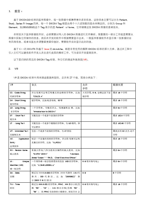
VR是DICOM标准中用来描述数据类型的,总共有27个值。
简单分类如下:
DICOM TAG分类和说明
Patient Tag
Study Tag
Series Tag
Image Tag。
DICOM的常用Tag分类和说明

1.前言:
基于DICOM3.0标准的医学图像中,每一张图像中都携带着许多的信息,这些信息主要可以分为Patient, Study, Series和Image四类。
每一个DICOM Tag都是由两个十六进制数的组合来确定的,分别为Group和Element。
如(0010,0010)这个Tag表示的是Patient’s Name,它存储着这张DICOM图像的患者姓名。
在研发关于医学影像软件时,必然需要对导入的DICOM图像进行文件解析,很重要的一部分工作就是需要从图像中获取它所储存的信息,然后在开发的软件中根据需要显示出来。
一般医学影像软件在显示某一张影像时会有四角信息,或者当医生在调整窗宽窗位值时,需要软件实时显示此时的值。
基于C++的DCMTK和基于Java的dcm4che,都是非常优秀的解释DICOM标准的第三方库,通过在工程中引入它们可以避免软件开发人员去进行底层的解析工作,可为项目开发提高效率。
以下是归纳的常见的DICOM Tag标签,和它们的描述和值表现(VR)。
2.VR
VR是DICOM标准中用来描述数据类型的,总共有27个值。
简单分类如下:
3.DICOM TAG分类和说明Patient Tag。
DICOM_札记

=====================================DICOM标准及其原理分析=====================================1>DICOM标准内容和范畴DICOM标准由多个文档组成,按照国际标准的规范书写.。
目前,DICOM标准共有18个章节和若干扩充部分2>DICOM3.0标淮的特点DICOM3.0标准是医学信息通信领域的国际标准协议,它的突出特点有:(1)DICOM3.0协议是一种基于TCP/IP的上层网络协议DICOM协议要求在数据的编码、传输之前,必须先进行连接协商以确认双方同意某些特定的条件,可以完成特定的通信功能。
DICOM连接协商成功称为建立了一个“关联"(Association)。
只有在建立“关联"之后,才能进行DICOM命令和数据的发送和接收。
(2)DICOM数据编码的特点◆标准定义了26种内部数据类型。
◆像素数据的编码支持JPEG图像压缩。
◆图像可以包含缩略图像和J下常图像,也可以有多帧格式。
◆DICOM标准支持多个字符集。
◆DICOM具有自己独特的数据模型。
◆DICOM通过“信息对象定义"IOD(Information 0bject Definition)的形式来完整地建立和定义医院环境下的数据模型。
◆DICOM使用“全局唯一标识”UID(Unique IdentifieO在网络环境下唯一地标识各种IOD信息对象,使之不致混淆(3)拥有完整、庞大的数据字典DICOM3.0标准拥有一个庞大的数据字典,其内容包含了几乎所有医疗环境下的常用数据,可以完整地描述各种医学设备、图像格式数据以及病人相关信息。
数据字典的条目以“数据元素’’(Data Element)为单位,每个数据元素描述一项数据内容,如病人姓名、检查日期、一幅图像的像素数据等都可以是一个数据元素。
数据字典具有可扩充性。
DICOM标准预留出数据字典的一部分,允许各厂家按照标准格式自定义新的数据元素。
DICOM图像浏览器

Image Viewer using Digital Imaging and Communications inMedicine (DICOM)Trupti N. BaraskarDepartment of Information Technology, Maharashtra Institute of Technology, Pune University, Maharashtra, India Email: trupti_001@, baraskartn@Mobile No. +91-9922789956, +91-20-25462867Abstract- Digital Imaging and Communications in Medicine is a standard for handling, storing, printing, and transmitting information in medical imaging. The National Electrical Manufacturers Association holds the copyright to this standard. It was developed by the DICOM Standards committee. The other image viewers cannot collectively store the image details as well as the patient's information. So the image may get separated from the details, but DICOM file format stores the patient's information and the image details. Main objective is to develop a DICOM image viewer. The image viewer will open .dcm i.e. DICOM image file and also will have additional features such as zoom in, zoom out, black and white inverter, magnifier, blur, B/W inverter, horizontal and vertical flipping, sharpening, contrast, brightness and .gif converter are incorporated.Keyword - Digital Imaging and Communication in Medicine (DICOM), National Electrical Manufacturers Association (NEMA), Information Object Definitions (IOD), Value Representation (VR).I.IntroductionDICOM stands for Digital Imaging and Communication in Medicine. The DICOM standard addresses the basic connectivity between different imaging devices and also the workflow in a medical imaging department. The DICOM standard was created by the National Electrical Manufacturers Association (NEMA) and it also addresses distribution and viewing of medical images. The standard comprises of 16 parts [1] and it is freely available at the NEMA website: ./dicom.html[2] .Within the innards of the standard are also contained a detailed specification of the file format for images. The latest version of the document is as of 2008[3]. In this article present a viewer for DICOM images DICOM Image File FormatThis present a brief description of the DICOM image file format. Like other image file formats, a DICOM file consists of a header, followed by pixel data. The header comprises of the patient name and other patient particulars and image details. Important in the image details are the image dimensions - width, height and image bits per pixel. All these details are hidden inside the DICOM file in the form of tags and their values. Before it gets into tags and values, a brief about DICOM itself and related terminology is in place. In what follows, this explains only those terms and concepts related to a DICOM file. In particular, this does not discuss the communication and network aspects of the DICOM standard. Everything in DICOM is an object - medical device, patient, etc. An object, as in object oriented programming is characterized by attributes. DICOM objects are standardized according to IODs (Information Object Definitions). An IOD is a collection of attributes describing a data object. In other words, an IOD is a data abstraction of a class of similar real world objects which defines the nature and attributes relevant to that class [4]. DICOM has also standardized on the most commonly used attributes and these are listed in the DICOM data dictionary [6]. An application which does not find a needed attribute name in this standardized list may add its own private entry, termed as a private tag; proprietary attributes are therefore possible in DICOM. Examples of attributes are study date, patient name, modality, transfer syntax UID, etc. As it can be seen, the attributes require different data types for correct representation. This “data type” is termed as Value Representation (VR) in DICOM. There are 27 such VRs defined[5], and these are AE, AS, AT, CS, DA, DS, DT, FL, FD, IS, LO, LT, OB, OF, OW, PN,SH, SL, SQ, SS, ST, TM, UI, UL, UN, US, and UT. For example, DT represents Date Time, a concatenated date time character string in the format YYYYMMDDHHMMSS.FFFFFF&ZZXX. An important characteristic of VR is its length, which should always be even. Characterizing an attribute are its tag, VR, VM (Value Multiplicity) and value. A tag is a 4 byte value which uniquely identifies that attribute. A tag is divided into two parts, the Group Tag and the Element Tag, each of which is of length 2 bytes. For example, the tag 0010 0020 (in hexadecimal) represents Patient ID, with a VR of LO (Long String). In this example, 0010 (hex) is the group tag, and 0020 (hex) is the element tag. The DICOM data dictionary gives a list of all the standardized group and element tags. Also important is to know whether a tag is mandatory or not. For data element type, five categories are defined - Type 1, Type 1C, Type 2, Type 2C, and Type 3. One more important concept is transfer syntax. In simple terms, it tells whether a device can accept the data sent by another device. EachCP1324,I nt e r nat i onal Conf e r e nc e on M e t hods and M ode l s i n Sc i e nc e and Te c hnol ogy (I CM 2ST-10)e di t e d by R. B. Pa t e l a nd B. P. Si ngh© 2010 A m e r i c a n I ns t i t ut e of Phys i c s 978-0-7354-0879-1/10/$30.00device comes with its own DICOM conformance statement, which lists all transfer syntaxes acceptableto the device. Transfer syntax tells how the transferred data and messages are encoded. Part [5 ] of the DICOM standard gives the transfer syntax as a set of encoding rules that allow application entities to unambiguously negotiate the encoding techniques (e.g., data element structure[8], byte ordering, compression) they are able to support, thereby allowing these application entities to communicate. (One more term here - Application Entity is the nameof a DICOM device or program used to uniquely identify it.)Transfer syntaxes for non-compressed images are:x Implicit VR Little Endian, with UID1.2.840.10008.1.2x Explicit VR Little Endian, with UID1.2.840.10008.1.2.1x Explicit VR Big Endian, with UID1.2.840.10008.1.2.2Images compressed using JPEG Lossy or Lossless compression techniques have their own transfer syntax UIDs. A viewer should be able to identify the transfer syntax and decode the image data accordingly; or display appropriate error messages if it cannot handle it. More points on a DICOM file, it is a binary file, which means that an ASCII-character-based text editor like notepad does not show it properly. A DICOM file may be encoded in Little Endian or Big Endian byte orders. Elements in a DICOM file are always in ascending order of tags. Private tags are always odd numbered. With this background, it is now time to develop into the DICOM File Format. A DICOM file consists of Preamble: comprising of 128 bytes, followed by, Prefix: comprising of the characters 'D', 'I', 'C', 'M', followed by, File Meta Header: This comprises, among others, of the Media SOP Class UID, Media SOP Instance UID, and the transfer syntax UID. By default, these are encoded in explicit VR, Little Endian. The data is to be read and interpreted depending upon the VR type. Data Set comprising of a number of DICOM Elements, characterized by tags and their values. The main functionality of a DICOM Image Reader is to read the different tags as per the transfer syntax and then use these values appropriately. An image viewer needs to read the image attributes - image width, height, bits per pixel and the actual pixel data. The viewer presented here can be used to view DICOM images with non-compressed transfer syntax. Open DICOM files with Explicit VR and Implicit VR Transfer Syntax, read DICOM files where image bit depth is 8 or 16 bits. Read a DICOM file with just one image inside it. Read a DICONDE file (a DICONDE file is a DICOM file with NDE - Non Destructive Evaluation - tags inside it). Display the tags in a DICOM file.Enable user to save a DICOM image as JPEG/GIF. This viewer is not intended to check whether all mandatory tags are present, open files with VR other than Explicit and Implicit - in particular, not to open JPEG compressed loss and lossless files. To read old DICOM files - requires the preamble and prefix for sure. Earlier DICOM files do not have the preamble and prefix, and just contain the string 1.2.840.10008 somewhere in the beginning. For the viewer, the preamble and prefix are necessary to read images which are not 8 bit or 16 bit in bit depth. In particular not to open color images, to read a sequence of images. Problem:Other file format used in modern times doesn’t have the facility to obtain the image of the patient and the related details together in the same document thus more storage space is required and difficulties are faced by the user.Solution:We can develop a viewer that that displays the patient details and image details in just one click and in just one file thus making it convenient for the end user. It also takes less storage space.II.Block Diagram Of DICOM FileStructureFigure 1. Block Diagram of DICOM file formatFile Header:The header consists of a 128 byte File Preamble, followed by a 4 byte DICOM prefix. The header may or may not be included in the file [9].Preamble Prefix128 bytes=??? ???4 bytes = ‘D’, ‘I’, ‘C’, ‘M’Table 1: DICOM File HeaderThe DICOM standard does not require any structure for the fixed size preamble. It is not required to be structured as a DICOM data element with a tag and a length. It is intended to facilitate access to the imagesand other data in the DICOM file by providing compatibility with a number of commonly used computer image file formats. If the File preamble is not used by an application profile or a specific implementation, all 128 bytes shall be set to 00H. This is intended to facilitate the recognition that the preamble is used when all 128 bytes are not set as specified above. The file preamble may for example contain information enabling a multi-media application to randomly access images stored in a DICOM data set. The same file can be accessed in two ways: First by a multi-media application using the preamble and second by a DICOM application which ignores the preamble. The four byte DICOM prefix shall contain the character string "DICM" encoded as uppercase characters of the ISO 8859 G0 Character Repertoire. This four byte prefix is not structured as a DICOM data element with a tag and a length.Sample output:Figure 2. Image view at DICOM file formatData Set:Data Element: A unit of information as defined by a single entry in the data dictionary [6]. An encoded Information Object Definition (IOD) attribute that is composed at a minimum three fields: Data Element Tag, Value Length, and Value Field. For some specific transfer syntaxes, a data element also contains a VR field where the value representation of that data element is specified explicitly.Data Element Tag: A unique identifier for a data element composed of an ordered pair of numbers (a group number followed by an element number).Data Element Type: Used to specify whether an attribute of an Information Object Definition. This translates to whether a Data Element of a Data Set is mandatory, mandatory only under certain conditions, or optional.Data Set: Exchanged information consisting of a structured set of Attribute values directly or indirectly related to Information Objects. The value of each Attribute in a Data Set is expressed as a Data Element. A collection of data elements ordered by increasingdata element tag number that is an encoding of the values of attributes of a real world object.Pixel Data: Graphical data (e.g., images or overlays) of variable pixel-depth encoded in the pixel data element, with value representation OW or OB. Additional descriptor data elements are often used to describe the contents of the pixel data element.Value Field: The field within a data element that contains the value(s) of that data element.Value Length: The field within a data element that contains the length of the value field of the data elementValue Representation (VR): Specifies the data type and format of the value(s) contained in the value field of a data element. A data set represents an instance of a real world information object. A data set is constructed of data elements. Data elements contain the encoded values of attributes of that object. The construction, characteristics, and encoding of a data set and its data elements are discussed here.Data Elements: A Data Element is uniquely identified by a Data Element Tag. The Data Elements in a Data Set shall be ordered by increasing Data Element Tag Number and shall occur at most once in a Data Set. There are 2 types of data elementStandard Data Elements have an even Group Number that is not (0000, eeee), (0002, eeee), (0004, eeee), or (0006, eeee). [5]Private Data Elements have an odd Group Number that is not (0001, eeee), (0003, eeee), (0005, eeee), (0007, eeee), or (FFFF, eeee).Although similar or related Data Elements often have the same Group Number; a Data Group does not convey any semantic meaningNote: A Data Element shall have one of three structures. Two of these structures contain the VR of the Data Element (Explicit VR) but differ in the way their lengths are expressed, while the other structure does not contain the VR (Implicit VR). All three structures contain the Data Element Tag, Value Length and Value for the Data Element. See Figure 2.Implicit and Explicit VR Data Elements shall not coexist in a Data Set and Data Sets nested within it. Whether a Data Set uses Explicit or Implicit VR, among other characteristics, is determined by the negotiated Transfer SyntaxFigure 3. Data set and Data elements structureData Element Field:A data element is made up of fields. Three fields are common to all three data element structures; these are the Data Element Tag, Value Length, and Value Field.A fourth field, Value Representation is only present in the two Explicit VR Data Element structures. The definitions of the fields are [6]:Data Element Tag: An ordered pair of 16-bit unsigned integers representing the group number followed by element number.Value Representation: A two byte character string contains the VR of the data element. The VR for a given data element tag shall be as defined by the data dictionary. The two characters VR shall be encoded using characters from the DICOM default character set.Value Length:A 16 or 32-bit (dependent on VR and whether VR is explicit or implicit) unsigned integer containing the explicit length of the value field as the number of bytes (even) that make up the value. It does not include the length of the data element tag, value representation, and value length fields.A 2-bit length field set to undefined length (FFFFFFFFH). Undefined lengths may be used for data elements having the Value Representation (VR) Sequence of Items (SQ) and Unknown (UN). For data elements with value representation OW or OB undefined length may be used depending n the negotiated transfer syntax.Value Field: An even number of bytes containing the Value(s) of the Data Element. The data type of value(s) stored in this field is specified by the data element's VR. The VR for a given data element tag can be determined using the data dictionary [6], or using the VR field if it is contained explicitly within the data element. The VR of standard data elements shall agree with those specified in the data dictionary. The value multiplicity specifies how many values with this VR can be placed in the value field. If the VM is greater than one, multiple values shall be delimited within the value field. The VMs of standard data elements are specified in the data dictionary value fields with undefined length are delimited through the use of sequence delimitation items and item delimitation data elements.RGB Color Model:A color in the RGB color model is described by indicating how much of each of the red, green, and blue is included. The color is expressed as an RGB triplet (r, g, b), each component of which can vary from zero to a defined maximum value. If all the components are at zero the result is black; if all are at maximum, the result is the brightest represent able white [11, 12, and 13].These ranges may be quantified in several different ways: From 0 to 1, with any fractional value in between. This representation is used in theoretical analyses, and in systems that use floating-point representations. Each color component value can also be written as a percentage, from 0% to 100%.ting, the component values are often stored as integer numbers in the range 0 to 255, the range that a single 8-bit byte can offer (by encoding 256 distinct values).High-end digital image equipment can deal with the integer range 0 to 65,535 for each primary color, by employing 16-bit words instead of 8-bit bytes. For example, the full intensity red is written in the different RGB notations as given table 2:Notation RGB tripletArithmetic(1.0, 0.0, 0.0) Percentage(100%, 0%, 0%)Digital 8-bit per channel(255, 0, 0)Table 2. RGB NotationIn this paper the digital 8-bit model as the input file is in binary for the RGB model and the ASCII code of the color has been used in almost all the algorithms of the features of the DICOM Image Viewer.III.System ArchitectureDescription:Parser: It is the first and the most important part of the project. It separates the data set and the image. The input to parser is .dcm file (i.e. a binary file) which is version 1.3, standardized by NEMA [10]. The output i.e. image and data set is then passed on to next stage. DICOM Header: The header is the part which makes DICOM different from all the other file formats. The header encompasses of patient details and image information like pixel representation, height, width, file data length etc. Thus in this stage we read patient details and image information so that they can be processed further.Display Details: The Header details which are processed in the above stage are displayed.Image Processing: In this stage, various features like Zoom In, Zoom Out, Blur, B/W inverter, Horizontal and Vertical flipping, Sharpening, Contrast, Brightness and .gif converter are incorporated. The final processed image in then passed onto the next stage.DICOM Image Viewer: The final image is displayed in this stage.IV.Implementation Images:Figure 5. Image and Text ViewerFigure 6. Invert ImageFigure 7. Blur imageFigure 8. White Inverter imageFigure 9. Zoom In ImageFigure 10. Sharpen ImageFigure 11. Flip Horizontal Image[10]Boqiang Liu, Minghui Zhu, Zhenwang Zhang, CongYin, Zhongguo Liu and Jason Gu* “Medical ImageConversion with DICOM” IEEE2007[11]Foley J., A. van Dam, Fejner S. Hughes J., Computergraphics - principles and practice, Second edition,Addison-Wesley, 1996[12]Gonzales R., R. Woods, Digital Image Processing,Adison Wesley, 1993[13]Marion A., An Introduction to Image Processing,Chapman and Hall, 1991Figure 11. Save Image FileV.ConclusionThe primary aim of this paper is to study “.dcm” fileformat and develop an image viewer with someenhanced features like Zoom, invert, brightness,sharpen and save image as “.gif” format.Implementation of algorithms e.g.. blur, horizontalflipping, vertical flipping black and white inversionetc. Benefits of the “.dcm” file format and imageviewer provides a greater leap to the present medicalscenario further helps for the future development ofvideo player for CT scan and MRI. Display the x-rayimage and the patient details together. Converts“.dcm” file to “.gif” file format. It is cost-effective inand helpful to health care industry. Because of theDICOM viewer images can be captured andcommunicated more quickly helps physician tomake diagnosis sooner and treatment decision can bemade quickly.References[1]Mario Mustra, Kresimir Delac, Mislav Grgic “Overviewof the DICOM Standard” 50th International SymposiumELMAR-2008, 10-12 September 2008, Zadar, Croatia[2]The DICOM Standard, /[3]Digital Imaging and Communications in Medicine(DICOM), NEMA Publications,"DICOM Standard",2008, available at:ftp:///medical/dicom/2008/[4]National Electrical Manufacturers Association “DigitalImaging and Communications in Medicine (DICOM)”Standards Publication PS 3.3-2004, 2009[5]National Electrical Manufacturers Association “DigitalImaging and Communications in Medicine (DICOM)”Standards Publication PS 3.5-2004, 2009[6]National Electrical Manufacturers Association “DigitalImaging and Communications in Medicine (DICOM)”Standards Publication PS 3.6-2004, 2009[7]National Electrical Manufacturers Association “DigitalImaging and Communications in Medicine (DICOM)”Standards Publication PS 3.7-2004, 2009[8]National Electrical Manufacturers Association “DigitalImaging and Communications in Medicine (DICOM)”Standards Publication PS 3.10-2004, 2009[9] “DICOM file structure.htmlCopyright of AIP Conference Proceedings is the property of American Institute of Physics and its content may not be copied or emailed to multiple sites or posted to a listserv without the copyright holder's express written permission. However, users may print, download, or email articles for individual use.。
Dicom基础概述

• Part3:信息对象定义
– 定义了应用于数字医学图像以及相关信息(如波形,格式化报告、 放射治疗药剂等)通信的真实世界实体的抽象说明。 – 定义了标准信息对象类和复合信息对象类; – 描述了现实世界模型及在信息对象定义中反映的相应信息模型。
DICOM - 2008文档结构
• Part4:服务类描述
– 1996版增加了媒体交换标准; – 新的图像定义, 比如:X线心血管成像, X光数字乳腺 成像…… – Modality Performed Procedure Step (MPPS), 结构化报 告……
DICOM Storage & Query/Retrieve
CT,MR CR,US, Sec Capt X线心血管成像 X线荧光镜成像 核医学 新的超声图像
• Part6:数据字典
– 定义了所有的DICOM数据单元以及UIDs。
• Part7:消息交换
– 定义了DICOM消息服务单元(DIMSE),以及所有的DICOM网络服务。
DICOM - 2008文档结构
• Part8:消息交换的网络通信支持
dictionary怎么读

dictionary怎么读
“Dictionary”是一个英语单词,发音为/ˈdɪkʃəneri/。
这个单词由三个音节组成:第一个音节是“Dic”,读作/ˈdɪk/,第二个音节是“tion”,读作/ˈʃən/,最后一个音节是“ary”,读作/ˈreri/。
在发音时,注意将重音放在第一个音节“Dic”上,并保持每个音节的清晰和准确。
要正确掌握“Dictionary”的发音,可以通过多听、多模仿来实现。
可以听英语母语者的发音示范,注意他们的发音细节和语调,然后尝试模仿。
此外,也可以通过在线发音工具或手机应用程序来辅助练习发音。
“Dictionary”是一个非常重要的工具,用于查找单词的定义、用法和发音等信息。
对于英语学习者来说,掌握它的发音和用法至关重要。
通过正确的发音和频繁的使用,我们可以更好地利用“Dictionary”来扩展词汇量、提高英语水平。
总之,正确掌握“Dictionary”的发音对于英语学习者来说非常重要。
通过多听、多模仿和频繁使用,我们可以更好地利用这个工具来提高英语水平。
DICOM协议目录

DICOM协议目录DICOM(Digitalimaging and Communications in Medicine)数字影像和通信标准DICOM3.0.2004在2004年11月发布。
DICOM 3.0标准共有18个部分,其各部分的内容概要如下:第一部分:引言与概述,简要介绍了DICOM的概念及其组成。
第二部分:DICOM兼容性声明。
声明DICOM要求制造商精确地描述其产品的DICOM 兼容性,即构造一个该产品的DICOM兼容性声明。
第三部分:DICOM信息对象定义。
介绍了lOD和SOP类。
第四部分:服务类,说明了14个服务类,服务类详细介绍了功能与信息对象上的命令及其产生的结果。
第五部分:数据结构及语意,描述了怎样对信息对象类和服务类进行构造和编码。
第六部分:数据字典,描述了所有信息对象是由数据元素组成的,数据元素是对属性值的编码。
第七部分:消息交换,定义了进行消息交换时相互通讯的医学图像应用实体所用到的服务和协议。
第八部分:消息交换的网络通讯支持,说明了在网络环境下的通讯服务和支持DICOM应用实体进行消息交换的必要的上层协议。
第九部分:消息交换的点对点通讯支持。
由于目前在实际中很少使用点对点通信,该部分在DICOM 2003版中已经被删除。
第十部分:介质存储与文件格式。
第十一部分:介质存储应用描述。
第十二部分:存储功能和用于数据交换的介质格式。
第十三部分:打印管理的点对点通讯支持。
该部分在DICOM 2003中也已被删除。
第十四部分:灰度图的标准显示(显示和打印)功能。
第十五部分:安全特性描述。
第十六部分:内容资源映射。
第十七部分:说明信息。
这部分包含标准化表格和信息附件中的说明信息。
第十八部分:WEB访问DICOM持久对象。
定义基于WEB的服务,用于访问DICOM持久对象。
提供从HTML页面或者XML文档访问DICOM持久对象的简单机制。
DICOM简介一dicom是什么?二dicom文件结构三如何编写dicom程序四利用开发包开发dicom程序五dcmtk使用介绍一dicom是什么?dicom全名是医学数字影像和通讯。
- 1、下载文档前请自行甄别文档内容的完整性,平台不提供额外的编辑、内容补充、找答案等附加服务。
- 2、"仅部分预览"的文档,不可在线预览部分如存在完整性等问题,可反馈申请退款(可完整预览的文档不适用该条件!)。
- 3、如文档侵犯您的权益,请联系客服反馈,我们会尽快为您处理(人工客服工作时间:9:00-18:30)。
using System.Collections.Generic;// Dicom Dictionary.// Written by Amarnath S, Mahesh Reddy S, Bangalore, India, April 2009.// This Dicom Dictionary does not contain all tags specified in Part 6 of the Standard, // but contains tags used 80 percent of the time.// Updated by Harsha T, Apr 2010// Inspired heavily by ImageJ// Update from July 2012: Checked with DICOM Standard Part 6, Version 2011namespace DicomImageViewer{class DicomDictionary{public Dictionary<string, string> dict = new Dictionary<string,string>(){{"00020002", "UIMedia Storage SOP Class UID"},{"00020003", "UIMedia Storage SOP Instance UID"},{"00020010", "UITransfer Syntax UID"},{"00020012", "UIImplementation Class UID"},{"00020013", "SHImplementation Version Name"},{"00020016", "AESource Application Entity Title"},{"00080005", "CSSpecific Character Set"},{"00080008", "CSImage Type"},{"00080010", "CSRecognition Code"},{"00080012", "DAInstance Creation Date"},{"00080013", "TMInstance Creation Time"},{"00080014", "UIInstance Creator UID"},{"00080016", "UISOP Class UID"},{"00080018", "UISOP Instance UID"},{"00080020", "DAStudy Date"},{"00080021", "DASeries Date"},{"00080022", "DAAcquisition Date"},{"00080023", "DAContent Date"},{"00080024", "DAOverlay Date"},{"00080025", "DACurve Date"},{"00080030", "TMStudy Time"},{"00080031", "TMSeries Time"},{"00080032", "TMAcquisition Time"},{"00080033", "TMContent Time"},{"00080034", "TMOverlay Time"},{"00080035", "TMCurve Time"},{"00080040", "USData Set Type"},{"00080041", "LOData Set Subtype"},{"00080042", "CSNuclear Medicine Series Type"},{"00080050", "SHAccession Number"},{"00080052", "CSQuery/Retrieve Level"},{"00080054", "AERetrieve AE Title"},{"00080058", "AEFailed SOP Instance UID List"},{"00080060", "CSModality"},{"00080064", "CSConversion Type"},{"00080068", "CSPresentation Intent Type"},{"00080070", "LOManufacturer"},{"00080080", "LOInstitution Name"},{"00080081", "STInstitution Address"},{"00080082", "SQInstitution Code Sequence"},{"00080090", "PNReferring Physician's Name"},{"00080092", "STReferring Physician's Address"},{"00080094", "SHReferring Physician's Telephone Numbers"},{"00080096", "SQReferring Physician Identification Sequence"}, {"00080100", "SHCode Value"},{"00080102", "SHCoding Scheme Designator"},{"00080103", "SHCoding Scheme Version"},{"00080104", "LOCode Meaning"},{"00080201", "SHTimezone Offset From UTC"},{"00081010", "SHStation Name"},{"00081030", "LOStudy Description"},{"00081032", "SQProcedure Code Sequence"},{"0008103E", "LOSeries Description"},{"00081040", "LOInstitutional Department Name"},{"00081048", "PNPhysician(s) of Record"},{"00081050", "PNPerforming Physician's Name"},{"00081060", "PNName of Physician(s) Reading Study"},{"00081070", "PNOperator's Name"},{"00081080", "LOAdmitting Diagnoses Description"},{"00081084", "SQAdmitting Diagnoses Code Sequence"},{"00081090", "LOManufacturer's Model Name"},{"00081100", "SQReferenced Results Sequence"},{"00081110", "SQReferenced Study Sequence"},{"00081111", "SQReferenced Performed Procedure Step Sequence"}, {"00081115", "SQReferenced Series Sequence"},{"00081120", "SQReferenced Patient Sequence"},{"00081125", "SQReferenced Visit Sequence"},{"00081130", "SQReferenced Overlay Sequence"},{"00081140", "SQReferenced Image Sequence"},{"00081145", "SQReferenced Curve Sequence"},{"00081150", "UIReferenced SOP Class UID"},{"00081155", "UIReferenced SOP Instance UID"},{"00082111", "STDerivation Description"},{"00082112", "SQSource Image Sequence"},{"00082120", "SHStage Name"},{"00082122", "ISStage Number"},{"00082124", "ISNumber of Stages"},{"00082129", "ISNumber of Event Timers"},{"00082128", "ISView Number"},{"0008212A", "ISNumber of Views in Stage"},{"00082130", "DSEvent Elapsed Time(s)"},{"00082132", "LOEvent Timer Name(s)"},{"00082142", "ISStart Trim"},{"00082143", "ISStop Trim"},{"00082144", "ISRecommended Display Frame Rate"},{"00082200", "CSTransducer Position"},{"00082204", "CSTransducer Orientation"},{"00082208", "CSAnatomic Structure"},{"00100010", "PNPatient's Name"},{"00100020", "LOPatient ID"},{"00100021", "LOIssuer of Patient ID"},{"00100022", "CSType of Patient ID"},{"00100030", "DAPatient's Birth Date"},{"00100032", "TMPatient's Birth Time"},{"00100040", "CSPatient's Sex"},{"00100050", "SQPatient's Insurance Plan Code Sequence"},{"00100101", "SQPatient's Primary Language Code Sequence"},{"00100102", "SQPatient's Primary Language Modifier Code Sequence"}, {"00101000", "LOOther Patient IDs"},{"00101001", "PNOther Patient Names"},{"00101005", "PNPatient's Birth Name"},{"00101010", "ASPatient's Age"},{"00101020", "DSPatient's Size"},{"00101030", "DSPatient's Weight"},{"00101040", "LOPatient's Address"},{"00101050", "LOInsurance Plan Identification"},{"00102000", "LOMedical Alerts"},{"00102110", "LOAllergies"},{"00102150", "LOCountry of Residence"},{"00102152", "LORegion of Residence"},{"00102154", "SHPatient's Telephone Numbers"},{"00102160", "SHEthnic Group"},{"00102180", "SHOccupation"},{"001021A0", "CSSmoking Status"},{"001021B0", "LTAdditional Patient History"},{"00102201", "LOPatient Species Description"},{"00102203", "CSPatient Sex Neutered"},{"00102292", "LOPatient Breed Description"},{"00102297", "PNResponsible Person"},{"00102298", "CSResponsible Person Role"},{"00102299", "CSResponsible Organization"},{"00104000", "LTPatient Comments"},{"00180010", "LOContrast/Bolus Agent"},{"00180015", "CSBody Part Examined"},{"00180020", "CSScanning Sequence"},{"00180021", "CSSequence Variant"},{"00180022", "CSScan Options"},{"00180023", "CSMR Acquisition Type"},{"00180024", "SHSequence Name"},{"00180025", "CSAngio Flag"},{"00180030", "LORadionuclide"},{"00180031", "LORadiopharmaceutical"},{"00180032", "DSEnergy Window Centerline"},{"00180033", "DSEnergy Window Total Width"},{"00180034", "LOIntervention Drug Name"},{"00180035", "TMIntervention Drug Start Time"},{"00180040", "ISCine Rate"},{"00180050", "DSSlice Thickness"},{"00180060", "DSKVP"},{"00180070", "ISCounts Accumulated"},{"00180071", "CSAcquisition Termination Condition"},{"00180072", "DSEffective Duration"},{"00180073", "CSAcquisition Start Condition"},{"00180074", "ISAcquisition Start Condition Data"},{"00180075", "ISAcquisition Termination Condition Data"}, {"00180080", "DSRepetition Time"},{"00180081", "DSEcho Time"},{"00180082", "DSInversion Time"},{"00180083", "DSNumber of Averages"},{"00180084", "DSImaging Frequency"},{"00180085", "SHImaged Nucleus"},{"00180086", "ISEcho Numbers(s)"},{"00180087", "DSMagnetic Field Strength"},{"00180088", "DSSpacing Between Slices"},{"00180089", "ISNumber of Phase Encoding Steps"},{"00180090", "DSData Collection Diameter"},{"00180091", "ISEcho Train Length"},{"00180093", "DSPercent Sampling"},{"00180094", "DSPercent Phase Field of View"},{"00180095", "DSPixel Bandwidth"},{"00181000", "LODevice Serial Number"},{"00181004", "LOPlate ID"},{"00181010", "LOSecondary Capture Device ID"},{"00181012", "DADate of Secondary Capture"},{"00181014", "TMTime of Secondary Capture"},{"00181016", "LOSecondary Capture Device Manufacturer"},{"00181018", "LOSecondary Capture Device Manufacturer's Model Name"}, {"00181019", "LOSecondary Capture Device Software Versions"},{"00181020", "LOSoftware Versions(s)"},{"00181022", "SHVideo Image Format Acquired"},{"00181023", "LODigital Image Format Acquired"},{"00181030", "LOProtocol Name"},{"00181040", "LOContrast/Bolus Route"},{"00181041", "DSContrast/Bolus Volume"},{"00181042", "TMContrast/Bolus Start Time"},{"00181043", "TMContrast/Bolus Stop Time"},{"00181044", "DSContrast/Bolus Total Dose"},{"00181045", "ISSyringe Counts"},{"00181050", "DSSpatial Resolution"},{"00181060", "DSTrigger Time"},{"00181061", "LOTrigger Source or Type"},{"00181062", "ISNominal Interval"},{"00181063", "DSFrame Time"},{"00181064", "LOCardiac Framing Type"},{"00181065", "DSFrame Time Vector"},{"00181066", "DSFrame Delay"},{"00181070", "LORadiopharmaceutical Route"},{"00181071", "DSRadiopharmaceutical Volume"},{"00181072", "TMRadiopharmaceutical Start Time"},{"00181073", "TMRadiopharmaceutical Stop Time"},{"00181074", "DSRadionuclide Total Dose"},{"00181075", "DSRadionuclide Half Life"},{"00181076", "DSRadionuclide Positron Fraction"},{"00181080", "CSBeat Rejection Flag"},{"00181081", "ISLow R-R Value"},{"00181082", "ISHigh R-R Value"},{"00181083", "ISIntervals Acquired"},{"00181084", "ISIntervals Rejected"},{"00181085", "LOPVC Rejection"},{"00181086", "ISSkip Beats"},{"00181088", "ISHeart Rate"},{"00181090", "ISCardiac Number of Images"},{"00181094", "ISTrigger Window"},{"00181100", "DSReconstruction Diameter"},{"00181110", "DSDistance Source to Detector"},{"00181111", "DSDistance Source to Patient"},{"00181120", "DSGantry/Detector Tilt"},{"00181130", "DSTable Height"},{"00181131", "DSTable Traverse"},{"00181140", "CSRotation Direction"},{"00181141", "DSAngular Position"},{"00181142", "DSRadial Position"},{"00181143", "DSScan Arc"},{"00181144", "DSAngular Step"},{"00181145", "DSCenter of Rotation Offset"},{"00181146", "DSRotation Offset"},{"00181147", "CSField of View Shape"},{"00181149", "ISField of View Dimensions(s)"},{"00181150", "ISExposure Time"},{"00181151", "ISX-ray Tube Current"},{"00181152", "ISExposure"},{"00181153", "ISExposure in uAs"},{"00181154", "DSAverage Pulse Width"},{"00181155", "CSRadiation Setting"},{"00181156", "CSRectification Type"},{"0018115A", "CSRadiation Mode"},{"0018115E", "DSImage and Fluoroscopy Area Dose Product"}, {"00181160", "SHFilter Type"},{"00181161", "LOType of Filters"},{"00181162", "DSIntensifier Size"},{"00181164", "DSImager Pixel Spacing"},{"00181166", "CSGrid"},{"00181170", "ISGenerator Power"},{"00181180", "SHCollimator/grid Name"},{"00181181", "CSCollimator Type"},{"00181182", "ISFocal Distance"},{"00181183", "DSX Focus Center"},{"00181184", "DSY Focus Center"},{"00181190", "DSFocal Spot(s)"},{"00181191", "CSAnode Target Material"},{"001811A0", "DSBody Part Thickness"},{"001811A2", "DSCompression Force"},{"00181200", "DADate of Last Calibration"},{"00181201", "TMTime of Last Calibration"},{"00181210", "SHConvolution Kernel"},{"00181242", "ISActual Frame Duration"},{"00181243", "ISCount Rate"},{"00181250", "SHReceive Coil Name"},{"00181251", "SHTransmit Coil Name"},{"00181260", "SHPlate Type"},{"00181261", "LOPhosphor Type"},{"00181300", "ISScan Velocity"},{"00181301", "CSWhole Body Technique"},{"00181302", "ISScan Length"},{"00181310", "USAcquisition Matrix"},{"00181312", "CSIn-plane Phase Encoding Direction"},{"00181314", "DSFlip Angle"},{"00181315", "CSVariable Flip Angle Flag"},{"00181316", "DSSAR"},{"00181318", "DSdB/dt"},{"00181400", "LOAcquisition Device Processing Description"}, {"00181401", "LOAcquisition Device Processing Code"},{"00181402", "CSCassette Orientation"},{"00181403", "CSCassette Size"},{"00181404", "USExposures on Plate"},{"00181405", "ISRelative X-Ray Exposure"},{"00181450", "CSColumn Angulation"},{"00181500", "CSPositioner Motion"},{"00181508", "CSPositioner Type"},{"00181510", "DSPositioner Primary Angle"},{"00181511", "DSPositioner Secondary Angle"},{"00181520", "DSPositioner Primary Angle Increment"},{"00181521", "DSPositioner Secondary Angle Increment"},{"00181530", "DSDetector Primary Angle"},{"00181531", "DSDetector Secondary Angle"},{"00181600", "CSShutter Shape"},{"00181602", "ISShutter Left Vertical Edge"},{"00181604", "ISShutter Right Vertical Edge"},{"00181606", "ISShutter Upper Horizontal Edge"},{"00181608", "ISShutter Lower Horizontal Edge"},{"00181610", "ISCenter of Circular Shutter"},{"00181612", "ISRadius of Circular Shutter"},{"00181620", "ISVertices of the Polygonal Shutter"},{"00181700", "ISCollimator Shape"},{"00181702", "ISCollimator Left Vertical Edge"},{"00181704", "ISCollimator Right Vertical Edge"},{"00181706", "ISCollimator Upper Horizontal Edge"},{"00181708", "ISCollimator Lower Horizontal Edge"},{"00181710", "ISCenter of Circular Collimator"},{"00181712", "ISRadius of Circular Collimator"},{"00181720", "ISVertices of the Polygonal Collimator"},{"00185000", "SHOutput Power"},{"00185010", "LOTransducer Data"},{"00185012", "DSFocus Depth"},{"00185020", "LOProcessing Function"},{"00185021", "LOPostprocessing Function"},{"00185022", "DSMechanical Index"},{"00185024", "DSBone Thermal Index"},{"00185026", "DSCranial Thermal Index"},{"00185027", "DSSoft Tissue Thermal Index"},{"00185028", "DSSoft Tissue-focus Thermal Index"},{"00185029", "DSSoft Tissue-surface Thermal Index"},{"00185050", "ISDepth of Scan Field"},{"00185100", "CSPatient Position"},{"00185101", "CSView Position"},{"00185104", "SQProjection Eponymous Name Code Sequence"},{"00185210", "DSImage Transformation Matrix"},{"00185212", "DSImage Translation Vector"},{"00186000", "DSSensitivity"},{"00186011", "SQSequence of Ultrasound Regions"},{"00186012", "USRegion Spatial Format"},{"00186014", "USRegion Data Type"},{"00186016", "ULRegion Flags"},{"00186018", "ULRegion Location Min X0"},{"0018601A", "ULRegion Location Min Y0"},{"0018601C", "ULRegion Location Max X1"},{"0018601E", "ULRegion Location Max Y1"},{"00186020", "SLReference Pixel X0"},{"00186022", "SLReference Pixel Y0"},{"00186024", "USPhysical Units X Direction"},{"00186026", "USPhysical Units Y Direction"},{"00181628", "FDReference Pixel Physical Value X"},{"0018602A", "FDReference Pixel Physical Value Y"},{"0018602C", "FDPhysical Delta X"},{"0018602E", "FDPhysical Delta Y"},{"00186030", "ULTransducer Frequency"},{"00186031", "CSTransducer Type"},{"00186032", "ULPulse Repetition Frequency"},{"00186034", "FDDoppler Correction Angle"},{"00186036", "FDSteering Angle"},{"00186038", "ULDoppler Sample Volume X Position (Retired)"},{"00186039", "SLDoppler Sample Volume X Position"},{"0018603A", "ULDoppler Sample Volume Y Position (Retired)"}, {"0018603B", "SLDoppler Sample Volume Y Position"},{"0018603C", "ULTM-Line Position X0 (Retired)"},{"0018603D", "SLTM-Line Position X0"},{"0018603E", "ULTM-Line Position Y0 (Retired)"},{"0018603F", "SLTM-Line Position Y0"},{"00186040", "ULTM-Line Position X1 (Retired)"},{"00186041", "SLTM-Line Position X1"},{"00186042", "ULTM-Line Position Y1 (Retired)"},{"00186043", "SLTM-Line Position Y1"},{"00186044", "USPixel Component Organization"},{"00186046", "ULPixel Component Mask"},{"00186048", "ULPixel Component Range Start"},{"0018604A", "ULPixel Component Range Stop"},{"0018604C", "USPixel Component Physical Units"},{"0018604E", "USPixel Component Data Type"},{"00186050", "ULNumber of Table Break Points"},{"00186052", "ULTable of X Break Points"},{"00186054", "FDTable of Y Break Points"},{"00186056", "ULNumber of Table Entries"},{"00186058", "ULTable of Pixel Values"},{"0018605A", "ULTable of Parameter Values"},{"00187000", "CSDetector Conditions Nominal Flag"},{"00187001", "DSDetector Temperature"},{"00187004", "CSDetector Type"},{"00187005", "CSDetector Configuration"},{"00187006", "LTDetector Description"},{"00187008", "LTDetector Mode"},{"0018700A", "SHDetector ID"},{"0018700C", "DADate of Last Detector Calibration"},{"0018700E", "TMTime of Last Detector Calibration"},{"00187010", "ISExposures on Detector Since Last Calibration"}, {"00187011", "ISExposures on Detector Since Manufactured"},{"00187012", "DSDetector Time Since Last Exposure"},{"00187014", "DSDetector Active Time"},{"00187016", "DSDetector Activation Offset From Exposure"},{"0018701A", "DSDetector Binning"},{"00187020", "DSDetector Element Physical Size"},{"00187022", "DSDetector Element Spacing"},{"00187024", "CSDetector Active Shape"},{"00187026", "DSDetector Active Dimension(s)"},{"00187028", "DSDetector Active Origin"},{"00187030", "DSField of View Origin"},{"00187032", "DSField of View Rotation"},{"00187034", "CSField of View Horizontal Flip"},{"00187040", "LTGrid Absorbing Material"},{"00187041", "LTGrid Spacing Material"},{"00187042", "DSGrid Thickness"},{"00187044", "DSGrid Pitch"},{"00187046", "ISGrid Aspect Ratio"},{"00187048", "DSGrid Period"},{"0018704C", "DSGrid Focal Distance"},{"00187050", "LTFilter Material"},{"00187052", "DSFilter Thickness Minimum"},{"00187054", "DSFilter Thickness Maximum"},{"00187060", "CSExposure Control Mode"},{"00187062", "LTExposure Control Mode Description"}, {"00187064", "CSExposure Status"},{"00187065", "DSPhototimer Setting"},{"0020000D", "UIStudy Instance UID"},{"0020000E", "UISeries Instance UID"},{"00200010", "SHStudy ID"},{"00200011", "ISSeries Number"},{"00200012", "ISAcquisition Number"},{"00200013", "ISInstance Number"},{"00200014", "ISIsotope Number"},{"00200015", "ISPhase Number"},{"00200016", "ISInterval Number"},{"00200017", "ISTime Slot Number"},{"00200018", "ISAngle Number"},{"00200020", "CSPatient Orientation"},{"00200022", "USOverlay Number"},{"00200024", "USCurve Number"},{"00200030", "DSImage Position"},{"00200032", "DSImage Position (Patient)"},{"00200037", "DSImage Orientation (Patient)"},{"00200050", "DSLocation"},{"00200052", "UIFrame of Reference UID"},{"00200060", "CSLaterality"},{"00200070", "LOImage Geometry Type"},{"00200080", "UIMasking Image"},{"00200100", "ISTemporal Position Identifier"},{"00200105", "ISNumber of Temporal Positions"},{"00200110", "DSTemporal Resolution"},{"00201000", "ISSeries in Study"},{"00201002", "ISImages in Acquisition"},{"00201004", "ISAcquisitions in Study"},{"00201040", "LOPosition Reference Indicator"},{"00201041", "DSSlice Location"},{"00201070", "ISOther Study Numbers"},{"00201200", "ISNumber of Patient Related Studies"},{"00201202", "ISNumber of Patient Related Series"},{"00201204", "ISNumber of Patient Related Instances"},{"00201206", "ISNumber of Study Related Series"},{"00201208", "ISNumber of Study Related Instances"},{"00204000", "LTImage Comments"},{"00280002", "USSamples per Pixel"},{"00280004", "CSPhotometric Interpretation"},{"00280006", "USPlanar Configuration"},{"00280008", "ISNumber of Frames"},{"00280009", "ATFrame Increment Pointer"},{"00280010", "USRows"},{"00280011", "USColumns"},{"00280030", "DSPixel Spacing"},{"00280031", "DSZoom Factor"},{"00280032", "DSZoom Center"},{"00280034", "ISPixel Aspect Ratio"},{"00280051", "CSCorrected Image"},{"00280100", "USBits Allocated"},{"00280101", "USBits Stored"},{"00280102", "USHigh Bit"},{"00280103", "USPixel Representation"},{"00280106", "USSmallest Image Pixel Value"},{"00280107", "USLargest Image Pixel Value"},{"00280108", "USSmallest Pixel Value in Series"},{"00280109", "USLargest Pixel Value in Series"},{"00280120", "USPixel Padding Value"},{"00280300", "CSQuality Control Image"},{"00280301", "CSBurned In Annotation"},{"00281040", "CSPixel Intensity Relationship"},{"00281041", "SSPixel Intensity Relationship Sign"},{"00281050", "DSWindow Center"},{"00281051", "DSWindow Width"},{"00281052", "DSRescale Intercept"},{"00281053", "DSRescale Slope"},{"00281054", "LORescale Type"},{"00281055", "LOWindow Center & Width Explanation"},{"00281101", "USRed Palette Color Lookup Table Descriptor"}, {"00281102", "USGreen Palette Color Lookup Table Descriptor"},。
