PD 模型
我行公司客户信用评级模型包括pd模型
我行公司客户信用评级模型包括pd模型信用评分模型是一种传统的信用风险量化模型,利用可观察到的借款人特征变量计算出一个数值(得分)来代表债务人的信用风险,并将借款人归类于不同的风险等级。
对个人客户而言,可观察到的特征变量主要包括收入、资产、年龄、职业以及居住地等;对法人客户而言,包括现金流量、各种财务比率等信用评分模型的关键在于特征变量的选择和各自权重的确定。
目前,应用最广泛的信用评分模型有线性概率模型(Linear Probability Model)、Logit模型、Probit模型和线性辨别模型(Linear Discriminant Model)线性概率模型:线性概率模型的命名是由于它的预测性;在自变量的值可用概率来解释时,应变量能以此概率假定值的单位。
这种模型,在其中应变量是一个虚设变量或双值变量,并用一个或一个以上的自变量的线性函数来表示。
该种模型有助于质的现象的分析。
线性概率模型是使用诸如会计比率之类的历史数据作为模型的输入数据,来解释以前的贷款偿还情况。
我们可以使用在过去贷款偿还中起重要作用的一些因素来预测新贷款的偿还概率。
过去的贷款通常划分为两类,即违约的(Zi=1)和不违约的(Zi=0)。
然后,我们通过对随机变量(Xij)的线性回归来进行估计,Xij表示第j个借款者的数量信息,如收入、财务杠杆或收益率等,通过如下形式的线性回归来估算模型:式中,Bj表示在过去的偿还情况中第j个变量的重要性。
如果我们得到变量j的估计Bj值,并且将其与对未来借款者所观测到的Xij值相乘,并进行加总,得到借款者违约的概率E(Zi)=(1一Pi)=预期的违约率,其中Pi是对贷款偿还的概率。
只要可以获得借款者Xij的当前信息,这种方法是非常直截了当的。
Logit模型:(Logit model,也译作“评定模型”,“分类评定模型”,又作Logistic regression,“逻辑回归”)是离散选择法模型之一,Logit模型是最早的离散选择模型,也是目前应用最广的模型。
PD模型大鼠纹状体中等多棘神经元树突棘运动依赖可塑性研究

PD模型大鼠纹状体中等多棘神经元树突棘运动依赖可塑性研究陈平1,2,刘晓莉1*,马婧1,3,乔德才1(1.北京师范大学体育与运动学院,北京100875;2.吉首大学体育科学学院,湖南吉首416000;3.国家体育总局体育科学研究所,北京100061)摘要:目的:揭示帕金森病(Parkinson’s disease,PD)模型大鼠纹状体中等多棘神经元(medium spiny neurons,MSNs)树突棘运动依赖可塑性发生的细胞靶点。
方法:清洁级SD大鼠随机分为3组:假手术组(Con组)、PD组和PD 运动组(PD+Ex组),采用神经毒素6-羟基多巴胺(6-Hydroxydopamine,6-OHDA)注射于大鼠右脑内侧前脑束(me‐dial forebrain bundle,MFB)建立偏侧损毁PD模型大鼠,假手术组于相同部位给予同等剂量的生理盐水作为对照组。
阿扑吗啡(apomorphine,APO)诱导旋转行为测试并结合黑质和纹状体酪氨酸羟化酶(tyrosine hydroxxylase,TH)免疫组织化学染色评价PD模型的可靠性。
PD+Ex组于手术后1周开始进行跑台训练干预(11m/min,30min/d,5d/w,共4周)。
采用爬杆实验评价模型大鼠的四肢协调能力;采用逆行神经示踪结合荧光素标记方法区分D1-MSNs和D2-MSNs;采用免疫印迹技术检测纹状体突触连接蛋白突触后致密物-95(postsynaptic density-95,PSD-95)和突触素(synaptophysin,Syn)的表达水平。
结果:APO诱导的旋转行为测试和TH免疫组织化学检测结果表明,PD大鼠模型可靠,成模率为75%。
爬杆实验结果表明,与Con组相比,PD组大鼠爬杆延迟时间显著延长(P<0.01);与PD组相比,PD+Ex组大鼠爬杆延迟时间显著缩短(P<0.05)。
逆行神经示踪结合荧光素标记结果表明,与Con组相比,PD组和PD+Ex组大鼠D1-MSNs树突棘密度无显著改变(P>0.05);PD组大鼠D2-MSNs树突棘密度显著降低(P<0.01);与PD组相比,PD+Ex组大鼠D2-MSNs树突棘密度显著增加(P<0.05)。
PDCAD戴明环
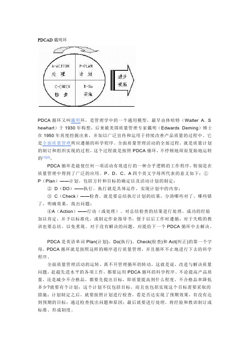
PDCAD戴明环PDCA循环又叫戴明环,是管理学中的一个通用模型,最早由休哈特(Walter A. S hewhart)于1930年构想,后来被美国质量管理专家戴明(Edwards Deming)博士在1950年再度挖掘出来,并加以广泛宣传和运用于持续改善产品质量的过程中。
它是全面质量管理所应遵循的科学程序。
全面质量管理活动的全部过程,就是质量计划的制订和组织实现的过程,这个过程就是按照PDCA循环,不停顿地周而复始地运转的[1][2]。
PDCA循环是能使任何一项活动有效进行的一种合乎逻辑的工作程序,特别是在质量管理中得到了广泛的应用。
P、D、C、A四个英文字母所代表的意义如下:①P(Plan)——计划。
包括方针和目标的确定以及活动计划的制定;②D(DO)——执行。
执行就是具体运作,实现计划中的内容;③C(Check)——检查。
就是要总结执行计划的结果,分清哪些对了,哪些错了,明确效果,找出问题;④A(Action)——行动(或处理)。
对总结检查的结果进行处理,成功的经验加以肯定,并予以标准化,或制定作业指导书,便于以后工作时遵循;对于失败的教训也要总结,以免重现。
对于没有解决的问题,应提给下一个PDCA循环中去解决。
PDCA是英语单词Plan(计划)、Do(执行)、Check(检查)和Act(纠正)的第一个字母,PDCA循环就是按照这样的顺序进行质量管理,并且循环不止地进行下去的科学程序。
全面质量管理活动的运转,离不开管理循环的转动,这就是说,改进与解决质量问题,赶超先进水平的各项工作,都要运用PDCA循环的科学程序。
不论提高产品质量,还是减少不合格品,都要先提出目标,即质量提高到什么程度,不合格品率降低多少?就要有个计划;这个计划不仅包括目标,而且也包括实现这个目标需要采取的措施;计划制定之后,就要按照计划进行检查,看是否达实现了预期效果,有没有达到预期的目标;通过检查找出问题和原因;最后就要进行处理,将经验和教训制订成标准、形成制度。
PD模型protocol

动物选择动物的种系非常重要,必须选择同一个种系的小鼠,即使是同一个种系不一个卖家的老鼠,也能够表现出对MPTP不同的敏感性,也会对实验结果产生影响.最多见的是:雄性,至少8周龄,体重至少22g,从同一个实验动物中心获得。
在MPTP导致的动物模型中最常见的一个问题是动物的急性死亡,发生在在MPTP第一次注射后的24h内。
这种急性死亡和多巴胺能系统的损伤无关,而是由于外周心脑血管的副作用,这种反应是剂量依赖性的并且在某些种系和雌性老鼠中更为明显。
注射时间安排:急性中毒-一天注射一针或者几针.亚急性或者慢性中毒-连续或者非连续的注射几天甚至一周,但是由于MPTP代谢很快,所谓的亚急性或者慢性中毒实际上是连续的损伤.最常用的方法:1,每两小时注射一次,每天注射八小时,共四次.依据剂量不同,纹状体区多巴胺损伤在40%(14mg/kg*4次)-90%(20mg/kg*4次),多巴胺能神经元在邢台上呈现非凋亡性质的死亡,纹状体多巴胺神经元至少死亡40-50%,多巴胺能神经元的损伤可以在注射完成之后持续7天.2.每天注射一次.30mg/kg,连续注射5天.可以导致40-50%的损伤,可以在注射完毕后持续21天.MPP+的含量在大概90min后达到高峰,在最后一针注射后8h后小时.这样,就可以设计时间点,类似50,70,110,130min和6,7,9,10h.设计神经退行性变的时间点:每个时间点5-6个老鼠,分别在最后一针注射完毕后第2,4,,7,14,21,30d,然后收集组织块儿,处理分析多巴胺能系统.最好订购小的MPTP,例如10或者100mg的,而不是大瓶的,这样就可以直接加盐水溶解,而不需要称量。
MPTP作用机理:MPTP为一种脂溶性的物质,分子量小,易透过血脑屏障,被星形胶质细胞,5-羟色胺神经元等细胞摄取后,在单胺氧化酶B(MAO-B)的作用下活化,转变为中间代谢产物MPFP+,生成有毒性的MPP+,释放到细胞外间隙,被多巴胺能神经元轴突末梢通过突触前膜多巴胺转运体摄取,逆向运输到黑质多巴胺神经元胞体,阻止线粒体呼吸链,导致能量衰竭,随后增加ROS.NO等自由基的释放,反应性胶质细胞增生等引起黑质多巴胺能神经细胞的死亡与凋亡.造模方法:选用对MPTP最敏感的C57BL/6J,小鼠,年龄在8-16周,体重在22-25g,雄性,腹腔注射MPTP·HCl(Sigma). 一个比较常用的方法是每2h注射一次,每天注射八小时,一共四次. 根据药物剂量的不同,纹状体区的多巴胺能损伤到40%(14 mg/kg per dose×4)-90%(20 mg/kg per dose×4).这种给药方法形成的损伤在注射完成之后可以持续7天.另一种比较公认的方法是每天注射一次,30mg/kg,连续注射5天,这种注射方法可以引起纹状体多巴胺神经元的坏死,达到40%-50%的减少,多巴胺能神经元的损伤可以在MPTP注射完成之后持续21天.在给药期间出现的一个最严重的问题是小鼠在接受第一次药物注射后的24h内会出现急性死亡,死亡率大于20﹪.这种死亡并非由于多巴胺能系统的损伤而引起,而是由于外周心血管的副作用,这种急性死亡是剂量依赖性的,在某些种属和雌性小鼠表现尤为突出在MPTP注射以后,MPP+含量将会在90min左右达到高峰,并且在最后一针注射完成8h后不再检测到有MPP+.注射完成后可以选择每5-6只老鼠为一个时间点,分别在第 4, 7, 14, 21和第30天检测多巴胺能神经元变性的情况.取材:①麻醉小鼠(戊巴比妥 35-45mg/kg)②经颈动脉灌流,起初以10ml/min的速度灌流生理盐水3分钟,然后用4﹪的多聚甲醛灌注8min.③轻轻的将脑组织取下,在多聚甲醛溶液中过夜后固定,溶液量至少要大于脑组织体积的十倍.④将脑块儿置于30﹪的蔗糖溶液中(0.1M PBS,PH 7.1-7.4),浸泡至少48h,使得脑块沉入容器底部.⑤冰冻切片机切脑片30-40um,进行免疫组化染色.高尔基染色:①将动物麻醉.②将已麻醉的动物固定在解剖台上,固定四肢,用左手持镊子假期腹部皮肤,右手持剪刀自腹部剪一小口,由此沿腹中线和胸骨剑突中线向上将皮肤剪至下颌,分离皮下组织,将皮肤翻向外侧,再沿腹中线和胸骨中线向上剪开胸骨,沿膈肌向两侧分开,并用止血钳将胸骨和胸部的皮肤前进,将止血钳翻向外侧,充分暴漏心脏.小心用镊子将心包打开.③将灌注针插入左心室,同时剪开右心耳,打开调节阀,灌注生理盐水,待血液流净时改为灌注固定液多聚甲醛,直至全身组织器官变硬为止。
pd模型评级对应的违约概率
pd模型评级对应的违约概率【最新版】目录1.PD 模型评级与违约概率的关系2.PD 模型评级的含义3.违约概率的定义与计算方法4.PD 模型评级对投资者与债市的影响正文PD 模型评级对应的违约概率在金融市场中,风险评估是至关重要的。
对债券发行人的评级,可以帮助投资者更好地了解其违约风险。
PD 模型评级就是其中一种常用的评级方式,它与违约概率有着紧密的关系。
本文将从 PD 模型评级的含义、违约概率的定义与计算方法以及 PD 模型评级对投资者与债市的影响等方面进行探讨。
首先,我们来了解一下 PD 模型评级的含义。
PD 模型,即违约概率模型,是根据大量的历史数据和先进的统计方法构建出来的一种风险评估工具。
它能够对债券发行人在未来一段时间内发生违约的概率进行量化,从而为投资者提供参考。
评级则是根据发行人的违约概率将其划分为不同的等级,例如 AAA、AA、A 等。
其次,我们需要了解违约概率的定义与计算方法。
违约概率,指的是债券发行人在一定时期内无法按照约定履行还款义务的概率。
PD 模型评级正是根据这个概率来划分评级的。
计算违约概率通常采用统计方法,如泊松分布、正态分布等,需要大量的历史数据作为支撑。
接下来,我们来看一下 PD 模型评级对投资者与债市的影响。
对于投资者而言,评级可以帮助他们更加准确地判断投资风险,从而作出更为明智的投资决策。
高评级的债券通常意味着较低的违约风险,因此更受投资者青睐。
而对于债市而言,评级可以提高市场的透明度,降低信息不对称带来的风险,从而促进市场的稳定发展。
总之,PD 模型评级与违约概率紧密相连,它是一种重要的风险评估工具。
通过评级,投资者可以更好地判断债券的违约风险,从而作出更为明智的投资决策。
帕金森病动物模型的制作的详细步骤及方法
帕金森病动物模型的制作的详细步骤及方法一、通过耗竭单胺类神经递质制作PD模型(一)利血平模型利血平是一种生物碱。
通过不可逆地封闭囊内单胺运输消耗中枢和外周单胺,从而影响细胞内囊泡的单胺再循环,于20世纪50年代被用于制作大鼠PD模型。
当啮齿类动物注射利血平后,囊内摄取的DA、5-羟色胺(5-HT)和去甲肾上腺素(NA)被封闭,在胞质内被降解,快速降低单胺水平,导致肌肉僵硬等PD的症状。
制备方法:应用雄性Wistar大鼠,腹腔内注射一定剂量的利血平后即可使其出现骨骼肌僵硬、震颤、姿势异常。
该模型制作方法简单。
其对人类大的贡献在于揭示出左旋多巴(L-DOPA)可作为治疗PD的潜在药物。
缺点是只能短暂模拟PD的症状,不能完全复制自发性PD 的病理变化。
(二)甲丨基丨苯丨丙丨胺模型苯丨丙丨胺丨类药物是一类具有潜在成瘾性的神经兴丨奋丨剂,具有促DA释放作用,大剂量应用对啮齿类动物和非人类的灵长类动物具有神经毒性,像利血平一样,它能够导致DA能神经元末梢DA的耗竭,对黑质神经元损伤作用较小。
Boirean等研究表明,该药可通过DA转运体被摄入细胞内,兴奋性氨基酸受体拮抗剂MK.801可阻断其毒性作用,体外实验提示能量代谢障碍、氧化应激和兴奋性氨基酸均与其毒性作用有关。
制作方法:用成年大鼠,按体重10~25mg/kg 皮下注射甲丨基丨苯丨丙丨胺,每天一次,连续4天。
用药后可出现短暂活动过多,随之是活动减少。
4~5d后可有“幻觉样”行为,表现为颤抖、舔毛、咬皮毛。
1周后检测即可发现黑质和纹状体中酪氨酸羟化酶(tyrosine hydroxylase, TH)活性降低,DA含量减少,但黑质 DA能神经元丢失不明显。
该模型制作方法简单,是一个急性损伤模型,症状的持续时间及稳定性方面尚不理想,也无PD的特征性组织病理学改变。
主要用于研究PD多巴胺耗竭后的纹状体生理、生化学改变,也可用于神经保护方面的研究。
二、神经毒素制作PD模型对PD患者大脑的研究显示,在氧化应激过程中存在线粒体复合物Ⅰ功能的抑制,因而这可能是黑质DA神经退行性变的重要组成部分。
PD需求模型术语

PD需求模型(RQM)术语释义
[仅供参考]
1、extend model definition: 拓展模型定义
2、model properties: 模型属性
3、detail: 细节
4、type: 四种类型释义:undefined: 未定义;design: 设计;functional:功能;technical:技术]
5、verification[验证]选项5种类型:undefined: 未定义;automated test: 自动测试;demonstration: 演示;manual test: 人工测试;mixed:混合测试。
6、related glossary term:相关术语词汇;
7、workload:工作量
8、user allocation:用户配置
9、glossary 词汇表
10、full description 完全描述
11、code:代码
12、priority:优先级[用数值大小表示,默认为1~5,自定义]
13、risk: 风险[默认:低、中、高]
14、RQM的status属性状态[默认5种:draft:草稿,defined:已定义;verified:已验证;to be reviewed:待复查,approved:己核准]
15、stereotype:构造类型
16、prolile:配置
17、add metaclasses:添加元类
18、repository:知识库
19、contents:目录
20、views:视图。
6-OHDA诱导的PD模型小鼠的运动及焦虑症状

第59卷 第3期2023年06月青岛大学学报(医学版)J O U R N A LO FQ I N G D A O U N I V E R S I T Y (M E D I C A LS C I E N C E S)V o l .59,N o .3J u n e 2023[收稿日期]2022-12-06; [修订日期]2023-05-20[基金项目]国家自然科学基金面上项目(32171132)[第一作者]陈凤华(1997-),女,硕士研究生㊂[通信作者]谢俊霞(1956-),女,博士,教授,博士生导师㊂E -m a i l :j x i a x i e @p u b l i c .qd .s d .c n ㊂石丽敏(1982-),女,博士,副教授,硕士生导师㊂E -m a i l :l i m i n s h i @q d u .e d u .c n㊂6-OH D A 诱导的P D 模型小鼠的运动及焦虑症状陈凤华,石丽敏,谢俊霞(青岛大学基础医学院生理学与病理生理学系,脑科学与疾病研究院,山东青岛 266071)[摘要] 目的 探讨6-羟基多巴胺(6-OH D A )诱导的单侧帕金森病(P D )模型小鼠的运动及焦虑症状㊂方法7周龄雄性C 57B L /6小鼠20只,随机分为对照组及模型组,每组10只㊂模型组小鼠通过左侧纹状体立体定位注入6-OH D A (2g /L ,2μL )制备P D 模型,对照组小鼠注入等量的生理盐水㊂2周后进行旷场实验检测小鼠的移动总距离和中心区探索时间,采用酪氨酸羟化酶(T H )免疫荧光染色检测黑质区多巴胺能神经元数目㊂结果 旷场实验结果显示,与对照组相比,模型组小鼠移动总距离明显减少,中心区探索时间明显增加,差异具有统计学意义(t =2.201㊁2.576,P <0.01)㊂免疫荧光染色结果显示,与对照组相比,模型组小鼠黑质区T H 阳性神经元的数目明显减少,差异有统计学意义(t =17.570,P <0.001)㊂结论 6-OH D A 诱导的单侧P D 模型小鼠黑质-纹状体系统功能受损,出现运动障碍但没有产生焦虑㊂[关键词] 帕金森病;羟多巴胺;纹状体;小鼠;症状评估[中图分类号] R 338.2 [文献标志码] A [文章编号] 2096-5532(2023)03-0321-04d o i :10.11712/jm s .2096-5532.2023.59.076[开放科学(资源服务)标识码(O S I D )][网络出版] h t t ps ://k n s .c n k i .n e t /k c m s 2/d e t a i l /37.1517.R.20230719.1611.001.h t m l ;2023-07-20 13:27:08M O T O RA N D A N X I E T YS Y M P T O M SI N A M O U S E M O D E L O F6-H Y D R O X Y D O P A M I N E -I N D U C E D P A R K I N S O N SD I S E A S EC H E N F e n g h u a ,S H IL i m i n ,X I EJ u n x i a (D e p a r t m e n t o f P h y s i o l o g y a n dP a t h o p h y s i o l o g y ,S c h o o l o f B a s i cM e d i c i n e ,Q i n g -d a oU n i v e r s i t y ,I n s t i t u t e o f B r a i nS c i e n c e s a n dD i s e a s e s ,Q i n gd a o 266071,C h i n a )[A B S T R A C T ] O b je c t i v e T o i n v e s t i g a t et h e m o t o ra n da n x i e t y s y m p t o m s i na m o u s e m o d e lo f6-h y d r o x y d o p a m i n e (6-O H D A )-i n d u c e du n i l a t e r a l P a r k i n s o n s d i s e a s e (P D ). M e t h o d s At o t a l of 20m a l e C 57B L /6m i c e ,a ge d7w e e k s ,w e r e r a n -d o m l y d i v i d e d i n t o c o n t r o l g r o u p a n dm o d e l g r o u p ,w i t h 10m i c e i n e a c h g r o u p .T h em i c e i n t h em o d e l g r o u p w e r e gi v e n s t e r e o t a c t i c i n j e c t i o no f 2μL6-O H D A (2g /L )i n t o t h e l e f t c o r p u s s t r i a t u mt o e s t a b l i s h am o d e l o f P D ,a n d t h o s e i n t h e c o n t r o l g r o u p w e r e g i -v e n i n j e c t i o no f a ne q u a l v o l u m e o f n o r m a l s a l i n e .T w ow e e k s l a t e r ,t h e o p e n f i e l d t e s tw a s u s e d t om e a s u r e t o t a lm o v i n g di s t a n c e a n d t i m e s p e n t i n t h e c e n t e r o f t h e o p e n f i e l d ,a n d t y r o s i n e h y d r o x y l a s e (T H )i mm u n o f l u o r e s c e n t s t a i n i n g w a s u s e d t om e a s u r e t h e n u m b e ro f d o p a m i n e r g i cn e u r o n s i nt h es u b s t a n t i an i g r a . R e s u l t s T h eo p e n f i e l dt e s t s h o w e d t h a t c o m pa r e dw i t ht h e c o n t r o l g r o u p ,t h em o d e l g r o u p h a d a s i g n i f i c a n t r e d u c t i o n i n t o t a lm o v i n g d i s t a n c e a n d a s i g n i f i c a n t i n c r e a s e i n t i m e s p e n t i n t h e c e n t e r o f t h e o p e n f i e l d (t =2.201,2.576;P <0.01).I mm u n o f l u o r e s c e n t s t a i n i n g s h o w e d t h a t c o m p a r e dw i t h t h e c o n t r o l g r o u p,t h em o d e l g r o u p h a d a s i g n i f i c a n t r e d u c t i o n i n t h en u m b e r o fT H -p o s i t i v en e u r o n s i n t h e s u b s t a n t i an i g r a (t =17.570,P <0.001). C o n c l u -s i o n I m p a i r e d f u n c t i o no f t h e s u b s t a n t i a n i g r a -c o r p u s s t r i a t u ms ys t e mi s o b s e r v e d i n am o u s em o d e l o f 6-O H D A -i n d u c e d u n i l a t e r a l P D ,w i t h t h e p r e s e n c e o fm o v e m e n t d i s o r d e r s ,b u tw i t h o u t t h e p r e s e n c e o f a n x i e t y.[K E Y W O R D S ] P a r k i n s o nd i s e a s e ;o x i d o p a m i n e ;c o r p u s s t r i a t u m ;m i c e ;s y m pt o ma s s e s s m e n t 帕金森病(P D )是仅次于阿尔茨海默病的第二大神经退行性疾病,其病理学特征为黑质致密带多巴胺能神经元选择性丢失和纹状体轴突末梢多巴胺含量减少[1-3]㊂其运动症状主要有静止性震颤㊁肌僵直㊁运动迟缓和姿势不稳等,非运动症状有嗅觉障碍㊁睡眠障碍㊁认知障碍㊁焦虑和疲劳等㊂动物模型在探究P D 发病机制和寻找潜在治疗靶点的过程中发挥着重要作用[4-6]㊂6-羟基多巴胺(6-O H D A )是一种儿茶酚胺选择性神经毒素,脑内纹状体注射6-O H D A 会引起相应的黑质-纹状体多巴胺系统进行性和部分受损,可用于制备稳定有效的大鼠P D 模型[7-9]㊂尽管6-OH D A 单侧损伤大鼠模型是P D 研究中最常用的模型之一,但随着光遗传和化学遗传技术的发展,6-O H D A 制备P D 模型也逐步应用于小鼠[10-14]㊂目前尚缺乏6-OH D A 注射诱导的P D模型小鼠的系统性研究㊂本实验通过单侧纹状体立体定位注射6-O H D A 制备小鼠P D 模型,观察其运动及焦虑症状,以期为P D 模型小鼠的基础研究提供实验证据㊂Copyright ©博看网. All Rights Reserved.322青岛大学学报(医学版)59卷1材料与方法1.1动物及主要试剂S P F级雄性C57B L/6小鼠,7周龄,体质量为(22ʃ2)g,购自北京维通利华公司㊂小鼠饲养于25ħ㊁12h昼夜循环光照的S P F级清洁环境中,可自由饮水㊁摄食㊁活动,适应环境1周后开始实验㊂6-O H D A购于中国A b s i n公司,L-A s c o r b i c a c i d以及地昔帕明购于美国S i g m a公司,酪氨酸羟化酶(T H)抗体购于美国M i l l i p o r e公司,其他试剂均为国产分析纯㊂1.2动物分组及处理将小鼠随机分为对照组和模型组,每组10只㊂术前30m i n小鼠腹腔注射地昔帕明25m g/k g㊂利用瑞沃德公司的呼吸麻醉机将小鼠麻醉后,固定在立体定位仪上㊂用耳杆适配器将小鼠固定好,调整高度使颅骨保持水平㊂剃除小鼠头部毛发,用碘附擦拭消毒,剪开头皮暴露颅骨的前囟和后囟㊂以前囟为零点,前囟前0.4mm㊁旁开1.8mm㊁深度-3.5mm定位坐标㊂模型组将2μL溶于2g/L抗坏血酸的6-O H D A(2g/L)按立体定位坐标注入左侧纹状体,流量6n L/s,注射完成后停针10m i n;对照组则以等量生理盐水代替6-O H D A㊂在整个手术过程中,用异氟烷麻醉小鼠并用加热垫维持体温㊂1.3旷场实验实验前小鼠置于测试环境中适应至少半小时㊂将小鼠放在一个27c mˑ27c mˑ35c m大小不透明测试盒的中央,摄像机放于盒子的正上方㊂利用S m a r t v3.0系统记录小鼠10m i n的活动情况㊂每只小鼠检测结束后,用体积分数0.75的乙醇清理旷场区域,并在测试时保持干燥㊂分析在10m i n的旷场实验中小鼠的移动总距离和中心区探索时间,评估小鼠的运动行为和焦虑程度㊂1.4脑组织切片及T H免疫荧光染色行为学检测结束后,腹腔注射阿佛丁(20m L/ k g)麻醉小鼠㊂经心灌注9g/LN a C l和多聚甲醛溶液(用0.1m o l/LP B S配制,p H值为7.2~7.4),小心取出鼠脑㊂将鼠脑置于多聚甲醛溶液中,4ħ固定6h,然后分别用200㊁300g/L的蔗糖溶液(用0.1m o l/LP B S配制)进行梯度脱水㊂用冷冻切片机(L e i c a,C M1950)进行冠状面连续切片㊂参照小鼠脑图谱,确定黑质区域㊂进行厚度为20μm的冠状面连续切片,每组10张,共4组㊂取一组完整脑片进行T H免疫荧光染色㊂将脑片置于多聚甲醛溶液中固定10m i n,用0.01m o l/ LP B S漂洗3次,每次10m i n㊂用含有体积分数0.05驴血清(J a c k s o n)的P B S T缓冲液室温封闭1h,然后置于用P B S T配制的一抗稀释液中4ħ摇床孵育过夜㊂次日,用0.01m o l/LP B S漂洗3次,每次10m i n㊂将脑片放于用P B S T配制的荧光二抗稀释液中室温孵育2h,之后用0.01m o l/LP B S 漂洗3次,每次10m i n㊂将脑片平铺于载玻片上,避光保存㊂免疫荧光染色实验中用到的一抗为a n-t i-t y r o s i n eh y d r o x y l a s e(1ʒ2000,r a b b i t),二抗为d o n k e y a n t i-r a b b i t555(稀释比为1ʒ500)㊂使用数字病理切片扫描系统(O L YM P U S,T o k y o,J a p a n, V S120)拍摄成像,应用O l y V I A软件对T H阳性神经元进行计数㊂1.5统计学分析应用G r a p h P a dP r i s m6软件进行统计学处理㊂实验所得计量资料结果以 xʃs形式表示,两组比较采用t检验㊂P<0.05表示差异有统计学意义㊂2结果2.16-O H D A对小鼠运动行为的影响旷场实验结果显示,与对照组小鼠相比,模型组小鼠移动总距离明显减少,中心区探索时间明显增加,差异有统计学意义(t=2.201㊁2.576,P<0.01)㊂见表1㊂表1两组小鼠在旷场中的活动情况比较( xʃs)组别n移动总距离(l/c m)中心区探索时间(χ/%)对照组94267.0ʃ160.95.618ʃ0.297模型组133570.0ʃ195.2*8.039ʃ0.886*与对照组比较,*t=2.201㊁2.576,P<0.01㊂2.26-O H D A对小鼠黑质T H阳性神经元的影响免疫荧光染色结果显示,对照组和模型组小鼠黑质区T H阳性神经元的数目分别为10852.0ʃ209.8和6072.0ʃ173.3(n=10),与对照组相比,模型组小鼠黑质区T H阳性神经元的数目明显减少,差异有统计学意义(t=17.570,P<0.001)㊂3讨论P D是常发生于中老年人的第二大神经退行性疾病,其主要病理改变为黑质多巴胺能神经元进行性丢失,其临床表现除肌僵直㊁运动迟缓等运动症状Copyright©博看网. All Rights Reserved.3期陈凤华,等.6-O H D A诱导的P D模型小鼠的运动及焦虑症状323外,还有嗅觉障碍㊁焦虑和抑郁等非运动症状㊂由于P D的病因病理尚未完全阐明,目前该病的治疗主要是对症治疗[5,15-16]㊂为了阐明P D的发病机制和寻找潜在治疗靶点,已经开发了许多动物模型[17-19]㊂6-O H D A可被黑质内含单胺氧化酶的多巴胺能神经元特异性摄取,并在单胺氧化酶的作用下转化成自由基损伤神经元,故被广泛应用于损伤黑质-纹状体多巴胺能系统制备P D模型[10,20-21]㊂长期以来6-O H D A多用于大鼠P D模型的制备,近年来随着光遗传学㊁化学遗传学的发展以及各种C r e小鼠的应用,6-O H D A也逐渐用于小鼠P D模型的制备㊂6-O H D A参与氧化应激反应,通过和多巴胺竞争,可与高亲和力的多巴胺转运体结合进入黑质-纹状体多巴胺能神经元,并迅速被氧化,生成大量的活性氧(R O S),发挥毒性作用损伤细胞㊂还有研究结果表明,6-O H D A可以抑制线粒体呼吸链的功能,从而引起神经毒性[10,22-24]㊂由于6-OH D A不能通过血-脑脊液屏障,因此必须通过立体定位技术将它直接注射到黑质㊁内侧前脑束或纹状体中㊂研究表明,6-O H D A单侧纹状体注射具有较大的优势:首先,注射到纹状体引起的多巴胺能神经元进行性丢失和区域性的病变与P D病理进展最为相似;其此,小鼠脑内纹状体是一个较大的区域,为立体定位注射减轻了难度[2,25-27]㊂T H是多巴胺合成的限速酶,其功能缺失或表达不足直接影响多巴胺的合成与分泌㊂因此,检测模型动物T H免疫阳性细胞的数目不仅可以反映多巴胺能神经元的数目和功能状态,同时还可评估模型多巴胺水平[28-30]㊂本实验采用单侧纹状体注射4μg6-OH D A的方法制备P D模型,结果显示,单侧纹状体注射2周后,损伤侧黑质T H阳性神经元减少了约44%,提示多巴胺能神经元丢失;同时模型小鼠出现运动缺陷,在旷场实验中的运动总距离减少㊂但是本实验中P D模型小鼠在旷场中心区探索时间与对照组小鼠相比显著增加,提示小鼠并未出现焦虑症状㊂以往有研究显示,纹状体注射5μg 以上6-OH D A,3周后小鼠黑质多巴胺能神经元丢失超过50%,并出现运动障碍以及焦虑等非运动症状[31-34]㊂与之相比,本实验中6-O H D A用药剂量低㊁作用时间较短,因此推测这可能是小鼠未出现焦虑症状的原因㊂综上,本研究通过单侧纹状体注射6-O H D A观察其对小鼠运动和焦虑症状以及黑质-纹状体系统功能的影响,结果表明4μg的6-O H D A单侧纹状体注射在2周后可以造成黑质-纹状体通路的部分损失,小鼠出现运动障碍㊂本实验为6-O H D A制备小鼠P D模型提供了良好的注射位点,为P D的研究提供了有效的实验工具㊂[参考文献][1]B A L E S T R I N O R,S C HA P I R A A H V.P a r k i n s o nd i s e a s e[J].E u r o p e a nJ o u r n a l o fN e u r o l o g y,2020,27(1):27-42.[2]J A N K O V I CJ,T A N E K.P a r k i n s o n sd i s e a s e:e t i o p a t h o g e-n e s i s a n d t r e a t m e n t[J].J o u r n a l o fN e u r o l o g y,N e u r o s u r g e r y,a n dP s y c h i a t r y,2020,91(8):795-808.[3]G R A Y S O N M.P a r k i n s o n sd i s e a s e[J].N a t u r e,2016,538(7626):S1.[4]T I E U K.A g u i d e t on e u r o t o x i c a n i m a lm o d e l s o fP a r k i n s o n sd i se a s e[J].C o l d S p r i n g H a r b o r P e r s p e c t i v e si n M e d i c i n e,2011,1(1):a009316.[5]C H I A SJ,T A N E K,C H A O Y X.H i s t o r i c a l p e r s p e c t i v e:m o d e l s o f P a r k i n s o n s d i s e a s e[J].I n t e r n a t i o n a l J o u r n a l o fM o-l e c u l a r S c i e n c e s,2020,21(7):2464.[6]MU S T A P H A M,MA T T A I B C N.M P T P-i n d u c e d m o u s em o d e l o f P a r k i n s o n sd i s e a s e:a p r o m i s i n g d i r e c t i o no f t h e r a-p e u t i c s t r a t e g i e s[J].B o s n i a n J o u r n a l o f B a s i c M e d i c a l S c i e n c e s,2021,21(4):422-433.[7]S A U E R H,O E R T E L W H.P r o g r e s s i v ed e g e n e r a t i o no fn i-g r o s t r i a t a ld o p a m i n en e u r o n sf o l l o w i n g i n t r a s t r i a t a lt e r m i n a ll e s i o n sw i t h6-h y d r o x y d o p a m i n e:ac o m b i n e dr e t r o g r a d et r a-c i n g a n di mm u n o c y t o c h e m i c a ls t ud y i nt h er a t[J].Ne u r o-s c i e n c e,1994,59(2):401-415.[8]P R Z E D B O R S K I S,L E V I V I E R M,J I A N G H,e t a l.D o s e-d e-p e n d e n t l e s i o n so f t h ed o p a m i n e r g i cn i g r o s t r i a t a l p a t h w a y i n-d u ce db y i n t r a s t r i a t a l i n j e c t i o n o f6-h y d r o x y d o p a m i n e[J].N e u-r o s c i e n c e,1995,67(3):631-647.[9]I R A V A N P O U R F,D A R G A H IL,R E Z A E I M,e ta l.I n-t r a n a s a l i n s u l i n i m p r o v e sm i t o c h o n d r i a l f u n c t i o n a n d a t t e n u a t e s m o t o r d e f i c i t s i nar a t6-O H D A m o d e l o fP a r k i n s o n sd i s e a s e [J].C N S N e u r o s c i e n c e&T h e r a p e u t i c s,2021,27(3):308-319.[10]S I MO L A N,MO R E L L IM,C A R T A AR.T h e6-H y d r o x y d o-p a m i n em o d e lo fP a r k i n s o n sd i s e a s e[J].N e u r o t o x i c i t y R e-s e a r c h,2007,11(3):151-167.[11]G U I MA RÃE SRP,L E A N D R O R I B E I R O D,D O SS A N T O SKB,e t a l.T h e6-h y d r o x y d o p a m i n e r a tm o d e l o fP a r k i n s o n sd i se a s e[J].J o u r n a l o fV i s u a l i z e dE x p e r i m e n t s,2021(176):1-17.[12]B O U C H A T T A O,A B Y F,S I F E D D I N E W,e ta l.P a i nh y-p e r s e n s i t i v i t y i na p h a r m a c o l o g i c a lm o u s e m o d e l o f a t t e n t i o n-d e f i c i t/h y p e r a c t i v i t y d i s o r d e r[J].P r o c e e d i n g so f t h eN a t i o n a lA c a d e m y o f S c i e n c e so f t h eU n i t e dS t a t e so fA m e r i c a,2022,119(30):e2114094119.Copyright©博看网. All Rights Reserved.324青岛大学学报(医学版)59卷[13]MA G N O L A V,T E N Z A-F E R R E R H,C O L L O D E T T I M,e t a l.O p t o g e n e t i c s t i m u l a t i o nof t h e M2c o r t e xr e v e r t sm o t o rd y s f u n c t i o n i nam o u s em o de l o fP a r k i n s o n sd i s e a s e[J].T h eJ o u r n a l o fN e u r o s c i e n c e:t h eO f f i c i a l J o u r n a l o f t h e S o c i e t y f o r N e u r o s c i e n c e,2019,39(17):3234-3248.[14]Z H A N G H,Z H A N GCK,Q UZ W,e t a l.S T N-A N T p l a s t i-c i t y i s c r u c i a l f o r t h em o t o r c o n t r o l i nP a r k i n s o n sd i se a s em o-d e l[J].S i g n a lT r a n s d u c t i o na n d T a r g e t e d T h e r a p y,2021,6(1):215.[15]A R M S T R O N G MJ,O K U N M S.D i a g n o s i s a n d t r e a t m e n t o fP a r k i n s o nd i s e a s e:ar e v i e w[J].J AMA,2020,323(6):548-560.[16]V I J I A R A T N AM N,S I MU N I T,B A N D MA N NO,e t a l.P r o-g r e s s t o w a r d s t h e r a p i e s f o r d i s e a s em o d i f i c a t i o n i nP a r k i n s o n sd i se a s e[J].T h eL a n c e tN e u r o l o g y,2021,20(7):559-572.[17]T A G U C H IT,I K U N O M,Y AMA K A D O H,e ta l.A n i m a lm o d e l f o r p r o d r o m a lP a r k i n s o n sd i s e a s e[J].I n t e r n a t i o n a l J o u r n a l o fM o l e c u l a r S c i e n c e s,2020,21(6):1961. [18]D A U E R W,P R Z E D B O R S K I S.P a r k i n s o n sd i s e a s e:m e c h a-n i s m s a n dm o d e l s[J].N e u r o n,2003,39(6):889-909.[19]K I NK,Y A S UH A R AT,K AM E D A M,e t a l.A n i m a lm o d e l sf o rP a r k i n s o n sd i s e a s e r e s e a r c h:t r e n d s i nt h e2000s[J].I n-t e r n a t i o n a l J o u r n a l o fM o l e c u l a r S c i e n c e s,2019,20(21):5402.[20]A S A N UMA M,H I R A T A H,C A D E TJL.A t t e n u a t i o no f6-h y d r o x y d o p a m i n e-i n d u c e dd o p a m i n e r g i c n i g r o s t r i a t a l l e s i o n s i ns u p e r o x i d e d i s m u t a s e t r a n s g e n i cm i c e[J].N e u r o s c i e n c e,1998, 85(3):907-917.[21]S C HWA R T I N G R K W,HU S T O N J P.U n i l a t e r a l6-h y d r o x y d o p a m i n el e s i o n so f m e s o-s t r i a t a ld o p a m i n en e u r o n sa n d t h e i r p h y s i o l o g i c a l s e q u e l a e[J].P r o g r e s s i nN e u r ob i o l o g y,1996,49(3):215-266.[22]K O N N O V AEA,S WA N B E R G M.A n i m a lm o d e l s o f P a r k i n-s o n sd i s e a s e[M]//S T O K E R T B,G R E E N L A N DJC.P a r-k i n s o n sd i s e a s e:P a t h o g e n e s i sa n dc l i n i c a la s p e c t s.B r i s b a n e(A U):C o d o nP u b l i c a t i o n sC o p y r i g h t.2018.[23]T H I R U G N A N AM T,S A N T HA K UMA R K.C h e m i c a l l y i n-d u ce dm o d e l so fP a r k i n s o n sd i s e a s e[J].C o m p a r a t i v e B i o-c h e m i s t r y a n dP h y s i o l o g y T o x i c o l o g y&P h a r m a c o l o g y,2022,252:109213.[24]T R O N C I E,F R A N C A R D OV.A n i m a lm o d e l s o f L-D O P A-i n-d u ce dd y s k i n e s i a:t h e6-O H D A-l e s i o n e dr a ta n d m o u s e[J].J o u r n a lo f N e u r a l T r a n s m i s s i o n(V i e n n a,A u s t r i a:1996), 2018,125(8):1137-1144.[25]T R I P A N I C H K U L W,J A R O E N S U P P A P E R C H E O.A m e-l i o r a t i n g e f f e c t s o f c u r c u m i n o n6-O H D A-i n d u c e d d o p a m i n e r g i cd e n e r v a t i o n,g l i a l r e s p o n s e,a n dS O D1r e d u c t i o n i nt h e s t r i a-t u mo f h e m i p a r k i n s o n i a nm i c e[J].E u r o p e a nR e v i e wf o rM e d i-c a l a n dP h a r m a c o l o g i c a l S c i e n c e s,2013,17(10):1360-1368.[26]V A R C I N M,B E N T E AE,M E R T E N SB,e t a l.A c u t e v e r s u sl o n g-t e r me f f e c t s o f6-h y d r o x y d o p a m i n e o n o x i d a t i v e s t r e s s a n dd o p a m i ne d e p l e t i o n i n t h e s t r i a t u mo fm i c e[J].J o u r n a l o fN e u-r o s c i e n c eM e t h o d s,2011,202(2):128-136.[27]K A B U T O H,N I S H I Z AWA M,T A D A,e t a l.Z i n g e r o n e[4-(4-h y d r o x y-3-m e t h o x y p h e n y l)-2-b u t a n o n e]p r e v e n t s6-h y d-r o x y d o p a m i n e-i n d u c e dd o p a m i n e d e p r e s s i o n i nm o u s e s t r i a t u ma n d i n c r e a s e s s u p e r o x i d e s c a v e n g i n g a c t i v i t y i n s e r u m[J].N e u-r o c h e m i c a lR e s e a r c h,2005,30(3):325-332.[28]C O L E T T ES,D A U B N E R.T y r o s i n eh y d r o x y l a s ea n dr e g u l a-t i o no f d o p a m i n es y n t h e s i s[J].A r c h i v e so fB i o c h e m i s t r y a n dB i o p h y s i c s,2011,508(1):1-12.[29]N A G A T S U T,N A K A S H I MA A,WA T A N A B E H,e ta l.N e u r o m e l a n i ni n P a r k i n s o n sd i s e a s e:t y r o s i n eh y d r o x y l a s ea n d t y r o s i n a s e[J].I n t e r n a t i o n a l J o u r n a l o fM o l e c u l a r S c i e n c e s,2022,23(8):4176.[30]N A G A T S U T,N A K A S H I MA A,I C H I N O S E H,e t a l.H u-m a n t y r o s i n e h y d r o x y l a s e i nP a r k i n s o n s d i s e a s e a n d i n r e l a t e dd i s o r de r s[J].J o u r n a l o fN e u r a lT r a n s m i s s i o n,2019,126(4):397-409.[31]A N T U N E S M S,C A T T E L A NS O U Z AL,L A D DF V L,e ta l.H e s p e r i d i na m e l i o r a t e sa n x i e t y-d e p r e s s i v e-l i k eb e h a v i o r i n6-O H D A m o d e l o fP a r k i n s o n sd i s e a s eb y r e g u l a t i n g s t r i a t a lc y t o k i n e a n dn e u r o t r o p h i c f a c t o r s l e v e l sa n dd o p a m i ne r g i c i n-n e r v a t i o nl o s s i nt h es t r i a t u m o fm i c e[J].M o l e c u l a rN e u r o-b i o l o g y,2020,57(7):3027-3041.[32]M E N D E S-P I N H E I R O B,S O A R E S-C U N HA C,MA R O T EA,e ta l.U n i l a t e r a l i n t r a s t r i a t a l6-h y d r o x y d o p a m i n e l e s i o n i n m i c e:ac l o s e rl o o ki n t on o n-m o t o r p h e n o t y p ea n d g l i a lr e-s p o n s e[J].I n t e r n a t i o n a l J o u r n a l o fM o l e c u l a rS c i e n c e s,2021, 22(21):11530.[33]L I U XJ,Y U H,C H E NBX,e t a l.C B2a g o n i s tGW842166xp r o t e c t e d a g a i n s t6-O H D A-i n d u c e d a n x i o g e n i c-a n d d e p r e s s i v e-r e l a t e db e h a v i o r s i nm i c e[J].B i o m e d i c i n e s,2022,10(8):1776.[34]MA S I N ID,P L E WN I AC,B E R T H O M,e t a l.A g u i d e t o t h eg e n e r a t i o no fa6-h y d r o x y d o p a m i n e m o u s e m o d e lo fP a r k i n-s o n s d i s e a s e f o r t h es t u d y o fn o n-m o t o rs y m p t o m s[J].B i o-m e d i c i n e s,2021,9(6):598.(本文编辑马伟平)作者书写文内标题须知本刊文内标题序号使用阿拉伯数字顺序编码,左顶格书写㊂标题一般可分为1~4级,即:1,2,3 ;1.1,1.2,1.3 ;1.1.1,1.1.2, 1.1.3 ;1.1.1.1,1.1.1.2,1.1.1.3 ㊂第5级标题可用(1)或①㊂1,2级标题均单独占行㊂请作者来稿时遵照执行㊂Copyright©博看网. All Rights Reserved.。
- 1、下载文档前请自行甄别文档内容的完整性,平台不提供额外的编辑、内容补充、找答案等附加服务。
- 2、"仅部分预览"的文档,不可在线预览部分如存在完整性等问题,可反馈申请退款(可完整预览的文档不适用该条件!)。
- 3、如文档侵犯您的权益,请联系客服反馈,我们会尽快为您处理(人工客服工作时间:9:00-18:30)。
SAGE-Hindawi Access to ResearchParkinson’s DiseaseVolume2011,Article ID327089,7pagesdoi:10.4061/2011/327089Review ArticleLipopolysaccharide Animal Models for Parkinson’s DiseaseMei Liu and Guoying BingDepartment of Anatomy and Neurobiology,College of Medicine,University of Kentucky,Lexington,KY40536,USACorrespondence should be addressed to Guoying Bing,gbing@Received24November2010;Accepted28February2011Academic Editor:Gilles J.GuilleminCopyright©2011M.Liu and G.Bing.This is an open access article distributed under the Creative Commons Attribution License, which permits unrestricted use,distribution,and reproduction in any medium,provided the original work is properly cited.Lipopolysaccharide(LPS),an endotoxin from Gram-negative bacteria,acts as a potent stimulator of microglia and has been used to study the inflammatory process in the pathogenesis of Parkinson’s disease(PD)and anti-inflammatory therapy for PD treatment.Here,we review the growing body of literature on both in vitro and in vivo LPS PD models.Primary cell cultures from mesencephalic tissue were exposed to LPS in vitro;LPS was stereotaxically injected into the substantia nigra,striatum,or globus pallidus of brain or injected into the peritoneal cavity of the animal in vivo.In conclusion,the LPS PD models are summarized as(1)local and direct LPS treatment and(2)systemic LPS treatment.Mechanisms underlying the PD models are investigated and indicated that LPS induces microglial activation to release a variety of neurotoxic factors,and damaged neurons may trigger reactive microgliosis,which lead to progressive dopaminergic neurodegeneration.1.IntroductionParkinson’s disease(PD)is the most prevalent neurode-generative movement disorder.In PD,clinical symptoms including tremor,rigidity,and bradykinesia are primarily resulted from the loss of dopamine-containing neurons in the substantia nigra pars compacta.Although the etiology and pathogenesis of PD remain not fully elucidated,many interacting pathological processes appear to contribute to dopaminergic neuron degeneration in the disease.Recently, inflammatory processes have been implicated as one of the active contributors to dopaminergic neuron damage in the development and progression of the disease[1, 2].In the central nervous system,microglia,the resident innate immune cells,play a major role in the inflammatory process.Typically microglia exist in a resting state char-acterized by ramified morphology and monitor the brain environment[3].In response to various pathogenic stimuli including inflammation,microglia are readily activated and undergo a transformation to amoeboid morphology with an upregulated catalogue of surface molecules[3–5].Activated microglia can serve diverse beneficial functions essential to neuron survival,which include cellular maintenance and innate immunity[6].However,uncontrolled activated microglia produces a variety of neurotoxic factors such as proinflammatory cytokines(interleukin-1(IL-1),tumor necrosis factor alpha(TNF-α),interleukin-6(IL-6)),nitro oxide(NO),prostaglandin E2,and superoxide,which lead to neuronal damage or death[1,7–10].Additionally,dam-aged neurons may emit injury signals to cause microglia activation,which used to be defined as reactive microgliosis [11].This microglial-neuronal interaction will be reinforced and become a self-amplifying cycle of neuronal injury and microglial activation,whichfinally leads to more neuronal damage and death.Importantly,clinical researches have reported that microglial activation was found in the nigros-triatal system of PD patients[12–14].Therefore,it is essential to study the inflammatory process in PD,which may help us understand the pathogenesis of the disease and eventually develop an effective therapeutic strategy.Over the last two decades,studies in animal models have demonstrated that inflammation induced by lipopolysac-charide(LPS)can replicate some characteristics of PD, including extensive activation of microglia and selective loss of dopaminergic neurons in the nigrostriatal system[15–19]. The history of understanding LPS starts in the late nineteenth century.LPS is found in the outer membrane of Gram-negative bacteria and acts as endotoxin.LPS from many Gram-negative bacteria species initiates acute inflammatory responses in mammals and induces a diverse range of effects, ranging from pyrexia to Gram-negative septic shock[20]. Thus,using different serotypes of LPS and their differentapplication routes may cause different outcomes[21].More-over,LPSs from different bacteria species share common fea-tures in their basic architecture,which consists of three cova-lently linked segments,a surface carbohydrate polymer(O-specific chain),a core oligosaccharide featuring an outer and inner region,and an acylated glycolipid(termed lipid A).The O-specific chain shows the most diversity and is the basis for serological specificity,while lipid A,which anchors the LPS molecule in the Gram-negative outer membrane,is the most conserved biochemical structure across different bacterial species[20].There is wide acceptance that the lipid A moiety is the innate immune stimulating or endotoxic component of LPS[22].In addition,it is documented that LPS-associated pathology results from the stimulation of host cell responses, in which LPS binds to specific receptors in order to elicit the release of cytokines and other inflammatory mediators.Sev-eral membrane-bound and soluble proteins have been shown to bind LPS;the most important appear to be CD14and LPS-binding protein(LBP)and the toll-like receptor(TLR)family which is a recently discovered group of transmembrane receptors[23,24].In the central nervous system,it is found that systemic LPS injection upregulated its membrane CD14receptor within specific cellular populations including microglia in the brain[25].Thereafter,microglia were identified as the major LPS-responsive cell in the brain.LPS binds to TLR4on microglia and induces microglial activation that results in neuronal damage[26,27].LPS acts as an endotoxin and elicits multiple pathological effects in human beings.One case report may uncover a potential link between LPS infection and the development of Parkinsonism.A22-year-old laboratory worker was acci-dentally exposed to10μg Salmonella minnesota LPS through an open wound and developed Parkinson’s syndrome with bradykinesia,rigidity,tremor,and cogwheel phenomenon three weeks later;damage to the substantia nigra and cerebral cortex was shown by positron emission tomography a few years after the accident[28].However,it is known that LPS from many bacterial species such as Salmonella, Pseudomonas,Vibrio,and Rhizobium can initiate acute inflammatory responses in mammals and induce a large and diverse range of effects,ranging from pyrexia and Gram-negative septic shock[29].There is another case report regarding the Salmonella endotoxin exposure.One middle-aged laboratory worker was self-administered intravenously a single large dose of endotoxin(1mg Salmonella minnesota LPS)and immediately developed a severe septic shock syndrome with multiple-organ dysfunction.The patient was successfully rescued in the emergency room,and there has been no follow-up report to date[30].Thus,further investigation and more epidemiologic data are needed to exploit the relationship between endotoxin and PD.In the current paper,we present a summary of a variety of LPS PD models and discuss their strengths and limitations, which may be helpful for the future LPS PD study.2.In Vitro Studies of LPS PD Model2.1.LPS Treatment to Cell Culture from Mesencephalic Tissue. Bronstein et al.in1995reported the comparison study between the dopaminergic neurotoxin6-hydroxydopamine (6-OHDA)and LPS in rat mesencephalic cultures[31]. Investigators found that,in the neuron-enriched cultures, 6-OHDA killed89%of the tyrosine hydroxylase-(TH-) immunopositive neurons,but LPS(50μg/mL)was not neurotoxic;however,in the mixed neuron-glial cultures,6-OHDA killed only27%of the TH-immunopositive neurons, but LPS killed70%of the TH-immunopositive neurons.This early experiment suggested that the dopaminergic neuro-toxicity of LPS is dependent on the presence of microglia. Subsequently,dopaminergic neurotoxicity of LPS was con-firmed by the other groups on rat mesencephalic mixed neuron-glial cultures and demonstrated that LPS induced microglial activation,and activated microglia released the proinflammatory and cytotoxic factors:NO,TNF-α,and IL-1β,which lead to dopaminergic neuron damage[32,33].In addition,the dopaminergic neurotoxicity of LPS was studied on mouse mesencephalic neuron-glial culture,and it was found that the neurotoxicity was mainly mediated through LPS-induced nicotinamide adenine dinucleotide phosphate (NADPH)oxidase activation on microglia,which generated reactive oxygen species production,which are neurotoxic factors[34].In the studies of LPS-treated primary cultures generated from forebrains of embryonic day17mice,the investigators found that the LPS neurotoxicity occurred through binding the signal-transducing receptor,TLR4;microglia are the major cells in the central nervous system that express TLR4.However,it is emphasized that the toxic effect of LPS on neurons is a general phenomenon,independent of neuronal subtype[26].Thus,LPS may cause neurotoxicity without selectivity for neuronal types.Since previous studies from primary microglia cultures have found LPS treatment induced the release of proinflammatory and cytotoxic factors from microglia[35,36],the investigators suggested that the activation of TLR4on microglia may initiate the intracellular signaling pathway of microglia,and result in the release of proinflammatory mediators which cause neuronal damage [26].3.In Vivo Studies of LPS PD Model3.1.Intranigral Injection of LPS.In order to study the response of the nigrostriatal system to inflammation,Bing et al.and Casta˜n o et al.independently reported the PD model of LPS intranigral injection in1998[15,16].After LPS was stereotaxically injected into the nigral area of rats, investigators found that LPS induced microglia activation and dopaminergic neuron loss in the substantia nigra[15, 16].In a following study,it was reported that LPS-induced dopaminergic neuronal damage was permanent,as observed one year postinjection.Moreover,there was no detectable damage to either the GABAergic or the serotoninergic neurons in the striatum and nigra after LPS injection, indicating that LPS selectively induced dopaminergic neuron death in the nigrostriatal system[37].Thereafter,more studies confirmed the results and also found the increased level of proinflammatory cytokines including IL-1β,TNF-α,IL-6,and NO in the substantia nigra after LPS injection,which may be causal factor for LPS-induced neuronal damage[38–40].In addition,the effects of intranigral LPSinjection on behavior and dopamine content and turnoverwere investigated and showed that LPS treatment enhancedlocomotor activity2-to3-fold and increased dopamine turnover ratios in comparison with control subjects.Thissuggests that LPS insult may induce a compensatory responseof dopaminergic system[41].3.2.Intrapallidal Injection of LPS.The globus pallidus is amajor integrative nucleus within the basal ganglia,with neu-rons projecting to striatum,subthalamic nucleus,entope-duncular nucleus,and substantia nigra.Thus,the globuspallidus is positioned to influence the nigrostriatal pathwayand function of the basal ganglia as a whole.LPS was injectedinto the globus pallidus of young and middle-aged rats.The results showed that microglial activation was found in bothglobus pallidus and substantia nigra,dopaminergic neuronswere significantly and progressively decreased in the substan-tia nigra,and locomotor deficits were detected in animal afterLPS injection[17].Moreover,the following study reportedan increased level of proinflammatory cytokines includingIL-1β,TNF-α,and IL-6,the elevated expression of inducible nitric oxide synthase,and the enhancedα-synuclein nitrationand oligomerization in the substantia nigra of LPS-injectedanimal[42].Interestingly,the above pathological changeswere much severer in middle-aged animals when compared with the younger animals after LPS treatment,supporting theview that aging itself is a risk factor for PD development[42].Inflammation promotes the release of neurotoxic factors and the development of synucleinopathy lesions thatfinally leadto dopaminergic neurodegeneration in PD model of LPSintrapallidal injection.Additionally,thefinding of abnormal α-synuclein may help us to explore the mechanisms under-lying progressive loss of dopaminergic neurons in LPS PDmodels.It is reported that aggregatedα-synuclein inducedmicroglial activation in a primary mesencephalic neuron-gliaculture system[43],thus the pathological process of reactive microgliosis may be triggered and microglial activation maybecome uncontrolled,which eventually result in progressivedopaminergic neurotoxicity.3.3.Intrastriatal Injection of LPS.In the nigrostriatal system,the cell bodies of dopaminergic neurons are located in thesubstantia nigra and their dopamine-containing terminals are distributed in the striatum.After LPS was injected into the striatum of rats,we detected a progressive degeneration of dopamine cell bodies in the substantia nigra and their axonal terminals in the striatum,a depletion of dopamine content in the striatum,cytoplasmic accumulation ofα-synuclein and ubiquitin in the nigral dopamine neu-rons,and behavioral deficits assessed by cylinder test and amphetamine-induced rotational behavior behavioral test [19,44–46].Molecular mechanisms underlying the neuro-toxicity of LPS intrastriatal injection included activation of microglia,impairment of mitochondria state III and state V respiration,and an increased release of proinflammatory mediators:IL-1β,TNF-α,IL-6,IL-1α,and NO,in both the substantia nigra and the striatum.This indicates that the inflammatory insult or stimuli in the striatum not only directly damaged the terminals of dopaminergic neurons in the striatum,but also indirectly damaged the cell bodies of dopaminergic neurons in the substantia nigra through an unknown retrograde signal transduction pathway[19,44, 45].3.4.Intraperitoneal/Systemic Injection of LPS.T o study how infectious disease through blood transmission affects the development of neurodegenerative disease in the central nervous system,LPS was systemically injected into animals. Early work reported that after systemic(intraperitoneal or intravenous)injection of LPS,LPS has the ability to target the brain in upregulating its membrane CD14receptor within specific cellular populations including microglia,which is likely to be responsible for the transcription of proinflam-matory cytokines:first within accessible structures from the blood and thereafter through scattered parenchymal cells during severe sepsis[25].In addition,early work also showed that intraperitoneal endotoxin even at a high dose(2mg/kg of LPS,which has cardiovascular effects,e.g.,a decrease in blood pressure)into rats did not disrupt blood-brain barrier(BBB)permeability,suggesting that intraperitoneal LPS administration is unlikely to contribute to the observed central nervous system mediated effects of endotoxin[47]. However,other studies have found that some cytokines including TNF-αand IL-1can be transported across the BBB by saturable transport systems,which are able to directly affect central nervous system functions[48,49]. Using the intraperitoneal injection of LPS in mice,Qin et al.reported that increased cytokine TNF-αdue to LPS insult was critical for the transfer of inflammation from the periphery to the central nervous system to induce microglial activation and dopaminergic neuron loss in the substantia nigra at7and9months posttreatment[18].Nevertheless, Byler et al.found that systemic LPS injection alone did not affect dopamine levels or Parkinsonian behavioral tests in mice whereas systemic LPS plus MPTP in combination induced the depletion of dopamine in the striatum and Parkinsonian behavioral deficits(reduced stride length)at 4months postinjection[50].In addition,MPTP treatment alone reduced striatal dopamine levels quickly but they recovered to normal levels later,addressing the point that nigrostriatal dopamine neurons may succumb after time to multiple toxic agents[50].4.Implication Using the Animal LPS PD ModelsA number of studies have suggested that microglial activation plays a key role in the initiation and progression of PD [8,12,13,51,52].LPS PD models provide us with a good tool to investigate the inflammatory process in PD development and anti-inflammation therapy for PD treatment.For example,naloxone,an antagonist of opioid receptors,provided the dopaminergic neuroprotective effects against LPS damage[32,38,53].Interleukin-10,a natural immune modulator,reduced LPS-induced dopaminergic neurotoxicity by inhibiting microglial activation[40,54]. Pioglitazone,an agonist of peroxisome proliferator-activatedFigure1:LPS induces progressive neurotoxicity.In response to LPS stimuli,microglial cells are readily activated.It is demonstrated that LPS binds to specific receptors,for example,CD14/TLR4/LBP receptor complex on the microglia,to induce microglial activation. Uncontrolled microglial activation produce a variety of neurotoxic factors such as proinflammatory cytokines(IL-1,TNF-α,IL-6),NO, PGE2,and O2−,which lead to neuronal damage or death through a cascade of events such as oxidative/nitrative stress,mitochondrial dysfunction,and apoptosis.Moreover,damaged neurons may emit injury signals to cause microglia activation,which is defined as reactive microgliosis.The injury signals could be neuromelanin andα-synuclein released by injured dopaminergic neurons.This microglial-neuronal interaction will be reinforced and become a self-amplifying cycle of neuronal injury and microglial activation,which mayfinally result in the neurodegenerative disease.receptor gamma,improved dopaminergic neuron survival by restoring mitochondrial function,decreasing the release of proinflammatory mediators and suppressing the oxida-tive stress[33,45,55,56].Minocycline,a semisynthetic second-generation tetracycline,exerts potential neuropro-tective effects by reducing the inflammatory response and inhibiting apoptotic cell death[57–59].Among all of these, minocycline is receiving a great deal of attention for its potent antiinflammatory and anti-apoptosis effects.It has been demonstrated that minocycline has few safety concerns and that it should be considered for a large phase III efficacy trial after phase II clinical trials in early PD patients[60,61]. Currently,minocycline is used in many ongoing clinical trials for various diseases including PD[62].5.Discussion of LPS PD ModelsMechanisms underlying the LPS PD models are investi-gated and indicated that LPS induces microglial activation, activated microglia release proinflammatory and neurotoxic factors such as IL-1,TNF-α,IL-6,and NO to cause neuronal damage[40,42,63],and damaged neuron may emit injury signals such as neuromelanin and abnormalα-synuclein to trigger reactive microgliosis[43,64,65].This neuronal-microglial interaction may be reinforced and become a self-amplifying cycle to result in progressive dopaminergic neu-rodegeneration(Figure1).Based on the application routes in these LPS PD models,we summarize them as follows: (1)LPS is directly and locally applied into the nigrostriatal system and its related structures,such as LPS treatment in mesencephalic cell culture systems in vitro and stereotaxic injection of LPS in nigra,striatum or globus pallidus in vivo;(2)LPS is systemically administered and selectively affects the nigrostriatal system,such as intraperitoneal injection of LPS in vivo.First,let us discuss the local and direct LPS treatment of PD models.Many studies have suggested that dopaminergic neurons are more vulnerable than others in the nigrostriatal system to inflammation-mediated neuro-toxicity owing to their precarious redox equilibrium and colocalization with a large population of microglia[2,66]. Thus,inflammatory responses induced by direct and local LPS treatment may selectively cause dopaminergic neuron damage in mesencephalic tissue in vitro and in the nigrostri-atal system in vivo.For stereotaxic injection of LPS in nigra, striatum,or globus pallidus in vivo,because of the smaller size of the nigral area compared with the striatum/globus pallidus area and the dense distribution of dopaminergic neurons in the nigra,intranigral injection itself may cause severe mechanical injury to neurons and glial cells in the nigral area whereas intrastriatal/intrapallidal LPS injection has the advantage of keeping intact the structure of the nigra for enabling the study of the toxic effect of inflammation on neurons.Moreover,intrastriatal/intrapallidal LPS treatment not only induces progressive dopaminergic neuron loss,but also leads to behavioral deficits in animal studies.Thus intrastriatal/intrapallidal LPS injection may be a better PD model in vivo.Next,let us discuss the systemic LPS treatment of PD model.There remains a puzzle how systemic treatment of LPS selectively induced dopaminergic neuron death in the nigrostriatal system of brain.We know that LPS acts as a potent stimulator of microglia and microglia density varies by brain region in human and animals.It has been reported that the level of microglial cells was high in the medulla oblongata and pons in comparison with that in the substantia nigra,hippocampus,thalamus,basal ganglia, and pedunculus cerebri in an adult normal human brain study[67].Likewise,microglial cells are not uniformly distributed in the normal adult mouse wson et al.reported that microglial densely populated areas include the hippocampus,olfactory telencephalon,basal ganglia,and substantia nigra in the adult mouse brain[68].Importantly, these studies demonstrate that the density of microglial cells in the substantia nigra is similar to that in the hippocampus,basal ganglia,and so on for both the human and mouse brains.In other words,microglia activation and subsequent proinflammatory cytokines release due to LPS insult may occur in several brain regions,but not in nigral area alone.For example,LPS is also widely used in experimental in vitro and in vivo models of inflammation and amyloidosis for Alzheimer’s disease[69,70].Thus,it needs further investigation for the selective dopaminergic neurodegeneration in the substantia nigra after systemic LPS treatment.In summary,bacterial endotoxin LPS used as a potent stimulator of glial cells,especially microglia, help us to study the molecular mechanism underlying inflammatory processes in neurodegenerative diseases in the central nervous system.Direct and local LPS treatment in the nigrostriatal system and its related structures may be better PD models to study the etiology and therapeutic strategies for inflammation in PD.AcknowledgmentThe authors greatly thank Dr.Wayne A.Cass for the helpful comments and suggestions.References[1]Y.S.Kim and T.H.Joh,“Microglia,major player in the braininflammation:their roles in the pathogenesis of Parkinson’s disease,”Experimental and Molecular Medicine,vol.38,no.4, pp.333–347,2006.[2]M.L.Block,L.Zecca,and J.S.Hong,“Microglia-mediatedneurotoxicity:uncovering the molecular mechanisms,”Nature Reviews Neuroscience,vol.8,no.1,pp.57–69,2007.[3]A.Nimmerjahn,F.Kirchhoff,and F.Helmchen,“Neuro-science:resting microglial cells are highly dynamic surveillants of brain parenchyma in vivo,”Science,vol.308,no.5726,pp.1314–1318,2005.[4]D.Davalos,J.Grutzendler,G.Y ang et al.,“ATP mediatesrapid microglial response to local brain injury in vivo,”Nature Neuroscience,vol.8,no.6,pp.752–758,2005.[5]B.P.Cho,D.Y.Song,S.Sugama et al.,“Pathological dynamicsof activated microglia following medial forebrain bundle transection,”Glia,vol.53,no.1,pp.92–102,2006.[6]W.J.Streit,“Microglia as neuroprotective,immunocompetentcells of the CNS,”Glia,vol.40,no.2,pp.133–139,2002. [7]R.B.Banati,S.E.Daniel,and S.B.Blunt,“Glial pathologybut absence of apoptotic nigral neurons in long-standing Parkinson’s disease,”Movement Disorders,vol.13,no.2,pp.221–227,1998.[8]ngston,L.S.Forno,J.T etrud,A.G.Reeves,J.A.Kaplan,and D.Karluk,“Evidence of active nerve cell degener-ation in the substantia nigra of humans years after1-methyl-4-phenyl-1,2,3,6-tetrahydropyridine exposure,”Annals of Neu-rology,vol.46,no.4,pp.598–605,1999.[9]C.F.Orr,D.B.Rowe,Y.Mizuno,H.Mori,and G.M.Halliday,“A possible role for humoral immunity in the pathogenesis of Parkinson’s disease,”Brain,vol.128,no.11,pp.2665–2674, 2005.[10]M.L.Block and J.S.Hong,“Microglia and inflammation-mediated neurodegeneration:multiple triggers with a com-mon mechanism,”Progress in Neurobiology,vol.76,no.2,pp.77–98,2005.[11]W.J.Streit,S. A.Walter,and N. A.Pennell,“Reactivemicrogliosis,”Progress in Neurobiology,vol.57,no.6,pp.563–581,1999.[12]P.L.McGeer,S.Itagaki,H.Akiyama,and E.G.McGeer,“Rateof cell death in parkinsonism indicates active neuropatholog-ical process,”Annals of Neurology,vol.24,no.4,pp.574–576, 1988.[13]P.L.McGeer,S.Itagaki,B.E.Boyes,and E.G.McGeer,“Reac-tive microglia are positive for HLA-DR in the substantia nigra of Parkinson’s and Alzheimer’s disease brains,”Neurology,vol.38,no.8,pp.1285–1291,1988.[14]K.Imamura,N.Hishikawa,M.Sawada,T.Nagatsu,M.Y oshida,and Y.Hashizume,“Distribution of major histo-compatibility complex class II-positive microglia and cytokine profile of Parkinson’s disease brains,”Acta Neuropathologica, vol.106,no.6,pp.518–526,2003.[15]G.Bing,X.Lu,N. A.Zheng,L.Jin,Y.Qi,and H.-C.Kim,“Microglia mediated dopaminergic cell death in the substantia nigra:a new animal model for Parkinson’s disease,”Neuroscience Abstracts,vol.24,p.44,1998.[16]A.Casta˜n o, A.J.Herrera,J.Cano,and A.Machado,“Lipopolysaccharide intranigral injection induces inflamma-tory reaction and damage in nigrostriatal dopaminergic system,”Journal of Neurochemistry,vol.70,no.4,pp.1584–1592,1998.[17]J.Zhang,D.M.Stanton,X.V.Nguyen et al.,“Intrapallidallipopolysaccharide injection increases iron and ferritin levels in glia of the rat substantia nigra and induces locomotor deficits,”Neuroscience,vol.135,no.3,pp.829–838,2005. [18]L.Qin,X.Wu,M.L.Block et al.,“Systemic LPS causeschronic neuroinflammation and progressive neurodegenera-tion,”Glia,vol.55,no.5,pp.453–462,2007.[19]D.Y.Choi,M.Liu,R.L.Hunter et al.,“Striatal neuroinflam-mation promotes parkinsonism in rats,”PLoS One,vol.4,no.5,Article ID e5482,2009.[20]J.Schletter,H.Heine, A.J.Ulmer,and E.T.Rietschel,“Molecular mechanisms of endotoxin activity,”Archives of Microbiology,vol.164,no.6,pp.383–389,1995.[21]T.Nedrebøand R.K.Reed,“Different serotypes of endotoxin(lipopolysaccharide)cause different increases in albumin extravasation in rats,”Shock,vol.18,no.2,pp.138–141,2002.[22]R.J.Ulevitch and P.S.T obias,“Recognition of Gram-negativebacteria and endotoxin by the innate immune system,”Current Opinion in Immunology,vol.11,no.1,pp.19–22,1999. [23]K.Takeda,T.Kaisho,and S.Akira,“T oll-like receptors,”Annual Review of Immunology,vol.21,pp.335–376,2003. [24]C.A.Janeway Jr.and R.Medzhitov,“Innate immune recog-nition,”Annual Review of Immunology,vol.20,pp.197–216, 2002.[25]croix,D.Feinstein,and S.Rivest,“The bacterial endo-toxin lipopolysaccharide has the ability to target the brain in upregulating its membrane CD14receptor within specific cellular populations,”Brain Pathology,vol.8,no.4,pp.625–640,1998.[26]S.Lehnardt,L.Massillon,P.Follett et al.,“Activation of innateimmunity in the CNS triggers neurodegeneration through a T oll-like receptor4-dependent pathway,”Proceedings of the National Academy of Sciences of the United States of America, vol.100,no.14,pp.8514–8519,2003.。