SILAC定量技术方案
silac定量蛋白质组学, 囊泡运输机制
在定量蛋白质组学的超酷世界中,有一个叫做SILAC(细胞文化中氨基酸的Stable Isotope Labeling)的游戏变化工具,它类似于研究蛋白质行为方式的秘密剂。
它通过潜入特殊,标签的氨基酸进入细胞的蛋白质组成,有点像卧底间谍。
在质谱学的帮助下,科学家们用普通的,无标签的间谍们来修正这些狡猾的间谍的数量,以便揭开蛋白质是如何变化的,作用有多快,甚至如何在蛋白质被制成后被修改的各种秘密。
这个技术被用来揭开从细胞如何相互交谈,如何决定自己长大后想要成为什么,甚至如何在疾病中无赖。
这就像解锁一个超级酷,蛋白质谜充满惊喜!
SILAC 定量蛋白质组学在研究细胞如何在小气泡中移动东西方面起到了超级的作用。
这个过程,叫做维辛运输,对于把蛋白质和脂质带到细胞中需要去的地方来说,是极为重要的。
使用SILAC,科学家可以测量蛋白质水平和周转量的变化,当它们与流体运输路径相混淆时。
这能帮助他们弄清楚这一切是如何运作的。
SILAC的研究发现了控制冰球交通的老板分子,展示了冰球的移动方式,并解释了蛋白质的微小变化会如何影响冰球的形成和与细胞其他部分的结合。
SILAC定量蛋白质组学的实施使我们在理解细胞内病毒运输的复杂过程方面取得了显著进展。
这种方法可以精确地测量蛋白质丰度和周转量的变化,从而发现以前未识别的调节器和车辆运输路径效应器。
SILAC在数量上评估遗传或药理学干预对车辆运输的影响的能力,在加深我们对这一基本细胞机制的了解方面发挥了关键作用。
随着定量
蛋白质组学领域继续取得进展,很明显,SILAC仍将是调查不同生物背景的蛋白质动力学和调控机制的不可或缺的技术。
SILAC定量蛋白质组学

2.4 SILAC研究蛋白质与RNA相互作用
千里之行 始于足下 感谢聆听
SILAC法。 1、标记:在缺乏Lys和Arg 的培养基中添加Lys和Arg的 同位素Lys(D4或13C6 15N4)、 Arg(13C6,或13C6 15N4)等培 养细胞,使蛋白质被标记成 “中型”或“重型”; 2、细胞处理,如药物处理; 3、细胞裂解、提取蛋白质; 4、等量混合对照与处理组 蛋白质、SDS-PAGE电泳、染 色; 5、割取蛋白质条带,胰蛋 白酶消化,质谱分析。
2.1 Conclusion
辉骏生物:fitgene/
免费服务热线:400-699-1663
2.2 SILAC研究蛋白质与蛋白质相互作用
J. Cell Biol. Vol. 183 No. 2 223–239,2019
2.3 SILAC研究蛋白质与DNA相互作用
Genome Res. 2009 19: 284-293
In3T3-Her2cells,198 proteins showed significant increases in phosphorylation,and 81 proteins showed a significant decrease
辉骏生物:fitgene/
免费服务热线:400-699-1663
1:SILAC定量蛋白质组学原理
1.1:SILAC定量蛋白质组学的原理 1.2:SILAC技术流程
1.2.1:培养基准备 1.2.2:细胞标记与测试 1.2.3: 实验处理 1.2.3:蛋白质分离与质谱分析 1.2.4:结果形式
辉骏生物:fitgene/
免费服务热线:400-699-1663
1.1:SILAC定量蛋白质组学原理
SILAC定量蛋白质组学

将SILAC技术应用于更多生物学问题研究中,如药物筛选、 疾病机制研究等。
智能化与自动化
开发基于人工智能和机器学习的SILAC数据分析方法和自动 化实验流程,提高实验效率和数据分析的智能化水平。
04
SILAC定量蛋白质组学研 究案例
案例一:肿瘤蛋白质组学研究
总结词
通过SILAC技术,对肿瘤细胞系进行蛋白质组学分析,发现与肿瘤发生、发展相关的关键蛋白质。
对分离后的蛋白质进行质谱分 析,比较标记和非标记蛋白质 的相对丰度。
细胞培养
将细胞培养在含有稳定同位素 标记的氨基酸的培养基中,使 细胞合成标记的蛋白质。
蛋白质分离
通过凝胶电泳或色谱技术将蛋 白质进行分离。
数据分析
对质谱数据进行处理和分析, 得出蛋白质的相对定量结果。
SILAC的优势与局限性
优势
SILAC技术具有高灵敏度、高准确性 和高重复性,适用于大规模蛋白质组 学研究,能够同时对多个蛋白质进行 定量分析。
互作验证
02
03
互作动力学
SILAC技术可以用于验证已知的 蛋白质相互作用,以及发现新的 相互作用。
此外,SILAC技术还可以用于研 究蛋白质相互作用的动态变化, 有助于深入了解细胞生理过程。
蛋白质修饰研究
修饰鉴定
利用SILAC技术可以鉴定出蛋白质的修饰,如磷酸化、糖 基化、乙酰化等。
01
修饰定量
详细描述
SILAC技术用于标记肿瘤细胞系中的蛋白质,通过比较正常细胞和肿瘤细胞的蛋白质表达谱,发现差 异表达的蛋白质,进而研究这些蛋白质在肿瘤发生、发展中的作用。
案例二:神经科学蛋白质组学研究
总结词
应用SILAC技术分析神经细胞蛋白质组, 揭示神经细胞功能和信号转导机制。
SILAC(定量蛋白质组学)技术原理

SILAC(定量蛋白质组学)技术原理
作为蛋白质组学研究中一种强有力的工具,质谱在过去很长一段时间内只是用于定性研究,鉴定蛋白质和**后修饰。
于是,科学家们不断开发出定量蛋白质组学方法,来了解细胞、组织或生物体的整体蛋白质动力学。
早期的蛋白质研究致力于鉴定并了解单个蛋白或蛋白复合物的功能,这些年,仪器和技术的进步大大促进了蛋白质组学研究。
这种蛋白质组学的研究,与当今热门的基因组、转录组和代谢组等领域的研究一起,让我们更好地了解了整体的生物过程,以及它们如何应对不同刺激,或在**状态下如何改变。
若想了解一个人患病之后,基因表达水平发生了怎样的改变,DNA芯片是一个常见的选择。
然而DNA 芯片也许并没有说出全部,因为基因表达的差异并不直接对应着蛋白表达的差异。
为了更准确地比较两个样本的蛋白水平,科学家们通常使用双向电泳外加质谱,然而流程复杂,通量低,重复性也欠佳。
为什么不能定量?因为各次运行之间,水解后的肽段在理化性质上表现出很大差异,从而导致质谱响应的变化。
此外,质谱只能对样品中的一部分肽段进行分析。
定量蛋白质组学又分为相对定量和定量。
相对定量方法(如SILAC、ICAT、ICPL等)是用来比较样品之间的蛋白或肽段丰度;而在未标记的样品中加标有已知浓度的同位素标记的合成肽段,就实现了目标肽段的定量。
显然,定量比相对定量更为理想,因为不同样品的肽段值也可用于比较相对蛋白变化。
然而,相对定量却更为常用,因为每个目的蛋白的定量都需要昂贵的试剂,也需要花费大量的时间来开发分析。
在相对定量方法中,SILAC又是比较常用的一种。
silac定量蛋白质组学

silac定量蛋白质组学摘要:I.介绍- 蛋白质组学- silac 定量蛋白质组学技术II.silac 技术的基本原理- 稳定同位素标记- 细胞培养条件下的应用III.silac 技术的应用- 蛋白质定量- 蛋白质组差异分析IV.silac 技术的优缺点- 优点- 高精度- 高效率- 缺点- 成本较高- 技术复杂V.总结- silac 技术的意义- 展望未来正文:I.介绍蛋白质组学是研究细胞或组织中所有蛋白质组成、表达和功能的一门科学。
近年来,随着蛋白质组学技术的不断发展,越来越多的研究者开始关注于定量蛋白质组学。
其中,silac 定量蛋白质组学技术是一种广泛应用的方法。
II.silac 技术的基本原理Silac,全称为“稳定同位素标记氨基酸在细胞培养条件下的应用”,是一种基于稳定同位素标记技术的定量蛋白质组学分析方法。
在这种方法中,研究人员首先将细胞培养在含有稳定同位素标记氨基酸的培养基中。
这些标记氨基酸会参与到蛋白质的合成过程中,从而使得蛋白质中带有标记。
随后,研究人员通过质谱分析,对带有标记的蛋白质进行定量分析。
III.silac 技术的应用Silac 技术广泛应用于蛋白质定量、蛋白质组差异分析等领域。
通过这种技术,研究者可以在同一实验条件下,对不同样本中的蛋白质进行精确定量,从而揭示蛋白质表达的差异。
此外,silac 技术还可以应用于蛋白质翻译后修饰的研究,以及对蛋白质表达调控机制的探究。
IV.silac 技术的优缺点Silac 技术具有较高的精度和效率,可以在短时间内得到大量蛋白质的信息。
然而,这种技术的成本较高,且技术复杂,需要专门的设备和技术支持。
此外,silac 技术还存在一定的局限性,例如,某些特殊类型的蛋白质(如糖基化蛋白质)在silac 技术中难以定量。
V.总结总的来说,silac 技术为定量蛋白质组学提供了有力的工具。
SILAC代谢标记定量实验步骤
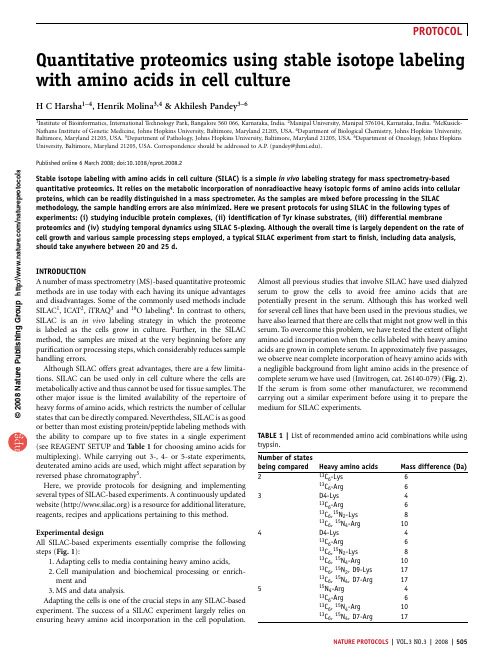
Quantitative proteomics using stable isotope labeling with amino acids in cell cultureH C Harsha 1–4,Henrik Molina 3,4&Akhilesh Pandey 3–6Instituteof Bioinformatics,International Technology Park,Bangalore 560066,Karnataka,India.Manipal University,Manipal 576104,Karnataka,India.McKusick-Nathans Institute of Genetic Medicine,Johns Hopkins University,Baltimore,Maryland 21205,USA.4Department of Biological Chemistry,Johns Hopkins University,Baltimore,Maryland 21205,USA.5Department of Pathology,Johns Hopkins University,Baltimore,Maryland 21205,USA.6Department of Oncology,Johns Hopkins University,Baltimore,Maryland 21205,USA.Correspondence should be addressed to A.P.(pandey@).Published online 6March 2008;doi:10.1038/nprot.2008.2Stable isotope labeling with amino acids in cell culture (SILAC)is a simple in vivo labeling strategy for mass spectrometry-based quantitative proteomics.It relies on the metabolic incorporation of nonradioactive heavy isotopic forms of amino acids into cellular proteins,which can be readily distinguished in a mass spectrometer.As the samples are mixed before processing in the SILAC methodology,the sample handling errors are also minimized.Here we present protocols for using SILAC in the following types of experiments:(i)studying inducible protein complexes,(ii)identification of Tyr kinase substrates,(iii)differential membraneproteomics and (iv)studying temporal dynamics using SILAC 5-plexing.Although the overall time is largely dependent on the rate of cell growth and various sample processing steps employed,a typical SILAC experiment from start to finish,including data analysis,should take anywhere between 20and 25d.INTRODUCTIONA number of mass spectrometry (MS)-based quantitative proteomic methods are in use today with each having its unique advantages and disadvantages.Some of the commonly used methods include SILAC 1,ICAT 2,iTRAQ 3and 18O labeling 4.In contrast to others,SILAC is an in vivo labeling strategy in which the proteome is labeled as the cells grow in culture.Further,in the SILAC method,the samples are mixed at the very beginning before any purification or processing steps,which considerably reduces sample handling errors.Although SILAC offers great advantages,there are a few limita-tions.SILAC can be used only in cell culture where the cells are metabolically active and thus cannot be used for tissue samples.The other major issue is the limited availability of the repertoire of heavy forms of amino acids,which restricts the number of cellular states that can be directly compared.Nevertheless,SILAC is as good or better than most existing protein/peptide labeling methods with the ability to compare up to five states in a single experiment (see REAGENT SETUP and Table 1for choosing amino acids for multiplexing).While carrying out 3-,4-or 5-state experiments,deuterated amino acids are used,which might affect separation by reversed phase chromatography 5.Here,we provide protocols for designing and implementing several types of SILAC-based experiments.A continuously updated website ()is a resource for additional literature,reagents,recipes and applications pertaining to this method.Experimental designAll SILAC-based experiments essentially comprise the following steps (Fig.1):1.Adapting cells to media containing heavy amino acids,2.Cell manipulation and biochemical processing or enrich-ment and3.MS and data analysis.Adapting the cells is one of the crucial steps in any SILAC-based experiment.The success of a SILAC experiment largely relies on ensuring heavy amino acid incorporation in the cell population.Almost all previous studies that involve SILAC have used dialyzed serum to grow the cells to avoid free amino acids that are potentially present in the serum.Although this has worked well for several cell lines that have been used in the previous studies,we have also learned that there are cells that might not grow well in this serum.T o overcome this problem,we have tested the extent of light amino acid incorporation when the cells labeled with heavy amino acids are grown in complete serum.In approximately five passages,we observe near complete incorporation of heavy amino acids with a negligible background from light amino acids in the presence of complete serum we have used (Invitrogen,cat.26140-079)(Fig.2).If the serum is from some other manufacturer,we recommend carrying out a similar experiment before using it to prepare the medium for SILAC experiments.p u o r G g n i h s i l b u P e r u t a N 8002©n a t u r e p r o t o c o l s/m o c .e r u t a n .w w w //:p t t h TABLE 1|List of recommended amino acid combinations while using trypsin.Number of statesbeing compared Heavy amino acidsMass difference (Da)2C 6-Lys 613C 6-Arg 63D4-Lys413C 6-Arg 613C 6,15N 2-Lys 813C 6,15N 4-Arg 104D4-Lys413C 6-Arg 613C 6,15N 2-Lys 813C 6,15N 4-Arg 1013C 6,15N 2,D9-Lys 1713C 6,15N 4,D7-Arg 17515N 4-Arg 413C 6-Arg 613C 6,15N 4-Arg 1013C 6,15N 4,D7-Arg 17NATURE PROTOCOLS |VOL.3NO.3|2008|505Below,we describe four SILAC experiments in detail with relevant examples.These are (A)SILAC for studying induced protein complexes,(B)SILAC for identification of Tyr kinase substrates,(C)SILAC for differential membrane proteomics and (D)studying temporal dynamics using SILAC for 5-plexing.(A)SILAC for studying induced protein complexes Cellular functions are often mediated through physical interactions invol-ving protein–protein,protein–RNA and protein–DNA molecules.Analyzing components of protein complexes in response to stimuli is an important step toward understanding the molecular mechan-isms involved in downstream signaling pathways.Although several biochemical methods have been used to characterize protein–protein interactions,they generally do not distinguish specific from nonspecific interactions.SILAC-based experiments are simple and allow one to distinguish specific from nonspecific interactions.Below,we describe an experiment where two conditions (resting and ligand induced)are compared (option A).(B)SILAC for identification of Tyr kinase substrates Tyr kinase signaling pathways play an important role in cellular growth and differentiation.Aberrant regulation of Tyr kinases and their sub-strates has been known to lead to several pathologic conditions including ing SILAC,it is not only possible to identify downstream substrates of Tyr kinases,but to also identify differ-entially regulated Tyr kinase signaling pathways in pathologic conditions such as cancers.A number of studies have already described the use of SILAC to study temporal dynamics of Tyr kinase signaling pathways.Here,we describe a simple SILAC protocol to identify Tyr kinase substrates in a ligand-induced system (option B).(C)SILAC for differential membrane proteomics Membrane proteins are attractive candidates to study,as they have the potential as biomarkers or drug targets.Membrane enrichment methods coupled to quantitative proteomics can be used to identify cell surface and other membrane-associated proteins that are differen-tially expressed between different states (option C).(D)Studying temporal dynamics using SILAC for 5-plexing It is now possible to carry out a 5-plex SILAC experiment with currently available stable isotopic forms of Arg.This is particularly useful in studying temporal dynamics of signaling pathways,to measure dose–response to pharmacological agents,monitor proteomic changes at various stages of cell differentiation,etc.Below,we describe a 5-state SILAC strategy to identify proteomic changes in the secretome of preadipocytes differentiating into adipocytes as an example (option D).CommentSILAC has been successfully used to carry out a wide variety of studies including temporal dynamics of signaling pathways 6,7,enzyme substrates 8,identification of cancer biomarkers 9and studying protein complexes 10.The success of a SILAC-based quantitative proteomic experiment depends on various criteria including the selection of heavy amino acids,the choice of protease,normal-ization of samples and depth of mass spectrometric analysis.Although most cell lines we have tested take approximately five cell divisions to incorporate heavy amino acids into the entire proteome,we recommend testing the same every time a new cell line is used.Save an aliquot of sample at every processing step and monitor the proteins by western blotting to check for sample loss and efficiency of the enrichment methods used.As SILAC completely relies on metabolic incorporation of the heavy amino acids and samples are mixed and processed together,the chances of error are considerably minimized when compared to other chemi-cal labeling methods.Metabolic conversion of Arg to Pro has been observed in some cell types 11.This probably occurs when there is an excess of Arg in the growth medium.Thus,an excess of heavy Arg (13C 6)provided for the cells grown in heavy medium could result in introduction of heavy Pro residues (13C 5),complicating the quantitation of Pro-containing peptides.T o overcome this hurdle,we recommend determining the optimum concentration of Arg required for every new cell line used.p u o r G g n i h s i l b u P e r u t a N 8002©n a t u r e p r o t o c o l s/m o c .e r u t a n .w w w //:p t thIn-solution digestionR e l a t i v e i n t e n s i t yMedium* Light* Heavy*Figure 1|Workflow of a stable isotope labeling with amino acids in cell culture (SILAC)-based quantitative proteomic experiment.Adapting the cell lines in heavy medium is a first step in any SILAC-based experiment.The lysates from light and heavy cell populations are mixed after normalizing the protein amounts and samples are processed together thereafter.Fractionation and enrichment methods vary based on the requirement and proteins may be subjected to either in-gel or in-solution digestion.The digested proteinsamples can be analyzed by any high-resolution mass spectrometer.LC,liquid chromatography;MS,mass spectrometry.506|VOL.3NO.3|2008|NATURE PROTOCOLSTrypsin is the most commonly used protease in proteomic experiments.Lys-C and Arg-C are other proteases that could be used.Although Lys-C seems to cleave C-terminus to Lys residues specifically,thespecificity of Arg-C is less predictable.Incases where Lys-C is used for generating peptides,only heavy Lys should be used for labeling.Although this is cost effective,there is one disadvantage of taking this approach,as it results in large peptides,which may not be optimal for MS/MS analysis.SILAC cannot be used on tissues.Although this is one of the disadvantages,there are other quantitative proteomicmethods,which can serve the purpose.T o provide researchers with updated informa-tion on SILAC,we update our website ()continuously.The website provides a list of all reagents that are necessary to set up a SILAC experiment along with relevant publications that describe SILAC for different applications.MATERIALSREAGENTS.DMEM.RPMI !CAUTION If the cells require media other than those mentionedabove,they should be prepared as per the formulation of the respective media.The light and heavy medium to be used in a SILACexperiment should be from the same manufacturer to avoid variability.(Some of the manufacturers who can provide custom SILAC media deficient in desired amino acids are Athena ES,Cambridge isotopes,Sigma andInvitrogen.)Amino acid concentrations for DMEM and RPMI are provided in Table 2..Sequencing grade trypsin (Promega).Agarose-conjugated antiphosphotyrosine Ab 4G10(Upstate Biotechnology),PY20(BIOMOL).Protein A agarose beads (Sigma).Dialysis tube (molecular weight cutoff 3,500;Pierce).NP-40(EMD Biosciences).Urea (Sigma).Sodium deoxycholate (Sigma).Sodium orthovanadate (Sigma).Complete protease inhibitor cocktail tablets (Roche).DC protein assay kit (Bio-Rad).Colloidal coomassie staining kit (Invitrogen).Acetonitrile (J.T.Baker).2-Mercaptoethanol (Sigma)!CAUTION This reagent is toxic if inhaled;it is advisable to use a chemical hood while using this reagent..Heavy amino acids (Cambridge Isotope Laboratories or Sigma-Aldrich)(see REAGENT SETUP).Modified RIPA (see REAGENT SETUP).Lysis buffer (see REAGENT SETUP).Sodium orthovanadate (see REAGENT SETUP).5ÂSDS-PAGE sample loading buffer (Laemmli)(see REAGENT SETUP)EQUIPMENT.Falcon tubes (15and 50ml).Microfuge tubes (1.5ml).Vacufuge (Eppendorf).Microcentrifuge with refrigeration (Z 16,000g ).Centrifuge with refrigeration (Z 16,000g and hold tubes 410ml).Quadrupole time of flight mass spectrometer.Liquid chromatography (LC)equipment (Agilent)(see EQUIPMENTSETUP)REAGENT SETUPHeavy amino acids Determining the heavy amino acid(s)and the protease to be used is an important step in planning a SILAC-based quantitativeproteomic experiment.As trypsin is the most commonly used protease for MS experiments,heavy Arg and heavy Lys are the recommended amino acids.Table 1provides most preferred heavy amino acid combinations and the respective mass differences.L -Lys 2HCl (4,4,5,5-D4),L -Lys:2HCl (13C 6),L -Lys:2HCl (13C 6,15N 2),L -Lys:2HCl (13C 6,15N 2,D9),L -Arg:HCl (15N 4),L -Arg:HCl (15N 4,D7),L -Arg:HCl (13C 6),L -Arg:HCl (13C 6,15N 4),and L -Arg:HCl (13C 6,15N 4,D7).!CAUTION Itp u o r G g n i h s i l b u P e r u t a N 8002©n a t u r e p r o t o c o l s/m o c .e r u t a n .w w w //:p t t h 6 DaDay 1Day 2Day 3Day 5Day 6Day 4100LVSDEMVVELIEK760.4R e l a t i v e i n t e n s i t y100R e l a t i v e i n t e n s i t y763.4763.9764.4760.9761.47600100R e l a t i v e i n t e n s i t y00761762763m/z764765760761762763m/z 764765100R e l a t i v e i n t e n s i t y0760761762763m/z 764765100R e l a t i v e i n t e n s i t y0760761762763m/z 764765100R e l a t i v e i n t e n s i t y760761762763m/z 764765760761762763m/z764765Figure 2|Incorporation of heavy Lys (13C 6)in the presence of complete serum.Humanembryonic kidney 293T cells were switched to stable isotope labeling with amino acids in cell culture medium containing complete serum and supplemented with heavy Lys on day 0.An aliquot of cells was removed every 24h and the celllysates resolved by SDS-PAGE.The same band was excised for each time point,digested using trypsin and analyzed using mass spectrometry.Doubly charged ions at m/z 760.4and 763.4correspond to a peptide from the protein adenylate kinase 2.The observed mass difference is due to the incorporation of heavy Lys.As seen,by day 4,there is near complete incorporation of heavy Lys in the presence of complete serum.NATURE PROTOCOLS |VOL.3NO.3|2008|507is important to make sure all the heavy amino acids are as pure as possible (497%).Modified RIPA 150mM NaCl,50mM Tris–HCl,pH 7.4,1%NP-40,0.25%sodium deoxycholate,1mM EDTA,1mM sodium orthovanadate,1protease inhibitor cocktail tablet per 50ml modified RIPA buffer.!CAUTION Modified RIPA can be stored at room temperature (22–251C)without adding protease inhibitor tablet and sodium orthovanadate.We recommend adding both the components just before use and chilling the buffer at 41C.Lysis buffer 150mM NaCl,50mM Tris–HCl,pH 7.4,1%NP-40,1mM sodium orthovanadate,1protease inhibitor cocktail tablet per 50ml lysis buffer.!CAUTION Lysis buffer can be stored at room temperature without adding protease inhibitors or sodium orthovanadate.We recommend adding both just before use and chilling the buffer at 41C.Sodium orthovanadate Prepare 100mM sodium orthovanadate stock solu-tion,adjust the pH to 10using 1N HCl and boil in a microwave until it turns colorless.Add this to a final concentration of 1mM to modified RIPA or lysis buffer just before use.53SDS-PAGE sample loading buffer (Laemmli)0.3M Tris–HCl,pH 6.8,10%SDS,12.5%vol/vol 2-mercaptoethanol,50%glycerol,0.016%bromo-phenol blue.!CAUTION 5ÂSample buffer without 2-mercaptoethanol can be prepared and stored at room temperature.Add to protein samples to obtain a final concentration of 1Âalong with 2-mercaptoethanol.Boil the protein samples at 951C for 10min just before loading.EQUIPMENT SETUPLC equipment 1100series HPLC system equipped with a capillary pump,micro-vacuum degasser,microwell-plate autosampler.PROCEDUREAdapting cells in media containing heavy amino acids1|Determine the media requirements of the cell line to be used in the experiment.Obtain sufficient quantity of medium containing naturally abundant isotopic forms of amino acids (light medium )along with the same medium lacking the desired amino acids (e.g.,medium lacking Arg and Lys)to be substituted as heavy isotopic forms (heavy medium ).m CRITICAL STEP The amount of media required is determined by the number of cells required for an experiment.It is recommended to obtain complete medium and the custom medium lacking required amino acids from the same manufacturer to avoid variability.2|Prepare the light medium by adding supplements as required (serum,antibiotics,etc.).Refer to for additional details.3|Prepare the heavy medium for SILAC experiments by dissolving heavy amino acids in the suggested concentration (Table 2).Add supplements as required and filter using 0.22-m m filter.m CRITICAL STEP Ensure that the supplements are not a source of free amino acids (e.g.,those containing crude extracts).4|Expand the control cell population in light medium and the test cell population in heavy medium to obtain the cells required for the experiment.The cells grown in heavy medium should undergo a minimum of five divisions to obtain near complete incorporation of heavy amino acids into the entire proteome (Fig.2).For convenience,we grow the cells to be induced in heavy medium when using an inducible system.!CAUTION Reversing the labeling (uninduced in heavy medium and induced in light medium)can also be used if desired.We grow cells to be induced in heavy medium only as a matter of convenience to look at the data and we do not observe any change by reversing the label.Apart from growth in light or heavy medium,the cells should be treated identically to avoid variability.5|Perform one of the following protocols for SILAC-based experiments:(A)SILAC for studying induced protein complexes,(B)SILAC for identification of Tyr kinase substrates,(C)SILAC for differential membrane proteomics or (D)studying temporal dynamics using SILAC for 5-plexing.(A)SILAC for studying induced protein complexes(i)Choose an appropriate cell line that responds to a stimulus.Grow one cell population in light medium and adapt another cell population of the same cell type in heavy medium containing 13C 6-Arg plus 13C 6Lys.m CRITICAL STEP See Table 1for choosing the appropriate heavy amino acid(s)to be used if three or more conditions are to be compared.p u o r G g n i h s i l b u P e r u t a N 8002©n a t u r e p r o t o c o l s/m o c .e r u t a n .w w w //:p t t h TABLE 2|A partial list of stable isotopic forms of Lys and Arg that are commercially available.Stable isotopic amino acids Concentration in DMEM Concentration in RPMI Mass difference (Da)L -Lys:2HCl (4,4,5,5-D4)178.00mg l À1(0.798mM)61.14mg l À1(0.274mM)4L -Lys:2HCl (13C 6)179.6mg l À1(0.798mM)61.68mg l À1(0.274mM)6L -Lys:2HCl (13C 6,15N 2)181.2mg l À1(0.798mM)62.23mg l À1(0.274mM)8L -Lys:2HCl (13C 6,15N 2,D9)188.4mg l À1(0.798mM)64.69mg l À1(0.274mM)17L -Arg:HCl (15N 4)85.4mg l À1(0.398mM)246.79mg l À1(1.15mM)4L -Arg:HCl (13C 6)86.2mg l À1(0.398mM)249.1mg l À1(1.15mM)6L -Arg:HCl (13C 6,15N 4)87.8mg l À1(0.398mM)253.7mg l À1(1.15mM)10L -Arg:HCl (15N 4,D7)88.2mg l À1(0.398mM)254.8mg l À1(1.15mM)11L -Arg:HCl (13C 6,15N 4,D7)90.6mg l À1(0.398mM)261.7mg l À1(1.15mM)17If any media other than DMEM or RPMI is being used,the amount of heavy isotopic amino acid should be calculated accordingly.508|VOL.3NO.3|2008|NATURE PROTOCOLS(ii)Expand each cell population in five 15-cm dishes and grow them until they are B 85%confluent.The example here assumes an overexpression system.!CAUTION The number of cells required is decided by the amount of target protein expression.In overexpressionsystems,the cell number required is lower as compared to an experiment where an endogenous protein is being studied.(iii)Wash the cells three times using either 5ml of PBS or serum-free medium and allow them to starve overnight (B 12h)inB 20ml of the corresponding serum-free medium.This is to reduce the signaling levels in cells to a basal level.!CAUTION The cells growing in light medium should be starved in serum deficient light medium while the cells growing in heavy medium should be starved in serum deficient heavy medium.(iv)Stimulate the cells grown in heavy medium with the appropriate ligand for an optimized time;leave the control cellpopulation untreated.m CRITICAL STEP Ligand concentration and the duration of treatment should be determined by a small-scale experiment based on methods such as immunoblotting before initiating the SILAC experiment.(v)Aspirate the medium and lyse the cells using 1ml of chilled lysis buffer per cell culture dish and place the dishes at 41C for 15min.m CRITICAL STEP The choice of lysis buffer in a co-immunoprecipitation experiment is important.Do not use buffers with denaturing detergents such as SDS.The optimal detergent to be used for lysis should be determined by a small-scale experiment.(vi)Scrape the cells after 15min and transfer the lysates into individual Sorvall centrifuge tubes (15ml)maintained on ice.(vii)Place the lysates on ice with occasional vortexing for 15–20min.(viii)Centrifuge the lysates at 16,000g for 10min at 41C.(ix)Transfer the supernatant into fresh tubes.(x)Measure the protein concentration of cell lysates using standard methods such as Lowry or Bradford and mix the lysates in 1:1proportion based on protein amounts.(xi)Preclear the lysates by incubating with 300m l of protein A or G agarose beads for 4h at 41C.m CRITICAL STEP This step is essential to reduce the nonspecific binding of proteins to beads during immunoprecipitation.(xii)Centrifuge the lysates at 16,000g for 2min and transfer the supernatant to a fresh tube.(xiii)Immunoprecipitate the protein of interest by incubating the lysate with an appropriate amount of agarose-conjugatedAb directed against the protein of interest at 41C overnight.m CRITICAL STEP The amount of Ab required is determined by the amount of target protein present in the sample.We routinely start with B 100m g of Ab for every 50mg of total protein.Alternatively,Ab coupled to magnetic beads (Dynabeads)can also be used at this step.(xiv)Centrifuge the lysates at 16,000g for 1min and aspirate the supernatant.(xv)Wash the beads three times using 5ml chilled lysis buffer.(xvi)Elute the immunoprecipitated protein either by competitive elution or by boiling the beads with sample buffer for10min.Resolve the protein sample on SDS-PAGE.m CRITICAL STEP We use competitive elution wherever possible.If elution is carried out using some salts or peptides,we recommend dialyzing the eluate before proceeding to the next step.Wherever competitive elution is not possible,elution by low pH or boiling of beads can be used as an alternative strategy.We suggest saving fractions at each processing step during the entire experiment to help with troubleshooting if need arises.(xvii)Stain the gel using colloidal coomassie.’PAUSE POINT The protein gel can be stored at 41C up to a week before continuing with the next step.(xviii)Digest the protein bands using trypsin as described in Step 6A.’PAUSE POINT The trypsin digests can be dried and stored at À801C until further analysis.(B)SILAC for identification of Tyr kinase substrates(i)Choose a cell line that responds to a known activator of Tyr kinase signaling (e.g.,Hela for epidermal growth factor and NIH3T3for platelet-derived growth factor).m CRITICAL STEP Determine the appropriate ligand concentration and the treatment time by immunoblotting-based experiments using antiphosphotyrosine Ab.Ligand concentration and treatment time that provides appreciable difference between ligand treated and untreated conditions should be chosen.(ii)For a 2-state experiment,start adapting the cells to heavy medium containing 13C 6-Arg and 13C 6Lys.(iii)Expand light and heavy cells in twenty 15-cm dishes until they are B 85%confluent.The number of cells thatwould yield a total of 100mg protein after combining light and heavy samples is a good starting material for Ab-based enrichment of Tyr phosphoproteins.(iv)Wash the cells three times using either 5ml of PBS or serum-free medium and allow them to starve overnight (B 12h)inB 20ml serum-free medium.p u o r G g n i h s i l b u P e r u t a N 8002©n a t u r e p r o t o c o l s/m o c .e r u t a n .w w w //:p t t h NATURE PROTOCOLS |VOL.3NO.3|2008|509m CRITICAL STEP This step is to reduce the basal levels of phosphorylation in the cells.The cells must be washed and starved in the corresponding serum-free medium (light medium for the control cell population and heavy medium for the cells that are going to be treated with the ligand).(v)Stimulate the cells grown in heavy medium with the appropriate ligand for an optimized time;leave the control cell population untreated.(vi)Aspirate the medium from each dish and lyse the cells in 1ml chilled modified RIPA buffer,place the dishes at 41C forB 15min.!CAUTION The cell culture dishes can be placed either on ice or in a cold room maintained at 41C.Modified RIPA is recommended for cell lysis by the manufacturer of the 4G10antiphosphotyrosine Ab (Upstate Biotechnology).(vii)Scrape the cells and transfer the lysates from cells grown in light and heavy medium into individual Sorvall centrifugetubes and vortex.(viii)Place the tubes on ice for 20min with occasional vortexing.(ix)Centrifuge the lysates at 16,000g for 10min at 41C.(x)Transfer the supernatant into fresh tubes.(xi)Measure protein concentration of the lysates using standard Lowry or Bradford method and mix them 1:1based onprotein amounts.m CRITICAL STEP It is important to normalize the protein amounts before proceeding,failing to do so will bias the experiments and the results will not be reliable.(xii)Preclear the lysates by incubating with 400m l of protein A agarose beads for 4h with gentle rocking at 41C.(xiii)Centrifuge the lysates at 16,000g for 1min and transfer the supernatant to a fresh tube.(xiv)Add 400m g of agarose-conjugated antiphosphotyrosine Ab (4G10)and 150m g of agarose-conjugatedantiphosphotyrosine Ab (PY20).(xv)Incubate the lysates at 41C overnight with gentle rocking.(xvi)Pellet the agarose beads by centrifuging at 16,000g for 1min and aspirate the supernatant.(xvii)Wash the beads three times with 5ml chilled modified RIPA,then spin and discard the supernatant each time.(xviii)Transfer the beads to a 2-ml microfuge tube and incubate with 1bed volume of 100mM phenyl phosphate prepared inPBS.Rock the tubes gently at room temperature for 15min.(xix)Collect the supernatant into a fresh tube after centrifuging the tubes at 16,000g .(xx)Repeat Steps 5B(xviii)and (xix)and pool the eluates.(xxi)Transfer the eluate into dialysis tube (molecular weight cutoff 3,500)and dialyze at least two times against 4l of waterfor 2h each time followed by an overnight dialysis at 41C,to remove phenyl phosphate.m CRITICAL STEP Failing to dialyze will affect the migration of the protein sample on SDS-PAGE and produce smears.(xxii)Collect the dialyzed eluate into fresh microfuge tubes and concentrate it in a vacufuge to B 100–150m l.(xxiii)Add appropriate volume of 5ÂSDS-PAGE sample buffer to bring the final concentration to 1Â.Add reducing agent andresolve on SDS-PAGE.(xxiv)Stain the gel using colloidal coomassie.’PAUSE POINT The protein gel can be stored at 41C up to a week before continuing with the next step.(xxv)Digest the protein bands using trypsin as described in Step 6A.’PAUSE POINT The trypsin digests can be dried and stored at À801C until further analysis.(xxvi)Analyze tryptic digests using MS.(C)SILAC for differential membrane proteomics(i)Grow five 15-cm dishes each of control cell population and test cell population in light and heavy medium,respectively,until they are 85%confluent.!CAUTION For a two-state comparison,we recommend using 13C 6-Arg and 13C 6-Lys.See Table 1for choosing the appropriate heavy amino acid(s)to be used if three or more conditions are to be compared.(ii)Wash the cells three times using either 5ml of PBS or serum-free medium and allow them to starve overnight (B 12h)in B 20ml serum-free medium.(iii)Blend the cells in appropriate amount of high salt buffer (2M NaCl,10mM HEPES,pH 7.5,1mM EDTA,pH 8.0)using adounce homogenizer and incubate on ice for 30min.!CAUTION If the two samples are derived from the same cell type,and the cell number is equalized before seeding,they can be mixed and processed together from this step.In this case,skip Steps 5C(viii)–(x).If the samples are derived from two different cell types,we recommend not mixing at this stage and following the described procedure accordingly.(iv)Centrifuge the disrupted cells at 250,000g for 45min at 41C.(v)Discard the supernatant and resuspend the pellet in 0.1M Na 2CO 3,pH 11and incubate on ice for 30min.(vi)Centrifuge at 250,000g for 45min at 41C.Discard the supernatant.(vii)Repeat Steps 5C(iii)–(vi).p u o r G g n i h s i l b u P e r u t a N 8002©n a t u r e p r o t o c o l s/m o c .e r u t a n .w w w //:p t t h 510|VOL.3NO.3|2008|NATURE PROTOCOLS。
SILAC
SILAC技术介绍及操作陈宁稳定同位素标记原理体内标记体外标记通过代谢标记、酶反应Array或化学反应,将重/轻同位素标签标记于不同样品的蛋白质或肽段,等量混合后通过色谱、SDS-PAGE的分离,然后进行质谱鉴定。
根据质谱谱图中成对峰的面积之比可判断出同一肽段在不同样品中的含量变化,根据二级谱图对肽段进行序列测定从而鉴定蛋白质。
优点1)样本需求量少,通常每个样品只需要几十微克的蛋白量;2)采用质谱定量,定量结果准确且批次变异小、重复性好;3)体内代谢标记与SDS-PAGE或色谱分离技术相结合,兼容疏水性蛋白和偏碱性蛋白,不受蛋白性质限制;4)多个样品混合后同时进行分离、酶切和鉴定,后续实验对样品的影响是一致,减少了实验操作和仪器设备产生的影响;5)由于该技术属于体内标记,标记效果稳定,其标记效率不受裂解液的影响,不仅适合于进行全细胞蛋白的分析,还适合于膜蛋白的鉴定和定量。
应用1) 多个样品全细胞蛋白或亚细胞蛋白的差异比较;如:前列腺癌微粒体蛋白的差异比较、HCV相关的细胞脂阀蛋白研究等2) 同一样品不同条件下全细胞蛋白或亚细胞蛋白的差异比较如:厚朴酚刺激前后的Hela细胞蛋白质变化、凋亡过程中细胞核蛋白变化等3) 将SILAC与IP技术相结合,可用于研究特定蛋白质的相互作用蛋白分析;4) 通过对质谱谱图的解读,还能进行磷酸化位点的确认和磷酸化蛋白的定量分析。
应用范围活体培养的细胞或低等有机体(如蠕虫),对于疾病研究中常用的组织样品、体液样品等无法分析SILAC操作参考1. SILAC-标记培养液的准备2. 标记效率的确定3. 标记培养4. 蛋白的分离与鉴定5. MSQuant软件分析差异蛋白1. SILAC-标记培养液的准备1.1 试剂(根据实验选择)•缺陷培养基:Lysine 缺陷型DMEM / RPMI-1640培养基、Arginine 缺陷型DMEM / RPMI-1640培养基、Methionine 缺陷型DMEM / RPMI-1640培养基; 氨基酸缺陷型DMEM/RPMI-1640培养基•“heavy AA”(标记型):13C6-Arginine 、13C 615N 4-Arginine 、13C 6-Lysine 、13C 615N 4-Lysine 、methyl-13C 2H 3-Methionine 、D3-Leucine 等•“light AA”(非标记型):L-Arginine ,L-Lysine ,L-Methionine 等•透析型血清1.2 细胞培养液的配制•在缺陷型培养液的基础上加入“heavy / light AA”,可根据细胞状态酌量添加透析型血清含量。
SILAC蛋白组
SILAC蛋白组
SILAC蛋白组或SILAC定量蛋白组学,是指利用SILAC技术对蛋白质组进行定量研究。
百泰派克生物科技提供基于SILAC标记的定量蛋白组分析服务。
SILAC
在基于质谱(MS)的定量蛋白质组学中,SILAC(Stable Isotope Labeling By Amino Acids In Cell Culture)是通过在细胞培养过程中加入稳定同位素标记的
氨基酸对样品进行标记后结合质谱检测,对样品进行蛋白质组学分析的方式是一种简单、可靠而又强大的方法。
SILAC通过正常代谢过程对细胞蛋白进行标记,在新
合成的蛋白质中加入非放射性的、稳定的含同位素的氨基酸。
SILAC提供了准确的
相对定量,不需要任何化学衍生或操作,即可实现蛋白质组学分析。
SILAC属于体
内标记技术,使样品更接近自然状态,其标记效率高达100%,且标记效果稳定,
适合于全细胞蛋白分析,以及膜蛋白的鉴定和定量,每个样本只需要几十微克的蛋白量。
SILAC蛋白组分析
SILAC定量蛋白组分析的实验流程是:在细胞培养基中加入轻、中或重型稳定同位
素标记的必需氨基酸赖氨酸(Lys)和精氨酸(Arg),在细胞生长过程中,新合成的蛋白质带上稳定同位素标签。
培养一段时间后,等量混合细胞培养物中各类型蛋白质,酶解后进行质谱分析。
由于质量差异,可以在质谱仪中区分带有不同稳定同位素组成的化学性质相同的肽对。
通过比较一级质谱图中不带标记和带同位素标记的质谱峰型的面积大小即可进行相对定量,同时二级谱图还可以对肽段进行序列测定从而进行蛋白鉴定。
SILAC蛋白组。
SILAC定量技术方案
SILAC定量技术方案——样品制备1. 细胞培养和蛋白提取(1). MEM培养基的准备(配制400 mL )①制备氨基酸储液:分别在20mL L-Arginine缺陷型MEM培养基中配置0.30mM(50% 正常浓度)的13C6 L-Arginine hydrochloride 和13C6, 15N4 L-Arginine hydrochloride 储存液(均称取26 mg),待充分溶解后,分别用0.22pm的无菌滤器过滤。
②分别加入20mL除菌后的氨基酸储液、10%的透析胎牛血清、1%的青链霉素、2mM的L-Glutamine和2mM的丙酮酸钠并用缺陷型MEM培养基定容到400 mL,贮存于4°C。
每种完全培养基中的氨基酸浓度见表4-1:表4-1 MEM 培养基中轻、中、重L-Arginine的浓度(2).细胞培养将A、B、C三种细胞复苏后,分别接种于10cm培养皿中,在常规RPMI 1640培养基(添加10%的透析胎牛血清和1%的青链霉素)中培养HL-7702,分别使用仅含有13C6 L-Arginine hydrochloride 禾口13C6, 15N4 L-Arginine hydrochloride 的MEM培养基培养B和C,经过5-6个细胞世代的培养后,将细胞数量扩增到107-108左右(5-6盘)。
(3).蛋白提取用500卩全细胞裂解液(50 mM Tris,pH 7.4; 150mM NaCl ;1%Triton-100 ; 1mM AEBSF ; 20 卩1/ 卩g apro ;n20 p 1/ 卩g leupeptin)2. 蛋白质含量测定(1) . 采用改进的Bradford 法,将BSA 分别稀释为从0-1.40mg/ml 范围内若干浓度,待测样品稀释10 倍、20倍,各取20 丄加80 让0.12M HCl ,轻轻混匀。
(2) . 各管再加入3.5ml 过滤的稀释4 倍的dye reage nt 轻轻混匀,静置5min 后测A595 值。
silac实验步骤
silac实验步骤英文回答:The SILAC (Stable Isotope Labeling by Amino Acids in Cell Culture) experiment is a widely used technique in proteomics research. It allows for the quantitative analysis of protein expression and dynamics in cells. In this experiment, cells are grown in media containing either light or heavy isotopically labeled amino acids. The heavy amino acids contain stable isotopes, such as 13C or 15N, which can be easily distinguished from the light amino acids. The cells incorporating the heavy amino acids will then synthesize proteins with the labeled amino acids, while the cells grown in light media will synthesize proteins with the normal, unlabeled amino acids.After the cells have been labeled, they can be treated differently to induce changes in protein expression or dynamics. For example, one group of cells can be treated with a drug, while another group can be left untreated as acontrol. The cells are then harvested and the proteins are extracted. The proteins from the different groups are mixed together and subjected to proteomic analysis, such as mass spectrometry.The mass spectrometry analysis allows for the identification and quantification of the proteins in each sample. The heavy and light isotopically labeled proteins can be distinguished based on their mass-to-charge ratio. The abundance of each protein in the treated and control samples can be compared, allowing for the identification of proteins that are differentially expressed or regulated in response to the treatment.SILAC experiments have been used in a wide range of biological studies. For example, researchers have used SILAC to study the effects of drug treatments on cancer cells. By comparing the protein expression profiles of treated and untreated cells, they were able to identify proteins that are involved in drug resistance or sensitivity. SILAC has also been used to study protein dynamics, such as the turnover rates of specific proteinsin response to different cellular stimuli.中文回答:SILAC(细胞培养中氨基酸稳定同位素标记)实验是蛋白质组学研究中广泛使用的技术。
- 1、下载文档前请自行甄别文档内容的完整性,平台不提供额外的编辑、内容补充、找答案等附加服务。
- 2、"仅部分预览"的文档,不可在线预览部分如存在完整性等问题,可反馈申请退款(可完整预览的文档不适用该条件!)。
- 3、如文档侵犯您的权益,请联系客服反馈,我们会尽快为您处理(人工客服工作时间:9:00-18:30)。
SILAC定量技术方案一样品制备1.细胞培养和蛋白提取(1).MEM培养基的准备(配制400mL)①制备氨基酸储液:分别在20mL L-Arginine缺陷型MEM培养基中配置0.30mM(50%正常浓度)的13C6L-Arginine hydrochloride和13C6,15N4L-Arginine hydrochloride储存液(均称取26mg),待充分溶解后,分别用0.22μm的无菌滤器过滤。
②分别加入20mL除菌后的氨基酸储液、10%的透析胎牛血清、1%的青链霉素、2mM的L-Glutamine和2mM的丙酮酸钠并用缺陷型MEM培养基定容到400mL,贮存于4°C。
每种完全培养基中的氨基酸浓度见表4-1:表4-1MEM培养基中轻、中、重L-Arginine的浓度Cell lines Amino Acids M. W.Culture Media Amount(concentration)A L-Arginine174.2RPMI1640241.9mg/L(1.15mM)B13C6L-Arginine216.7MEM65.0mg/L(0.3mM) C13C6,15N4L-Arginine220.7MEM65.0mg/L (0.3mM)(2).细胞培养将A、B、C三种细胞复苏后,分别接种于10cm培养皿中,在常规RPMI1640培养基(添加10%的透析胎牛血清和1%的青链霉素)中培养HL-7702,分别使用仅含有13C6L-Arginine hydrochloride和13C6,15N4L-Arginine hydrochloride的MEM培养基培养B和C,经过5-6个细胞世代的培养后,将细胞数量扩增到107-108左右(5-6盘)。
(3).蛋白提取用500μl全细胞裂解液(50mM Tris,pH7.4;150mM NaCl;1%Triton-100;1mM AEBSF;20μl/μg aprotinin;20μl/μg leupeptin)2.蛋白质含量测定(1).采用改进的Bradford法,将BSA分别稀释为从0-1.40mg/ml范围内若干浓度,待测样品稀释10倍、20倍,各取20μL加80μL0.12M HCl,轻轻混匀。
(2).各管再加入3.5ml过滤的稀释4倍的dye reagent,轻轻混匀,静置5min后测A595值。
(3).取A595值对BSA标准浓度作标准曲线,再根据待测样品的A595值,从BSA标准曲线中确定其浓度。
(4).同位素标记样品与非标记样品按照蛋白含量1:1混合。
3.SDS-PAGE分离和染色(1).电泳条件:4%浓缩胶,12%分离胶;(2).2×SDS上样缓冲液的配制:2mL Tris-HCL buffer(0.5M,pH6.8);2mL甘油;1mL巯基乙醇;4mL10%SDS溶液;适量水补足体积至10mL,混匀即得。
(3).电泳条件:4%浓缩胶,12%分离胶,先恒流10mA,运行时间0.5h,再于20mA运行至溴酚蓝前沿到达分离胶底部。
(4).胶体考马斯亮蓝染色方法如下:用10%甲醇,7%乙酸溶液固定30min,然后用胶体考马斯亮蓝溶液(0.12%G-250,10%硫酸铵,10%磷酸,20%甲醇)染色过夜,用蒸馏水或10%甲醇水溶液脱色,清洗胶数遍即可获得背景清晰的染色效果。
4.胶内酶解、肽段提取(1).用干净的解剖刀将胶按照从高分子量到低分子量全覆盖切成3-4mm宽度的胶条,大约切成20个条带左右。
然后将每个胶条切成1-2mm2大小的胶块。
(2).100μL50%乙腈/25mmol/L碳酸氢氨浸泡胶粒,超声10min,弃去溶液,重复1-2次至胶粒的蓝色褪尽。
(3).真空离心干燥至胶粒完全脱水,加入10-15μl10ng/μL的胰蛋白酶(25mmol/L碳酸氢铵溶解),4°C冰箱放置30-40min,吸走多余酶液或补充25mM碳酸氢铵覆盖胶粒,37°C保温18-20小时。
(4).80-100μl5%TFA40°C提取1h,期间超声两次,取出上清。
80-100μl2.5%TFA/50%ACN30°C提取1h,期间超声两次,合并上清,离心干燥。
二质谱分析与数据库检索1.LC-LTQ-FT分析1).毛细管色谱系统在Agillent1100系统上进行,由自动上样器上样,上样体积为30μl,上样流速为10μl/min;色谱洗脱在由二元泵系统上进行,洗脱下来的组份经过ESI离子源直接进入质谱,液相色谱条件为:流动相A:0.1%FA-98%水溶液;流动相B:0.1%FA-80%ACN溶液。
洗脱条件:0-90min,流动相比例由2%B 线性升至40%B;90-105min,流动相比例由40%B线性升至100%B;维持15min 后,以100%流动相A平衡色谱柱30min。
流动相流速为300nL/min。
2).质谱仪为线性离子阱-傅里叶变换离子回旋共振质谱仪(LTQ-FT,Thermo Finnigan),磁场强度为7特斯拉。
雾化N2气流速(sheath gas)12L/min;碰撞气为高纯氩气;喷雾电压(spray voltage)为3.2kV;毛细管温度(capillary temperature)为160°C;毛细管电压(capillary voltage)为2.8kV。
分析柱为PicoFrit TM反相柱(BioBasic C18,5μm,75μm i.d.x10cm,15μm i.d.spray tip,New Objective,Woburn,MA,USA)。
一级质谱的数据依赖的二级质谱扫描模式(Data Dependent MS/MS Scan);依次选取一级质谱中离子强度最强的5个离子进行CID二级串联质谱。
采用串联质谱扫描的动态排除功能(dynamic exclusion),设置排除时间为3min。
离子自动增益控制设置为:一级质谱扫描为5×10⁵个电荷,二级串联质谱为1×10⁴个电荷。
一级质谱的在m/z为400处的分辨率为100000。
离子传输毛细管温度为200°C;从电喷雾电压1.8KV。
二级串联质谱母离子窗口为4Da,归一化碰撞能量为35%;质谱的一级全扫描质荷比范围是400-2000(Mass range,m/z400-2000)。
三.数据库检索1.应用DTA supercharge将raw文件转成mgf文件(1).DTAsupercharge软件可以从网上下载,,网站定期有版本更新。
(2).安装前需要安装Xcalibur,Bioworks,不同版本的DTAsupercharge对Xcalibur的版本要求不同。
安装时若版本不符会有提示。
安装Xcalibur时要安装XDK功能,见下图A.还需要 Framework的支持(1.1或2.0,不同版本要求不同)B.功能:把raw文件提出dta文件再合并成mgf文件。
C.界面如下:(3).转换单个raw文件:DTA processing标签下,选择raw文件。
然后点“Convert”菜单下的“Start Conversion”(4).批量转换:把指定文件夹下所有的raw文件进行转换:automation菜单下选择“set parameters for processing several raw files”设定这些raw文件所在的文件夹。
然后点“process raw files in folder”这样会把指定文件夹下所有的raw文件转换成dta和mgf文件。
2.Mascot搜库使用Mascot检索合并好的mgf文件。
设置检索参数:(1).所用数据库;(2).可变修饰设置为半胱氨酸烷基化修饰(+57.0214Da),甲硫氨酸(Met)氧化(+15.99Da),13C6L-Arginine修饰(+6.02Da),13C6,15N4L-Arginine修饰(+10.01Da)。
(即将SILAC同位素的修饰设置为可变修饰)(3).最大胰酶漏切位点为1个,一级母离子误差20ppm,二级碎片离子误差0.8Da。
(4).将搜库的网页结果以peptide summary的方式保存。
(5).这样每个mgf文件就得到1个html格式的搜库结果。
四.数据定量分析1.Mascot检索后的肽段定量分析是由MSQuant完成。
2.软件在/,网站定期更新,可下载最新版本。
3.软件安装与datasupercharge一样,需要在装Xcalibur时安装XDK功能,还需要 Framework的支持。
4.软件在使用时,将raw文件与其相对应的Mascot搜索结果html文件相关联(associated)。
5.MSQuant定量软件的参数设置主界面示意图如下:肽段 Mascot 打分卡值蛋白 Mascot 打分卡值SILAC 标记模式定量形式(一般用 full scan 定量)肽段定量结果设置 SILAC 定量分析的参数:主要是设置肽段 mascot 打分卡值;SILAC 定量标记同位素氨基酸模式;以及定量形式(定量形式一般采用 full scan 定量的形式)。
设置完定量参数后可以进行定量分析了。
6.定量分析主界面如下:保留时间及峰强度定量质谱图重构色谱图在定量软件结果的基础上需要作手工校正,即对主界面上肽段定量结果SD偏差大的肽段定量结果进行手工检查和修正,A.检查定量肽段的质谱峰有没有指认错误;B去掉定量偏差大的个别错误定量结果;C.如果一个蛋白有多个肽段定量,个别定量结果偏离太大的可以去除。
下图是肽段的具体参数(包括打分和修饰形式)以及肽段的 MS/MS 图谱:7.定量的结果 Msquant 可以以 excel 的形式输出。