
罗氏地高辛说明书
DIG DNA Labeling and Detection Kit 采用地高辛-dUTP进行随机引物DNA标记,碱性标记,利用NBT/BCIP 酶联免疫,随时即用。
此试剂盒可对10ng-3μgDNA进行25次标记反应,
可检测10×10cm2面积的杂交膜50张
指导手册
1 前言
1.1 内容表
1 前言
1.1 内容表
1.2 试剂盒成分
2.介绍
2.1 产品简介
3. 操作步骤和所需材料
3.1 在你开始前
3.2 流程图
3.3 地高辛标记DNA
3.4 标记效率的确定
3.5 DNA的转移和固定
3.6 杂交
3.7 免疫检测
3.8 DNA印记的洗脱和再杂交
除了上表中列出的试剂外,你必须准备一些溶液。
在下表中,你可以找到不同操作程序需要准备的设备概要。
2.介绍
2.1 产品概况
实验原则
此试剂盒采用DIG(地高辛),一种甾类半抗原,steroid hapten去标记DNA
应用
地高辛标记DNA探针可以用于:所有类型的滤膜杂交
样品材料
至少100bp的DNA片段
线性的质粒,cos质粒或者DNA
超螺旋的DNA
实验时间
检测数量
一个试剂盒足够用于:
25个标准的标记反应,每个反应的膜板DNA不超过3μg;
并可以检测50张10×10cm2的杂交膜。
质量控制
根据操作步骤中的说明对未标记的对照DNA 【pBR328】进行标记,并按
照下面的标准检测操作方法在点杂交中用0.1pg 同源DNA点在膜上16h后成色
显影检测。
试剂盒的储存和稳定性
未开封的试剂盒在保质期内可以稳定的存放在-15℃到-25℃(保质期印在标签上)。
在干冰中运输。
一旦开封,请根据下表选择适宜的储存条件。
采用southern杂交从1μg人胎盘DNA(经BglⅡ或EcoRⅠ酶切)中检测到单拷贝基因。
3.实验步骤和所需材料
3.1 在你开始前
主要的操作要求
此表中描述了地高辛标记和检测中主要的提示:
3.3 地高辛标记DNA
介绍
随机引物标记的DNA带有地高辛-11-dUTP,这一标记采用的是地高辛高效引物试剂,这是一种含有随机六碳糖,dNTP混合物的5×浓度标记混合物,其中dNTP混合物包括碱性标记的地高辛-11-dUTP,标记级的Klenow酶和适合的反应缓冲液。
附加设备和所需试剂
水浴
冰水混合物
此表中列出了成分,储存条件和除了试剂盒成分外所需试剂的用途。
模板DNA:模板DNA所需特征:
如果你想要完成基因组的southern 杂交,你应该利用琼脂糖凝胶电泳将插入载体的模板DNA从载体上分离出来。
为了从凝胶中分离DNA,你可以用琼脂糖凝胶DNA提取试剂盒(the Agarose
Gel DNA Extraction Kit)进行大小在400bp-5kbp范围内DNA片段的回收。
这一试剂盒既可以用于标准琼脂糖凝胶也可用于低熔点琼脂糖凝胶。
然后,不需要进一步纯化即可对DNA片段进行高效的地高辛标记。
然后标记好的探针应该用高效的PCR产物纯化试剂盒(High Pure PCR Product Purification Kit)进行纯化,以去除残留的琼脂糖颗粒。
步骤
此步骤用于标记10ng-3μg DNA 。
可以通过扩大所有成分和体积标记更大量的DNA(最高达10μg)。
标记反应的产量
表1:
此表向您展示了在适宜条件下地高辛高效引物标记的产量。
在标准反应中每
个实验采用1μg DNA 作为模板。
1小时内,大约有15%的核苷酸参与到了0.8μg
新合成的地高辛标记DNA中。
20小时后消耗了大约38%的核苷酸。
1小时和反应20小时的差异。
地高辛标记DNA的产量由放射线示踪器进行测定,并通过斑点杂交进行证实(10个独立标记实验的平均值)。
3.4 标记效率的确定
介绍
地高辛标记DNA产量的确定对于获得最佳的,具有可重现性的杂交结果十分重要。
在杂交混合物中探针浓度过高容易产生背景,而太低浓度则容易导致信号弱。
实验原则
检测标记探针质量的好方法是直接检测法。
所需附加溶液的制备
请找出下表中的成分和所需制备的试剂。
下表中的缓冲液也可以在地高辛洗涤和封阻缓冲液无DNA酶和RNA酶系列产品中获得。
试剂盒工作溶液的制备
下表中列出试剂盒工作溶液的制备方法:
稀释系列
根据合成核苷酸的预期产量开始下面的的系列稀释,标记的探针和地高辛标记的对照DNA(Vial 4)应该稀释到1ng/μL。
利用第3.3章中图表可以最好的估计出在你的探针中地高辛标记DNA的预期产量。
产量取决于起始时的模板量和孵育时间。
注意:表1中给出的产量是采用高纯度的DNA模板在最佳条件下生产出的。
按照下表中的描述对你标记的探针和你的对照DNA进行一系列的梯度稀释。
操作步骤
下面的步骤描述的是直接检测。
注意:在全部的步骤中,用足够的缓冲液体积完全覆盖膜。
结果分析
比较对照与你的标记反应产生的点的强度差异,并计算出地高辛标记DNA 的量。
如果你的探针稀释后量达0.1pg的点与对照DNA的点均可见,那么说明标记的探针达到了预期的标记效率,能够满足杂交所需的探针浓度。
3.5 DNA的转移和固定
转移方法和膜
凝胶电泳的标准操作方法,胶的变性和中和均参照参考文献中Sambrook等的文章。
胶中不加入EB会更好,因为EB可能引起背景不均匀的问题。
所有普通类型的DNA转移方法都适用于后续的地高辛杂交实验;
在实验中,在20×SSC中采用毛细管转移的方法将DNA转移到带有正电荷的尼龙膜上可以获得最好的结果。
注意:碱性转移(如在0.4M NaOH中)不适用于地高辛标记分子量marker 的转移。
固定操作
将DNA固定到膜上可以通过下面的任何一种操作:
膜的储存
请根据下表选择膜的储存方法。
3.6杂交
需要的附加设备DIG EASY Hyb
冰水浴
震荡水浴
或杂交炉
耐高温的塑料或者玻璃盒,培养皿,滚筒瓶或者可密封的塑料袋
注意:不要用敞口的容器盛放杂交缓冲液
杂交温度
适宜的杂交温度是根据GC含量和探针与靶片段的相似百分数计算获得的,具体的公式如下:
Tm =49.82+0.41(%G+C)-(600/I) {I=能够杂交上的片段的长度,以碱基对进行计算}
T opt.= Tm -(20~25℃)
这一给出数字的方程式是根据杂交溶液中含有50%甲酰胺的标准方程式计算得到的。
用于地高辛杂交液进行杂交的实际杂交温度T opt.要比计算出的Tm低20-25℃。
T opt.可以作为一个严谨的杂交温度,允许探针和靶序列之间有18%的错配。
当你的探针与靶序列的相似度低于80%时,你应该相应的降低T opt(大约每1%的错配要降低1.4℃),同时相应的调整严谨洗涤步骤(即提高SSC浓度和降低洗涤温度)。
操作步骤请参照下表进行实验。
杂交液的储存
含有地高辛标记探针的地高辛杂交液可以储存在-15到-25℃,可以反复使用几次,在每次使用前需要68℃新鲜变性10分钟。
注意:不可以将杂交液煮沸。
严谨洗涤
对于大部分DNA:DNA应用,用0.5×SSC进行严谨洗涤已经足够。
针对每个探针来说,正确的洗涤条件应该依据经验来确定。
对于人基因组DNA,采用的条件是0.5×SSC,65℃。
探针长度大于150bp且GC含量高时,应在68℃下进行洗涤。
对于长度为100bp或更短的探针,洗涤温度应该降低。
此表描述了怎样完成后面的杂交洗涤。
3.7 免疫检测
需要附加的试剂
请找出下表中的成分和所需制备的试剂。
下表中的缓冲液也可以在地高辛洗涤和封阻缓冲液无DNA酶和RNA酶系列产品中获得。
试剂盒工作溶液的制备
下表中列出试剂盒工作溶液的制备方法:
操作步骤
此表描述了完成一张100cm2膜的免疫检测方法。
注意:所有的孵育均是在15-25℃下,振荡完成的。
如果膜还需要再次与探针杂交,那么任何时候都不要让膜干。
3.8 DNA印记的洗脱和再次探针杂交
主要信息印记中的碱性标记形式的DIG-11-dUTP 能够更容易更有效的被洗脱,以用于再一次的杂交实验。
所需附加的仪器和试剂
DMF
2×SSC
0.2N NaOH, 0.1% SDS
操作步骤
此操作描述的是膜的洗脱。
注意:如果计划印记需要洗脱和再次杂交,那么膜在任何时候都不能干。
洗脱后的膜需要的储存条件
一旦膜被洗脱,可以将膜放在马来酸缓冲液中或者2×SSC中准备再用。
5 附录
7.0,用水补足体积至1L。
dig RNA labeling kit 罗氏

Number of Tests 1 kit is sufficient for 2× 10 labeling reactions. Kit Contents Bottle Label 1 pSPT18 DNA Contents Including Function • 40 l (0.25 mg/ml) • Cloning/transcription vector; subclones are transcribed into RNA probes by T7 or SP6 RNA polymerase • 40 l • [0.25 mg/ml] • Cloning/transcription vector; subclones are transcribed into RNA probes by T7 or SP6 RNA polymerase
Labeled control • 100 l RNA* • (100 ng/l) • DIG-labeled “antisense” Neo RNA (7) (760 bases), transcribed with T7 RNA polymerase from 4 l (equivalent to 1 g) Pvu II-linearized pSPT18-Neo DNA (vial 3) according to the standard protocol. The transcription assay was not treated with DNase I. After ethanol precipitation, it was dissolved in dimethylpyrocarbonatetreated water. • The solution contains approx. 10 g of DIG-labeled Neo RNA and 1 g pSPT18Neo template DNA • For semi-quantitative estimation of DIGlabeled RNA and use as a hybridization control • contains DNA fragments of 798 and 3,281 bp. Unlabeled con- • 20 l, unlabeled control RNA, trol RNA • [200 g/ml] • Unlabeled Neo poly (A) “sense” RNA in dimethylpyrocarbonate-treated water. • The Neo poly (A) RNA is synthesized by in vitro transcription and is 1 kb long • Target RNA to practice RNA/RNA hybridizations; when applied to a membrane, this RNA will hybridize with the labeled control RNA (vial 5) NTP labeling mixture* • 40 l nucleotide mixture, • 10 ×, [10 mM ATP, 10 mM CTP, 10 mM GTP, 6.5 mM UTP, 3.5 mM DIG-11-UTP, pH 7.5 (20oC)] • 40 l • 10 × conc. • 40 l • [10 U/l] • Degrades DNA template after the labeling reaction
Roche(罗氏)-地高辛系统产品选择指南
Labeling
PCR DIG Probe Synthesis Kit DIG Northern Starter Kit In Vitro Transcription DIG RNA Labeling Kit (SP6/T7) DIG RNA Labeling Mix
according to the current quality procedures. With DIG-labeled probes, you can easily detect single-copy genes on Southern blots, unique mRNAs on northern blots, or rare recombinants in bacterial colonies or viral plaques.
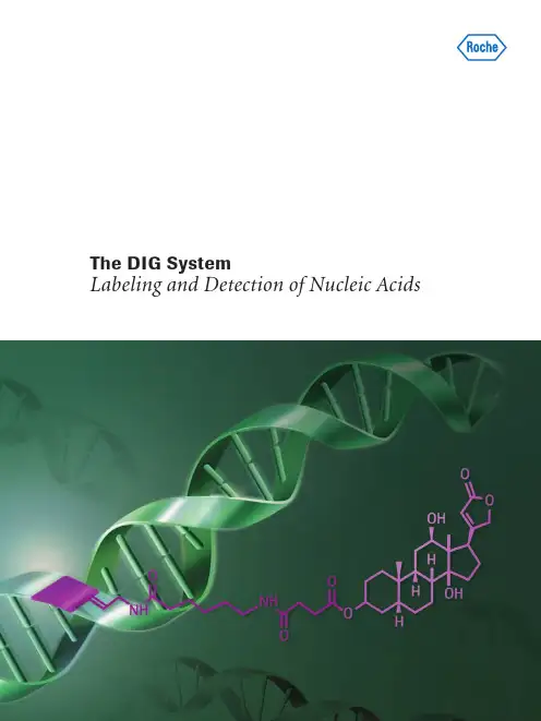
The DIG System
Labeling and Detection of Nucleic Acids
The DIG System Specifically Label and Detect Nucleic Acids
Publishable results require high level specific detection and low background. Do your hybridizations have nonspecific signals and high background? The DIG System is ideal for nucleic acid labeling. Flexibly use colorimetric, luminescent or fluorescent signal detection. Achieve high sensitivity and low background in very short exposure times
地高辛 (Digoxin)使用说明书

地高辛 (Digoxin)使用说明书地高辛 (Digoxin) 使用说明书地高辛是一种常用的心脏疾病治疗药物,主要用于治疗心力衰竭和心律失常。
本说明书将详细介绍地高辛的用法、剂量、注意事项和可能的副作用,帮助患者正确使用地高辛,从而达到最佳疗效。
一、药物名称和成分药物名称:地高辛(Digoxin)成分:每片含地高辛0.25毫克。
二、适应症地高辛适用于下列疾病的治疗:1. 心力衰竭:用于改善心脏收缩力,增加心脏排血量。
2. 心房颤动:用于控制心脏节律,减少心房颤动的发作并减轻症状。
三、用法和剂量1. 使用方法:口服。
最好与饭后服用,以减少不适感。
2. 常用剂量:成人一般剂量为每日0.125-0.25毫克,根据患者具体情况,可在医生指导下微调剂量。
3. 用量调整:用地高辛治疗时,应根据患者的病情和耐受性进行用量调整。
请遵循医生的指导,并定期检查药物浓度和心脏功能。
四、注意事项1. 专科医生指导:地高辛使用需要在专科医生的监护下进行,不得擅自调整剂量或停药。
2. 定期检查:使用地高辛期间,应定期检查心脏功能、药物浓度等指标,以确保疗效和安全性。
3. 过敏反应:如果出现过敏反应,如皮疹、荨麻疹、呼吸困难等症状,请立即就医。
4. 注意相互作用:地高辛有一些药物相互作用可能导致心脏毒性增加,如抗心律失常药物、某些抗生素等,请告知医生所用的所有药物。
5. 年老体弱者:对于年老体弱、肝肾功能减退者,应特别谨慎使用,避免出现中毒症状。
五、可能的副作用地高辛的使用可能会出现以下副作用:1. 胃肠道反应:如恶心、呕吐、腹泻等,特别是在剂量过高的情况下。
2. 心脏毒性:如心律失常、心搏骤停等,请密切关注心脏状况,若出现异常请立即就医。
3. 颜面潮红:部分患者使用地高辛后可能出现颜面潮红的不适感。
4. 视觉障碍:可能出现视觉模糊、视物有影等,若出现此类问题,请及时就医。
六、妊娠和哺乳期使用妊娠和哺乳期妇女慎用地高辛,应在医生的指导下使用。
口服地高辛注意事项

口服地高辛注意事项地高辛是一种常用的强心利尿药物,主要用于治疗心力衰竭和心律失常等心脏疾病。
使用地高辛需要注意以下几个方面:1.注意给药剂量:地高辛的剂量需要根据患者的年龄、体重、肾功能等因素进行个体化调整。
一般来说,初始剂量一般为0.125毫克,然后根据患者的病情和耐受性逐渐调整剂量。
一次最大剂量不应超过0.25毫克。
老年人和肾功能受损的患者需要减量使用。
2.定期监测血药浓度:地高辛的治疗窗口比较窄,血药浓度过高或过低都会影响药效和安全性。
因此,治疗期间需要定期监测地高辛血药浓度,一般在开始治疗后5至7天测定第一次血药浓度,如果达到治疗目标就不需要再次测定,如果没有达到目标则每隔4至7天测定一次,直至达到治疗目标。
3.注意药物相互作用:地高辛与其他药物之间存在一定的相互作用,可能会增加或降低地高辛的血浓度,导致不良药物反应或降低药效。
常见的相互作用药物包括一些抗心律失常药物、利尿剂、抗生素、非甾体抗炎药等。
使用地高辛时要告知医生所有正在使用的药物,以便医生调整药物剂量或选择其他药物。
5.避免长期应用:地高辛是一种长期应用的药物,但由于其与其他药物的相互作用和血药浓度的监测需要,长期使用地高辛可能会增加药物治疗的复杂性和药物错误的风险。
因此,在使用地高辛时要定期复查病情,如有需要可以选择其他替代药物。
6.慎用于孕妇和哺乳期妇女:地高辛穿过胎盘屏障进入胎儿血液,并可以通过乳汁排泄,因此对于孕妇和哺乳期妇女应该慎用地高辛。
如果有需要,则需在医生的指导下进行。
7.注意个体差异:地高辛的药代动力学存在个体差异,不同患者对地高辛的代谢和排泄能力不同,因此需要根据患者的实际情况进行个体化的用药调整。
总之,地高辛是一种常用的心脏疾病药物,但在使用时需要注意剂量调整、药物相互作用、监测血药浓度、观察不良反应等因素。
在用药过程中要密切配合医生的指导和监测,以确保药物的安全有效使用。
地高辛作业指导书 (2)

采用竞争法原理,整个过程18分钟完成。
第1步:10l标本与钌(Ru)标记的抗地高辛单克隆抗体混匀,形成夹心复合物,其数量取决于标本中分析物的浓度。
第2步:加入链霉亲和素包被的微粒和生物素标记的地高辛衍生物,后者与钌标记的单克隆抗体上未占据的位点结合,形成抗体-半抗原复合物。形成的完整复合物通过生物素与链霉亲和素间的反应结合到微粒上。
4. Lindenbaum J, Mellow MH, et al. Variation in Biologic Availability ofDigoxin from Four Preparations. New Engl J Med 1971;285:1344-1347.
5. Lindenbaum J, Butler VP Jr., Murphy JE,CresswellRM.Correlation of Digoxin-Tablet Dissolution Rate with BiologicalAvailability. Lancet 1973;1:1215-1217.
12.参考文献
1. Hoffman BF, Bigger JT Jr. In: Gilman AG, Goodman LS, Gilman A,eds.The Pharmacological Basis of Therapeutics. 6th ed.NewYork,NY: MacMillan; 1980:729-760.
8.参考范围
通常地高辛的治疗浓度是0.9-2.0ng/ml(1.2-2.6nmol/l)。高于2.0ng/mL(2.6nmol/l)时一般考虑中毒。但中毒和非中毒剂量之间有交叉。因此,临床诊断应根据临床和实验室的数据综合考虑。各实验室应提供合适的报告形式以及一致的异常结果报告程序。如有必要,各实验室应自己测定一个参考值。
罗氏地高辛标记说明书 英文版

For life science research only. Not for use in diagnostic procedures.FOR IN VITRO USE ONLY.DIG High Prime DNA Labeling and Detection Starter Kit IFor color detection with NBT/BCIP.Random primed DNA labeling with digoxigenin-dUTP,alkali-labile and detection of hybrids by enzyme immunoassayCat. No. 11 745 832 910Store at Ϫ15 to Ϫ25° C Kit for 12 labeling reactions of 10 ng-3 g DNAand detection of 24 blots of 100 cm²Instruction ManualVersion November 20091.Preface1.1Table of Contents1.Preface (2)1.1Table of Contents (2)1.2Kit contents (3)2.Introduction (5)2.1Product overview (5)3.Procedures and required materials (8)3.1Before you begin (8)3.2DIG-DNA Labeling (9)3.3Determination of labeling efficiency (12)3.4DNA transfer and fixation (15)3.5Hybridization (17)3.6Immunological detection (19)3.7Stripping and reprobing of DNA blots (21)4.Results (22)4.1Typical results (22)5.Appendix (23)5.1Trouble shooting (23)5.2References (24)5.3Ordering Information (25)1.2Kit contentsBottle/ Cap Label Contentincluding function1DIG-HighPrime •50 l DIG-High Prime• 5 × conc. labeling mixture containingoptimal concentrations of random primers, nucle-otides, DIG-dUTP (alkali-labile),Klenow enzyme and buffer components •ready-to-use•clear, viscous solution•for efficient random primed labeling of DNA2DIG-labeledControl DNA •20 l•[5 g/ml] pBR328 DNA(linearized with Bam HI)•clear solution•determination of labeling efficiency3DNA DilutionBuffer •3×1ml•[50 g/ml fish sperm DNA in 10 mM Tris-HCl, 1 mM EDTA; pH 8.0 at +25°C]•clear solution4Anti-Digoxigenin-AP Conjugate •100 l•[750 U/ml]•from sheep, Fab-fragments,conjugated to alkaline phosphatase •clear solution5NBT/BCIP• 6 × 1 ml•50x conc. stock solution [18.75 mg/ml nitrobluetetrazolium chloride and 9.4 mg/ml 5-bromo-4-chloro-3-indolyl-phosphate in67% (v/v) DMSO]•can vary between light yellow and brown, clearsolution•reacts with alkaline phosphatase.6Blockingsolution • 4 × 100 ml•10 × conc.•yellow, viscous solution7DIG Easy HybGranules •to give 4 × 100 ml (preparation page 17)•Hybridization solutionAdditional equipment and reagents requiredIn addition to the reagents listed above, you have to prepare several solutions.In the table you will find an overview about the equipment which is needed for the different procedures.Detailed information is given in front of each procedure.Procedure Equipment Reagents3.2 DIG-DNA labeling Water bath•H2O, sterile, doubledistilled water•EDTA, 0.2 M, pH 8.0,sterile3.3 Semi-quantitativedetermination oflabeling efficiencyNylon membranes positivelycharged*•DIG Wash andBlock Buffer Set*•TE-bufferor•Washing buffer•Maleic acid buffer•Detection buffer•TE-buffer3.4 DNA transferand fixation•UV-transilluminator•commercial availableUV-cross linker• 2 × SSCor•10 × SSC3.5 Hybridization•Nylon membranes, positivelycharged*•Hybridization bags*or•Temperature resistant,sealable plastic bags or rollerbottlesNote: Do not use open trays whenworking with DIG Easy Hyb buffer3.6 Immunologicaldetection•Container of appropriate size inrelation to filter size•Hybridization bags*•DIG Wash andBlock Buffer Set*•TE-bufferor•Washing buffer•Maleic acid buffer•Detection buffer•TE-buffer3.7 Stripping andreprobing of DNAblots•Large tray•Water bath•10 × SSC•10% SDS•0.2 M NaOH2.Introduction2.1Product overviewTest principle The DIG High Prime DNA Labeling and Detection Starter Kit I uses digoxigenin(DIG), a steroid hapten, to label DNA probes for hybridization and subsequentcolor detection by enzyme immunoassay [1].Stage DescriptionDNA labeling DIG-labeled DNA probes are generated with DIG-High Primeaccording to the random primed labeling technique. DIG-High Primeis a specially developed reaction mixture containing digoxigenin-dUTP, alkali-labile (Fig. 2) and all reagents, including enzymenecessary for random primed labeling, premixed in an optimized5 × concentrated reaction buffer.Hybridization DIG-labeled probes are used for hybridization to membraneblotted nucleic acids according to standard methods. The use of thealkali-labile form of DIG-11-dUTP enables easier and more efficientstripping of blots for rehybridization with a secondDIG-labeled probe.Immunological detection The hybridized probes are immunodetected with anti-digoxigenin-AP, Fab fragments and are then visualized with the colorimetric substrates NBT/BCIP.ApplicationDIG-labeled DNA probes can be used:•for all types of filter hybridization•for single copy gene detection in total genomic DNA, even from organisms with high complexity, e.g. human, barley, and wheat.Sample material•DNA fragments of at least 100 bp •linearized plasmid, cosmid or DNA •supercoiled DNAAssay timeThis table lists the reaction time of the single steps Number of tests1 kit is sufficient for•12 standard labeling reactions of up to 3 g template DNA and detection of•24 blots of 100 cm 2.Quality ControlUsing unlabeled control-DNA (pBR 328), labeled as described in the protocol, 0.1 pg of homologous DNA is detected in a dot blot after 16 h color develop-ment (1 pg of homologous DNA can be detected after 1h color development).Step Reaction time DNA labeling 1 h-O/N Hybridization6 h or O/N Immunological detection 1.5 h Color development0.5 - 16 hKit storage/ stabilityThe unopened kit is stable at -15 to -25° C until the expiration date printed on the label. Shipping conditions on dry ice.Once opened, please refer to the following table for proper storage.Sensitivity and specificityA single copy human gene is detected in a Southern blot of 1 g digested pla-centa DNA.Note: Sensitivity depends both on the concentration of labeled DNA in the hybridization and on the time of color reaction.AdvantagesThis table describes benefits and features of the kit.Kit componentStorageAnti-Digoxigenin-AP Conjugate cap 42-8° C, stableBlocking solution bottle 6•unopened, stable at 15-25° C•once opened, it should be aliquoted and stored at -15 to -25° C or at 2-8° C up to one month when keeping sterile•working solution should always be prepared fresh NBT/BCIP cap 6•2-8° C,stable•or at least 4 weeks at 15-25° CNote: During shipment of the kit on dry ice, a precipitate may occur which is easily dissolved by briefly warming to 37°CBenefitFeatureAccurate and fastThe use of premixed DIG-High Prime mini-mizes the hands-on-time required to label DNA probes and increases yields and reproducibility.Sensitive Single-copy genes can be detected in total human DNA complex and plant genomes.Time-savingDIG-labeled probes can be stored for at least one year. Hybridization solutions can be reused 3 – 5 times, depending on the amount of labeled probe used for signal generation in each hybridization.3.Procedures and required materials3.1Before you beginGeneral handling rec-ommendationsThis table describes general hints for DIG labeling and detection.OverviewRecommendation GuidelineWork under clean conditions•Autoclave DIG System solutions•Filter-sterilize solutions containing SDS•Tween 20 should be added to previouslysterilized solutionsUse clean incubation trays Rigorously clean and rinse laboratory traysbefore each use.Membrane handling requirements•Wear powder-free gloves•Handle membrane only on the edges andwith clean forcepsSection 3.2DIG-DNA labeling↓Section 3.3Quantification of labeling efficiency↓Section 3.4DNA fixation↓Section 3.5Hybridization↓Section 3.6Immunological detection↓Section 3.7Stripping and reprobing of DNA blots3.2DIG-DNA LabelingIntroduction DNA is random primed labeled with Digoxigenin-11-dUTP using DIG-HighPrime, a 5x concentrated labeling mixture of random hexamers, dNTP mix con-taining alkali-labile Digoxigenin-11-dUTP, labeling grade Klenow enzyme andan optimized reaction buffer.Additional equipment and reagents required •water bath•ice/waterThis table lists composition, storage and use of the required reagents in addi-tion to kit componentsTemplate DNA The following table lists the recommended features of the template DNALabeling of DNA isolated from agaroseIf you intend to perform genomic Southern blotting, you should separate thetemplate insert DNA from the vector by agarose gel electrophoresis.Excise the DNA fragment from an agarose gel. For best results use either our High Pure PCR Product Purification Kit * or our Agarose Gel DNA ExtractionKit* to separate the DNA from the agarose.Solution Composition Storage Use Water Autoclaved, double distilled water15-25° C,stableDilution of DNAEDTA0.2 M ethylenediamino- tetraacetic acid,pH 8.015-25° C,stableStopping thelabeling reactionFeature DetailPurity For plasmid DNA use the High Pure Plasmid Isolation Kit * for purification.When other commercially available purification kits are used, we recommendto do an additional phenol/chloroform extraction to remove residual protein.This step is also necessary when templates have been treated with restrictionor other modifying enzymes before labeling.Size To obtain optimal results, template DNA should be linearized andshould have a size of 100 or larger. Template DNA >5 kb should be restric-tion-digested using a 4 bp cutter (e.g. Hae III) prior to labeling.Amount With the procedure described below principally 10 ng – 3 g of template can be labeled, however, please check in the given table the necessaryamount of probe needed for your size of blot. By scaling up of all volumes andcomponents accordingly this procedure can be used for labeling of largeramounts. If single-copy gene detection in complex genomes is performed atleast 300 ng of template DNA (probe concentration: 25 ng/ml hybridizationsolution) should be labeled.Procedure This procedure is designed for 10 ng-3 g of DNA. Larger amounts (up to 10g) can be labeled by scaling up of all components and volumes.Yield of labeling reac-tion Table 1:This table shows you the yield of DIG-High Prime labeling under optimal condi-tions.In the standard reaction with 1 g DNA per assay approx. 15% of the nucle-otides are incorporated into about 0.8 g of newly synthesized DIG-labeled DNA within 1 h and approx. 38% of the nucleotides into about 2 g after 20 h. Using DIG-High Prime solution, reactions were performed with increasingamountsof different template DNAs for 1 h and 20 h. The yield of DIG-labeled DNA was determined by incorporation of a radioactive tracer and confirmed by a dot blot (Average of 10 independent labeling assays)Step Action1Add1g template DNA (linear or supercoiled) and autoclaved, double distilled water to a final volume of 16 l to a reaction vial.2Denature the DNA by heating in a boiling water bath for 10 min and quickly chilling in an ice/water bath.Note: Complete denaturation is essential for efficient labeling.3•Mix DIG-High Prime (vial 1) thoroughly and add 4 l to the denatured DNA, mix and centrifuge briefly.•Incubate for 1 h or O/N at 37° C.Note: Longer incubations (up to 20 h) will increase the yield of DIG-labeledDNA (see table below ).4Stop the reaction by adding 2 l 0.2 M EDTA (pH 8.0) and/or by heating to 65°C for 10 min.Note: The length of the DIG-labeled fragments obtained with DIG-High Prime range from 200 bp to 1000 bp or larger, depending on the length of theoriginal template.T emplate DNA 1 h20 h10 ng 45 ng 600 ng30 ng 130 ng1050 ng100 ng 270 ng1500 ng300 ng 450 ng2000 ng 1000 ng 850 ng2300 ng3000 ng1350 ng2650 ng.Fig. 2: Yield of DIG-labeled DNA from different amounts of template DNA after 1 and 20 h incu-bation of the DIG-High Prime reaction at 37°C.3.3Determination of labeling efficiencyIntroduction Determination of the yield of DIG-labeled DNA is most important for optimaland reproducible hybridization results. Too high of a probe concentration in thehybridization mix causes background, while too low of a concentration leads toweak signals.Test principle The preferred method for quantification of labeled probes is the direct detec-tion method.Preparation of additional solutions requiredPlease find in the following table composition and preparation of additionalreagents required. The following buffers are also available in the DIG Wash and Block Buffer Set, DNase and RNase free.*Please note: These solutions are also used in the detection procedure ofchapter 3.6. and can be prepared in larger quantities.Stage Description1• A series of dilutions of DIG-labeled DNA is applied to a small strip of nylon membrane positively charged*.•Part of the nylon membrane is preloaded with defined dilutions of DIG-labeled control DNA (vial 2) which are used as standards.2•The nylon membrane is subjected to immunological detection with anti-digoxigenin-AP conjugate (vial 4) and the premixed stock solutionof NBT/BCIP (vial 5).•The intensities of the dilution series of DIG-labeled DNA and control DNA are compared.Solution Composition / Preparation Storage andstabilityUseWashingbuffer0.1 M Maleic acid, 0.15 M NaCl;pH 7.5 (20° C); 0.3% (v/v) Tween 2015-25° C,stableRemovalof unboundantibody Maleic acidbuffer0.1 M Maleic acid, 0.15 M NaCl;adjust with NaOH (solid) to pH 7.5(20° C)15-25° C,stableDilution ofBlocking solutionDetectionbuffer0.1 M T ris-HCl, 0.1 M NaCl,pH 9.5 (20° C)15-25° C,stableAdjustment ofpH to 9.5TE-buffer10 mM T ris-HCl, 1 mM EDTA,pH 8.015-25° C,stableStopping colorreactionPreparation of kit working solutionsThe following table shows the preparation of kit working solutions ProbequantificationLabeled probes and the DIG-labeled control DNA (vial 2) must be diluted to 1 ng/l, according to the expected yield of synthesized nucleic acid to start the dilution series below. The expected yield of DIG-labeled DNA in your probe can best be estimated by using the table 1 in chapter 3.2. The yield depends on the starting amount of template and incubation time.Note: The yields given in table 1 were achieved under optimal conditions with highly purified template DNA.Dilution seriesPrepare a dilution series of your labeled probe and your control DNA as described in the table:Solution Composition/preparationStorage and stability Use Blocking solutionPrepare a 1 × working solution by diluting the 10x Blocking solution (vial 6) 1:10 in Maleic acid buffer.Always prepare fresh Blocking of unspecific binding sites on the membrane Antibody solutionCentrifuge Anti-Digoxigenin-AP (vial 4) for 5 min at 10 000 rpm in the original vial prior to each use, and pipet the necessary amount carefully from the surface. Dilute Anti-Digoxigenin-AP 1: 5 000 (150 mU/ml) in Blocking solution.12 h at 2-8° CBinding to the DIG-labeled probeColor-substrate solutionAdd 40 l of NBT/BCIP stock solution (vial 5) to 2 ml of Detection buffer.Note: Store protected from light!always prepare freshVisualization of antibody-binding TubeDNA (l)From tube #DNA Dilution Buffer (vial 3)(l)DilutionFinal concentration1diluted original1 ng/l 2211981:10010 pg/l 3152351:3.3 3 pg/l 452451:101 pg/l 553451:100.3 pg/l 654451:100.1 pg/l 755451:100.03 pg/l 856451:100.01 pg/l9-50-0Procedure The following procedure describes the direct detection.Note: Use sufficient buffer volumes to cover the membrane completely duringall steps.Analyzing the resultsCompare the intensity of the spots out of your labeling reaction to the control and calculate the amount of DIG-labeled DNA. If the 0.1 pg dilution spots of your probe and of the control are visible, then the labeled probe has reached the expected labeling efficiency (pls. see table 1 in 3.2.) and can be used in the recommended concentration in the hybridization.The following spots should be visibleStep Action1Apply a1l spot of tubes 2-9 from your labeled probes and the labeled control to the nylon membrane.2Fix the nucleic acid to the membrane by cross linking with UV-light or baking for 30 min at 120 ° C.3•Transfer the membrane into a plastic container with 20 mlMaleic acid buffer.•Incubate under shaking for 2 min at 15-25° C.4Incubate for 30 min in 10 ml Blocking solution.5Incubate for 30 min in 10 ml Antibody solution.6Wash with 10 ml Washing buffer, 2 × 15 min.7Equilibrate 2-5 min in 10 ml Detection buffer.8Incubate membrane in 2 ml freshly prepared color substrate solution in a appropriate container in the dark. Do not shake during color development.Note: The color precipitate starts to form within a few minutes. The membrane can be exposed to light for short time periods to monitor color development.9Stop the reaction, when desired spot or band intensities are achieved, by washing the membrane for 5 min with 50 ml of sterile double dist. wateror with TE-buffer.Results can be documented by photocopying the wet filter or by photography.Incubation time Appearance5-10 min30 pg spot30 min 3 pg spot3.4DNA transfer and fixationTransfer methods and membranes Standard protocols for gel electrophoresis, denaturation and neutralization of the gel are described in Sambrook et al. (2). Gels lacking ethidium bromide are preferred, because ethidium can cause uneven background problems. All com-mon types ofDNA transfer methods are suitable for subsequent DIG hybridization (4,5).In our experience, best results are obtained when gels are blotted by capillary transfer with 20 × SSC on nylon membranes*, positively charged.Note: Alkali transfer (e.g., in 0.4 M NaOH) is not suitable for the transfer of DIG-labeled molecular weight markers*.Fixation procedure Fix the DNA to the membrane by any of the following procedures:Storage of the membranePlease refer to the following table.IF you want to...THEN...UV-crosslinking(nylon membrane)•Place the membrane on Whatman 3MM-paper soakedwith 10 × SSC.•UV-crosslink the wet membrane without priorwashing.•After the UV-crosslinking, rinse the membrane brieflyin double distilled water and allow to air-dry.bake at 120° C(nylon membrane)•Wash the membrane briefly in 2 × SSC.•Bake the nylon membrane at 120° C for 30 min oraccording to the manufacturer`s instructions.bake at 80° C(nylon membrane)•Wash the membrane briefly in 2 × SSC.•Bake at 80° C for 2 h under vacuum.IF...THEN...you want to go e the membrane immediately for prehybridization. you want to work later on store the membrane dry at 2-8° C.3.5HybridizationAdditional equipment required •ice/water•shaking water-bath•or hybridization oven•temperature resistant plastic or glass boxes, petri dishes, roller bottles or sealable plastic bags.Note: Do not use open containers with DIG Easy Hyb buffer.Preparation ofDIG Easy Hyb working solution Add carefully 64 ml sterile double distilled water in two portions to the DIG Easy Hyb Granules (bottle 7), dissolve by stirring immediately for 5 min at 37° C.Hybridization temperature The appropriate hybridization temperature is calculated according to GC con-tent and percent homology of probe to target according to the following equa-tion:Tm= 49.82 + 0.41 (% G + C) - (600/l) [l = length of hybrid in base pairs]Topt.=Tm–20 to 25° C(The given numbers of the equation were calculated according to a standard equation for hybridization solutions containing formamide, 50%.) The actual hybridization temperature T opt. for hybridization with DIG Easy Hyb is 20-25° C below the calculated T m value. T opt. can be regarded as a stringent hybridization temperature allows up to 18% mismatches between probe and target. When the degree of homology of your probe to template is less than 80%, you should lower Topt. accordingly (approx. 1.4°C below Tmper 1 % mismatch) and also adjust the stringent washing steps accordingly (i.e. increase SSC concentration and lower washing temperature).Procedure Please refer to the following table.Storage of hybridization solution DIG Easy Hyb containing DIG-labeled probe can be stored at Ϫ15 to Ϫ25° C and be reused several times when freshly denatured at 68°C for 10 min before use.Note: Do not boil DIG Easy Hyb.Stringency washes For most DNA:DNA applications, a stringency wash with 0.5 × SSC is sufficient. The correct post washing conditions have to be determined empirically for each probe.•For human genomic DNA use 0.5 × SSC and 65° C.•Probes > 150 bp and with a high G/C content should be washed at 68° C.•For shorter probes around 100 bp or shorter, the wash temperature must belowered.This table describes how to perform post-hybridization washes.Step Action1•Pre-heat an appropriate volume of DIG Easy Hyb (10 ml/100 cm2 filter) to hybridization temperature (37 - 42° C).•Prehybridize filter for 30 min with gentle agitation in an appropri-ate container.Note: Membranes should move freely, especially if you use severalmembranes in the same prehybridization solution.2Denature DIG-labeled DNA probe (about 25 ng/ml) by boiling for5 min and rapidly cooling in ice/water.Note: As DIG-11-dUTP is alkali-labile, DNA probes cannot be dena-tured by alkali treatment (NaOH).3Add denatured DIG-labeled DNA probe to pre-heated DIG Easy Hyb(3.5 ml/100 cm2 membrane) and mix well but avoid foaming(bubbles may lead to background).4•Pour off prehybridization solution and add probe/hybridization mixture to membrane.•Incubate 4 hours - O/N with gentle agitation.Step Action1Wash 2 × 5 min in ample 2x SSC, 0.1% SDS at 15-25° C under con-stant agitation.2Wash 2 × 15 min in 0.5x SSC, 0.1% SDS (prewarmed to wash temper-ature) at 65-68° C under constant agitation.3.6Immunological detectionAdditional reagents required Please find in the following table composition and preparation of additional reagents required. The following buffers are also available in the DIG Wash and Block Buffer Set, DNase and RNase free*.Preparation of kit working solutions In the following table the preparation of kit working solutions is described.Solution Composition / Preparation Storage andstabilityUseWashingbuffer0.1 M Maleic acid, 0.15 M NaCl;pH 7.5 (20° C); 0.3% (v/v) Tween2015-25° C,stableWashing ofmembraneMaleic acidbuffer0.1 M Maleic acid, 0.15 M NaCl;adjust with NaOH (solid) to pH7.5 (20° C)15-25° C,stableDilution ofBlockingsolution Detectionbuffer0.1 M Tris-HCl, 0.1 M NaCl,pH 9.5 (20° C)15-25° C,stableAlkalinephosphatasebufferTE-buffer10 mM Tris-HCl, 1 mM EDTA, pH8.015-25° C,stableStoppingcolor reactionSolution Composition / Preparation Storage andstabilityUseBlockingsolutionPrepare a 1x working solution bydiluting 10 × Blocking solution(vial 6) 1:10 with Maleic acidbuffer.Alwaysprepare freshBlocking ofunspecificbinding sitesAntibodysolutionCentrifuge Anti-Digoxigenin-AP(vial 4) for 5 min at 10 000 rpm inthe original vial prior to each use,and pipet the necessary amountcarefully from the surface. DiluteAnti-Digoxigenin-AP 1:5000 (150mU/ml) in Blocking solution.12 h at 2-8° C Binding to theDIG-labeledprobeColor-substratesolutionAdd 200 l of NBT/BCIP stocksolution (vial 5) to 10 ml of Detec-tion buffer.Note: Store protected from light!Alwaysprepare freshVisualizationof antibody-bindingProcedure This table describes how to perform the immunological detection ona 100 cm2 membrane.Note: All incubations should be performed at 15-25° C with agitation. If themembrane is to be reprobed, do not allow the membrane to dry at any time.Storage of membrane Please refer to the following table.Step Action1After hybridization and stringency washes, rinse membrane briefly (1-5) min in Washing buffer.2Incubate for 30 min in 100 ml Blocking solution.3Incubate for 30 min in 20 ml Antibody solution.4Wash 2 x 15 min in 100 ml Washing buffer.5Equilibrate 2-5 min in 20 ml Detection buffer.6Incubate membrane in 10 ml freshly prepared color substrate solu-tion in a appropriate container in the dark. Do not shake duringcolor development.Note: The color precipitate starts to form within a few minutes andthe reaction is usually complete after 16 h. The membrane can beexposed to light for short time periods to monitor color development.7Stop the reaction, when desired spot or band intensities are achieved, by washing the membrane for 5 min with 50 ml of steriledouble dist. water or with TE-buffer.Results can be documented by photocopying the wet filter or by pho-tography.IF...THEN...you want to reprobe the membrane the membrane should not dry off at anytime, store in sealed plastic bag.Note: If you want to maintain the color,store membranes in TE buffer do notallow the membrane to dry.you don´t want to reprobe dry the membrane at 15 to 25°C for stor-age.Note: Color fades upon drying, to revita-lize the color, wet the membrane in TEbuffer.3.7Stripping and reprobing of DNA blotsGeneral The alkali-labile form of DIG-11-dUTP enables easier and more efficient strip-ping of blots for rehybridization experiment.Additional reagents required •Dimethylformamid (DMF)•0.2 N NaOH, 0.1% SDS (w/v)• 2 × SSCProtocolStorage of stripped membrane Please refer to the following table.Note: When stripping and rehybridization of blots is planned, the membrane should not dry off at any time.CAUTION: Work under a fume hoodStep Action1•Heat DMF in a large glass beaker in a water bath under a fume hood to 50-60° C.•Incubate the membranes in the heated DMF until the blue color precipitate is removed from the filter.CAUTION: DMF is volatile and can be ignited above 67° C.2Rinse membrane briefly in double distilled water.3Wash for 2 × 15 min in 0.2 N NaOH, SDS, 0.1% (w/v) at 37° C under constant agitation.4Equilibrate briefly in 2 × SSC.5Prehybridize and hybridize with a second probe.Once the membrane is stripped, it can be stored in Maleic acid buffer or2 × SSC until used again.4.Results4.1Typical resultsGenomic Southern blotFigure 3: This figure shows you the detection of a single-copy gene (ß-actin) in total human DNA using the standard protocol.Size (kb)1234215.1/5.04.33.52.01.91.61.40.950.830.562.551.0 。
地高辛中文操作说明
所用试剂盒为Roche公司的DIG High Prime DNA Labeling and Detection Starter Kit I,具体操作过程如下:①向一个反应管中加入1μg 模板DNA(线性或超螺旋)和高压灭菌的双蒸水,终体积达16μL;②沸水浴10min以变性DNA,然后迅速插入冰水混合物中;注意:完全变性对于标记的有效性是十分重要的。
③充分混合DIG-High Prime(1号管),并取4μL加入到变性DNA中,混合并简单离心;④37℃孵育,过夜;⑤65℃加热10min,终止反应。
2)探针的纯化与回收用PCR产物快速回收试剂盒(上海生工)回收标记好的探针,同时除去随机引物和未掺入的DIG-dUTP。
具体操作步骤如下所示:①在PCR反应液中加入5倍体积的Buffer B3,充分混匀;②8,000×g离心30秒。
倒掉收集管中液体;③加入500μl Wash Solution,9,000 ×g 离心30秒,倒掉收集管中液体;④重复步骤4一次;⑤空柱于9,000 ×g离心1分钟;⑥将吸附柱放入一个干净的 1.5 ml离心管中,在吸附膜中央加入15-40μl Elution Buffe r,室温静置1分钟后,离心1分钟。
保存管中DNA溶液。
⑦电泳检测探针的完整性,紫外分光光度法测定探针浓度,稀释到1 ng/μL 。
注:标记好的探针最好立即使用,最长使用时间一般不宜超过3天。
标记好的探针可以保存在-20℃。
3)检测探针标记的效率直接标记法:将地高辛标记DNA的一系列稀释梯度点到一小块带正电荷的尼龙膜上。
尼龙膜部分区域已经点好一系列稀释梯度的地高辛标记的对照DNA(2号管)作为标准。
具体操作步骤:(注意在全部的步骤中,用足够的缓冲液体积完全覆盖膜)①分别从你标记的探针和标记对照DNA的2-9号管中取出1 μL点在尼龙膜上;②通过120℃烘烤30min的方法将核酸固定在膜上;③将膜转移到一个装有20mL 马来酸缓冲液的塑料容器中,在15-25℃中振荡孵育2min;④在10mL 封阻液中孵育30min;⑤在10mL 抗体溶液中孵育30min;⑥用10mL洗涤缓冲液洗涤两次,每次15min;⑦在10mL检测缓冲液中平衡2-5min;⑧将膜上带有DNA的一面朝上,放在一个折叠夹上(或者杂交袋),向膜上加入2mL的新鲜配制的的底物显色液,均匀的铺平底物,且膜上不能有气泡。
地高辛——精选推荐
地高辛Digoxin【其它名称】强心素、异羟基洋地黄毒苷、Digoxine、Digoxinum、Lanoxin Vanoxm【临床应用】1.用于充血性心力衰竭。
对高血压、瓣膜病、先天性心脏病所引起的充血性心力衰竭疗效较好。
对继发于严重贫血、甲状腺功能低下或维生素B1缺乏症的充血性心力衰竭则疗效差;对肺源性心脏病、心肌严重缺血、活动性心肌炎或心肌外机械因素所致心力衰竭疗效也差。
2.用于控制快速性心房颤动、心房扑动患者的心室率及室上性心动过速。
【药理】1.药效学本药为毛花洋地黄中提纯制得的中效强心苷,治疗剂量时有两方面作用:(1)增加心肌收缩力和速度。
由于本药抑制细胞膜上的Na+-K+-ATP酶,减少钠钾交换,使细胞内Na+增加,从而肌膜上Na+、Ca2+交换趋于活跃,使细胞内Ca2+增多,作用于收缩蛋白,增加心肌收缩力和速度。
(2)对心肌电生理的影响。
通过直接对心肌细胞和间接通过迷走神经的作用,降低窦房结自律性;提高浦肯野纤维自律性;减慢房室结传导速度;缩短心房有效不应期;缩短浦肯野纤维有效不应期。
大剂量时,增加交感神经活性,这可能与地高辛的心脏毒性有关。
2.药动学本药口服吸收约50%-75%。
生物利用度:片剂为60%-80%、酏剂为70%-85%、胶囊剂为90%以上。
吸收后广泛分布到各组织,部分经胆道吸收人血,形成肠-肝循环。
其表观分布容积为6-10L/kg。
本药蛋白结合率低(为20%-25%)。
口服后0.5~2小时起效,2-6小时作用达高峰。
静脉注射后5-30分钟起效,l-4小时作用达高峰,持续作用6小时。
治疗血药浓度为0.5-2.0ng/ml,半衰期为32-48小时。
在体内代谢少,主要以原形由肾排泄,比洋地黄毒昔排泄快,尿中排出量为用量的50%-70%。
也可从粪便排泄。
【注意事项】1.禁忌症 (1)对任何强心昔制剂中毒者。
(2)室性心动过速、心室颤动。
(3)梗阻性肥厚型心肌病。
(4)预激综合征伴心房颤动或扑动。
地高辛标记及检测试剂盒说明书
仅仅研究于生命科学,不适合在诊断过程使用只能在体外使用地高辛DNA标记和检测试剂盒1和NBT/BCIP一起用于颜色检测通过酶联免疫法采用地高辛-dUTP进行随机引物DNA标记,碱性标记和检测货号:11 745 832 910此试剂盒储存条件-15o C—-25o C此试剂盒可对10ng-3ugDNA进行12次标记反应可检测100cm2面积的杂交膜24张指导手册2009.11版1前言1.1内容表1前言 (2)1.1内容表 (2)1.2试剂盒内容 (3)2简介 (5)2.1产品概述 (5)3步骤和所需材料 (8)3.1开始之前 (8)3.2地高辛DNA标记 (9)3.3 标记效率的测定 (11)3.4 DNA 的转移和固定 (14)3.5杂交 (16)3.6免疫检测 (18)3.7 DNA印记的洗脱和再杂交 (20)4结果 (21)4.1典型的结果 (21)5附录 (23)5.1故障排除 (23)5.2引用 (24)5.3订购信息 (25)1.2 试剂盒内容附加仪器与所需试剂除了上表中列出的试剂外,你必须准备一些溶液。
在下表中,你可以找到不同操作程序需要准备的设备概要。
*标记的产品可以从Roche Applied Science 获得2 介绍2.1 产品概况实验原则此试剂盒采用地高辛(DIG),一种甾类半抗原,去标记DNA探针从而通过酶免疫分析应用地高辛标记DNA探针可以用于:所有类型的滤膜杂交总基因组DNA中单拷贝基因的检测,甚至对于高度复杂的生物也可以进行检测,如人,大麦和小麦。
样品材料至少100bp的DNA片段线性的质粒,cos质粒或者λDNA超螺旋的DNA实验时间此表格列出了每一步实验所需的反应时间检测数量一个试剂盒足够用于:不超过3ug的模板DNA的12个标准的标记反应和100cm2有24个斑点的检测质量控制根据操作步骤中的描述,对未标记的对照品DNA(PBR328)进行标记,0.1pg同源DNA 在点杂交中通过16h的显色来检测(1pg同源DNA可以通过1小时的显色进行检测)。
地高辛-安全技术说明书MSDS
第1部分化学品及企业标识化学品中文名:地高辛化学品英文名:DigoxinCAS号:20830-75-5分子式:C41H64O14分子量:780.95产品推荐及限制用途:工业及科研用途。
第2部分危险性概述紧急情况概述:吞咽致命。
造成严重眼刺激。
吸入致命。
长期或反复接触会对器官造成伤害。
对水生生物毒性极大。
GHS危险性类别:急性经口毒性类别2严重眼损伤/眼刺激类别2急性吸入毒性类别1特异性靶器官毒性反复接触类别1危害水生环境——急性危险类别1标签要素:象形图:警示词:危险危险性说明:H300吞咽致命H319造成严重眼刺激H330吸入致命H372长期或反复接触会对器官造成伤害H400对水生生物毒性极大防范说明:•预防措施:——P264作业后彻底清洗。
——P270使用本产品时不要进食、饮水或吸烟。
——P280戴防护手套/穿防护服/戴防护眼罩/戴防护面具。
——P260不要吸入粉尘/烟/气体/烟雾/蒸气/喷雾。
——P271只能在室外或通风良好处使用。
——P284[在通风不足的情况下]戴呼吸防护装置——P273避免释放到环境中。
•事故响应:——P301+P310如误吞咽:立即呼叫解毒中心/医生——P330漱口。
——P305+P351+P338如进入眼睛:用水小心冲洗几分钟。
如戴隐形眼镜并可方便地取出,取出隐形眼镜。
继续冲洗。
——P337+P313如仍觉眼刺激:求医/就诊。
——P304+P340如误吸入:将人转移到空气新鲜处,保持呼吸舒适体位。
——P310立即呼叫解毒中心/医生——P314如感觉不适,须求医/就诊。
——P391收集溢出物。
•安全储存:——P405存放处须加锁。
——P403+P233存放在通风良好的地方。
保持容器密闭。
•废弃处置:——P501按当地法规处置内装物/容器。
物理和化学危险:无资料健康危害:吞咽致命。
造成严重眼刺激。
吸入致命。
长期或反复接触会对器官造成伤害。
环境危害:对水生生物毒性极大。