纳米金作固定相
纳米金的制备与性能ppt课件

制备生长溶液
25 ℃将CTAB(5mL, 0.2M)与AgNO3(0.05-0.25mL, 0.004M)混合 ;
加入HAuCl4(5mL,0.0005M),再加入AA(70uL,0.0788M),溶 液由深黄变为无色,生成生长溶液;
生长溶液和种子溶液混合,保持温度27-30 ℃,静置生长。
Secondary growth of NRS1 at different injection rates
(a) NRSF and (b) NRS1 are affected by varying (a) optical absorbance spectra and TEM images of
the concentration of AgNO3 in the primary GS (b) NRS1 and NRSF using (c) 1.25× (d)2.5×
电化学法
金片作阳极,铂片 做阴极,两点极浸 入到含有CTAB和 TCAB溶液中 ;
在超声和恒温( 36℃)下电解, 金从阳极溶出并于 阴极-电解质溶液 界面得金棒;
浸入银片来控制 长径比
LANGMUIR. 1999,15, 701-709
13
Sonochemistry
Angew. Chem. 2006, 118, 1134–1137
DNA 检测
靶向药物
图1 超灵敏DNA 检测示意图
图3 缩氨酸自组装和作为纳米反应器的反 应过程模拟
图2 Ag core Au shell结构免疫实验示意图
17
图1 基于CuO –金纳米颗粒标记的抗体 和Click 反应的免疫检测方法示意图
生物医学
疾病诊断
图2 基于罗丹明B-金纳米颗粒检测 ( 通过颜色变化和荧光) 乙酰胆碱酯酶 的设计策略
纳米金材料在生物传感器制备中的应用指南
纳米金材料在生物传感器制备中的应用指南近年来,纳米技术的快速发展为生物传感器制备领域带来了巨大的机遇和挑战。
纳米金材料作为一种重要的功能材料,具有卓越的电子、光学、磁学和化学性质,广泛应用于生物传感器的制备中。
本文将详细介绍纳米金材料在生物传感器制备中的应用指南,帮助读者理解纳米金材料的优势和潜力,以及其在生物传感器制备中的具体应用。
一、纳米金材料的优势纳米金材料具有许多优势,使其成为生物传感器制备中的理想选择。
首先,纳米金颗粒具有极大的比表面积,可以提供更多的反应接触点,从而增加生物传感器的灵敏度和响应速度。
其次,纳米金材料的表面与生物分子具有良好的亲和性,可以实现高效的生物分子固定化,并提供更好的电子传递通道。
此外,纳米金材料能够与生物分子之间的相互作用进行表征和调控,进一步提高生物传感器的选择性和稳定性。
综上所述,纳米金材料的优势使其成为生物传感器制备领域的研究热点。
二、纳米金材料在生物传感器制备中的应用1. 纳米金颗粒在电化学传感器中的应用纳米金颗粒在电化学传感器中的应用广泛,并在电化学生物传感器方面表现出了良好的性能。
例如,纳米金颗粒可以被修饰在电极表面,作为载体来固定生物分子,如抗体、生物素等,从而实现对生物分子的高灵敏度检测。
此外,纳米金材料还可以增强电化学信号,提高传感器的检测灵敏度。
因此,纳米金颗粒在电化学传感器中的应用可以实现对多种生物分子的检测,包括蛋白质、DNA、细胞等。
2. 纳米金材料在光学传感器中的应用纳米金材料在光学传感器中的应用是另一个重要的领域。
纳米金颗粒具有表面等离子共振现象,使其在光学传感器中具有优异的性能。
通过调节纳米金颗粒的形状、大小和组合方式,可以实现对不同波长的光的吸收和散射,从而实现了多种检测方法。
例如,纳米金颗粒可以被修饰在光学纤维表面,实现对生物分子的定量检测。
此外,纳米金材料还可以被用作光敏材料,通过光刺激来调控其表面等离子共振现象,实现对生物分子的快速响应检测。
纳米金材料的制备与性能研究

纳米金材料的制备与性能研究随着科技的不断进步,纳米材料的应用领域也在不断拓宽。
其中,纳米金材料作为一种有着独特性能的纳米材料,在能源、光电、催化等领域具有广阔的应用前景。
本文将探讨纳米金材料的制备方法以及其在性能研究方面的应用。
纳米金材料的制备有多种方法,其中较常见的是湿化学法和物理法。
湿化学法主要包括化学还原法、溶胶-凝胶法和电化学沉积法等。
化学还原法是指将金离子还原为金纳米颗粒,通过在反应溶液中加入还原剂,如氨水、甲醛等,可得到具有不同形貌和尺寸的纳米金颗粒。
溶胶-凝胶法则通过控制溶胶的成分和凝胶的温度、pH值和反应时间等参数,实现纳米金材料的制备。
电化学沉积法则是将金属离子通过外加电压的作用沉积到电极上,形成纳米金材料。
物理法主要包括溅射法、热蒸发法和激光蚀刻法等。
溅射法是将金属靶材置于真空腔内,通过高能粒子轰击金属靶材使其释放出金原子,再以惰性气体或惰性气氛控制金原子的运动,从而得到纳米金材料。
热蒸发法则是通过高温将金属材料蒸发,使其沉积在基底上形成纳米金材料。
激光蚀刻法则是利用激光束对金属材料进行蚀刻,形成纳米级小孔,然后将大孔在高温条件下迅速冷却,从而得到具有纳米尺寸的金材料。
除了制备方法外,纳米金材料的性能研究也是科学家们关注的热点。
纳米金材料由于其特殊的尺寸效应和表面效应,表现出与宏观金材料不同的物理、化学和生物学性能。
其中,表面等离子体共振现象是纳米金材料的重要性能之一。
当入射光与纳米金颗粒表面的自由电子振荡频率相匹配时,会发生等离子体共振现象,极大地放大了光的吸收和散射,从而使得纳米金材料具有优异的光学性能。
这一性能使得纳米金材料在光学传感器、光催化等领域具有广泛的应用前景。
此外,纳米金材料还具有优异的电学性能。
由于纳米金颗粒的特殊结构,其载流子具有较高的迁移率,因此纳米金材料在传感器、储能器件和显示器件等领域有着广泛的应用。
此外,纳米金材料在催化领域的应用也备受瞩目。
纳米金材料具有较大的比表面积和优异的催化活性,因此在催化剂的研究中具有广泛的应用前景。
纳米金催化 -回复

纳米金催化-回复纳米金催化技术是一种利用纳米尺度的金颗粒作为催化剂,用于促进化学反应速率和增强反应选择性的方法。
纳米金催化技术在化学合成、环境保护、能源转换等领域具有广泛应用前景。
本文将从纳米金催化的概念、合成方法、催化机理以及应用等方面详细介绍。
一、纳米金催化的概念纳米金催化是指利用纳米尺度的金颗粒作为催化剂,通过吸附、活化和断裂等表面反应过程,促进化学反应的进行。
纳米金催化具有较高的催化活性、选择性和稳定性,与传统的催化剂相比,具有更大的比表面积、更多的表面活性位点和更短的传质距离,因此能够在低温、低压和温和的条件下实现高效催化。
二、纳米金催化剂的合成方法纳米金催化剂的合成方法多种多样,常用的包括化学还原法、溶胶凝胶法、微乳液法、光还原法等。
其中,化学还原法是最常用的合成方法之一。
该方法通过还原剂将金离子还原成金原子,并在溶液中形成纳米颗粒。
溶胶凝胶法则通过氧化金胶体溶液的凝胶过程制备纳米金颗粒,微乳液法则是利用表面活性剂稳定形成的微乳液中沉淀出纳米金颗粒。
光还原法是利用光照射还原剂溶液中的金离子,形成纳米金颗粒。
三、纳米金催化的机理纳米金催化的机理主要包括吸附、活化和断裂三个过程。
首先,在纳米金颗粒表面,反应物分子通过物理吸附或化学吸附与金颗粒发生相互作用。
吸附过程可以通过吸附能力、吸附位点密度和吸附活性等因素来影响催化反应的进行。
然后,吸附的反应物分子在金颗粒表面发生活化,通过吸附位点上催化剂与反应物分子之间的化学键形成和断裂,促进反应物的转化。
最后,活化后的反应物分子脱附离开金颗粒表面,形成生成物。
四、纳米金催化的应用纳米金催化技术在化学合成、环境保护、能源转换等领域具有广泛的应用前景。
在化学合成方面,纳米金催化已被用于各类有机反应,如有机合成、偶联反应、氧化反应等。
纳米金催化对于复杂有机分子的合成具有较高的选择性和效率。
在环境保护方面,纳米金催化技术可应用于有机污染物降解和废水处理等领域,通过催化氧化反应,将有毒有害物质转化为无害的物质。
纳米金的制备及形态控制研究
这些性能都是 因为它 的小 尺寸效应为 基础 的。通 常, 这 样 的纳米 粒 子体 系具 有很 高 的 比表 面能 , 具有
很 强 的 团聚趋 势 。一 旦 团聚 发 生 , 金 属 纳 米 粒 子体 系就 失去 了许 多其 独 特 的性 能 , 最 终 失 去 实 际 应用
定 程 度 的危 害 。
粒子。使 用紫外一 可见光谱研 究在反应体 系中添加表面活性 剂对于所制备产物 的形貌尺寸 , 稳 定性产生的影响 。结果
表明: 阴离子及 非离子表面活性荆的添加有利于减缓银镜反应 速度 , 阴 离子型表 面活性荆体 系可获得尺寸为 1 5『 1 r nAu
纳米粒子, 并呈现一定的分散 性; 在 非离子型表面活性剂体 系中可获得 5 ~1 0 n m球 形 A u纳米粒子 , 分散性较好 。
有 力 工具 。纳 米金 的小 尺 寸效应 使其 具有 独 特 的光
( U V _ 1 实验试 剂
吸收 特征 。在 可 见 光 谱 5 2 0 n m处, 纳 米 金 会 产 生
一
最 大吸 收峰 , 而 这个 最 大 吸 收峰 会 随 着 金 纳 米粒
作 者简介 : 杨
通信作者 :王
荣( 1 9 8 6 一) , 女, 陕西西安人 , 硕士研究生 , 主要从事纳米催 化剂材料的研 究 。
胸, E - ma i l : t a o t a o 5 7 1 @h o t ma i l . c o n r
1 7 4
浙
江
理
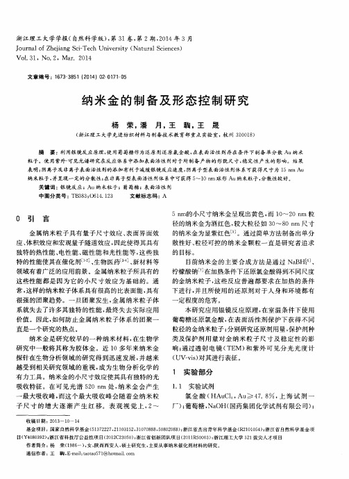
5 n m的小 尺寸 纳米 金 呈现 出 黄色 , 而 1 0  ̄2 0 n m 粒 径 的纳 米金 为酒 红色 , 较 大 粒径 如 3 0 ~8 0 n m 尺 寸 的纳 米金 为显 紫 红色 l _ 5 ] 。通 过简 单方 法制 备 出单分 散性 好 、 粒径 可 控 的纳 米 金 颗 粒 一直 是 研 究 者追 求
DNA修饰纳米金溶胶的稳定性

第57卷 第4期 化 工 学 报 Vol 157 No 14 2006年4月 Journal of Chemical Industry and Engineering (China ) April 2006研究论文DNA 修饰纳米金溶胶的稳定性杨 薇,李 韦华,张金利(天津大学化工学院,天津300072)摘要:纳米金在分子生物学、生物传感器等领域具有广阔的应用前景.针对纳米金在电解质溶液中易形成不可逆聚集的问题,通过紫外光谱、TEM 、Zeta 电位测试等表征,研究了DNA 分子修饰对纳米金溶胶稳定性的影响.结果表明,所制备的纳米金粒子的初始Zeta 电位与粒径有关,平均粒径为13nm 的金粒子对应-4414mV 的电位;当加入钠离子缓冲液后,纳米金粒子迅速聚集沉积.紫外光谱的动力学特性曲线表明,经过与巯基相连的DNA 修饰后,纳米金粒子能够在钠离子缓冲液中稳定存在.关键词:纳米金;DNA 修饰;胶体;粒子团聚中图分类号:TB 383;Q 523文献标识码:A 文章编号:0438-1157(2006)04-0970-05Stabilit y of gold nanoparticles modified wit h DNAYAN G Wei ,L I Wei ,ZHAN G Jinli(S chool of Chemical Engineering and Technology ,Tianj in Universit y ,Ti anj in 300072,China )Abst ract :G old nanoparticles show promising applications in t he field of molecular biology and R &D of biological sensors.Aiming at overcoming t he irreversible aggregation of nano 2gold in electrolyte solutions ,t his work investigated t he effect of DNA modification on nano 2gold stability in t he presence of sodium ions ,t hrough t he characterization of UV ,TEM and Zeta potential measurement s.It was indicated t hat t he Zeta potential of nano 2gold depended on t he diameter of particles.Nano 2gold particles wit h diameter of 13nm were stable at t he Zeta potential of -4414mV ,while irreversible aggregation of particles appeared quickly when a sodium buffer was added to t he solution.UV spect ra of kinetic properties for t he DNA modified nano 2gold particles reflected t he high stability of t he system wit h t he addition of sodium ions.Key words :nano 2gold ;DNA modification ;colloid ;particle aggregation 2005-07-04收到初稿,2005-10-20收到修改稿.联系人:张金利.第一作者:杨薇(1980—),女,硕士研究生.基金项目:国家自然科学基金项目(20576090,20476077);教育部春晖计划资助.引 言纳米材料因其具有体积效应、量子效应等特性而日益受到人们的关注,在生物、化学、免疫学等领域具有广泛的应用前景.而纳米金颗粒在生物体系检测中的应用研究是近年来的一个热点课题.纳米金胶体溶液对应的特异性吸光系数值比普通有机发色团要高出3~5个数量级,因此极低浓度的纳 Received date :2005-07-04.Corresponding aut hor :Prof.ZHAN G Jinli.E -mail :zhangjinli @tju 1edu 1cnFoundation item :supported by t he National Natural Science Foundation of China (20576090,20476077)and t he Chunhui Program.米金(10-9mol ・L -1)即可以用肉眼直接观察[1].纳米金颗粒还可与氨基发生非共价的静电吸附,或与巯基形成强的Au 2S 共价键,从而使得胶体金能够与生物活性分子相互结合,以形成生物体系检测的探针.可见纳米金在分子生物学、生物传感器等领域具有深远的研究意义.纳米金可以通过弱的相互作用与生物大分子结合,也可以通过化学键与生物大分子偶联,而不改变生物大分子的生物活性,目前被广泛应用于免疫组织染色的电镜观察研究,如免疫金和生物素金银染色法的定性、定位以至定量研究,均采用了“抗体2抗原2抗体2纳米金”夹心法[2].新近研究表明,金纳米颗粒也可与脱氧核糖核酸分子作用,并且利用纳米金胶体的特殊颜色和独特的生物活性,在DNA的识别与检验方面展示出极具应用价值的成果.例如,Mirkin等[3]利用纳米金与DNA片段在组装分子的引导下可形成超分子结构的特点,建立了用巯基化寡核苷酸探针来检测特定多核苷酸序列的新方法,这种方法将有力地推进DNA传感器以及DNA芯片的研制.Brown等[4]利用不同粒径的纳米金与巯基修饰的DNA结合,并与脱氧核酶(DNAzyme)协同作用,研究了一种高选择性的Pb2+检测方法.可见,利用纳米金的比色效应,可设计出理想的生物检测微传感器[528].然而在胶体金溶液的应用过程中,还存在着纳米金溶胶稳定性受环境因素影响严重的问题,在电解质溶液中易形成不可逆聚集,从而影响其后续使用[9].基于纳米金能够与巯基形成强的Au2S共价键,本文利用与巯基相连的DNA分子对纳米金进行修饰,通过紫外光谱、TEM、Zeta电位测试等表征,研究了DNA分子修饰对纳米金溶胶稳定性的影响.1 实验材料和方法111 主要试剂11111 纳米金制备试剂 柠檬酸三钠(>9910%);四氯金酸(99199%);011mol・L-1 NaCl缓冲溶液(0101mol・L-1Tris2Acetate, p H=710);其中氯化钠为分子生物级纯度;所有溶液均采用三次蒸馏的去离子水配制.11112 巯基修饰DNA 经HPL C纯化后的巯基修饰寡核苷酸:5′2HS2(C H2)62CAC GA GT T GA2 CA23′(简记为SH2DNA)(大连宝生物工程公司).112 纳米金溶胶的制备金溶胶由HAuCl4经柠檬酸三钠溶液还原而成[10].一定浓度的HAuCl4加热至沸腾,在剧烈搅拌下快速加入定量的柠檬酸三钠溶液.混合物加热至沸腾,在剧烈搅拌下快速加入定量的柠檬酸三钠溶液.015h后移去热源逐渐冷却,使用0145μm的滤膜过滤后避光保存.实验中采用了两种典型的HAuCl4与柠檬酸三钠的摩尔配比1∶3188和1∶31348.113 DNA对金纳米粒子的修饰将SH2DNA与纳米金粒子混合,使SH2DNA 的最终浓度为3×10⁻⁶mol・L-1,纳米金浓度约为819×10-9mol・L-1,室温下放置16h后,逐渐向溶液中滴加011mol・L-1NaCl缓冲液(p H= 710)并置于摇床中,大约48h后Na+达到约0105 mol・L-1,所得溶液体系可稳定存在.114 纳米粒子表征方法利用Tecnai G2F20场发射透射电子显微镜(TEM)(Philip s公司)对纳米金的粒径进行表征.采用紫外2可见光光谱(Varian Cary UV300型,美国Varian公司)对纳米金溶液进行200~600nm波长范围的扫描,表征样品对应的特征吸收峰.金纳米粒子对应的消光系数ε520值取为217×108 L・mol-1・cm-1[11],根据朗伯2比尔定律以及溶胶在520nm处的吸光度,可以计算出金溶胶的浓度.使用Zeta电位粒度仪(Zetasizer3000HS型,英国Malvern公司)表征金溶胶的Zeta电位. Zeta电位的数值能够指示出胶体体系的稳定性,如果纳米粒子具有较高绝对值的正或负Zeta电位,则彼此之间的排斥力可以使其稳定地存在于溶液中.2 实验结果与讨论211 纳米金溶液的表征通过透射电镜对制得的纳米金溶胶进行表征的结果如图1所示,可见,纳米粒子粒径分布均匀,图1(a)中的粒子平均粒径为13nm,溶液呈现酒红色,其对应的制备条件是HAuCl4与柠檬酸三钠的摩尔比为1∶3188;图1(b)的平均粒径为24nm,溶液呈现深红色,制备条件是HAuCl4与柠檬酸三钠的摩尔比为1∶31348.图2是对上述金胶体溶液的紫外光谱图,由图可见,在523nm左右有很强的吸收峰,且随纳米粒子粒径的减少,峰强度增大.另外,尽管溶液中不存在DNA序列,纳米金溶液在260nm处也有一定的紫外吸收.这表明对于DNA分子与纳米金粒子共存的溶液,不能单纯通过260nm处紫外特・179・ 第4期 杨薇等:DNA修饰纳米金溶胶的稳定性(a )diameter =13nm(b )diameter =24nmFig 11 TEM images of gold nanoparticleswith uniform diameter distributionFig 12 UV 2vis spectra of gold nanoparticleswith different particle diameters征吸收峰来表征DNA 的浓度.采用Zeta 电位粒度仪测定了金溶胶的Zeta 电位,测试表明,13nm 和24nm 的金溶胶在p H =3135的溶液中,Zeta 电位分别是-4414mV 和-3413mV.通常Zeta 值大于30mV 或小于-30mV 时说明溶胶体系相当稳定[12],因此说明所制备的纳米金胶体溶液很稳定.212 Na +离子对金溶胶的稳定性影响纳米金溶胶中的各粒子间主要是通过彼此间的静电斥力来稳定存在于溶液之中,如果向溶液中快速加入大量的带异种电荷的离子会中和金粒子表面的电性,使相邻粒子间距缩小,易导致如图3所示的纳米金粒子间的紧密堆积,进而造成不可逆沉积作用.实验中向溶液中快速加入大量的011mol ・L -1NaCl 溶液(最终溶液中Na +浓度约0105mol ・L -1)时,发现加入盐缓冲液后,溶液中的纳米金会发生聚集并最终沉积为黑色沉淀物,同时酒红色溶液变澄清.Fig 13 TEM of gold nanoparticles aggregatein 011mol ・L -1Na +buffer将011mol ・L -1NaCl 缓冲液(p H =710)与13nm 金溶胶快速混合后(最终Na +浓度约0105mol ・L -1),每隔6min 在200~600nm 波长范围内扫描一次获得其吸光度变化曲线,如图4所示.可见,520nm 处纳米金的吸收峰强度随时间增加而下降,而670nm 处的吸收峰强度随时间增加而增大,这是因为溶液中的纳米金粒子在阳离子加入后不断聚集,形成了不可逆沉积.然而,文献报道的纳米金与抗体或核酸分子相结合用于生物检测的过程中,必须在一定的盐离子环境下才能实现其功能;例如带负电磷酸骨架的DNA 或RNA 分子需要通过溶液中的一定浓度的阳离子来稳定其活性构象,并最终发挥序列杂交与识别的功能.可见,不仅溶液中的盐离子,核酸分子或某些带电的蛋白分子本身就是多价带电离子[9],它们均能对纳米金颗粒产生影响.因此,必须研究溶液中纳米金粒子稳定存在的条件,以拓增其应用领域.・279・化 工 学 报 第57卷 Fig 14 UV 2vis spectra of gold nanoparticles (13nm )without SH 2DNA after additionof 011mol ・L-1Na+buffers213 DNA 修饰的纳米金溶胶的稳定性对于DNA 修饰后的纳米金溶胶,同样采用212节的方式,用紫外光谱表征加入011mol ・L -1Na +缓冲液后溶液的吸光度变化曲线,如图5所示,由图可知,Na +的加入没有导致DNA 修饰的纳米金粒子的沉积.Fig 15 UV 2vis spectra of gold nanoparticles (13nm )with SH 2DNA after addition of 011mol ・L -1Na +buffers (in time range of 0—230min )依据图4中纳米金沉积的特性曲线,分别计算出不同时间下DNA 修饰前后纳米金在520nm 与670nm 处的峰强度比值,示于图6,可见未经DNA 修饰的金溶胶不能稳定地存在于盐溶液中,盐离子加入后A 520nm /A 670nm 的比值迅速下降;而DNA 修饰的金溶胶则可以稳定地存在于离子缓冲溶液中.这种DNA 修饰的金溶胶将在有离子存在的核酸分子变性2复性、杂交等实验研究中有极好的应用前景.3 结 论(1)实验制备了两种不同粒径的纳米金粒子,并且测得其Zeta 电位分别为-4414mV 和-3413Fig 16 Effect of SH 2DNA on goldnanoparticles aggregates(Extinction ratios of A 520nm /A 670nm werenormalized for comparison )mV ,说明所制备的纳米金胶体溶液很稳定.而加入离子缓冲液后,A 520nm /A 670nm 的比值迅速下降,纳米金粒子出现了显著的沉积.(2)对于DNA 修饰的纳米金溶胶,离子缓冲液的加入没有导致纳米金粒子的沉积.说明DNA 修饰的金溶胶可以稳定地存在于离子缓冲溶液中,它将在盐离子存在的核酸分子变性2复性、杂交等实验研究中有极好的应用前景.致谢:衷心感谢L u Y i 教授(University of Illinois atUrbana 2Champaign )对本课题的指导.References[1] Liu J ,L u Y.Accelerated color change of gold nanoparticlesassembled by DNAzymes for simple and fast colorimetricPb 2+detection.J.A m.Chem.S oc 1,2004,126(39):12298212305[2] Deng Y ongpei (邓永沛),Zhao Hongqiu (赵红秋),JiangLong (江龙).Applications of nanogold particles inbiomimetic engineering.China B asic S cience (中国基础科学),2000,9:11217[3] Mirkin C A ,Lot singer R L ,Mucic R C ,Storhoff J J.ADNA 2based met hod for rationally assembling nanoparticles into macroscopic materials.N at ure ,1996,382:6072609[4] Brown A K ,Li J ,Pavot C M 2B ,L u Y.A lead 2dependentDNAzyme wit h a two 2step mechanism.B iochemist ry ,2003,42(23):715227161[5] Swearingen C B ,Wernette D P ,Cropek D M ,L u Y ,BohnP W.Immobilization of a catalytic DNA molecular beacon on Au for Pb (Ⅱ):Detection.A nal.Chem 1,2005,77(2):4422448[6] Pavlov V ,Xiao Y ,G ill R ,Dishon A ,K otler M ,Willner I.Amplified chemiluminescence surface detection of DNA and Telomerase activity using catalytic nucleic acid labels.A nal.Chem 1,2004,76(7):215222156・379・ 第4期 杨薇等:DNA 修饰纳米金溶胶的稳定性[7] Storhoff J J ,Lazarides A A ,Mucic R C ,Mirkin C A ,Let singer R L ,Schatz G C.What controls t he optical properties of DNA 2linked gold nanoparticles assemblies ?J.A m.Chem.S oc 1,2000,122(19):464024650[8] Liu J ,Lu Y.Optimization of a Pb 2+2directed goldnanoparticle/DNAzyme assembly and it s application as a colorimetric biosensor for Pb 2+.Chem.M ater.,2004,16(17):323123238[9] Demers L M ,Mirkin C A ,Mucic R C ,Reynolds ⅢR A ,Let singer R L ,ElghanianR ,Viswanadhem G.Afluorescence 2based met hod fordetermingt hesurfacecoverageandhybridization efficiency oft hiol 2capped oligonucleotides bound to gold t hin films and nanoparticles.A nal.Chem.,2000,72(22):553525541[10] Storhoff J J ,Elghanian R ,Mucic R C ,Mirkin C A ,Let singer R L.One 2pot colorimetric differentiation of polynucleotides wit h single baseimperfections using gold nanoparticle probes.J.A m.Chem.S oc 1,1998,120(9):195921964[11] Jin R ,Wu G ,Li Z ,Mirkin C A ,Schatz G C.What controlst he melting properties of DNA 2linked gold nanoparticle assemblies ?J.A m.Chem.S oc 1,2003,125(6):164321654[12] Li Xiang (李翔),Dai Yatang (戴亚堂),Deng Yun (邓赟).Research on t he particle 2size distribution of nano 2titanium dioxideby photon correlation spect roscopy.J ournal ofS ichuan Universit y (Engi neeri ng S cienceEdition ),2004,36(4):62266・479・化 工 学 报 第57卷 。
纳米金粒子的制备及其在生物传感器中的应用
纳米金粒子的制备及其在生物传感器中的应用纳米金粒子是指金属黄金在100纳米以下的微小颗粒,因其独特的光学、电学、磁学和化学等性质而引起研究者的极大兴趣。
在近年来的科学研究中,纳米金粒子被广泛应用于医学、电子、光电、生物传感、光学传感、热传感等领域。
其中,纳米金粒子在生物传感器中的应用具有广阔的应用前景。
一、纳米金粒子的制备纳米金粒子的制备方法有多种,如物理气相沉积、化学气相沉积、溶液法、微反应系统等。
其中,溶液法是制备纳米金粒子最为常用的方法之一。
通过选择不同的还原剂、保护剂、模板等条件,可以制备出晶体形貌不同的纳米金粒子,如球形、棒形、八面体形等。
此外,纳米金粒子亦可通过激光蚀刻法等方法制备。
二、纳米金粒子在生物传感器中的应用在生物传感器中,纳米金粒子作为生物反应器、识别元素和信号放大器等重要角色。
其具有以下应用:1. 生物传感器纳米金粒子在生物传感器中可以作为载体搭载生物分子,例如抗体、DNA探针、酶等,来检测特定物质。
当前,基于纳米金粒子的免疫传感技术被广泛应用于免疫识别、抗菌药物检测、酶活性测定等领域。
2. 生物成像利用纳米金粒子的高度表面增强拉曼散射效应,可以普及成像领域,例如在细胞成像、分子成像等方面有广泛应用。
3. 传感器信号放大器纳米金粒子在生物传感器中作为信号放大器,可以增强传感器的灵敏度和快速响应。
近年来,许多人体检测设备和检测仪器中采用了这一技术。
4. 气体传感器纳米金粒子在气体传感器中可以自身吸附气体,如H2,CO和NO2等。
当吸附的气体只有实际质量的0.01%时,其性质发生了明显改变,可以用作气体传感器探测吸附的气体。
三、纳米金粒子存在的问题尽管纳米金粒子在生物传感器中有着广泛的应用前景,但同时也存在一些问题。
首先,纳米金粒子人工制备过程中可能存在产生有害化合物的风险,例如使用还原剂亚硫酸钠和棕榈酸钠等。
其次,纳米金粒子的使用需考虑是否对人类健康有副作用,例如纳米金粒子可能被身体吸收进入人体,对人体器官造成损伤。
纳米金的制备与表征[1]
制备纳米金,主要还 是采用还原剂,如柠檬酸 钠、硼氢化钠等,还原氯 金酸。氯金酸在还原剂作 用下,可聚合胶体 状态,故称为胶体金。用 还原法可以方便地从氯金 酸制备各种不同粒径、也 就是不同颜色的胶体金。
纳米金标记DNA后可作为纳米金探针进行核 酸核算检测。纳米金颗粒随直径的变化会呈现出 不同的颜色,此特征可用于核酸杂交检测。纳米 金标记DNA用于检测是一种具有很高选择性和灵 敏度的新的比色检测多核苷酸的方法,是利用纳 米金标记的寡核苷酸探针和靶序列杂交形成伸展 的金纳米颗粒—多核苷酸的多聚网络结构,并由 此引发粒子光学性质的变化,产生多核苷酸的杂 交信号。金纳米粒子在水中形成分散系俗称胶体 金,可以与巯基之间形成很强的Au-S共价键这使 得胶体金可与含巯基生物活性分子形成探针,可 用于多种生物体系的检测中。
固相检测模式及其应用
Taton等创立了一种将纳米金探针用于识别靶基因的固相检 测模式。 该模式采用预化学处理过的普通玻片或硅片等作为 固相支持物。
纳米金探针结合银增强法的固相检测模式
在固相支持物上修饰一段与靶基因序列一端互补的寡核苷酸链,我们称 其为捕获探针; 加入待测样品,与支持物上的捕获探针进行杂交; 将未 杂交上的核酸洗去; 加入表面修饰了寡核苷酸链的纳米金(13nm)探针, 该寡核苷酸链序列与靶基因的另一端互补; 纳米金探针与靶基因杂交并 冲洗后,在固相支持物上形成一种由捕获探针、靶基因、纳米金探针三 种成分组成的夹心结构。 最后,在存在靶基因的区域将出现红色斑点。 为了增强检测信号,可以在固相支持物上滴入由坏血酸和硝酸银溶液组 成的银增强液。 由于纳米金颗粒具有催化性,能催化溶液中的Ag+ 还原 成银,被还原出来的银附着在金颗粒的表面形成一层银壳,起到增强信 号的作用。在没有金颗粒的区域,只有极少的银离子存在,从而保证了 银染色的特异性。 信号的强弱与靶基因的量呈正比。 靶基因越多,支 持物表面被固定的纳米金颗粒越多,形成的银壳越厚,检测信号越强。
纳米金的产品介绍和应用(Gold...
纳米金的产品介绍和应用(Gold...纳米金的产品介绍和应用(Gold Nanoparticles Overview and Application)Gold Nanoparticles纳米金是一种以氯金酸(HAuC14)为主要材料,通过还原来制备成的胶体金(colloidalgold),它通常是一种金颗粒的悬浮液,其粒径为1-100nm不等,颜色呈紫红色。
该产品可被应用于诊断探针、免疫印迹、治疗药物、药物传送等等。
胶体金颗粒也是Gold Nanoparticles纳米金颗粒的结构,实际上是由一个金(Au)做为核心,其Au核心的外围包裹的内外二层离子层,内层离子层带负离子auc12,其作用是紧紧链接金核(Au),外层离子层带正离子H,其作用是均匀的分散在胶体间的溶液中,以维持稳定的悬浮状态。
Gold Nanoparticles纳米金颗粒的性状一般小于30纳米的都会呈现是规律的圆球形状,如果大于30纳米的胶体金(Gold Nanoparticles)一般是呈现的椭圆状的。
颜色上来讲也有比较细微的划分,一般是2-5nm间的会呈现橙黄色,8nm-25nm的会呈现酒红色,30nm-100nm的是呈现紫红色。
光吸收性胶体金在可见光范围内有一单一光吸收峰,这个光吸收峰的波长(λmax)在510~550nm范围内,随胶体金颗粒大小而变化,大颗粒胶体金的λmax偏向长波长,反之,小颗粒胶体金的λmax 则偏于短波长。
以下列表是纳米金粒子的大小,个数和SPR波长列表:Particle Size (nm) Particle Conc. (Particles/mL) SPR Wavelength (nm) (mg/mL) 2nm 1.5x10E14 Not measured 0.1mg/ml3nm 1.5x10E14 512~515 0.1mg/ml5nm 5.0x10E13 515~520 0.1mg/ml10nm 5.7x10E12 515~520 0.1mg/ml15nm 1.4x10E12 517~522 0.1mg/ml20nm 7.0x10E11 525 0.1mg/ml30nm 2.0x10E11 527 0.1mg/ml40nm 9.0x10E10 530 0.1mg/ml50nm 4.5x10E10 535 0.1mg/ml60nm 3.1x10E10 540 0.1mg/ml80nm 2.6x10E10 553 0.1mg/ml100nm 1.1x10E11 572 0.1mg/ml纳米金颗粒Gold Nanoparticles应用包括有:1:纳米金应用于毛细管电泳检测尿液中8-OHdG2:纳米金应用于蛋白质纤维染色的研究3:纳米金应用于肺癌靶向诊疗的研究进展4:纳米金应用于肿瘤诊疗的研究进展5:纳米金颗粒在仿生工程中的应用6:纳米金生物探针及其应用7:纳米金在生物标记分析中的应用进展8:纳米金在光学和电化学传感器中的应用西安瑞禧生物是国内知名的纳米产品试剂供应商,我公司提供各种不同的金纳米系列产品、银纳米系列产品、磁性纳米颗粒系列产品、聚苯乙烯微球系列产品、金纳米棒系列产品、功能性琼脂糖珠产品、和荧光量子点系列产品。
基于双巯基化合物和纳米金固定生物分子的研究
基于双巯基化合物和纳米金固定生物分子的研究摘要:利用自组装方法,将双巯基化合物通过形成金硫键修饰至金电极表面,再利用双巯基化合物的另一个-SH基,吸附纳米金颗粒形成纳米金的膜。
运用循环伏安法对此修饰电极进行研究。
发现双巯基化合物的最佳修饰时间为2小时;纳米金的最佳修饰速度为6小时;当扫描速度为100mv/s时固定抗体抗原的效果最好;而抗体抗原浓度为1:4时固定效果最佳。
关键词:纳米金修饰电极自组装循环伏安法一、引言自组装单分子膜是使用含有各种活性官能团的分子,以化学键的形式与相应的基底(如Au、Ag、Cu、Pt、Si等)相互作用从而形成的自组装膜。
目前研究最多的是巯基化合物在金电极表面的自组装及应用分析。
由于金表面无自然氧化膜,稳定性好,而且与二硫化合物或硫醇形成的自组装体系具有良好的稳定性,因而以Au-S键为基础的自组装体系往往成为研究的首选体系。
分子自组装技术是80年代新兴的、基于分子自组装作用,在固体表面自然形成高度有序的分子层的方法。
该技术具有制备方法简单、膜结构有序、性能稳定等优点,提供了在分子水平上方便地构造理想界面的手段,对实现优良功能材料的分子设计具有指导作用,在生物仿生、生物传感器、润滑、非线型光学等众多领域有广泛的应用前景,已成为当今学术界一项极有意义的研究课题。
在众多自组装单分子膜种类中,硫醇类在金基底上的自组装因具有成膜条件较易控制、有序性强、吸附杂质少、选择性高等特点成为了研究最多和最具代表性的体系,在基础及应用研究领域都受到广泛重视。
这些技术主要是针对自组装单分子膜形成后稳定状态下结构和性质的探识。
但是只有了解和认识溶液中分子自组装的动态的物理和化学过程,研究自组装单分子膜有关的成膜过程的动态信息,才能更好地选择条件并控制膜的制备从而获得高质量自组装单分子膜。
总之,自组装技术越来越显示出其不可比拟的优越性。
自组装技术提供了分子水平上方便构造理想界面的手段,在润滑、防腐、催化、刻蚀、电子得失反应、分子器件、非线性光学众多领域有广泛的应用前景,从而成为近年来界面化学与材料化学领域研究的特点。
- 1、下载文档前请自行甄别文档内容的完整性,平台不提供额外的编辑、内容补充、找答案等附加服务。
- 2、"仅部分预览"的文档,不可在线预览部分如存在完整性等问题,可反馈申请退款(可完整预览的文档不适用该条件!)。
- 3、如文档侵犯您的权益,请联系客服反馈,我们会尽快为您处理(人工客服工作时间:9:00-18:30)。
Monolayer-Protected Gold Nanoparticles as a Stationary Phase for Open Tubular Gas ChromatographyGwen M.Gross,†David A.Nelson,‡Jay W.Grate,‡and Robert E.Synovec*,†Center for Process Analytical Chemistry,Department of Chemistry,Box351700,University of Washington, Seattle,Washington98195,and Pacific Northwest National Laboratory,Richland,Washington99352The use of a thin film of monolayer-protected gold nano-particles(MPNs)as a stationary phase for gas chroma-tography(GC)is reported.Deposition of a MPN film was obtained in a2-m,530-µm-i.d.deactivated silica capillary using gravity to force the solution containing the MPN material through the capillary.By SEM analysis,the average film thickness was determined to be60.7nm. The retention behavior for the dodecanethiol MPN column was studied using four compound classes(alkanes,al-cohols,aromatics,ketones),and retention orders were objectively compared to a commercially available column (AT-1,100-nm film thickness).Separation of an eight-component mixture was performed using both isothermal and temperature-programming methods with the dode-canethiol MPN phase and compared to an isothermal separation with the AT-1phase.The AT-1phase separa-tion had an efficiency,N,of6200(k′)0.33)while the dodecanethiol MPN phase separation had an efficiency, N,of5700(k′)0.21)for the same analyte,octane.The reduced plate height,h,for octane was found to be less than1at the optimum linear flow velocity,indicating the MPN column operated near the optimum possible per-formance level.Robustness of the MPN phase is also discussed with consistent performance observed over several months.Overall,MPNs appear promising as a stationary-phase material for GC and as an experimental platform to study their thermodynamic and mass-transfer properties.Nanoparticles and nanoparticle-based materials are attracting great interest for their unique properties and potential for application in diverse areas.1-3Use of nanoparticles for chromato-graphic applications has proven to be advantageous.For example, capillary electrophoresis has found applications of nanoparticles as a pseudostationary phase,enhancing selectivity,as well as a production method for nanoparticles.4-6Monolayer-protected nanoparticles(MPNs)are of particular interest because the surface monolayer stabilizes them relative to aggregation and their properties are influenced by the structure of the monolayer-forming molecules.More recently,the use of high-performance liquid chromatography for the separation of different MPNs based upon nanoparticle size and the chosen monolayer was reported.7 An ordered arrangement of the thiol linkages with uniform, monolayer coverage can be obtained on flat gold surfaces.8 However,with small-diameter MPNs,the curvature of the gold core leads to a larger population of core surface gold atoms,which are defect sites in2D self-assembled monolayers.9This disorder is a desired effect,promoting a noncrystalline interparticle structure,facilitating fast mass transfer via diffusion.Studies of dodecanethiol MPNs have shown the closest core-core spacing is equal to the extended chain length of a single dodecanethiol monolayer,demonstrating the chains interpenetrate to form a disordered packing structure.3Thus,a thin MPN film should function as an efficient stationary phase for gas chromatography (GC).It is envisaged that similar chromatographic efficiencies relative to commercial stationary phases can be realized.Since a wide range of thiol-based organic monolayers is available,ample chemical selectivity could be achieved for different MPN-based GC stationary phases.This report initiates a GC-based platform for thermodynamic and mass-transfer studies of MPNs.With a firm understanding of the chemical selectivity and mass-transfer properties,one could potentially devise MPNs that are tuned for a particular GC application of interest or for other chemical analyzers and sensors as well.The characterization of the MPNs is of considerable interest. In particular,Au-based MPNs have been utilized in a sensor format,10but a greater understanding of their properties as a*Corresponding author.E-mail:Synovec@.†University of Washington.‡Pacific Northwest National Laboratory.(1)Brust,M.;Fink,J.;Bethell,D.;Schiffrin,D.;Kiely,C.J.Chem.Soc.,Chem.Commun.1995,1655-1656.(2)Brust,M.;Bethell,D.;Kiely,C.;Schiffrin,ngmuir1998,14,5425-5429.(3)Templeton,A.;Wuelfing,M.;Murray,R.Acc.Chem.Res.2000,33,27-36.(4)Neiman,B.;Grushka,E.;Ovadia,L.Anal.Chem.2001,73,5220-5227.(5)Sunderhaus,J.;Steinbock,B.;Steinbock,O.Abstr.Pap.Am.Chem.Soc.2002,223,360-PHYS Part2.(6)Viberg,P.;Jornten-Karlsson,M.;Petersson,P.;Spegel,P.;Nilsson,S.Anal.Chem.2002,74,4595-4601.(7)Jimenez,V.L.;Leopold,M.C.;Mazzitelli,C.;Jorgenson,J.W.;Murray,R.W.Anal.Chem.2003,75,199-206.(8)Sabapathy,R.;Bhattacharyya,S.;Leavy,M.;Cleland,W.;Hussey, C.Langmuir1998,14,124-136.(9)Hostetler,M.;Wingate,J.;Zhong,C.;Harris,J.;Vachet,R.;Clark,M.;Londono,J.;Green,S.;Stokes,J.;Wignall,G.;Glish,G.;Porter,M.;Evans, N.;Murray,ngmuir1998,14,17-30.(10)Grate,J.W.;Nelson,D.A.;Skaggs,R.Anal.Chem.2003,75,1868-1879.Anal.Chem.2003,75,4558-45644558Analytical Chemistry,Vol.75,No.17,September1,200310.1021/ac030112j CCC:$25.00©2003American Chemical SocietyPublished on Web08/05/2003sensing medium was desired,serving as impetus for this work. Previous characterization has been done using techniques such as electron microscopies and thermal gravimetric analysis as well as other spectroscopic and thermochemical techniques.9-11The GC platform reported herein can be used to further characterize the gold-based MPNs in a way that is different from the studies done on nanoparticles to ing techniques previously established for materials characterization,the sorptive properties of the nanoparticles as well as their chemical retention behavior can be explored.12-14Gas chromatographic measurements provide a reliable,reproducible,and efficient platform for measuring retention behavior of many compounds on a given MPN phase and,hence,for evaluating chemical selectivity.Reported herein is the development of a stationary phase consisting of dodecanethiol-protected gold nanoparticles and initial characterization of this phase by and for capillary GC.Successful deposition of a thin film of MPNs within a capillary for GC was explored for the first time as well as the methodology for that deposition.Dodecanethiol MPN was coated in a thin film, nominally60nm thick,in a530-µm-i.d.capillary with2-m length.Separations of different compound classes and a test mixture,as well as characterization studies of the nanoparticle stationary phase,were completed.The characterization studies included exploration of the retention behavior of the stationary phase and chromatographic efficiency.An objective comparison between the novel dodecanethiol MPN phase and a commercial AT-1GC stationary phase was completed.Mass-transfer behavior of the dodecanethiol MPN phase was studied by observing the band broadening of several analytes as a function of linear flow velocity. The retention and mass-transfer behavior provided by the dode-canethiol MPN phase will serve as a benchmark for comparison with future MPN-based stationary phases.EXPERIMENTAL SECTIONReagents and Chemicals.All chemicals were reagent grade or a higher grade.Many of the bulk reagents were purchased from Fisher(Fisher Scientific,Fairlawn,NJ).Chromatographic standards from PolyScience(AccuStandard,Inc.,New Haven,CT) were used as analytes for the GC separations with the exception of samples containing benzene and hexane(both Fisher),anisole, octane,decane(all Aldrich,Milwaukee,WI),and chlorobenzene (J.T.Baker,Phillipsburg,NJ)(Table1).Water used in the production of the column was filtered using a NANOpure II filter system(Barnstead/Thermolyne Corp.,Dubuque,IA).The GC carrier gas was hydrogen and was supplied by a hydrogen generator(Whatman Inc.,Kent,U.K.).Nanoparticle Synthesis.The method used for the synthesis of the gold-based MPNs was taken from Wolhtjen and Snow15 but was initially developed by Brust.16The basic formula for the synthesis isA1:1molar ratio of gold to thiol was used,with a dodecanethiol monolayer.Tetraoctylammonium bromide(4.5g)was dissolved in17mL of toluene.A solution consisting of65mL of water with 1.03g of hydrogen tetrachloroaurate was added to the toluene solution in a three-neck round-bottom flask attached to a nitrogen bubbler system.An additional3mL of water was used to completely transfer the gold-containing solution to the flask. Dodecanethiol(0.50g)was dissolved in2mL of toluene and added to the flask.Sodium borohydride(0.79g)was taken from under vacuum and dissolved in50mL of water while stirring.This solution was then added dropwise to the flask over a5-min period. Once the addition was complete,the flask was capped and allowed to stir for3.5h,after which time the solution was transferred to a separation funnel.The toluene layer was washed twice with150 mL of water.The product was placed under a nitrogen stream overnight prior to purification.Purification was done using350mL of ethanol,allowing the gold MPNs to precipitate from solution.The solution was centrifuged and the solvent decanted.Two additional30-mL ethanol washes were completed in the same way.The product was dissolved in5mL of dichloromethane and transferred to a tared vial.An additional3mL of dichloromethane was used to completely transfer the dodecanethiol MPN product to the tared vial.The solvent was evaporated off under a stream of nitrogen, and a product yield of0.78g was achieved.The purity of the product was tested using thin-layer chromatography(TLC)and Fourier transform infrared spectroscopy(FT-IR)(Vector33, Bruker Optics,Billerica,MA).Of particular interest was the presence of residual thiol or disulfide present in the ing a9:1dichloromethane/methanol solution,the TLC plates were run and then stained with iodine.No additional spots were detected with the iodine stain,indicating that neither residual thiol nor disulfide was present at detectable levels in the final product. The FT-IR spectra(KBr pellet)of the MPN product were consistent with the literature and confirmed the absence of any(11)Warner,M.;Reed,S.;Hutchison,J.Chem.Mater.2000,12,3316-3320.(12)Abraham,M.;Poole,C.;Poole,S.J.Chromatogr.,A1999,842,79-114.(13)Abraham,M.H.;Andonian-Haftvan,J.;Du,C.M.;Diart,V.;Whiting,G.;Grate,J.W.;McGill,R.A.J.Chem.Soc.,Perkin Trans.21995,369-378.(14)Abraham,M.;Ballantine,D.;Callihan,B.J.Chromatogr.,A2000,878,115-124.(15)Wohltjen,H.;Snow,A.W.Anal.Chem.1998,70,2856-2859.(16)Brust,M.;Walker,M.;Bethell,D.;Schiffrin,D.;Whyman,R.J.Chem.Soc.,mun.1994,801-802.HAuCl4‚3H2O+RSH98(C8H17)4NBrNaBH4Au:SRTable1.Twelve Components Chosen for Evaluation ofthe Dodecanethiol Monolayer-Protected GoldNanoparticle Stationary Phase acompound bp(°C)k′(MPN)log L16k′(AT-1)hexane690.01 2.6680.04ethanol780.02 1.4850.01benzene800.06 2.7860.083-pentanone1020.060.121-butanol1180.16 2.6010.10octane1260.21 3.6770.34chlorobenzene1320.65 3.6570.451-pentanol1380.43 3.1060.273-heptanone1480.420.66anisole1540.82 3.8900.803-octanone1680.71 1.59decane174 1.47 4.686 1.99a The retention factors,k′,were determined using methanol as thedead time marker.For comparison,the elution data for two othernonpolar stationary phases are included:log L16and AT-1.The elutionorder for hexadecane as a stationary phase is given by the log L16values.Analytical Chemistry,Vol.75,No.17,September1,20034559significant amount of phase-transfer agent in the final product.17,18However,additional purification steps may be warranted to fullyeliminate the phase-transfer agent.19The final dodecanethiol MPN product was black and stored dry in a sealed vial.Characterization of the nanoparticles was done using thermal gravimetric analysis (TGA;model STA409A,Netzsch,Selb,Germany)and transmission electron microscopy (TEM;model JEM 2010,JEOL,Tokyo,Japan)with TEM software (Digital Micrograph,Gatan Inc.,Pleasanton,CA).Characterization results are presented in Figure ing TGA,it was determined that 75.09%by mass of the nanoparticles was due to the gold core with most of the monolayer loss occurring between 200and 300°C (Figure 1C).While the synthesis mole ratio for gold to thiol was 1:1,the final product mole ratio calculated from the TGA was ∼3:1.The presence of nanoparticles was confirmed by TEM (Figure 1A)using a copper grid airbrushed with a dilute solution (0.2%by mass)of the nanoparticles dissolved in dichloromethane.The TEM image indicates the nanoparticle core size was polydisperse with diameters ranging from about 1.5to 5nm (Figure 1B)with an average of 3.2nm.Column Preparation.Deactivated silica capillary,530-µm i.d.with a 2-m length,was used for the production of the open tubular MPN column (Supelco,Bellefonte,PA).The capillary was washed with three 50-µL volumes of methylene chloride and with three volumes of water.The methylene chloride washes were evapo-rated from the capillary using a nitrogen stream while the water washes were evaporated from the capillary by baking in an oven at 300°C for 1h.This same treatment was used for a “blank”column (a column without MPN stationary phase to compare with the column with the dodecanethiol MPN phase,i.e.,the MPN column).Sufficient dodecanethiol MPN material was placed in a 100-µL conical insert inside a standard injection vial (Agilent Tech-nologies,Palo Alto,CA),and 50µL of methylene chloride was added.The resulting nanoparticle solution was introduced to prepare the MPN column via capillary action,resulting in a pluglength of ∼4cm.With the column in a vertical position,gravitational force was utilized to move the solution plug and the nanoparticles were deposited in the capillary via evaporation.When the solution plug reached the far end of the capillary,the column was inverted and the process repeated.Additional solution volumes were added until,via visual inspection,a uniform brown color was achieved.To help avoid MPN phase clumping a low-intensity hot air gun (Conair ProStylus 1500,Conair Corp.,Stamford,CT)was used as a low heat source for faster evaporation of the solvent.Several MPN columns were produced with varying shades from light tan to black.The uniform brown for the thin-film MPN column displayed minimal clumping with no observable bare spots and resulted in very reproducible GC separations.Determination of stationary-phase thickness,coverage,and con-firmation of stationary-phase presence were done using SEM as will be described later.SEM Imaging of the MPN Stationary Phase within the Capillary Column.All images were obtained using a LEO 982field emission SEM (LEO Electron Microscopy Ltd.,Cambridge,England).Prior to analysis,four randomly selected pieces of capillary column,three containing MPNs and one blank,were mounted and then coated with a thin platinum layer using a Ladd electronic sputter coating machine (Ladd Research,Williston,VT).Images were obtained in both the normal and backscatter modes.Elemental analysis of the visible stationary phase was performed in the backscattering mode.Chromatographic Instrumentation.All chromatograms were obtained with an Agilent 6890gas chromatograph using a standard FID detector and injector with ChemStation computer control (Agilent Technologies,Palo Alto,CA).Data were imported into Matlab (MathWorks,Natick,MA)for subsequent analysis.Chromatographic Experiments.All chromatograms were obtained with an injection source and FID temperature of 200°C and a hydrogen carrier gas.Samples were introduced by head-space injection.A 2-mm depth of sample containing the compo-nents of interest was placed in the bottom of a standard injection vial,below the injection depth of the syringe into the vial.An injection size of 0.5µL was used with a split injection of 50:1or 1:1as specified.The injection volume and split procedure did not introduce significant chromatographic band broadening.The vial(17)Hostetler,M.J.;Stokes,J.J.;Murray,ngmuir 1996,12,3604-3612.(18)Cai,Q.Y.;Zellers,E.T.Anal.Chem.2002,74,3533-3539.(19)Waters,C.A.;A.J.,M.;Johnson,K.A.;Schiffrin,mun.2003,540-541.Figure 1.Characterization of the synthesized dodecanethiol monolayer protected gold nanoparticles.(A)Transmission electron micrograph of a thin film of the dodecanethiol MPNs.(B)Histogram of the measured core size distribution from the TEM data.(C)Thermal gravimetric analysis plot indicating the loss of the dodecanethiol monolayer from the gold core.4560Analytical Chemistry,Vol.75,No.17,September 1,2003was not heated,so the amount of each component present in the injected sample was determined by component volatility at rooming samples prepared in this manner did result in sample-to-sample concentration variation for each component. However,this sample preparation and injection procedure was satisfactory for chromatographic evaluation of retention time, efficiency,N,and reduced plate height,h,for the components of interest.Due to the relatively short length and wide diameter of the MPN column,a short restrictor column(100µm i.d.,0.5m in length)consisting of a methyl-deactivated silica capillary followed the MPN column to avoid FID blowout(Supelco).The oven temperature was50°C unless otherwise noted.Additional reten-tion due to the restrictor column was found to be insignificant.The MPN stationary phase was compared to a commercial nonpolar AT-1stationary phase(Alltech Associates,Inc.,Deerfield, IL).All parameters were identical for the comparison except stationary-phase thickness.The AT-1phase thickness of0.10µm was as near as possible to the MPN phase thickness,nominally 0.060µm.The commercial column dimensions and chromato-graphic conditions were the same as those used for the MPN column separation:530-µm i.d.and2-m length,temperature of 50°C,and linear flow velocity of22cm/s.RESULTS AND DISCUSSIONUsing SEM analysis,the presence of the gold-containing dodecanethiol MPN stationary phase within the capillary GC column was confirmed.Stationary-phase thickness was measured at random spots along the column.Visually,the majority of the surface area of the inside wall was a uniform brown(∼97%),while a minor fraction of the surface area contained small black clumps (∼3%).Both uniform and nonuniform areas were imaged using the SEM.For a typical location on the capillary wall appearing uniform,the stationary-phase thickness was60.1nm(Figure2). There was very little variation((2nm)around the capillary circumference in these predominating uniform regions.For a rare area of nonuniformity,the average thickness was55.7nm,ranging from12.7to115.2nm.No bare areas of silica capillary were observed during any of the SEM measurements.The average film thickness for the full length of column was60.7nm.The retention characteristics of the dodecanethiol MPN stationary phase were studied using four different compound classes(Figure3).The analytes studied had a boiling point range from69to174°C(Table1).It should be noted that the analytes are not retained strictly upon the basis of boiling point.3-Octanone has a higher boiling point(168°C),but the less polar anisole, with a boiling point of154°C,is more highly retained.Retention order is based upon boiling point only within a homologous series of compounds.This can be seen within the three homologous series of alkanes,ketones,and alcohols.Additional support for the separation due to the MPN stationary phase was obtained using the“blank”column.This experiment addressed the pos-sibility of additional retention if exposed silica areas were present within the MPN column.Each of the four compound classes (Table1)were analyzed with the blank column under identical conditions as the MPN column.The bare silica capillary did not contribute significantly to the net analyte retention.This experi-ment confirmed the separations achieved with the MPN column were not due to bare silica areas,consistent with the SEM results.An expanded study of the retention behavior for the alkane homologous series was also done(C10-C16).The log of the retention factor(k′)was plotted against log L,16the Oswald solubility coefficient(gas-hexadecane partition coefficient at25°C).For straight-chain alkanes,log of k′is directly related to the log L16(Figure4).14The slope of the line is the l value of the stationary phase,used to describe the stationary phase in the solvation parameter model.12,14,20The l term is a measure of the hydrophobicity of the stationary phase and is an indicator of the ability of a stationary phase to separate members of a homologous series.12The solvation model is temperature dependent and so a change in the slope of the line was seen for the three temperatures examined(100,75,50°C).The k′values at25°C were extrapolated from the other three temperature series and plotted against the log L16values.The linearity of the data indicates the dodecanethiol MPN stationary phase is well correlated with the nonpolar n-alkane series used(R2of∼0.99for all temperature series)and behaves as expected by the model.The increased slope(l value) indicated the stationary phase separates n-alkanes better with a decrease in temperature.Lower temperatures are not always conducive to GC analysis,however,so it is useful to know that extrapolation of retention values can be done for the MPN stationary phase for use in future studies using the solvation model.From the original12analytes(Table1),8analytes were selected to demonstrate a more complex mixture separation.Each of the four compound classes were represented in the mixture. An acceptable isothermal separation was obtained with the dodecanethiol MPN column in a little over30s as shown in Figure 5A,comparing reasonably with the commercial AT-1column separation in ing temperature programming,the same mixture was separated with the MPN column in just over 25s with improved resolution(Figure5B).The isothermal separation was done at50°C,far below the boiling point of the higher boiling analytes in the mixture.It is not surprising that an improvement in the separation was observed for the later-eluting(20)Du,C.M.Thesis.The Application of Physiochemical Desciptors to theCharacterisation of Liquid and Solid Phases.University College,London, 1995.Figure2.SEM image of the dodecanethiol MPN stationary phasewithin the capillary.Area A is the silica capillary.Area B is the gold-based dodecanethiol MPN stationary phase,60.1nm thick at thising a random piece of column,this image was obtainedusing an end-on cross-sectional view.Analytical Chemistry,Vol.75,No.17,September1,20034561peaks (Figure 5B),even for the short temperature program (40-80°C at 70°C/min).Band broadening and separation efficiency were examined for the dodecanethiol MPN column.The reduced plate height,h ,was plotted versus the linear flow velocity,u ,for two aromatic analytes,chlorobenzene (k ′)0.65)and anisole (k ′)0.82).The van Deemter plots obtained for the two analytes were satisfactory (Figures 6and 7).21The reduced plate height,h ,is the experi-mentally determined plate height,H ,divided by the inside diameter of the ing reduced plate height plots,future comparisons between different systems with varying column diameters will be straightforward.The minimum reduced plateheight,h min ,is obtained at the optimum linear flow velocity.A h min less than or equal to 1is indicative of a high-performance open tubular GC system.22The theoretically obtainable h min for open tubular chromatography under ideal conditions is 0.8.23,24Thus,the h min values of 0.90(chlorobenzene)and 0.88(anisole)obtained from Figures 6and 7,respectively,are indicative of an efficient open tubular system.The h min near the optimum and the relatively shallow slope of h at high linear flow velocity both indicate the MPN column performance was consistent with the thin-film stationary phase being used (∼60nm).The efficiency of the MPN column separations was compared to commercial AT-1column separations.To compare the novel nanoparticle stationary phase with a commercial stationary phase it was decided that the two columns should be as dimensionally identical as possible.The AT-1phase was chosen since it is a nonpolar phase based on 100%poly(dimethylsiloxane).The AT-1phase thickness was 100nm compared to 60nm for the MPN column.Both columns were 2m long with 530-µm internal diameters.The slight difference in film thickness was not anticipated to hamper the ing the same experi-(21)Giddings,J.C.Unified Separation Science ;Wiley:New York,1991.(22)Grant,D.Capillary Gas Chromatography ;John Wiley &Sons:New York,1996.(23)Knox,J.J.Chromatogr.Sci.1980,18,453-461.(24)Novotny,M.Anal.Chem.1988,60,A500.Figure 3.Chromatographic separations using the dodecanethiol MPN column of four different compound classes:(A)alkanes,(B)aromatics,(C)alcohols,and (D)ketones.The extra peak eluting before 3-pentanone in (D)is an impurity peak.The separations were obtained using a 2-m,530-µm-i.d.column with dodecanethiol MPN phase nominally 60nm thick,at 50°C with a 0.5-µL headspace injection using a 50:1split on the inlet,operated under constant pressure conditions at 25psi (∼22cm/s).Figure 4.The log of the retention factor (k ′)versus log L 16,the Oswald solubility coefficient (gas -hexadecane partition coefficient at 25°C),for n -alkanes C10-C16.The k ′data were experimentally obtained at 50,75,and 100°C.The k ′values at 25°C were extrapolated from the other three temperature data sets and plotted against the log L 16values.4562Analytical Chemistry,Vol.75,No.17,September 1,2003mental parameters as initially used for the MPN column charac-terization,the AT-1column was used to separate an alkane mixture (hexane,octane,decane)and the eight-component mixture previously described (Figure 5).For octane as the representative analyte,the AT-1column had an efficiency,N ,of 6200(k ′)0.33)while the MPN column had an efficiency of 5700(k ′)0.21),indicating the dodecanethiol MPN column at its optimum is operating essentially the same as the commercial AT-1stationary phase.There were slight retention order differences obtained with the two phases,however,as indicated by the eight-component separations (Figure 5A and C).Note that the relative peak heights for each analyte in the two separations vary slightly due to the injection procedure employed.The AT-1column separation occurred in the same time frame as the MPN column separation.However,two of the components not resolved for the AT-1column separation,benzene (k ′)0.08)and 1-butanol (k ′)0.10),were resolved by the MPN column.In addition,the analyte peaks are distributed differently in the two chromatograms,indicating there is some difference in the chemical retention behavior despite both stationary phases being primarily nonpolar.The retention differ-ences are summarized in Table 1and compared to the log L 16values that indicate the elution order when hexadecane is applied as the stationary phase.Retention order variation is observed when all three stationary phases are compared.The variation in retention order for the AT-1and MPN phases is not surprising,since it was not anticipated that they be identical.However,a more in-depth analysis of the intermolecular interactions that contribute to the observed retention with the dodecanethiol MPN phase is warranted.Of particular interest in the MPN stationary-phase development is not only how it compares thermodynamically and kinetically with commercially available stationary phases but also the robust-ness of the phase.Little to no degradation in the MPN-based separations was observed even after a large number of injections,∼650sample runs to date.The hexane,octane,and decane separation was used as a “benchmark”for the MPN column performance at least once every 4weeks.The benchmark runs indicated little to no degradation occurred over time.It is known from thermal gravimetric analysis of the MPNs that,at temper-atures greater than ∼175°C,the monolayer begins to rapidly strip off the gold cluster (Figure 1C).Since we desired to conclude all initial characterization studies on the same MPN column,this temperature was used as the upper limit for separationmethodFigure 5.Eight-component mixture (subset of Table 1)separations with the dodecanethiol MPN column:(A)isothermal at 50°C and (B)temperature programmed,40-80°C at 70°C/min.The boiling point range was 78-174°C with the following elution order:ethanol,benzene,1-butanol,3-heptanone,chlorobenzene,3-octanone,anisole,and decane.(C)Isothermal separation at 50°C on the AT-1commercial column.Elution order:ethanol,benzene,1-butanol,chlorobenzene,3-heptanone,anisole,3-octanone,and decane.All other parameters were the same as those in Figure3.Figure 6.A van Deemter plot for chlorobenzene (k ′)0.65)on the dodecanethiol MPN column.Reduced plate height,h ,versus linear flow velocity,u ,is plotted.The standard deviation error bars were calculated from sets of five runs.Column parameters: 1.7m,530µm i.d.with MPN phase,operated at 50°C.Sample was introduced via a 200°C inlet under constant-pressure conditions with a sample size of 0.5µL with a 1:1split on the injectedvapor.Figure 7.A van Deemter plot for anisole (k ′)0.82)on the dodecanethiol MPN column.All parameters are the same as those stated in Figure 6.Analytical Chemistry,Vol.75,No.17,September 1,20034563。