兔股骨髁临界性骨缺损动物模型制备及临界骨缺损值
兔股骨缺损模型的建立
中国临床康复第加蕾身亨5朋2006-02—10出版ChineseJournalofClinicalRehabilitation.Februarylo2006V01.10No.5兔股骨缺损模型的建立柑张元平1,崔继秀1,裴国献2,王永刚2,郭飞87・基础研究・1解放军兰州军区临潼疗养院第四疗区骨科,陕西省西安市710600;2南方医科大学南方医院创伤骨科,广东省广州市510515张元平★,男,1970年生,河南省孟津市人,汉族,2005年解放军第一军医大学毕业,硕士,主治医师,主要从事骨组织工程和创伤骨科方面的研究。
zyp87518@163.corn通讯作者:裴国献,博士,教授,南方医科大学南方医院创伤骨科,广东省广州市510515国家高技术发展计划重大专项课题(编号2003AA205010)*中图分类号:R681文献标识码:A文章编号:167l-5926(2006)05—0087—03收稿13期:2005一12—06修回日期:2005一12—25(05—50—11-9091/ZS・Q)EstablishingofrabbitmodelsoffemoraldefectZhangYuan—pin91,CuiJi-xJC,PeiGuo—xian2,WangYong—gan92,GuoFeil1DepartmentofOrthopaedics,FourthRecuperationRegion,LintongRecuperationCenterofLanzhouMilitaryAreaCommandofChinesePLA,Xi’an710600。
ShaanxjProvince.China;2DepartmentofOrthopaedicsandTrauma,NanfangHospital,SouthernMedicalUniversity,Guangzhou510515.GuangdongProvince.ChinaZhangYuon—ping★.Master,Attendingphysician.DepartmentofOrthopaedics,FourthRecuperationRegion,LintongRecuperationCenterofLanzhouMilitaryAreaCommandofChinesePLA.Xi’an710600.ShaanxiProvince,Chinazy087518@163.eomCorrespondenceto:PeiGuo—xian,Doctor,‘Professor,DepartmentofOrthopaedicsandTrauma,NanfangHospital,SouthernMedicalUniversity,Guangzhon510515.GuaugdoneProvince.ChinaSupportedby:theKeySpecialTopicofNationalHigh—TechnologyDevelopmentProgramofChinaf863Progranll,No.2003AA205010*Received:2005-12-lD6Accepted:2005-12-25AbstractAIM:Traditionallytibiaorradiusbonedefectmodelbochhasthedisadvantageofinevitableoftheosteogenetictowardsbonedefectfromtheperiosteumofulnaorfibulaintheprocessofhealing.Toestablishasortofrabbitmodelwith1.5cmlongfemoralbonedefectforavoidingtheflawsoastoprovideamorescientificbonedefectmodel.METHoDS:TheexperimentwasconductedattheanimalcenterofNanfangHospitalofFirstMilitaryMedicalUniversityofChinesePLAbetweenMarch2004andDecember2005.SixNewZealandrabbits,aged6months.wereselectedandallmadethemiddlepieceofleftthighsegmentalfemoralbonedefectswith1.5cmlong.Astraightanterolateralincisionwasmadeparallelwiththelongaxisoftheskin,subcutaneoustissueanddeepfasciabetweenrectusfemoilsandvastuslateralismusclebutperiosteumwasnotcut.Boneplateswithfourapertureswereappliedforinteriorfixationandweresetattheanterolateralfemar.Intendingradianofplatewas50一80.whichmadeplateandradianoftheanterolateralfemarinosculate.Fourlagscrewswerefixedinorderafterboringwithelectricdrill.ORethinsteelwireringwasaddedbetweentwolagscrewsofthetwosidesinordertofixfirmly.Oneofthefemur’sextremitywassawedwithasawbetweenthesecondandthirdapertureofthesteelplate,andmeasured1.5calengthwitharuler.markedtheotherosteotomysiteandsawedagain.Correspondingperiosteumwascutyet.Thus,acuratesegmentalboneandperiosteumdefectweremade.Theincisionwasflushedwithsaline.Afterovcrsewingtheincisionwereenswathedbydressing.Postoperativeanimkalswereinjected400000npenneeillintodefendinfectionandtheirpsychosis,takingfood,activityandhealingconditionsofincisionwereobserved.At6.12and18weekspostoperative.2rabbitswerekilled,respectively.Surfacechangeofdefectregiou,formationofscab,changeofextremitiesoftwosidesandplerosisofbonedefectwereobservedgenerally.Anteroposteriorradiographwasusedtoobservethehealingofbonedefectatleftsideoffemarineachanimal.RESULTS:SixNewZealandrabbitswereallinvolvedintheresultanalysis.(DGeneralconditionofanimalafteroperation:Mentalstatusoftherabbitsatthel“dayafteroperationwasbadslightly.withSmallquantityofeatingandlessactivity.Limpingwassignificantatthelimbafteroperation,withoutskineruption,orfearingofcold,orfebrile.After2days,normalactivitywasdonegradually,andeatingalsoincreasedwithbettermentalstatus.(∞Generalobservationatthedefectregionafteroperation:Softgranulationtissueappearedatthebonedefectregion6weeksafteroperationwithoutobviousbonycallus.Twelveweeksafteroperation,thebonedefectsformedfewcallusandwereenwrappedbysofttissue.At18weekspostoperatively.bonycallusincreasedthanbefore.buttherestillwasnobonycolligation.Thebonedefectsstillnohealedbvbonecallus.(勤Observationofradiographsofa11animals:At6weekspostoperative,thebonedefectspresentedslightlydottedradioresistantimagesclearly.At12weekspostoperatively,theboneendsbecameblurry,areassurroundingthebonedefectspresentedsmallpatchyradioresistantimages.Thebonedefectsthathadnothealedyet,showedclearshadow.At18weekspostoperatively,thebonedefectshowedbnightshadow,andtheboneendsbecameossified,andmedullarycavityclosedandthebonedefectshadnothealedyet.CoNCLUSIoN:Bonedefectmodelmadebyfemoralbonecanavoidthemembranousboneformation.Anatomicsiteatnerveandvesselisstablerelativelywithsmalldissociation.Operationofestablishingmodelsissimpiewithgoodunityandreliabilityofmodels,whichcanobservetheplerosisofbonedefectbetter.ZhangYP,CuiJX,PeiGX,WangYG,GuoF.Establishingofrabbitmodelsoffemoraldefed.ZhongguoLinchuangK矾捌钆2006:1015l=87—9IChina)张元平,崔继秀,裴国献,王永刚,郭飞兔股骨缺损模型的建立U】中国临床康复,2006,1015):87-9[www.zglckfcorn]摘要目的:传统的兔桡骨骨缺损模型和胫骨骨缺损模型在骨的愈合过程中,不能排除与其紧密相临的尺骨或腓骨骨膜向骨缺损区成骨。
兔桡骨或尺骨骨缺损是研究骨缺损常用的动物模
3 讨论 兔桡骨或尺骨骨缺损是研究骨缺损常用的动物模型2,3。
火器性骨缺损动物模型制作困难的原因是:因为试验研究的需要,为了控制发射物的速度和质量,采用的是不同质量的钢珠而不是标准的子弹,因此53式滑膛枪的口径和钢珠直径不相符,发射出的钢珠轨道也不十分规则。
弹速高时,由于钢珠的能量大,容易导致尺桡骨双骨折。
从本实验也可看出,当弹速大于650m/s时,虽然着弹点准确,但双骨折的发生率高。
而弹速低于550m/s时,由于弹道不稳,着弹点不易控制,不但容易导致双骨折,而且骨缺损太短,不能满足研究的需要。
本实验采用0137g钢珠致伤动物,射击距离为5m,当弹速为600~650m/s时,双骨折的发生率低,骨缺损范围约115cm,符合骨缺损动物模型的要求4,且动物生命体征平稳,能长期存活。
该模型的成功制作,为火器性骨缺损的理论及修复研究打下了一定的实验基础。
关键词:动物模型;骨缺损;火器伤 中图法分类号:R2332;R826.68 文献标识码:B参考文献:1李主一.火器伤外科学M.北京:人民军医出版社,1993.440.2G og olewski S,Pineda L,Büsing C M.Bone regeneration in segmental de2 fects with res orbable polymeric membranes:IV.D oes the polymer chemical com position affect the healing process J?Biomaterials,2000,21(15): 2513-2520.3Perka C,Schultz O,S pitzer R S,et al.Segmental bone repair by tissue2 engineered periosteal cell transplants with biores orbable fleece and fibrin scaffolds in rabbitsJ.Biomaterials,2000,21(11):1145-1153.4齐 欣,刘建国,黄岚峰,等.生物可降解活性材料修复兔桡骨缺损的实验研究J.中国修复重建外科杂志,2001,15(4):202-205.(编辑 陈聪连)技术方法文章编号:100025404(2004)0420354202家兔瞬膜条件反射简易模型的制作方法Construction of simple model of eyeblink conditioning in rabbits刘 勇,王登高,余争平,张广斌 (第三军医大学预防医学系劳动卫生学教研室,重庆400038) 提 要:目的 探讨家兔瞬膜条件反射简易模型的制作方法。
骨缺损动物模型的研究进展
骨缺损动物模型的研究进展骨缺损是骨科临床工作中较棘手的问题。
目前临床修复骨缺损的常规方式为自体骨移植和异体骨移植,但两者均有一定的弊端。
自体骨移植造成供区损伤,给患者带来额外的负担;异体骨移植则有传播疾病的风险,且存在排异反应。
而且对于大范围骨缺损来说,二者都存在骨量来源有限的问题。
近年来,各种骨组织代替材料的研究日益兴起,而骨缺损模型的建立是这些研究的基础。
笔者根据近年来国内外对骨缺损动物模型的研究,从动物种类及年龄的选择,实验动物骨缺损部位的选择及缺损长度的选择等方面对骨缺损动物模型选择综述如下。
1动物种类的选择目前常用于制作骨缺损模型的动物有小鼠,大鼠,兔,犬等。
骨缺损动物模型理论上是选择越大型的动物越好,比如犬,猪,羊等。
一则越大型的动物骨折愈合机制与人类越相似;再则越大型的动物制作过程中越易于操作。
然而大型动物也有实验成本过高,样本数量难以保证的现实缺点,并且动物越高等,自我恢复能力越弱,不利于骨折断端骨痂生长情况的短期观察,极易造成实验周期过长,所以使用大型动物并不常见。
小鼠模型价格低廉,来源广泛,小鼠自身具有较强的抗感染及耐受手术的能力,且围手术期易于管理,因此小鼠模型具有一定优势。
但其也有一定的局限性:①小鼠自身修复速度比人类快,因此小鼠骨缺损修复模型仅可作为骨缺损修复的分子机制和骨栲料短期修复及生物相容性的初步研究;②作为骨替代植入材料在体内的短期评估较有优势,但不能做长期评估。
③小鼠骨结构中不含有哈佛系统,不能完全模拟人类的骨骼重建过程,因此不宜用在研究哈佛系统的形态、功能和组织性质方面的实验。
④小鼠过于瘦小,操作不便,模型手术要求术者具备一定的显微外科手术基础。
大鼠形态比小鼠大,手术操作较为方便,比小鼠更有优势。
兔是目前做骨缺损模型最常用的动物。
目前最常用的品种是新西兰大白兔,其来源广泛,生物信息已经充分了解,也比较容易饲养,耐受手术和抗感染能力也较强。
兔相比于鼠类最大的优势就是兔体型大,操作比鼠类方便,而且经济方面也在能接受的范围内.因此较为常用。
建立兔桡骨缺损动物模型的实验研究
空国些直疸堕迨苤盍!!!!生箜垄鲞筮!塑g!也』g盟垦!i!坐望!!!!:垫盟!:!!!!!体的必需微量元素,同时也与某些疾病的发生、发展有着密切的关系。
它在人体内保持相对平衡,这种平衡一旦被破坏,就会引起疾病。
因此我们在从化学元素角度来讨论大骨节病的同时,也要考虑到多方面的共同作用。
体内微量元素含量受食物、水、空气等诸多因素的影响。
当地环境中缺少微量元素硒,应及时给予补充,以满足少年儿童的生长需要,如用1g/L亚硒酸钠口服液治疗大骨节病获得明显疗效怛J。
毛发作为人体的排泄器官之一,其中微量元素含量与人体内含量呈相关性口J。
发样采集容易方便,易被人们所接受,且发样的处理简单,检测结果相对稳定准确,因此是一种对大骨节病防治极为理想的观察手段。
参考文献[1]祁嘉义,主编.临床元素化学[M].北京:化学工业出版社,2000.124—126.[2]苗健,高琦,许思来,主编.微量元素与相关疾病[J].郑州:河南医科大学出版社,1997.120~121.[3]王学琳.毛发微量元素式样的预处理[J].微量元素与健康研究,1997,14(2):41~42.(收稿日期:2007—08一03)(编辑翟俊民)建立兔桡骨缺损动物模型的实验研究王玉学1,李郑民1,毕郑钢2中图分类号:R681.7文献标识码:A文章编号:1001—1889(2008)01—0060—02由于创伤、感染和肿瘤等造成的骨缺损是骨科临床工作中经常遇到的问题。
目前临床常用的骨缺损修复手段是自体骨移植和异体骨移植,但前者会给病人带来额外的损伤,后者涉及难以克服的免疫排斥反应和疾病传播,而且二者皆存在着来源有限的问题。
近年来,骨组织工程技术的发展为解决这个难题带来了希望,而骨缺损模型的建立是骨组织工程研究的基础,如何能够保证在不同的个体建立相同的骨缺损动物模型,为骨组织工程的研究能够得出科学数据提供基础,是一个值得探讨的问题。
1材料与方法1.1材料1.1.1实验动物体重(2.5kg左右)接近成年日本大耳白兔30只,雌雄不限。
必看知识储备!如何有效构建骨缺损的动物模型
必看知识储备!如何有效构建骨缺损的动物模型动物模型实验服务项目简介:动物疾病模型主要用于生理病理、毒理药理及药效药代方面的研究。
借助于动物模型模拟人类疾病类型,可以有意识地改变那些在自然条件下不可能或不易排除的因素,以便更准确地观察模型的实验结果并与人类疾病进行比较研究,有助于更方便,更有效地认识人类疾病的发生发展规律,研究防治措施。
我平台已成功构建包括肿瘤、糖尿病、骨科疾病、消化系统疾病、呼吸系统疾病、神经系统疾病等百余种成熟的动物疾病模型;并可开展多项毒理学及药代动力学研究,无论是在药理药效、还是药物的安全性和生物利用度评价方面都可提供整体解决方案。
还可针对研究课题进行深入沟通,设计整体方案,并提供交付质量高、交付周期可控、有价格优势的全面技术服务。
7月27日至8月31日享活动8.5折优惠:【安心放暑假,实验不用愁】指南针生物暑期活动如期而至,你的顾虑,我们都预判到了!由各种意外事故造成的骨缺损是目前临床中相对棘手的问题。
近年来,人工制备的生物材料逐渐成为了骨组织工程的替代物,而骨缺损模型的建立则是这些研究的基础。
为此,本期为大家带来骨缺损动物模型的研究进展,希望能够对大家的动物实验模型起到一定的参考价值~依据骨缺损的原因可以将其分为三类:机械性骨缺损、炎症性骨缺损和肿瘤性骨缺损。
(1)机械性骨缺损机械性骨缺损可用于模拟创伤或手术导致部分骨受损或缺失的情况。
依据骨缺损的部位,机械性骨缺损主要有4种类型:颅骨缺损、长骨节段性缺损、部分皮质缺损和松质骨缺损模型,其中颅骨缺损和节段性骨缺损在文献中描述和应用最为广泛。
(2)炎症性骨缺损炎症性骨缺损是指骨缺损的同时伴有炎症,炎症常导致骨坏死、骨折延迟愈合、骨不连以及内置物植入的失败,其治疗是临床研究热点。
炎症性骨缺损动物模型的建立为其提供了有效的研究方法,可为解决相应临床问题提供宝贵的理论基础。
目前大多数实验采用在慢性骨髓炎模型的基础上清创制造骨缺损的方法构建炎症性骨缺损模型。
幼兔桡骨临界性骨缺损动物模型的建立
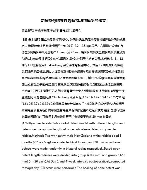
幼兔桡骨临界性骨缺损动物模型的建立芮敏;郑欣;王帆;李东亚;李成宇;曹伟;刘洪;郭开今【摘要】目的建立幼兔桡骨不同尺寸骨缺损模型,确定幼兔桡骨临界性骨缺损长度.方法选取健康3月龄雄性新西兰兔20只(2.2~2.5 kg),采用左右侧配对设计的方法在双侧桡骨中段分别制作15 mm及20 mm桡骨骨缺损模型,按骨缺损长度分为A组(15 mm)及B组(20 mm),每组各20侧.分别于术后第1天,术后第4、8、12周行CT检查,应用CT-Hedberg评分评估骨愈合情况.于术后12周处死所有实验兔,取出尺桡骨标本,通过大体观察及HE染色组织学观察分析缺损区骨愈合情况.结果术后实验兔均存活,术后第12周大体观察:A组19例(95%)桡骨断端有连续性骨痂生成,新生骨表面光滑,塑形良好;B组缺损断端髓腔封闭,缺损区由纤维组织填充.术后第12周CT图像可见A组皮质骨塑性完全,B组断端及缺损尺侧均有新骨生成,髓腔封闭.术后各时间点CT-Hedberg评分:A组(3.0±0.6,3.9±0.3,4.0±0.2)与B组(1.6±0.5,2.7±0.6,2.9±0.6)间差异有统计学意义(P<0.05).组织学结果:A组缺损已修复完全,新生骨组织内可见血管再生,B组缺损区由纤维组织填充.结论在进行幼龄兔骨缺损研究时,可选择3月龄雄性新西兰兔桡骨干构建20 mm长骨缺损.%Objective To establish a radial defect model with different lengths and determine the optimal length of bone critical-size defects in juvenile rabbits.Methods Twenty healthy male New Zealand white rabbits aged 3 months (2.2 ~2.5 kg) were selected.And 15 mm and 20 mm radial bone defects were made randomly in bilateral radius respectively.Based upon defect length,radiuses were divided into group A (15 mm) and group B (20 mm) (n =20 each).At Day 1 and 4-week intervals postoperatively,computed tomography (CT) scans were performed.The healing of bone defect wasevaluated with CT-Hedberg scores.The rabbits were sacrificed at 12 weeks postoperatively and forearms were harvested for gross observations and histological analysis of new bone formation.Results All rabbits survived after operation.Continuous bridged callus with a smooth surface were observed in 19 (95%) in group A.However,both ends were closed in group B and defect area was filled with fibrous tissue.In group A,perfectly plastic cortical bone was observed on CT images at 12 weeks postoperatively.And small amount of callus was found at both ends and ulnar side of radius in group B with closed marrow cavity.CT-Hedberg score comparison showed a significant difference between groups A and B at each timepoint postoperatively (3.0 ± 0.6,3.9 ± 0.3,4.0 ± 0.2 vs 1.6 ± 0.5,2.7 ± 0.6,2.9 ± 0.6,P < 0.05).Histological analysis showed that bone defect in group A had been repaired perfectly and neonatal revascularization was also present.In group B,the ends of defects were closed and defect area was filled with fibrous tissue.Conclusion A model of radial bone defect has been established in juvenile rabbits.And 20 mm is the most ideal length.【期刊名称】《临床小儿外科杂志》【年(卷),期】2017(016)002【总页数】5页(P189-193)【关键词】桡骨;骨/损伤;模型,动物;兔【作者】芮敏;郑欣;王帆;李东亚;李成宇;曹伟;刘洪;郭开今【作者单位】徐州医科大学附属医院骨科江苏省徐州市,221000;徐州医科大学附属医院骨科江苏省徐州市,221000;徐州医科大学附属徐州市立医院影像科江苏省徐州市,221000;徐州医科大学附属医院骨科江苏省徐州市,221000;徐州医科大学附属医院骨科江苏省徐州市,221000;徐州医科大学附属徐州市立医院影像科江苏省徐州市,221000;徐州医科大学附属徐州市立医院影像科江苏省徐州市,221000;徐州医科大学附属医院骨科江苏省徐州市,221000【正文语种】中文儿童时期骨髓炎清创、严重创伤、巨大骨肿瘤切除术后等,均易导致肢体大段骨缺损,后期可产生肢体短缩、成角畸形,造成肢体功能障碍,严重影响儿童生长发育,如何修复大段骨缺损仍是小儿骨科医生面临的重大难题[1,2]。
ACM复合rBMSC修复兔股骨内髁关节软骨缺损的价值
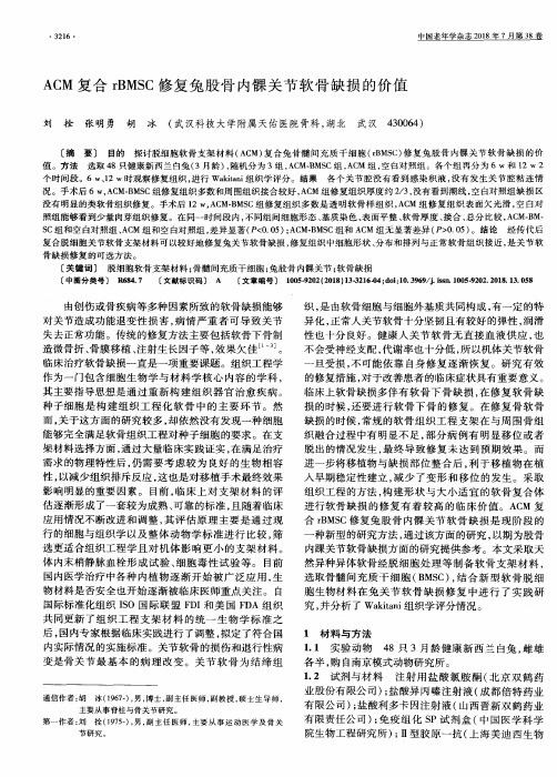
ACM 复合 rBMSC修 复 兔 股 骨 内髁关 节 软骨 缺 损 的价值
刘 拴 张 明 勇 胡 冰 (武 汉科 技大 学 附属 天佑 医院 骨科 ,湖北 武 汉 430064)
[摘 要 ] 目的 探讨脱细胞软 骨支 架材料 (ACM)复合兔 骨髓 间充 质 干细胞 (rBMSC)修 复兔股 骨 内髁关 节软 骨缺损 的价 值 。方 法 选 取 48只健康新西 兰白兔(3月龄 ),随机分 为 3组 ,ACM—BMSC组 ,ACM 组 ,空 白对照组。各个组再分 为 6 w和 12 w 2 个 时间段。6 W、12 W时观察修复组织 ,进行 Wakitani组织学评 分。结果 各 个关 节腔没有 看到感 染积 液 ,没 有发 生关节 腔粘 连情 况 。手术后 6 W,ACM—BMSC组修 复组织多数和周 围组织接合较好 ,ACM组 修复组织厚度 约 2/3,没有看到潮线 ,空 白对照组缺 损 区 没有 明显 的类软骨组织修 复。手术 后 12 W,ACM.BMSC组修 复组织 多数是 透明软 骨样 组织 ,ACM 组修 复组织 表面欠 光滑 ,空 白对 照组能 够看到少量 肉芽组织修 复。在 同一时 间段 内 ,不 同组 间细胞形态 、基质染色 、表面平整 、软骨厚度 、接合 、总分 比较 ,ACM—BM· sc组 和空 白对照组 、ACM组 和空 白对照组 ,差异显著 (P<0.05);ACM-BMSC组和 ACM组无 显著差异 (P>0.05)。结论 经传 代后 复合脱 细胞 关节软骨支架 材料可 以较好地修复兔关 节软骨缺损 ,修复组织 中细胞形状 、分布和排列与正 常软骨 组织接 近 ,是关 节软 骨 缺 损 修 复 的 可 选 方 法 。
[关键词 ] 脱细胞软骨支架 材料 ;骨髓 间充质干细胞 ;兔股骨 内髁关 节 ;软骨 缺损 [中图分类号] 11684.7 [文献标识码] A [文章 编号] 1005-9202(2018)13-3216-04;doi:10.3969/j.issm 1005-9202.2018.13.058
生物活性玻璃对家兔股骨髁骨缺损的修复实验研究
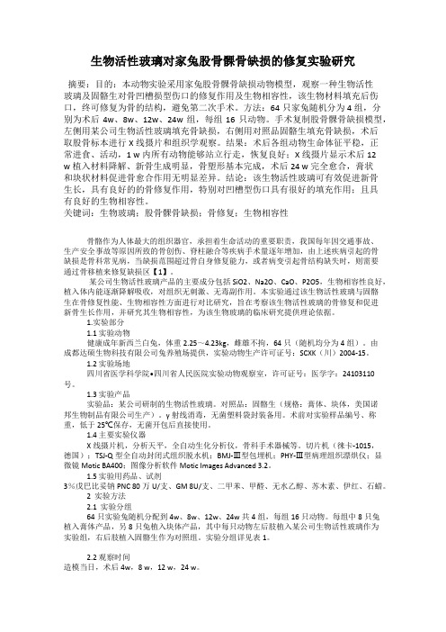
生物活性玻璃对家兔股骨髁骨缺损的修复实验研究摘要:目的:本动物实验采用家兔股骨髁骨缺损动物模型,观察一种生物活性玻璃及固骼生对骨凹槽损型伤口的修复作用及生物相容性,该生物材料填充后伤口,终可修复为骨的结构,避免第二次手术。
方法:64只家兔随机分为4组,分别为术后4w、8w、12w、24w组,每组16只动物。
手术复制股骨髁骨缺损模型,左侧用某公司生物活性玻璃填充骨缺损,右侧用对照品固骼生填充骨缺损,术后取股骨标本进行X线摄片和组织学观察。
结果:术后各组动物生命体征平稳,正常进食、活动,1 w内所有动物能够站立行走,恢复良好;X线摄片显示术后12w植入材料降解、新骨生成明显,骨塑形基本完成,术后24 w完全愈合,膏状和块状材料促进骨愈合作用无明显差异。
结论:该生物活性玻璃可有效促进新骨生长,具有良好的的骨修复作用,特别对凹槽型伤口具有很好的填充作用;且具有良好的生物相容性。
关键词:生物玻璃;股骨髁骨缺损;骨修复;生物相容性骨骼作为人体最大的组织器官,承担着生命活动的重要职责,我国每年因交通事故、生产安全事故等原因所致的骨创伤、脊柱融合等疾病手术量逐年增加,由上述疾病引起的骨缺损是骨科常见病,当缺损范围超过骨自身修复能力,或者病变引起骨结构缺失时,则需要通过骨移植来修复缺损区【1】。
某公司生物活性玻璃产品的主要成分包括SiO2、Na2O、CaO、P2O5,生物相容性良好,植入体内能逐渐降解吸收,对组织无刺激、无毒副作用。
本实验通过该生物活性玻璃与固骼生在骨修复性能、生物相容性方面进行对比研究,旨在考察该生物活性玻璃的骨修复和促进新骨生长作用,并研究其生物相容性,为该生物玻璃的临床研究提供理论依据。
1.实验部分1.1 实验动物健康成年新西兰白兔,体重2.25~4.23kg,雌雄不拘,64只(随机均分为4组)。
由成都达硕生物科技有限公司兔养殖场提供,实验动物生产许可证号:SCXK(川)2004-15。
1.2 实验场地四川省医学科学院•四川省人民医院实验动物观察室,许可证号:医学字:24103110号。
- 1、下载文档前请自行甄别文档内容的完整性,平台不提供额外的编辑、内容补充、找答案等附加服务。
- 2、"仅部分预览"的文档,不可在线预览部分如存在完整性等问题,可反馈申请退款(可完整预览的文档不适用该条件!)。
- 3、如文档侵犯您的权益,请联系客服反馈,我们会尽快为您处理(人工客服工作时间:9:00-18:30)。
《中国组织工程研究》 Chinese Journal of Tissue Engineering Research·研究原著·徐石庄,男,1986年生,江苏省连云港市人,汉族,徐州医科大学在读硕士,主治医师,主要从事骨关节疾病研究。
通讯作者:赵凤朝,博士,副教授,徐州医科大学,江苏省徐州市 221000文献标识码:B投稿日期:2019-09-09 送审日期:2019-09-10 采用日期:2019-10-19 在线日期:2020-01-04Xu Shizhuang, Master candidate, Attending physician, Department of Orthopedics, Affiliated Hospital of Xuzhou Medical University, Xuzhou 221000, Jiangsu Province, ChinaCorresponding author: Zhao Fengchao, MD, Associate professor, Department of Orthopedics, Affiliated Hospital of Xuzhou Medical University, Xuzhou 221000, Jiangsu Province, China兔股骨髁临界性骨缺损动物模型制备及临界骨缺损值徐石庄1,王 进2,潘文振1,刘 磊1,杨冠杰1,赵凤朝1 (1徐州医科大学附属医院骨科,江苏省徐州市 221000;2苏州大学附属张家港医院,江苏省苏州市 215000)DOI:10.3969/j.issn.2095-4344.2614 ORCID: 0000-0002-1257-965X(徐石庄)文章快速阅读:文题释义:临界性骨缺损:首先定义为自然状况下骨缺损不进行任何处理无法自愈的最短的骨缺损尺寸。
随后考虑到观察实验动物完整的生命周期是非常困难的,将临界性骨缺损值定义为在实验期间物种不能自行愈合的最短骨缺损尺寸。
动物模型:是在医学研究中建立的模拟人类疾病表现的动物,骨组织工程中建立临床相关的测试动物模型来研究材料的生物相容性、降解、力学性能以及与宿主组织的相互作用,是体外实验和人体临床试验之间的关键一步。
摘要背景:兔股骨远端骨缺损模型被研究者们广泛用于骨缺损替代骨组织工程材料的测试,但对于兔股骨髁圆柱形骨缺损模型的大小文献报道不一,直径分布在5-9 mm ,深度8-12 mm ,目前尚无统一的标准。
目的:建立兔股骨髁不同尺寸骨缺损模型,确定兔股骨髁临界性骨缺损尺寸。
方法:6月龄雄性新西兰白兔18只,随机分为3组,每组各6只,分别建立骨缺损模型,骨缺损直径依次为5,6,7 mm ,深度均为10 mm ,双侧手术,共计12侧。
分别于术后第1天及术后第4,8,12周行CT 扫描及三维重建,CT-Hedberg 评分评价骨缺损愈合情况;于术后12周处死新西兰白兔,取出股骨髁缺损样本,通过大体观察和苏木精-伊红染色分析缺损区愈合情况。
实验方案经徐州医科大学实验动物道德伦理委员会批准。
结果与结论:①术后所有兔均存活,术后12周大体观察示:直径5 mm 组缺损由新生骨组织充填,股骨髁塑形良好,骨缺损基本完全修复;直径6 mm 组、直径7 mm 组骨缺损区可见明显凹陷,新生骨组织较少,骨缺损未修复;②CT 图像示:术后第4,8周,直径5 mm 组缺损区逐渐减小,断端桥接;直径6 mm 、直径7 mm 组缺损区仅周边有少量新生骨长入,缺损面积较前稍减小;术后第12周可见直径5 mm 组皮质骨结构完整、连续,骨缺损基本完全修复;直径6 mm 组骨缺损部分修复;直径7 mm 组缺损未修复,仍可见明显缺损空腔存在;③CT-Hedberg 评分显示,术后各时间点直径6 mm 组评分显著低于直径5 mm 组(P < 0.05);与直径7 mm 组比较差异无显著性意义(P > 0.05);④组织学结果示:术后12周直径5 mm 组缺损区出现排列不规则的骨小梁结构,并可见大量新生骨组织填充,其他2组在骨缺损周边可见部分新生骨小梁存在,但缺损区新生骨组织填充较少;⑤结果说明,在12周的实验观察期内,在缺损深度同为10 mm 的条件下,直径>6 mm 的股骨髁缺损未能自行愈合,而直径<6 mm 的股骨髁缺损基本完全修复。
此结果符合临界骨缺损的标准,故直径6 mm 可作为兔股骨髁临界骨缺损值。
关键词:兔;股骨髁;临界性骨缺损;缺损尺寸;动物模型 中图分类号:R446;R496;R318Preparing an animal model of critical femoral defect in rabbit femoral condyle and the critical bone defect sizeXu Shizhuang 1, Wang Jin 2, Pan Wenzhen 1, Liu Lei 1, Yang Guangjie 1, Zhao Fengchao 1 (1Department of Orthopedics, Affiliated Hospital of Xuzhou Medical University, Xuzhou 221000, Jiangsu Province, China; 2Zhangjiagang Hospital of Soochow University, Suzhou 215000, Jiangsu Province, China)徐石庄,王进,潘文振,刘磊,杨冠杰,赵凤朝. 兔股骨髁临界性骨缺损动物模型制备及临界骨缺损值[J].中国组织工程研究,2020,24(20):3191-3195. DOI:10.3969/j.issn.2095-4344.2614AbstractBACKGROUND: Rabbit model of distal femoral bone defect has been widely used to test bone tissue engineering materials for bone defects. However, there is no uniform standard for the size of the cylindrical bone defect model of the rabbit femoral condyle, which ranges 5-9 mm in diameter and 8-12 mm in depth.OBJECTIVE: To establish the bone defect model of adult rabbit femoral condyle with different sizes and to determine the critical bone defect size of the femoral condyleMETHODS: Eighteen male New Zealand White rabbits aged 6 months were randomly divided into three groups according to the diameter of bone defect: 5 mm diameter group, 6 mm diameter group, and 7 mm diameter group. The defect depth was 10 mm. These rabbits underwent bilateral radial surgery, a total of 12 sides. Computed Tomography (CT) scan and three-dimensional reconstruction were performed at 1 day, 4, 8, 12 weeks after surgery. The CT-Hedberg score was used to evaluate the healing of bone defects. The rabbits were sacrificed at 12 weeks after surgery, and the femoral condyle specimens were taken out. Healing of the defect was analyzed by gross observation and hematoxylin-eosin staining. The study protocol was approved by the Animal Ethics Committee of Xuzhou Medical University.RESULTS AND CONCLUSION: All rabbits survived after surgery. The gross observation showed that the defect of 5 mm diameter group was filled with new bone tissue, the femoral condyle was well shaped, and the bone defect was completely repaired. In 6 mm and 7 mm diameter groups, depressed deformation was obviously observed in the defect area, with less new bone tissue, and the defect was was not repaired. The CT images showed that the defect area of 5 mm diameter group gradually decreased, and the broken ends of the defect were bridged. In the defect area of 6 mm and 7 mm diameter groups, only a small amount of new bone tissue was implanted, and the defect area was slightly reduced. At the 12th week after surgery, the cortical bone structure of 5 mm diameter group was intact and continuous, the femoral condyle was well shaped, and the bone defect was completely repaired. The defects of 6 mm and 7 mm diameter groups were partially or not repaired, and the defect cavity was still visible in the 7 mm diameter group. The CT-Hedberg scores of 6 mm diameter group were significantly lower than those of 5 mm diameter group at different time points (P < 0.05), and there was no significant difference in the CT-Hedberg scores between 6 mm and 7 mm diameter groups (P > 0.05). Histological results showed that there were irregular trabecular structures in the defect area of 5 mm diameter group, with a large amount of new bone tissue. In the other two groups, there were some new bone trabeculae around the bone defect, but the defect area was less filled with new bone tissue. During the 12-week observation period, the femoral condyle defect with a diameter of > 6 mm and a depth of 10 mm could not heal spontaneously, while the defect with a diameter of < 6 mm could be completely repaired, which met the criteria of critical bone defect. Therefore, the diameter of < 6 mm could be used as the critical bone defect size of rabbit femoral condyle.Key words: rabbit; femoral condyle; critical bone defect; defect size; animal model0 引言Introduction在全膝关节翻修或者复杂初次膝关节置换中经常会遇到股骨远端骨缺损的情况,如果股骨远端骨缺损得不到处理将会导致膝关节翻修和置换手术的失败[1]。