微生物染色(英文)
专业英语(微生物降解)

分类法 Taxonomy 形态学 Morphology 微生物生理学 Microbial physiology 鉴别染色-格兰氏染色 Differential stains-Gram stain 格兰氏阳性 Gram-positive 格兰氏阴性 Gram-negative
Thank you!
• • • • • • • • • • • • •
4、用什么方法检测、监测 were detected by were analyzed by was assayed polarographically(用副词) The compounds were visualized under UV light at 254 nm. Phenolic compounds were detected by spraying with Gibb’s reagent. The metabolites were analyzed by reversed-phase HPLC. Growth was monitored spectrophotometrically at 600 nm. DDT dioxygenase was assayed polarographically in a Gilson oxygraph with oxygen electrode. 2,3-Dihydroxybiphenyl-1,2-dioxygenase was assayed spectrophotometrically by measuring increase in absorbance at 432 nm. were recorded with/on+仪器厂家、型号 Mass spectra were recorded withJeol, MS-DX303 Mass spectrometer operated at 70 ev. The proton nuclear magnetic resonance (1HNMR) spectra were recorded on a Bruker AMX-300MHz spectrometer with TMS as internal standard.
微生物5 抗酸染色

原理:
由于分歧杆菌的胞壁含大量脂质,主要是分枝菌酸, 它包围在肽聚糖的外面,所以分枝杆菌一般不易着色, 可采用加温、延长着色时间或提高染料浓度等手段使其 着色。但分枝菌酸与染料结合后,就很难被酸性脱色剂 脱色。故分歧杆菌又称为抗酸杆菌,这种染色方法又成 为抗酸染色。
延长时间:5min 提高染料浓度:初染用的酸复红浓度是革兰染色酸复红浓 度的10倍 盐酸酒精:脱色能力远远强于95%酒精
原理:
齐-尼氏抗酸染色法是在加热条件下使分枝菌 酸与石炭酸复红牢固结合成复合物,用盐酸酒 精处理也不脱色。当再加碱性美兰复染后,分 枝杆菌仍然为红色,而其他细菌及背景中的物 质为兰色。
注意:不要使染液沸腾,若蒸发减少,可待玻片 稍冷却后适当补充。
(2)待玻片冷却后,用水缓慢冲洗,用吸水纸吸 干。
(3)脱色:滴加3%盐酸酒精,轻晃玻片,直至 无红色液体流下为止,一般作用1—3min*,水 洗,吸干。
(4)复染:用碱性美兰溶液复染1min,用水冲 洗后吸干。
(5)
*:不同文献报道不同,范围在30s至3min
П、漂浮集菌法。取晨痰2—3ml放入100ml三角瓶内,加1—2倍量 40g/lNaOH溶液,经103.43Pa高压灭菌20—25min(或煮30min)。 冷却后滴加汽油0.3ml,瓶口盖玻璃纸加塞,塞紧瓶口,置振荡器 或手摇振荡10min,再加蒸馏水至满瓶口而又不外溢,静置10— 15min,将已编号的洁净载玻片盖在瓶口上,静置15—20min,取 下载玻片并迅速将载玻片翻转至浸膜向上,或用接种环取瓶口液面 物涂于载玻片上,自然干燥,火焰固定后做抗酸染色或金胺“0”荧 光染色,镜检。
Giemsa染色,X1000
恙虫热立克次体, X1000
常用微生物染色方法

WOIRD格式革兰染色抗酸染色:分支杆菌属,诺卡放线菌属。
石炭酸复红染色,金永染色。
芽孢染色:用于区分细菌的芽孢和营养细胞。
结果芽孢染成绿色,营养细胞为红色。
晶体染色:观察苏云金杆菌不同变种的晶体形状大小。
结果晶体染成紫色,芽孢为透明椭圆形,仅见具有轮廓的折光体。
鞭毛染色:有赖夫生染色法、银盐染色法,西萨-基尔染色法等。
一般以西萨-基尔(Cesares-Gill)染法效果最佳。
异染颗粒染色:用于白喉棒状杆菌染色,直接涂片或改良阿伯特法染色。
墨汁荚膜染色:观察菌体有无荚膜,如新型隐球菌。
碳素墨汁。
镀银染色:螺旋体吉姆萨染色:螺旋体荧光染色魏申染色(wayson)染色:鼠疫耶尔森杆菌。
铬酸P、A、S真菌类染色。
结果:曲霉菌、念球菌属、细胞浆菌属、隐球菌属为红紫色。
曲霉菌丝周边紫红色,弹力纤维紫色,背景绿色。
Mann氏甲基蓝伊红染色法:病毒包涵体染色。
Negri氏小体红色。
Nacchiavello氏染色:病毒包涵体染色。
结果:包涵体、立克次氏体均染红色,其余组织呈蓝色。
Crocott-Gomori氏六胺银法:新型隐球菌碘染色法吉姆萨染色:衣原体乳酸酚棉蓝染色,糖原染色:真菌,前者适用于所有真菌,呈蓝色。
吉姆萨染色,瑞氏染色:鞭毛虫,滴虫,锥虫,疟原虫,弓形虫,隐孢子虫等原虫。
三色染色,碘染色:阿米巴等原虫。
荧光素吖啶橙染色:疟原虫。
金胺-酚染色:隐孢子虫。
甲苯胺蓝,四胺银染色:耶氏肺孢子虫。
专业资料整理。
微生物英文词汇

microorganism微生物;bacterium细菌;Gram stain革兰染色;coccus球菌;bacillus 杆菌;vibro弧菌;peptidoglycan肽聚糖;teichoic acid磷壁酸;lipopolysaccharide(LPS) 脂蛋白;lipid A 脂质A;L formed bacteria L型细菌;mesosome中介体;plasmid质粒formed bacteria L型细菌;mesosome中介体;plasmid质粒;capsule, 荚膜;flagellum,鞭毛;pilus, 菌毛;spore,芽胞Pyrogen,热原质;toxin,毒素;growth curve, 生长曲线Sterilization灭菌;disinfection消毒;autoclave压力蒸汽灭菌器;bacteriophage噬菌体;virulent phage毒性噬菌体;temperate phage 温和噬菌体transposable element转座元件;transposon转座子;mutation突变;transformation 转化;conjugation接合;transduction转导;lysogenic conversion溶原性转换pathogenicity 致病性;virulence毒力;exotoxin外毒素;endotoxin内毒素;normal flora正常菌群;conditioned pathogen机会致病菌;opportunistic infection机会性感染specimens标本;isolation分离;identification鉴定;artificial active immunization 人工主动免疫;artificial passive immunization人工被动免疫;vaccine疫苗;toxoid 类毒素;antitoxin抗毒素Staphlococcus葡萄球菌;Staphlococcus A protein葡萄球菌A蛋白;coagulase血浆凝固酶;enterotoxin肠毒素;Strptococcus链球菌;pyrogenic exotoxin致热外毒素;streptolysin链球菌溶素;hyaluronidase透明质酸酶E.coli大肠埃希菌;ETEC肠产毒性大肠埃希菌;EIEC肠侵袭性大肠埃希菌;EPEC肠致病性大肠埃希菌;EHEC肠出血性大肠埃希菌;LT不耐热肠毒素;Shigella 志贺菌;Salmonella沙门菌Vibrio cholerae 霍乱弧菌;Clostridium tetani 破伤风梭菌;C.perfringens 产气荚膜梭菌;C.botulinum 肉毒梭菌mycobacterium tuberculosis结核分枝杆菌;acid-faststain抗酸染色;BCG卡介苗;OT test结核菌素试验;actinomycetes放线菌mycoplasma支原体;chlamydia衣原体;inclusion body网状体;rickettsia立克次体;spirochete螺旋体;fungus真菌;hypha菌丝;spore孢子capsid衣壳;envelope包膜;nucleocapsid核衣壳;replicative cycle复制周期;adsorption吸附;Penetration)穿入;uncoating脱壳;biosynthesis生物合成;defective viruse缺陷病毒;abortive infection顿挫感染cytopathic effect,CPE细胞病变效应;slow virus infection慢发病毒感染;persistent infection持续性病毒感染;interferon,IFN干扰素influenza virus流感病毒;hemagglutinin,HA血凝素;neuraminidase,NA 神经氨酸;antigenic drift抗原性漂移;antigenic shift抗原性转变;hepatitis B virus乙肝病毒;Dane颗粒;HBsAg乙肝表面抗原;HBIG高效价乙肝免疫球蛋白HIV人类免疫缺陷病毒;AIDS艾滋病;prion朊粒。
Negative staining 阴性(负)染色法

• 實驗注意事項
– 進行細菌的染色時,樣本於玻片上之塗抹務求 均勻,以免影響染色的效果。 – 進行陰性染色法時,染色前皆必須先確定玻片 樣本呈現乾燥之狀態方可進行染色,否則易影 響實驗之最終結果。 – 進行陰性染色法時,染劑固定步驟不宜以熱固 定的方式進行,需以自然風乾的方法操作。
• 實驗結果
– 請畫出以陰性染色法觀察之菌體。
• 材料與儀器
– Bacteria
• 大腸桿菌(Escherichia coli) • 金黃色葡萄球菌(Staphylococcus aureus) • 枯草芽孢桿菌(Bacillus subtilis)
– Staining agent
• Nigrosine
– An acidic stain which can not be stained onto bacterial cell membrane which contain negative-charged particles.
• 染色:將製備完成之玻片樣本置於染色架上,加上一 滴染劑,在加上一滴水,以接種環於塗抹菌體於染色 液中,靜置約3~5分鐘,使菌體上色。 • 可用另一張載玻片,輕放在染液上,將其染液拉開。 • 於無菌操作台內風乾。
• 乾燥:將玻片上多餘的水分輕輕甩乾,並利用濾紙將 其吸乾,接著置於無菌操作台內使其自然風乾(此時 不進行火烤加熱風乾,而以自然風乾為主)後備用。 • 鏡檢:先以低倍數光學顯微鏡找出菌體,然後再逐步 換成高倍數接物鏡最終以油鏡進行菌體的觀察並繪圖 記錄之。
• 討論
– 比較不同菌種間的異同處
– 設備儀器及其他用具
• • • • • • • • • 光學顯微鏡 酒精燈 載玻片 接種環 蒸餾水 染色架 濾紙 75%酒精 乳頭滴管
微生物英文词汇

微生物英文词汇active immunity(主动免疫);active transport(主动运输);Alcohol fermentation(乙醇发酵);aerobe(好氧微生物);aflatoxin(黄曲霉毒素);AIDS(爱滋病);Ames test(艾姆氏实验);anabolism(合成代谢);anaerobe(厌氧微生物);antibiotic(抗生素);antibody(抗体);antigen(抗原);antigenic determinant(抗原决定基);antimetabolite(抗代谢物);antiseptic(防腐剂);antiserum(抗血清);antitoxin(抗毒素);arthrospore(节孢子);ascospore(子囊);asepsis(无菌);autoantibody(自身抗体);autoantigen(自身抗原);autoimmune disease(自身免疫疾病);bacteriophage(噬菌体);bacteriostatic(抑菌);binary fission(二分裂);broad spectrum(广谱);Capsid(衣壳);capsomer(衣壳粒):capsule(荚膜):Catabolism(分解代谢):cell-mediated immune(细胞介导免疫):chemoautotroph(化能自养菌):chemotaxis(趋化性):Chemotherapy(化学治疗剂):chitin(几丁质):complement(补体):Conldia(分生孢子):Conjugation(接合):Colony(菌落):Contaminant(污染物):Culture(培养物):differential medium(鉴别培养基):differential stain(鉴别染色):Disinfection(消毒):ELISA(酶联免疫):endospore(芽孢):endotoxin(内毒素):enriched medium(加富培养基):enveloped virus(包膜病毒):essential nutrient(必须营养):eucaryotic cell(真核细胞):Exotoxin(外毒素):Facultative(兼性的):Fermentation(发酵):Flagellum(鞭毛)Genotype(表型):Glycolysis(糖酵解):Gram stain(革兰氏染色):Granulocyte(粒细胞):growth factor(生长因子)Halophlle(嗜盐菌):H antigen(H-抗原):helper T cell(辅助T-细胞):Heterotroph(异养菌):Immunity(免疫):immunogen(免疫原):immune system(免疫系统):immunoglobulin(免疫球蛋白):Inclusion(内含物):Infection(感染):infectious disease(感染性疾病):Inflammation(发炎):Inoculation(接种):Interferon(干扰素):Isolation(分离):Latency(潜伏):L form(L-型菌):Lipopolysaccharide(脂多糖,LPS):Lysis(溶解):lysosome (溶酶体):病毒学virology噬菌体学bacteriophagology细菌学bacteriology鉴定细菌学determinative bacteriology系统细菌学systematic bacteriology真菌学mycology原生生物学protistology原生动物学protozoology普通微生物学general microbilogy微生物分类学microbial taxonomy微生物生理学microbial physiology微生物生物化学microbial biochemistry微生物遗传学microbial genetics微生物生态学microbial ecology古微生物学paleomicrobiology土壤微生物学soil microbiology水生微生物学aquatic microbiology海洋微生物学marine microbiology悉生生物学gnotobiology医学微生物学medical microbiology兽医微生物学veterinary microbiology农业微生物学agricultural microbiology工业微生物学industrial microbiology石油微生物学petroleum microbiology食品微生物学food microbiology乳品微生物学diary microbiology瘤胃微生物学rumen microbiology诊断微生物学diagnostic microbiology病原学etiology国际微生物学会联合会International Union of Microbiological Societies, IUMS中国微生物学会Chinese Society for Microbiology, CSM世界培养物保藏协会World Federation for Culture Collection, WFCC中国微生物菌种保藏管理委员会China Committee for Culture Collection of Microorganisms,CCCCM美国模式培养物保藏所American Type Culture Collection, A TCC自然发生说,无生源说spontaneous generation, abiogenesis原界urkingdom始祖生物progenote古始生物界archetista古细菌archaebacteria原生生物protista原生动物protozoan原生植物protophyte真核生物eukaryote原核生物prokaryote裂殖植物schizophyte微生物microorganism数值分类法numerical taxonomy模式目type order模式科type family模式属type genus模式种type species模式株type strain真菌fungi捕食真菌predacious fungi虫道真菌ambrosia fungi地下真菌hypogeal fungi虫生真菌entomogenous fungi菌根真菌mycorrhizal fungi木腐菌wood-decay fungi霉菌mold, mould半知菌imperfect fungi子囊菌ascomycetes粘菌slime mold, slime mould壶菌chytrid卵菌oomycetes接合菌zygomycetes担子菌basidiomycetes核菌pyrenomycetes盘菌cup fungi块菌truffles锈菌rust fungi蘑菇mushrooms毒蘑菇poisonous mushroom酵母菌yeast无孢子酵母菌asporogenous yeasts 有孢子酵母菌sporogenous yeasts 黑粉菌smut fungi双态性真菌dimorphic fungi毛外癣菌ectothrix毛内癣菌endothrix完全真菌perfect fungi黑粉病smut disease锈病rust disease菌丝hypha菌髓trama假菌丝体pseudomycelium气生菌丝体aerial mycelium基内菌丝体substrate mycelium球拍状菌丝体racquet mycelium 结节状菌丝nodular mycelium梳状菌丝pectinafe mycelium螺旋菌丝spiral mycelium匍匐菌丝stolon次生菌丝体secondary mycelium有隔菌丝septate hypha无隔菌丝nonseptate hypha生殖菌丝体reproductive mycelium 营养菌丝体vegetative mycelium不育菌丝体sterile mycelium菌丝体mycelium黄癣菌丝favic chandelier mycelium 产囊丝ascogenous hypha产囊体ascogonium原植体thallus粘菌体aethalium合胞体syncytium虫菌体hyphal body盾状体clypeus子实体fruiting body产孢体gleba子实层体hymenophore子实层hymenium子实下层subhymenium菌丝层subiculum菌丝段hyphal fragment菌丝束coremium菌丝索funiculus菌核sclerotium器菌核pycnosclerotium菌环annulus菌裙indusium菌盖pileus顶体apicle藏卵器oogonium雄器antheridium[锈菌]性孢子器pycnium锈子器aecium精子器spermogonium囊状体cystidium粉孢子梗oidiophore小梗sterigma接合孢子柄zygosporophore孢囊柄sporangiophore配囊柄suspensor孢子梗sporophore分生孢子梗conidiophore雄器柄androphore帚状枝penicillus瓶梗phialide梗基metulae芽孔germ pore芽管germ tube芽缝germ slit孢丝capillitium周丝periphysis类周丝periphysoid侧丝paraphysis拟侧丝pseudoparaphysis类侧丝paraphysoid[孢子]外壁exosporium外生菌根ectomycorrhiza内生菌根endomycorrhiza内外生菌根ectendomycorrhiza泡囊丛枝菌根vesicular-arbuscular mycorrhiza 刺突spike弹丝elater刚毛seta微体microbody泡囊vesicle隔膜septum假隔膜pseudoseptum分生孢子盘acervulus分生孢子座sporodochium精子团spermatium囊基膜hypothallus囊层基hypothecium囊层被epithecium囊间丝hamathecium囊托apophysis囊领collarette囊轴columella孔口ostiole菌托volva孢子角cirrus孢子球spore ball孢子印spore print聚簇cluster[菌丝]融合anastomosis[孢子]切落abjunction[孢子]缢断abstriction多态[现象] polymorphism缢缩[作用] constriction粉孢子oidium孢子spore掷孢子ballistospore厚壁孢子chlamydospore环痕孢子annellospore节孢子arthrospore卷旋孢子helicospore腊肠形孢子allantospore孔出孢子porospore星形孢子staurospore线形孢子scolecospore砖格孢子dictyospore侧生孢子aleuriospore芽生孢子blastospore瓶梗孢子phialospore无梗孢子thallospore分生孢子conidium大分生孢子macroconidium小分生孢子microconidium节分生孢子arthroconidium芽分生孢子blastoconidium器孢子pycnidiospore无隔孢子amerospore双胞孢子didymospore多隔孢子phragmospore休眠孢子hypnospore顶生孢子acrospore顶生厚壁孢子fuseau内分生孢子endoconidium担孢子basidiospore双孢担孢子dispore同形孢子isospore柄生孢子stylospore[锈菌]性孢子pycniospore产雄器孢子androspore锈孢子aeciospore夏孢子urediniospore, aeciospore 冬孢子teliospore四分孢子tetraspore粘孢子myxospore多核孢子coenospore孢囊孢子sporangiospore子囊孢子ascospore多核细胞coenocyte分生孢子果conidiocarp分生孢子器pycnidium孢[子]囊sporangium柱孢子囊merosporangium四分孢子囊tetrasporangium原孢子囊prosporangium多核孢子囊coenosporangium 休眠孢子囊hypnosporangium 子囊ascus接合孢子zygospore拟接合孢子azygospore原囊壁子囊prototunicate ascus 单囊壁子囊unitunicate ascus 双囊壁子囊bitunicate ascus子囊果ascocarp子囊壳perithecium闭囊壳cleistothecium闭囊果cleistocarp盘状子囊果discocarp孢囊果sporangiocarp[接]合子zygote单性合子azygote多核合子coenozygote异形合子heterozygote合子核zygotonucleus游动合子planozygote担子basidium半担子hemibasidium隔担子heterobasidium无隔担子holobasidium有隔担子phragmobasidium内生担子endobasidium原担子protobasidium上担子epibasidium下担子hypobasidium同担子homobasidium担子果basidiocarp担子体basidiophore配子gamete原配子progamete雄配子androgamete雄核发育androgenesis同形配子isogamete异形配子heterogamete游动配子zoogamete多核配子coenogamete配子囊gametangium配子母细胞gametocyte同形配子囊isogametangium原配子囊progametangium小孢子囊sporangiole微包囊microcyst足细胞foot cell脚胞foot cell固着器holdfast附着枝hyphopodium吸盘sucker锁状细胞clamp cell锁状联合clamp connection偶核细胞zeugite卵球oosphere卵质ooplasm孢原质sporoplasm卵配子oogamete卵孢子oospore球状胞sphaerocyst子囊腔locule子囊盘apothecium子囊座ascostroma缝裂壳hysterothecium下子座hypostroma包被peridium子座stroma壳心centrum拟包被pseudoperidium无融合生殖apomixis同宗配合homothallism准性生殖parasexuality异宗配合heterothallism同配生殖isogamy异配生殖heterogamy无配生殖apogamy配囊交配gametangial copulation 交配型mating type全型holomorph夏孢子期uredostage冬孢子堆teleutosorus, telium夏孢子堆uredinium子囊孢子形成ascosporulation孢子形成sporulation细菌bacteria薄壁[细]菌类gracilicutes硬壁[细]菌类fermicutes疵壁[细]菌类mendosicutes无壁[细]菌类tenericutes柔膜细菌mollicutes真细菌eubacteria暗细菌scotobacteria无氧光细菌anoxyphotobacteria生氧光细菌oxyphotobacteria放线菌actinomycetes螺[旋]菌spirilla粘细菌slime bacteria鞘细菌sheathed bacteria柄细菌caulobacteria弧菌vibrio根瘤细菌root nodule bacteria硫酸盐还原菌sulfate reducting bacteria硫细菌sulfur bacteria铁细菌iron bacteria紫色无硫细菌purple nonsulfur bacteria产甲烷菌methanogen硝化细菌nitrobacteria反硝化细菌denitrifying bacteria固氮细菌nitrogen fixing bacteria甲基营养菌methylotrophic bacteria产乙酸菌acetogen同型[产]乙酸细菌homoacetogenic bacteria光合作用细菌photosynthetic bacteria产氢产乙酸细菌hydrogen-producing acetogenic bacteria 同型发酵乳酸菌homofermentative lactic bacteria异型发酵乳酸菌heterofermentative lactic bacteria产氢菌hydrogenogens产气菌aerogen不产气菌anaerogen发光细菌luminous bacteria产色细菌chromogenic bacteria化能异养菌chemoheterotrophic bacteria化能自养菌chemoautotrophic bacteria光能异养菌photoheterotrophic bacteria光能自养菌photoautotrophic bacteria化能有机营养菌chemoorganotrophic bacteria 化能无机营养菌chemolithotrophic bacteria 光能有机营养菌photoorganotrophic bacteria 光能无机营养菌photolithotrophic bacteria有机营养菌organotrophic bacteria无机营养菌lithotrophic bacteria贫[营]养细菌oligotrophic bacteria一氧化碳营养菌carboxydotrophic bacteria自养菌autotrophic bacteria异养菌heterotrophic bacteria光养菌phototrophic bacteria需氧菌aerobe微需氧菌microaerobe耐氧菌aerotorelant bacteria厌氧菌anaerobe兼性厌氧菌facultative anaerobe专性厌氧菌obligate anaerobe溶原性细菌lysogenic bacteria腐生菌saprophytic bacteria苛求菌fastidious microorganism极端细菌extreme bacteria嗜压菌barophilic bacteria嗜盐菌halophilic bacteria嗜铁菌siderophilic bacteria嗜高渗细菌osmophilic bacteria微嗜氮菌oligonitrophilic bacteria嗜冷[细]菌psychrophilic bacteria嗜酸菌acidophilic bacteria嗜硫菌thiophilic bacteria中温菌mesophilic bacteria耐热细菌thermophilric bacteria氢营养菌hydrogenotrophic bacteria肠道细菌intestinal bacteria类菌体bacteroid细菌小体bacteriosome微生子gonidium蓝细菌cyanobacteria[蓝细菌]连锁体hormogonium类囊体thylakoid藻胆蛋白体phycobilisome静息孢子akinete滑行gliding异形[囊]胞heterocyst化学型chemotype化学变型chemovar血清型serotype血清变型serovar致病型pathotype致病变型pathovar生物型biotype生物变型biovar形态型morphotype形态变型morphovar革兰氏阳性菌Gram-positive bacteria 革兰氏阴性菌Gram-negative bacteria 球菌coccus双球菌diplococcus四联球菌tetrads八叠球菌sarcina球杆菌coccobacillus杆菌rod双杆菌diplobacillus棒状菌corynebacteria[细菌]毛状体trichome单鞭毛菌monotricha周[鞭]毛菌peritricha丛[鞭]毛菌lophotricha两端单[鞭]毛菌amphitrichate单端丛[鞭]毛菌cephalotricha滑行细菌gliding bacteria细菌L-型L-form of bacterium菌落colony酵母型菌落yeast type colony类酵母型菌落yeast like colony次生菌落secondary colony粗糙型菌落rough colony光滑型菌落smooth colony丝状型菌落filamentous type colony 子菌落daughter colony深层菌落deep colony粘液型菌落mucoid colony巨大菌落giant colony侏儒型菌落dwarf colony菌苔lawn菌胶团zoogloea菌膜pellicle[菌]醭mycoderm, pellicle群游现象swarming菌柄stipe[菌体]附器appendage鞭毛flagellum周质鞭毛periplasmic flagella轴丝axial filament菌毛pilus性丝sex pilus外生孢子exospore内生孢子endospore芽孢spore芽孢形成sporulation终端芽孢terminal spore近端芽孢subterminal spore中生芽孢central spore前芽孢forespore[芽孢]皮层cortex芽孢外膜exitine芽孢内膜intine外壁exine伴胞晶体parasporal crystal菌蜕ghost鞘sheath荚膜capsule粘液层slime layer微荚膜microcapsule壁膜间隙periplasmic space原生质体protoplast原生质球spheroplast气泡gas vacuole甲烷粒体methanochondria间体mesosome载色体chromatophore鞭毛基体flagellar basal body异染质volutin异染粒matachromatic granules致死颗粒killer particle紫膜purple membrane噬菌体bacteriophage无囊盖类inoperculatae超显微微生物ultramicroscopic organism 真菌噬菌体mycophage噬藻体phycophage烈性噬菌体virulent phage温和噬菌体temperate phage前原噬菌体preprophage原噬菌体prophage隐性前噬菌体cryptic prophage营养期噬菌体vegetative phage载体噬菌体carrier phageλ噬菌体lambda particles phage [可]诱导噬菌体inducible phage同源免疫噬菌体homoimmune phage 噬菌体分型bacteriophage typing噬菌体型phagetype噬菌体变型phagevar噬斑plaque[噬菌体]聚合头部polyhead[噬菌体]聚合尾鞘polysheath[噬菌体]伞毛fimbrium[噬菌体]颈须whisker[噬菌体]先导蛋白pilot protein[噬菌体]尾丝抗原fiber antigen[噬菌体]顶体apex[噬菌体]基片插孔base-plate hub [噬菌体]基片丝base-plate fibril [噬菌体]基片楔突base-plate wedge [噬菌体]串联体concatemer[噬菌体]颈部collar[噬菌体]顶部壳粒apical capsomere [噬菌体]尾丝tail fiber[噬菌体]畸形体monster[噬菌体]颈圈connector[噬菌体]髓部core[噬菌体]头部head[噬菌体]尾部tail[噬菌体]尾管tail tube[噬菌体]尾鞘tail sheath类病毒viroid病毒virus真病毒euvirus亚病毒subvirus原病毒provirus拟病毒virusoid卫星病毒satellite virus假型病毒pseudotype virus慢病毒slow virus辅助病毒helper virus过客病毒passenger virus多分体病毒multicomponent virus昆虫痘病毒entomopox virus, EPV颗粒体症病毒granulosis virus, GV多角体病毒polyhedrosis virus核型多角体病毒nuclear polyhedrosis virus, NPV质型多角体病毒cytoplasmic polyhedrosis virus,CPV 多粒包埋型病毒multiple embedded virus单粒包埋型病毒singly embedded virus伴随病毒associated virus浓核病毒densovirus,DNV内源病毒endogenous virus潜伏病毒latent virus肠道病毒enterovirus艾柯病毒ECHO virus虫媒病毒arbovirus腺病毒adenovirus腺伴随病毒adeno associated virus真菌病毒mycovirus肿瘤病毒oncovirus逆[转]录病毒retro virus坏死病毒necrosis virus虹彩病毒irido virus泛嗜性病毒pantropic virus毒株strain原[生小]体elementary body包含体inclusion body顾氏小体Guarnieri's bodies内氏小体Negri's body病毒[粒]体virion裸露病毒[粒]体naked virion假病毒体pseudovirion立体对称cubical symmetry二十面体对称icosahedral symmetry螺旋对称helical symmetry[病毒]五邻体pentomer,pentons[病毒]六邻体hexonmer,hexons复合对称complex symmetry包膜突起peplomerbody包膜envelope, peplos蛋白质包膜protein envelope[病毒]包膜抗原envelope antigen[病毒]壳体capsid[病毒]壳粒capsomer, capsomere二十面[体]壳体icosahedron capsid 核心core核壳nucleocapsid病毒原质体viroplasma病毒束virus bundle多角体polyhedron多角体蛋白polyhedrin颗粒体granule颗粒体蛋白granulin类核nucleoid内含颗粒inclusion granuleX体X-body[病毒]早期蛋白early protein[病毒]晚期蛋白late protein负链negative strand正链positive strand复制子replicon病毒发生基质virogenic stroma衣原体chlamydia[衣原体]始体initial body立克次氏体rickettsia假肽聚糖pseudopeptidoglycan肽聚糖peptidoglycan磷壁酸teichoic acid胞壁酸muramic acid2,6-吡啶二羧酸dipicolinic acid, DPA 脂多糖类lipopolysaccharides多糖包被glycocalyx鞭毛蛋白flagellin菌毛蛋白pilin杀白细胞素leucocidin豆血红蛋白leghaemoglobin藻胆蛋白phycobiliprotein藻青蛋白phycocyanin藻红蛋白phycoerythrin藻青素cyanophycin藻蓝素algocyan, leucocyan藻胆素phycobilin藻红[胆]素phycoerythrobilin藻蓝胆素phycocyanobilin藻青素颗粒cyanophycin granule别藻蓝素allophycocyanin类葫萝卜素carotenoids细菌淀粉粒granulose聚β羟基丁酸盐poly-β-hydroxy butyrate葡萄球菌A蛋白staphylococcal protein A, SPA 纯化蛋白衍生物purified protein derivative, PPD [葡萄球菌]凝固酶staphylocoagulaseβ[细胞]溶素β-lysinα淀粉酶α-amylase通透酶permease胞内酶intracellular enzyme胞外酶extracellular enzyme果胶酶pectinase逆[转]录酶reverse transcriptase凝固酶coagulase受体破坏酶receptor destroying enzyme, RDE透明质酸酶hyaluronidase纤维素酶cellulase链道酶streptodornase,SD链激酶streptokinase,SK神经氨酸酶neuraminidase青霉素酶penicillinase溶菌酶lysozyme[细菌]紫膜质bacteriorhodopsin菌紫素bacteriopurpurin[细]菌[叶]绿素bacteriochlorophyll自溶素autolysin亲菌素bacteriotropin攻击素aggressin抑殖素ablastin粘附素adhesin菌红素bacterioerythrin灵菌毒素prodigiosus toxin细菌素bacteriocin麻风菌素lepromin葡萄球菌素staphylococcin伞菌氨酸agarfitine苏云金菌素thuricin肠球菌素enterococcin布氏菌素brucellin大肠菌素colicin, colicine丁香假单胞菌素syringacin黄色粘球菌素xanthacin链球菌素streptocin流产菌素abortin绿脓[菌]素pyocyanin红假单胞菌素rhodopseudomonacin 绿脓菌荧光素pyofluorescein白喉毒素diphtheria toxin杯伞素clitocybine白细胞溶素leucolysin表皮溶解毒素epidermolytic toxin 产气荚膜梭菌素perfringocin肠毒素enterotoxin毒蝇碱muscarine肺炎球菌毒素pneumotoxin鬼笔[毒]环肽phalloidin根霉蝶呤rhizopterin肺炎[链]球菌溶血素pneumolysin 黑粉菌酸ustilagic acid分枝菌酸mycolic acid齿孔酸eburicoic acid根霉促进素rhizopin蘑菇素agaricin蘑菇酸agaricinic acid红斑毒素erythrogenic toxin黄曲霉毒素aflatoxin菌丝酰胺mycelianamide绿脓杆菌溶血素pyocyanolysin葡萄球菌溶血毒素staphylolysin真菌毒素mycotoxin曲霉毒素aspertoxin赭曲毒素ochratoxin曲酸kojic acid破伤风[菌]痉挛毒素tetanospasmin 溶葡萄球菌素lysostaphin破伤风[菌]溶血素tetanolysin溶纤维蛋白溶酶fibrinolysin溶血素hemolysin鼠疫菌素pesticin神经毒素neurotoxin杀[细]菌素bactericidin外毒素exotoxin内毒素endotoxin细菌毒素bacteriotoxin血凝素hemagglutinin杂色曲霉素A versicolorin A柄曲霉素sterigmatocystin毒植物素phytotoxin真菌醇mykol链球菌溶血素streptolysin剥脱性毒素exfoliative toxin细菌荧光素bacteriofluorescein[放线菌]土臭味素geosmins土壤杆菌素agrobacteriocin产甲烷[作用] methanogenesis生物转化bioconversion生长因子growth factor420 因子factor 420V 因子V factorX 因子X factormixed culture(混合培养):monoclonal antibody(单克隆抗体):Monocyte(单核细胞):Mutagen(诱变剂):Mutation(突变)Mycelium(菌丝体):narrow spectrum(窄谱):negative stain(负染色):nitrogen fixation(固氮):Nucleocapsid(核衣壳):Nucleoid(拟核):Nutrient(营养物质):Obligate(专性的):Parasite(寄生):Pasteurization(巴斯德消毒):Pathogen(病原体):Saprophytes(腐生型)Pathogenidty(致病性):Pathology(病原学):passive transport(被动扩散);Penicillins(青霉素):Peptidoglycan(肽聚糖):Plasmids(质粒)periplasmic space(周质空间):Phage(噬菌体):Phenotype(表型):Photoautotroph(光能自养菌):Pilus(性丝);prophage(前噬菌体):Protoplast(原生质体):Pseudohypha(假菌丝):Psychrophile(嗜冷菌):respiratory chain(呼吸链):reverse transcriptase(逆转录酶):SCP(单细胞蛋白):selective media(选择培养基):Serotyping(血清型):sexual reproduction(有性繁殖)Spheroplast(球形体):spike(刺突):Spirillum(螺菌):Spirochete(螺旋体):Sporangium(孢囊):Sterilization(灭菌):A Strain(菌株):subcellular vaccine(亚单位疫苗):superoxide ion(超氧离子):suppressor T cell(抑制T细胞):temperate phage(温和噬菌体):thermal death point(致死温度):thermal death time( 热致死时间):Therrnophlle(嗜热菌):Toxoid(类毒素):Transduction(转导):Transformation(转化):Transposon(转座):V accine(免疫法):V irold(类病毒):Zygospore(接合孢子)。
临床微生物学检验理论课:02细菌鉴定试验-涂片染色

涂片抗酸染色结果观察
镜检时,油镜下仔细查遍整个涂片或至少100个视野; 抗酸性菌呈红色,其它细菌及细胞呈蓝色; TB为细长,着色不均匀,可2个以上的菌形成V、Y、T
等或平行排列等。
涂片抗酸染色结果报告
结果报告,1984年国内统一暂行规定报告方式: 未找到抗酸菌-; 找到抗酸杆菌1或2个,全视野发现1或2个; 找到抗酸杆菌+,全视野发现3-9个; 找到抗酸杆菌++,全视野发现10-99个; 找到抗酸杆菌+++,每视野发现1-9个: 找到抗酸杆菌++++,每视野发现10个以上。
(二)蛋白质和氨基酸的代谢试验
吲哚(靛基质或蛋白胨水)试验: 阳性:色氨酸酶,分解蛋白胨水中的色氨酸生成吲哚,
加入指示剂形成红色界面; 阴性:加入指示剂不变色; 区分普通变形杆菌(+),奇异变形杆菌(-)。
尿素分解试验: 阳性:尿素分解酶分解尿素产生大量的氨,使培养基
呈碱性,加入指示剂为红色; 阴性:加入指示剂不变色。
4、墨汁染色查隐球菌
新生隐球菌广泛分布于自然界,也可存在于人体体表、 口腔及肠道中;
经呼吸道侵入人体,由肺经血行播散时,可侵犯所有脏 器组织,主要侵犯肺、脑及脑膜;
好发于细胞免疫功能低下者,如AIDS、恶性肿瘤、糖尿 病、器官移植及大剂量使用糖皮质激素者;
临床上怀疑脑膜炎时,常抽取脑脊液检查细菌真菌、抗 酸菌、隐球菌。
3、涂片抗酸染色:检查抗酸杆菌
据世界卫生组织估计,全球每年增加结核病人约800万 1000万,死亡病人约300万;
全人类的三分之一即17亿人曾感染过结核杆菌; 结核病人主要见于发展中国家,尤其经济落后地区; 我国是结核病高发国家,广东地区发病率较高。
医学微生物学英文
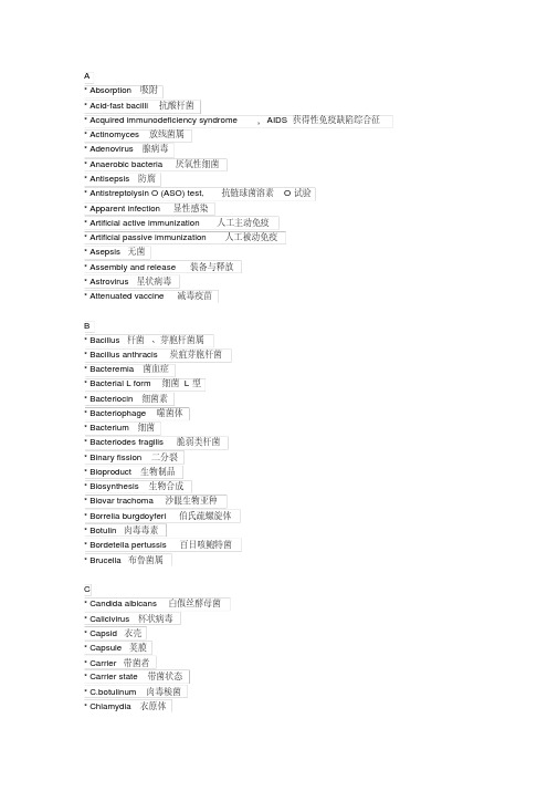
A* Absorption 吸附* Acid-fast bacilli 抗酸杆菌* Acquired immunodeficiency syndrome,AIDS 获得性免疫缺陷综合征* Actinomyces 放线菌属* Adenovirus 腺病毒* Anaerobic bacteria 厌氧性细菌* Antisepsis 防腐* Antistreptolysin O (ASO) test, 抗链球菌溶素O试验* Apparent infection 显性感染* Artificial active immunization 人工主动免疫* Artificial passive immunization 人工被动免疫* Asepsis 无菌* Assembly and release 装备与释放* Astrovirus 星状病毒* Attenuated vaccine 减毒疫苗B* Bacillus 杆菌、芽胞杆菌属* Bacillus anthracis 炭疽芽胞杆菌* Bacteremia 菌血症* Bacterial L form 细菌L型* Bacteriocin 细菌素* Bacteriophage 噬菌体* Bacterium 细菌* Bacteriodes fragilis 脆弱类杆菌* Binary fission 二分裂* Bioproduct 生物制品* Biosynthesis 生物合成* Biovar trachoma 沙眼生物亚种* Borrelia burgdoyferi 伯氏疏螺旋体* Botulin 肉毒毒素* Bordetella pertussis 百日咳鲍特菌* Brucella 布鲁菌属C* Candida albicans 白假丝酵母菌* Calicivirus 杯状病毒* Capsid 衣壳* Capsule 荚膜* Carrier 带菌者* Carrier state 带菌状态* C.botulinum 肉毒梭菌* Chlamydia 衣原体—* Chronic infection 慢性感染* Classical biotype 古典生物型* Clostridium 梭菌属* Coagulase 凝固酶* Coccus 球菌* Colony 菌落* Conditional pathogen 条件致病菌* Conjugation 接合* Corynebacterium diphtheriae 白喉棒状杆菌* Coxsackievirus 柯萨奇病毒* Coxiella burnetii 贝纳柯克斯体* C.perfringens 产气荚膜梭菌* C.pneumoniae 肺炎衣原体* Cryptococcus neoformans 新生隐球菌* C.tetani 破伤风梭菌* C.trachomatis 沙眼衣原体* Culture medium 培养基* Cytomegalovirus,CMV 巨细胞病毒* Cytopathic effect,CPE 细胞病变效应D* Darkfeild microscope 暗视野显微镜* Defective virus 缺陷病毒* Dengue virus 登革病毒* Dermatophytes 皮肤癣菌* Diplococcus 双球菌* Disinfectant 消毒剂* Disinfection 消毒* Dysbacteriosis 菌群失调* Dysentery bacterium 痢疾杆菌E* Eclipse period 隐蔽期* Ehrlichia 埃立克体属* El tor biotype Eltor 生物型* Encephalitis B virus 乙型脑炎病毒* Endogenous infection 内源性感染* Endotoxemia 内毒素血症* Endotoxin 内毒素* ECHO , Enteric cytopathogenic human orphan virus ECHO病毒* EAggEC, Enteroaggregative E.coli 肠凝集性大肠埃希菌* EHEC, Enterohemorrhagic E.coli 肠出血性大肠埃希菌* EIEC, Enteroinvasive E.coli 肠侵袭性大肠埃希菌* EPEC, Enteropathogenic E.coli 肠致病性大肠埃希菌* ETEC, Enterotoxigenic E.coli 肠产毒性大肠埃希菌* Enterotoxin 肠毒素* Enterovirus 肠道病毒* Envelope 包膜* Epstein-Barr virus,EBV EB病毒* Escherichia coli 大肠埃希菌* Exogenous infection 外源性感染* Exotoxin 外毒素* Extracellular bacteria 胞外菌F* Facultative anaerobe 兼性厌氧菌* Fertility factor,F factor 致育因子* Filtration 滤过除菌法* Fimbriae 菌毛* Flagellum 鞭毛* Fungus 真菌G* Generalized infection 全身感染* Gene transfer 基因转移* Genus 菌属* Gonococcus 淋球菌* Gram stain 革兰染色* Growth curve 生长曲线* Growth factor 生长因子H* Haemophilus influenzae 流感嗜血杆菌* Hantavirus 汉坦病毒* Helicobacter pylori 幽门螺杆菌* Helical symmetry 螺旋对称* Hemagglutinin ,HA 血凝素* α-hemolytic streptococcus 甲型(α)溶血链球菌* β-hemolytic steptococcus 乙性(β)溶血性链菌* HAV, Hepatitis A virus 甲型肝炎病毒* HBV, Hepatitis B virus 乙型肝炎病毒* HCV, Hepatitis C virus 丙型肝炎病毒* HDV, Hepatitis D virus 丁型肝炎病毒* HEV, hepatitis E virus 戊型肝炎病毒* HGV, hepatitis G virus 庚型肝炎病毒* Horizontal transmission 水平传播* Herpes simplex virus,HSV 单纯疱疹病毒* Human herpes viruses,HHV 人疱疹病毒* Human immunodeficiency viru ,HIV 人类免疫缺陷病毒* Human papillomavirus,HPV 人乳头瘤病毒属* Hyaluronidase 透明质酸酶* Hypha 菌丝I* Icosahedral symmetry 二十面体对称* Inactivated vaccine 灭活疫苗* Inactivation 灭活* Inapparent infection 隐性感染* Inclusion bodies 包涵体* Infection immunity 传染免疫* Influenza virus 流行性感冒病毒* Innate immunity 天然免疫* Integration 整合* Interferon 干扰素* Invasiveness 侵袭L* Latent infection 潜伏感染* Legionella 军团菌属* Leptospira 钩端螺旋体* Lipid A 脂质A* Lipopolysaccharide, LPS 脂多糖* Lyme disease 莱姆病* Lysogenic phage 溶原性噬菌体* Lysogenic bacterium 溶原性细菌* Lysogenic conversion 溶原性转换* Lysozyme 溶菌酶M* Measles virus 麻疹病毒* Medical microbiology 医学微生物学* Medical virology 医学病毒学* Meningococcus 脑膜炎球菌* Metachromatic granule 异染颗粒* Microaerophilic bacterium 微需氧菌* Microbiology 微生物学* Microorganism 微生物* M.leprae 麻风分枝杆菌* Mold 霉菌* M. Pneumoniae 肺炎支原体* M.tuberculosis 结核分枝杆菌* Mumps virus 腮腺炎病毒—* Mycelium 菌丝体* Mycobacterium 分枝杆菌* Mycoplasma 支原体N* Negri body 内基小体* Negative staining 负染* Neisseria 奈瑟菌属* Neuraminidase, NA 神经氨酸酶* N.gonorrhoeae 淋病奈瑟菌* N.meningitidis 膜炎奈瑟菌* Nonpathogenic bacterium,nonpathogen 非病原菌* Nonspecific immunity 非特异性免疫* Normal flora 正常菌群* Nosocomial infction 医院内感染* Nuclear material 核质* Nuclocapsid 核衣壳O* Obligate aerobe 专性需氧菌* Obligate anaerobe 专性厌氧菌* Opportunistic pathogenesis 机会致病性* Orthomyxoviridae 正粘病毒科* Outer membrane protein,OMP 外膜蛋白P* Parvovirus 细小病毒* Pasteurization 巴氏消毒法* Pathogenic bacterium,pathogen 病原菌* Pathogenicity 致病性* Peptostreptococcus 消化链球菌属* Peptidoglycan 肽聚糖* Persistent viral infection 持续性病毒感染* Phagocytosis 吞噬作用* Picornaviridae 小RNA病毒科* Pilus 菌毛* Pinocytosis 吞饮作用* Plasmid 质粒* Pneumocystis carinii 卡氏肺孢菌* Pneumococcus 肺炎球菌* Poliovirus 脊髓灰质炎病毒* Prion ( prion protine,PrP ) 朊粒* Prokaryotae 原核生物界* Prophage 前噬菌体—* Pyemia 脓毒血症* Pyogenic coccus 化脓性球菌*Pyrogen 热原质R* Rabies virus 狂犬病病毒* Respiratory syscytial virus, RSV 呼吸道合胞病毒* Retroviridae 逆转录病毒科* Rickettsia 立克次体* Rotavirus 轮状病毒* Rubella virus 风疹病毒S* Salmonella 沙门菌属* S.boydii 鲍氏志贺菌* S.dysenteriae 痢疾志贺菌* Septicemia 败血症* Serological diagnosis 血清学诊断* S.flexneri 福氏志贺菌* Shigella 志贺菌属* Slow virus infection 慢发病毒感染* S.paratyphi A 甲型副伤寒沙门菌* Specific polysaccharide 特异多糖* Spike 刺突* Spiral bacterium 螺形菌* Spirochete 螺旋体* S.pneumoniae 肺炎链球菌* Spore 芽胞*S.sonnei 宋内氏志贺菌* Staphylococcus 葡萄球菌属* Sterilization 灭菌* Stormy fermentation 汹涌发酵* Strain 菌株* Streptococcus 链球菌属* γ-streptococcus 丙型(γ)链球菌* StreptolysinO, SLO 链球菌溶素O* S.typhi 伤寒沙门菌* Subclinical infection 亚临床感染* Subvirus 亚病毒* Sulfur granule 硫磺样颗粒T* Teichoic acid 磷壁酸* Temperate phage 温和噬菌体—* Tetanospasmin 破伤风痉挛毒素* Tetanus antitoxin,TAT 破伤风抗毒素* Titer 效价或滴度* Toxemia 毒血症* Toxin 毒素* Toxoid 类毒素* Treponema pallidium 苍白密螺旋体* Transduction 转导* Transformation 转化* Type 型* Typhoid fever,or enteric fever 肠热症U* Uncoating 脱壳* Ureaplasma.Urealyticum 溶脲脲原体V* Vaccine 疫苗* Varicella-zoster virus VZV 水痘-带状疱疹病毒* Vertical transmission 垂直传播* Vibrio cholerae 霍乱弧菌* Virion 病毒体* Virology 病毒学* Virulence 毒力* Virulent phage 毒性噬菌体* Virus 病毒* V.parahemolyticus 副溶血性弧菌W* Weil-Felix reaction 外斐反应* Widal reaction 肥达反应Y* Yersina 耶尔森菌Z* Zoonosis 动物源性疾病。
- 1、下载文档前请自行甄别文档内容的完整性,平台不提供额外的编辑、内容补充、找答案等附加服务。
- 2、"仅部分预览"的文档,不可在线预览部分如存在完整性等问题,可反馈申请退款(可完整预览的文档不适用该条件!)。
- 3、如文档侵犯您的权益,请联系客服反馈,我们会尽快为您处理(人工客服工作时间:9:00-18:30)。
Role of Special Histochemical Stains in Staining MicroorganismsRashmil Saxena, BFA, HT(ASCP)CMDivision of TransplantationDepartment of Surgery, Indiana UniversityIndianapolis, IN, USAM icroorganisms encountered in routine pathology specimens include bacteria, fungi, protozoa and viruses1. Several histochemical stains help to visualize the first three groups of organisms; however, histochemical stains do not offer an advantage over H&E in the visualization of viruses and immunohistochemistry is the preferred method for this purpose. Histochemical stains also help to identify and classify bacteria, fungi and protozoa.The Giemsa and Gram’s stains help to visualize bacteria as well as classify them on their morphological characteristics. Thus bacteria can be classified into cocci or bacilli and cocci can be further classified into diplococci, staphylococci and streptococci based on their appearances on the Gram and Giemsa stains. The Gram stain also classifies bacteria into Gram-positive and Gram-negative organisms depending upon whether they take up the Gram stain or not; this classification is clinically useful and helps in therapeutic decisions. Some bacteria may not be adequately visualized with the Gram’s and Giemsa stains. Of these, the clinically most significant ones are mycobacteria and spirochetes. Mycobacteria stain with carbol fuschin and resist decolorization with acid-alcohol, leading to their designation as “acid-fast bacilli”. Spirochetes can be stained with a variety of silver stains such as the Warthin-Starry, Dieterle and Steiner stains. Finally, due to the large number of gastrointestinal biopsies in routine practice, a large number of stains are available for visualization of the Gram-negative bacillus, Helicobacter pylori. These include Giemsa, Alcian yellow - toludine blue, Diff-Quik, Genta, and Sayeed stains. A large number of laboratories prefer immunohistochemistry for identification of Helicobacter pylori.The Giemsa stain highlights several protozoa such as toxoplasma, leishmania, plasmodium, trichomonas, cryptosporidia and giardia. Ameba can be highlighted by the PAS stain due to their large glycogen content. Histochemical stains for fungi are discussed separately in this publication.Special Stains for Detection of BacteriaGram StainUtility of the Stain: The Gram stain is used to stain both bacillary and coccal forms of bacteria (Fig. 1). The most basic classification of bacteria consists of dividing them into Gram-positive and Gram negative bacteria based on whether they take up the Gram’s stain or not. Although the exact mechanism of staining is not known, bacteria that have large amounts of peptidoglycan in their walls retain the methyl violet stain, i.e., they are gram positive, whereas those that have more lipids and lipopolysaccharides in their cell walls are Gram-negative. The definite diagnosis of a bacterial species requires culture but the Gram stain provides a good initial indication of the nature of infection.1 Some microbiologists also include viruses as microorganisms, but others consider these as non-living. Lwoff (1957). “The concept of virus”. J. Gen. Microbiol. 17 (2): 239–53.T echnicalFigure 1. Photomicrograph of ulcerated skinstained with Gram’s stain. The purple stain represents gram-positive bacteria which are seen as clumps (arrowhead) or as separate clusters of cocci (arrows). Everything other than gram-positive bacteria is stained pink by the carbol fuschin counterstain. The underlying structure of the skin cannot be seen. Figure 2.Giemsa stained section showinga gastric pit containing Helicobacter pylori which appear as delicate, slightly curved rod-shaped purple organisms (arrowheads). The stomach is inflamed and shows many neutrophils (arrows). The background is stained light pink by the eosin counterstain.Principles of Staining:The method consists of initial staining of the bacterial slide with crystal violet or methyl violet which stain everything blue. This is followed by Gram’s or Lugol’s iodine made up of iodine and potassium iodide, which act by allowing the crystal violet to adhere to the walls of gram-positive bacteria. Decolorization with an acetone-alcohol mixture washes away the methyl violet which is not adherent to bacterial cell walls. At this stage, Gram-positive bacteria stain blue while the Gram-negative bacteria are colorless. A carbol fuschin counter-stain is then applied which stains the Gram-negative bacteria pink. Modifications: The Brown-Hopps and Brown-Brenn stains are modifications of the Gram stain and are used for demonstration of gram negative bacteria and rickettsia.Giemsa StainUtility of the Stain:The Giemsa is used to stain a variety of microorganisms including bacteria and several protozoans. Like the Gram stain, the Giemsa stain allows identification of the morphological characteristics of bacteria. However, it does not help further classification into Gram-negative or Gram-positive bacteria. The Giemsa stain is also useful to visualize H. pylori(Fig. 2, 3; See also Fig. 4 for a high resolution H&E stain showing Giardia). The Giemsa also stains atypical bacteria like rickettsia and chlamydiae which do not have the peptidoglycan walls typical of other bacteria and which therefore do not take up the Gram stain. The Giemsa stain is used to visualize several protozonas such as toxoplasma, leishmania, plasmodium, trichomonas, cryptosporidia and giardia.Principles of Staining: The Giemsa stain belongs to the class of polychromatic stains which consist of a mixture of dyes of different hues which provide subtle differences in staining. When methylene blue is prepared at an alkaline pH, it spontaneously forms other dyes, the major components being azure A and B. Although the polychromatic stain was first used by Romanowsky to stain malarial parasites, the property of polychromasia is most useful in staining blood smears and bone marrow specimens to differentiate between the various hemopoeitic elements. Nowadays, the Giemsa stain is made up of weighted amounts of the azures to maintain consistency of staining which cannot be attained if methylene blue is allowed to “mature” naturally.Modifications: The Diff-Quick and Wright’s stains are modifications of the Giemsa stain.Carbol Fuschin Acid-Alcohol StainUtility of the Stain: The carbol fuschin stain helps to identify mycobacteria which are bacilli containing thick waxy cell walls (Latin, myco=wax). Several mycobacteria can cause human disease; the two most significant ones are M. tuberculosis and M.leprae causing tuberculosis and leprosy respectively. Mycobacteria have large amounts of a lipid called mycolic acid in their cell walls which resists both staining as well as decolorization by acid-alcohol once staining has been achieved. The latter property is responsible for the commonly used term “acid-fast bacilli”. Mycobacteria cannot be stained by the Gram stain because it is an aqueous stain that cannot penetrate the lipid-rich mycobacterial cell walls.Principles of Staining: The mycobacteral cell walls are stained by carbol fuschin which is made up of basic fuschin dissolved in alcohol and phenol. Staining is aided by the application of heat. The organisms stain pink with the basic fuschin. Staining is followed by decolorisation in acid-alcohol; mycobacteria retain the carbol fuschin in their cell wall whereas other bacteria do not retain carbol fuschin, which is extracted into the acid-alcohol. Counterstaining is carried out by methylene blue. Mycobacteria stain bright pink with basic fuschin and the background stains a faint blue. Care has to be taken to not over-counter stain as this may mask the acid-fast bacilli.Modifications: The 2 commonly used methods for staining of M. tuberculosis are the Ziehl-Neelsen and Kinyoun’s acid fast stains. The Fite stain is used for staining of M.leprae which has cell walls that are more susceptible to damage in the deparaffinization process. The Fite procedure thus includes peanut oil in the deparaffinization solvent to protect the bacterial cell wall. The acid used for decolorization in the Fite procedure is also weaker (Fig. 5).“ Silver stains are very sensitive for the stainingof bacteria and therefore most useful forbacteria which do not stain or stain weaklywith the Grams and Giemsa stains.”Figure 3.Giemsa stained section of small intestinal mucosa showing clusters of Giardia which stain purple (arrows) in the crypts. The background is stained faint pink by the eosin counterstain.Figure 4.An H&E section of an intestinal crypt showing clusters of Giardia (arrowheads). The oval shape and clustering gives them a “tumbling leaves” appearance. Faint nuclei can be seen in some organisms (arrow).Figure 5. Ziehl-Neelsen stained section oflymph node. The pink color demonstrates clusters of mycobacteria stained with carbol-fuschin (arrows). The stain has resisted decolorisation by acid-alcohol. Other cells in the background are stained light blue by themethylene blue counterstain.Figure 6. Warthin-Starry stain of stomachcontaining Helicobacter pylori which appear as black and slightly curved, rod-like bacteria (arrows). The background is stained light yellow.Table 1. Provides a summary of some special stains used in detecting microorganisms.Silver Stains (Warthin Starry Stain, Dieterle, Steiner Stains)Utility of the Stains: Silver stains are very sensitive for the staining of bacteria and therefore most useful for bacteria which do not stain or stain weakly with the Grams and Giemsa stains. Although they can be used to stain almost any bacteria, they are tricky to perform and are therefore reserved for visualizing spirochetes, legionella, bartonella and H. pylori.Principles of Staining: Spirochetes and other bacteria can bind silver ions from solution but cannot reduce the bound silver. The slide is first incubated in a silver nitrate solution for half an hour and then “developed” with hydroquinone which reduces the bound silver to a visible metallic form. The bacteria stain dark-brown to black while the background is yellow (Fig. 6).Auramine O- Rhodamine B StainThe auramine O-rhodamine B stain is highly specific and sensitive for mycobateria. It also stains dead and dying bacteria not stained by the acid-fast stains. The mycobacteria take up the dye and show a reddish-yellow fluorescence when examined under a fluorescence microscope. SummaryHistochemical stains available for demonstrating microorganisms include Giemsa stain, Grams stain, carbol fuschin acid-alcohol stain and a variety of silver stains such as Warthin-Starry, Dieterle and Steiner stains. The Gram stain allows classification of bacteria into Gram-positive and Gram-negative bacteria. The acid-alcohol stain allows classification of bacilli into acid-fast and non-acid-fast bacilli. These are both clinically useful classifications. The silver stains are very sensitive and help to visualize difficult-to-stain bacteria. Most protozoans are stained by the Giemsa stain. GlossaryBacteria are unicellular organisms that do not contain a nucleus or other membrane-bound organelles. Most bacteria have a rigid cell wall composed of peptidoglycan. Although there are exceptions, bacteria come in 3 basic shapes: round (cocci), rod-like (bacilli) and spiral (spirochetes). Bacteria cause a variety of infections in various organs.Bartonella is a Gram-negative bacillus which causes cat-scratch disease. The bacilli are transmitted to humans by cat-bite or cat-scratch.Chlamydia are Gram-negative bacteria which are unusual because they do not have typical bacterial cell walls. They are obligate intracellular parasites which means that they can only survive within cells. Chlamydia cause sexually transmitted diseases and pneumonia in humans.Helicobacter pylori is a Gram-negative bacteria which causes inflammation (gastritis) and ulcers of the stomach. The name derives from the Greek “helix” for spiral and “pylorus” for the distal end of the stomach.Legionella is a Gram-negative bacillus so named because it caused an outbreak of pneumonia in people attending a 1976 convention of the American Legion in Philadelphia.The organism was unknown till then and was subsequently named Legionella. It causes pneumonia.Mycobacteria are bacilli that have a thick and waxy (Latin, myco = wax) cell wall composed of a lipid called mycolic acid. This cell wall is responsible for the hardiness of this organism as well as for its staining characteristics. The waxy cell wall is hydrophobic and resists staining with aqueous stains like the Gram and Giemsa stains. It also resists decolorisation once stained. Mycobacteria causes tuberculosis, leprosy and infections in patients with AIDS.Protozoa (Greek, proton = first; zoa = animal) are unicellular organisms that have a membrane-bound nucleus and other complex membrane-bound organelles.Rickettsia are Gram-negative bacteria that like Chlamydia lack typical cell walls and are obligate intracellular parasites. The rickettsial diseases are primary diseases of animals (zoonosis) such as the deer which are transmitted to humans by bites of insects like fleas and ticks. Rickettsial diseases include typhus fever and Rocky Mountain Spotty Fever.Spirochetes are long Gram-negative bacilli with tightly-coiled helical shapes. Spirochetes cause syphilis, leptospirosis and Lyme’s disease.。