定点突变小鼠命名方法
基因工程小鼠命名规则
基因工程小鼠命名规则实验小鼠是目前应用最为广泛的一种实验动物,在探寻基因基础功能,疾病发病机制以及药物临床前筛选等方面有着十分重要的作用。
其原因在于小鼠和人的基因具有极高的相似度(小鼠99%的基因能在人类基因组中找到同源基因),同猴子、猪等实验动物相比,由于小鼠体型小,饲养管理方便,易于控制,生长繁殖快,因此拥有了大量的封闭群和近交系,成为目前用量最大,用途最广,品种最多,研究最清楚的实验动物。
目前,实验小鼠有许多不同的品系,在发表各种peper中所用的小鼠也不尽相同,各位伙伴是不是经常会疑惑自己的实验到底应该用哪种小鼠?本文接下来将重点介绍国内用量较大的几种小鼠品系,以供参考。
封闭群:非近交交配方式进行交配生产的一个实验动物种群,在不从其外部引入新个体的条件下,至少连续繁殖4代以上,称为一个封闭群;杂合率高,群体基因频率基本稳定,个体存在差异性。
①KM小鼠(昆明小鼠)1926年美国Rockfeller研究所培育,我国最开始引进到昆明,故称"昆明小鼠",一直是我国生产量、使用量最大的封闭群小鼠。
特点∶昆明小鼠面部剑突,触须较长,畏强光,体型较小。
肿瘤自发率较高。
根据KM小鼠的各种自发性肿瘤等特征,在肿瘤学研究试验中,通过诱导剌激,让其生成相应的肿瘤模型,主要作为研究人类肿瘤生长发育、转移和治疗参考应用。
由于其繁殖力强,生长速度快等特点,为人类研究多代遗传性疾病提供了快捷便利的研究条件,例如人类白化病、系统性红斑狼疮和尿崩症等人类遗传性疾病研究。
②ICR小鼠是国际通用的封闭群小鼠。
Hauschka用Swiss小鼠群选育而来,后美国癌症研究所(Institute of Cancer Researcch)分送各国饲养实验,各国称为ICR。
特点:适应性强,体格健壮,繁殖力强,生长速度快,实验重复性较好,是进行免疫药物筛选,复制病理模型较常用的实验动物。
其外周血液和骨髓细胞,具有较好的稳定性,是良好的血液学实验用动物。
实验小鼠标记编号的三种常用方法

实验小鼠标记编号的三种常用方法
做小鼠实验,需要对随机分组的小鼠进行编号标记,这是一个重要的准备工作。
但是在对小鼠标记的同时应保证编号不对动物生理或者实验反应产生影响,而且号码要清楚、易认、耐久和适用。
目前常用的方法主要有染色法、耳孔法、剪趾法等。
一、染色法
用毛笔将苦味酸(染成黄色)或中性红(染成红色)涂在动物的不同部位,各个部位所表示的号码如下图所示,用黄色表示个位数,红色表示十位数。
此方法适用短期实验的大小老鼠。
二、耳孔法
用耳号钳在耳上打洞或者用剪刀在耳边缘剪缺口,左耳为十位,右耳为个位。
各个部位所表示的号码如下图表示:
三、剪趾法
新生仔可根据前肢4趾,后肢5趾的切断位置来表示,后肢从左到右表示1-10号,前肢从左到右表示20-90号。
切断趾时,应切断其一段趾骨,不能只断趾尖,以防止伤口痊愈后辨别不清。
基因突变的命名规则和表示方法

基因突变的命名规则和表示方法基因突变听起来就像是基因在玩一场突然的变身游戏。
那这基因变了之后得有个名字呀,就像人有了新特点或者新身份得有个称呼一样。
先说说这命名规则吧。
基因的名字往往是和它的功能或者发现它的一些特殊情况有关。
比如说,要是有个基因和眼睛的颜色相关,那这个基因的名字可能就会带着和眼睛有关的字眼。
这就好比家里养的宠物,要是特别能抓老鼠,可能就叫它捕鼠小能手之类的名字。
有的基因是根据发现它的地方来命名的,像在某个特定的细胞里发现的基因,名字里可能就会有这个细胞的名字。
这就像在村子东边的井里发现了一条特别的鱼,就可以叫它东村井鱼,虽然名字不是那么科学范,但大概就是这么个意思。
再说说表示方法。
这基因发生突变了,得有个特殊的表示法让大家一看就知道怎么回事。
一种常见的表示就是用字母和数字的组合。
就好像给每个基因都编了个身份证号一样。
这个身份证号不是随便编的,每个数字和字母都有它的意义。
比如说,字母可能代表基因所在的大的家族或者类别,数字呢就像是这个家族里它的排行。
如果基因发生了突变,可能就会在这个身份证号后面加上一些特殊的标记。
这就像本来一个人叫张三,身份证号是123456,要是他突然变了个样,比如头发全白了,那可能就会在他身份证号后面加上个“白发”标记,变成123456 - 白发。
还有一种表示方法是画图。
就像画画来描述一个故事一样。
科学家们会画一个基因的结构,正常的基因结构画出来就像一个设计好的房子蓝图。
要是基因发生了突变,就在这个蓝图上把突变的地方标出来。
这就好比房子蓝图上,某个房间本来是卧室,现在因为基因突变变成了厨房,就在那个房间的位置画上厨房的标志。
这种画图的表示方法特别直观,就像看地图一样,一眼就能看出来基因哪里出了问题。
基因的命名规则和表示方法还有很多种,不同的领域可能会有不同的习惯。
这就像不同的地方有不同的方言,虽然有点不一样,但都是为了能把基因的突变这件事说清楚。
在我看来,基因突变的命名规则和表示方法虽然有点复杂,就像解开一团乱麻一样,但它非常重要。
小鼠10号染色体上致聋突变基因hml的精确定位

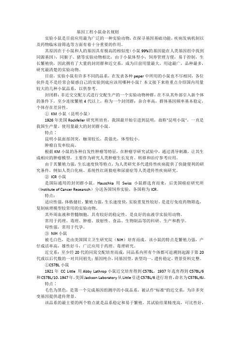
第25卷第3期2004年6月西安交通大学学报(医学版)Journal of Xi'an Jiaotong University (Medical Sciences )7ol8259o83Jun82004◇论 著◇小鼠10号染色体上致聋突变基因hml 的精确定位Qingyin Zheng ,Belinda S Harris ,Patricia F Ward-Bailey ,Heping Yu ,Roderick T Bronson ,Muriel T Davisson ,Kenneth R Johnson(The Jackson Laboratory ,Bar Harbor ,Maine 04609,USA )摘要:目的 定位小鼠致聋基因,识别决定其性状的有关突变,为人类耳聋基因研究提供动物模型。
方法 利用全基因组扫描来定位名为hml 可致小鼠听力丧失突变基因。
结果 ①hml 基因定位在小鼠10号染色体上,距中心粒约43cM 处。
根据已知的鼠-人同源同线性特点,提示人的同源基因位于12q22-q24;②获得了25个多态性微卫星标记,通过高分辨的小鼠图谱将3个已知人类基因进行了正确排列,并将hml 侯选基因限定在一个500kb 的区域内。
关键词:小鼠;耳聋;突变中图分类号:R764.21 文献标识码:A 文章编号:1671-8259(2004)03-0209-04Fine mapping of a deafness mutation hml on mouse chromosome 10Qingyin Zheng ,Belinda S Harris ,Patricia F Ward-Bailey ,Heping Yu ,Roderick T Bronson ,Muriel T Davisson ,Kenneth R Johnson (The Jackson Laboratory ,Bar Harbor ,Maine 04609,USA )ABSTRACT :Objective :o ;a<a ;ouse deafness gene ,identify t=e underlying ;utation and develo<a ;ouse;odel for =u;an deafness8Methods >enetic lin?age cross and geno;e scan @ere used to ;a<a novel ;utation na;ed =y<o<lasia of t=e ;e;Aranous laAyrint=(hml ),@=ic=causes =earing loss in ;utant ;ice8ResuIts ①hml @as ;a<<ed on ;ouse B=r 10(~43cM fro;t=e centro;ere ),suggesting t=at t=e =o;ologous =u;an gene is on 12C 22-C 24,@=ic=@as defined on t=e Aasis of ?no@n ;ouseD=u;an =o;ologies (EMFM ,2004)8②:=is study =as generated 25<oly;or<=ic ;icrosatellite ;ar?ers ,<laced 3?no@n =u;an genes in t=e correct order in a =ig=Dresolution ;ouse ;a<and narro@ed t=e hml candidate gene region to a 500?A area8KEY WORDS :;ouse ;deafness ;;utation Giogra<=y :Hingyin I=eng (1963D ),MJ ,;ale ,Kesearc=scientist8:el :+12072886609;LaM :+12072886149;N;ail :CyO @PaM8org1 Introduction1.1 Hereditary hearing impairment in humans and mice Genetic impairment of hearing affects about oneof every 2000children [1].Genetic analysis of mousedeafness mutations has already aided in the identification of human deafness genes.Depending on genetic back-ground mutations of one gene can cause recessive or dominant ,syndromic or nonsyndromic hearing loss.Mice homozygous for the shaker-1mutation (sh 1)are characterized by circling behavior and deafness.sh 1was shown by positional cloning to be a mutation of the Myo 7a gene ,which encodes an unconventional myosin-type protein [2].Subsequently ,the homologous MYO 7Agene in humans was shown to be responsible for both dominant (DFNA 11)and recessive (DFNB 2)forms ofhearing impairment [3],as well as for Usher syndrome type1B [4].1.2 Hypoplasia of the membranous labyrinth(hml )in mice ,a potential homology for human deafness DFNA 25 Here we describe a newly discov-ered mutation that causes hearing loss in mice.The map position on mouse Chr 10(~43cM from the centro-mere )suggests that the homologous human gene is on 12q22-q24,on the basis of known mouse-human homol-ogies (OMIM ,2004).A de novo deletion in a six-year-old boy with congenital hearing loss as well as mental and motor retardation provides a possible human homo-西安交通大学学报(医学版)第25卷logue syndrome of hml at a critical interval to13cM in the12q22-q24.1region where DFNA25resides.Pro-portional smaller body size represents a phenotype simi-lar to hml.Thus,identification of the hml gene will pro-vide insight into molecular mechanisms of inner ear de-velopment and formation.2 Materials and methods2.1Mice and linkage cross Hypoplasia of the membranous labyrinth(hml)is a recessive spontaneous mutation that arose in a colony of inbred BALB/cByJ mice at The Jackson Laboratory(TJL),Bar Harbor,Maine.Homozygous hml mice can be identified at10 days of age by their small size and unbalanced gait.The mouse strains CAST/Ei and BALB/cByJ-hml(abbrevia-tion CBy-hml),their F1hybrids,and F2intercross progenies are maintained in our research colonies at TJL.All animal procedures were approved by the Ani-mal Care and Use Committee(ACUC).Because neither sex of homozygotes CBy-hml/hml breed,the colony was maintained by progeny test and an outcross-intercross strategy was used for the mapping.Outcross:tested het-erozygotes(CBy-hml/!)were outcrossed with CAST/Ei and half of the resulting F1offspring were expected to carry the mutation-(CAST x CBy)-hml/!.F1hybrids were mated with known CBy-hml/!mice;if one or more offspring was born with a mutant phenotype,then the F1 was assumed to be an hml carrier.Intercross:F1hy-brids-(CAST x CBy)-hml/!x(CAST x CBy)-hml/! intercrosses were mated to generate F2intercross proge-ny.One fourth of the F2s were expected to be homozy-gous for the wildtype allele(!/!),1/2heterozygous (hml/!)and1/4homozygous for the mutation(hml/ hml).Phenotyping and the genome-wide scanning of these F2progeny were carried out as described below. 2.2 Genotyping Tail DNA was isolated according to Johnson[5].SSLP markers polymorphic between strains BALB/cByJ and CAST/Ei were selected at~15cM in-tervals across the mouse genome.A total of123SSLP markers were tested for the initial genome-wide screen. Additional markers were tested around loci that showed significant or suggestive linkage.For PCR amplifica-tion,100ng of DNA were used in a10µL volume con-taining50mmol KCl,10mmol Tris-HCl,pH8.3,2.5mmol MgCl2,0.2mmol oligonucleotides,200µmol dNTP and0.02U Ampli Taq DNA polymerase.The re-actions were subjected to the following temperature cyc-ling program:initial denaturation for2min at95℃;20 s at94℃,30s at50℃,40s at72℃for49cycles;followed by a7min extension at72℃.PCR products were separated by electrophoresis on a3%MetaPhor (FMC,Rockland,ME)agarose gel and visualized un-der UV light after staining with ethidium bromide.2.3 Light microscopy analysis BALB/cByJ(n= 8)and BALB/cByJ-hml/hml(n=8)mice were deeply anesthetized and transcardially perfused with phosphate-buffered saline(PBS)followed by Bouin’s fixative.The cochlea was removed and stored in Bouin’s fixative for 48h.Serial sections(7µm)of the cochlea were stained with hematoxylin and eosin(H&E)using standard pro-cedures.2.4ABR phenotyping A computer-aided evoked potential system(Intelligent Hearing System,IHS;Mi-ami,FL)was used to test mice for ABR thresholds as previously described[6].Mice were anesthetized with tri-bromoethanol(0.53mg・g-1body wt i.p.).Subder-mal needle electrodes were inserted at the vertex(ac-tive)and ventrolaterally to the right ear(reference)and to the left ear(ground).Specific acoustic stimuli were delivered binaurally through1cm plastic tubes chan-neled from high frequency transducers.Mice were tested with click stimuli and also with8,16and32kHz tone pips at varying intensity,from low to high(10~90dB SPL).An ABR threshold was determined for each stim-ulus frequency by identifying the lowest intensity which produced a recognizable ABR pattern(at least two con-sistent peaks)[6].3 Results3.1Mutant mouse phenotype Two mutant mice were observed in the progeny from a mating pair in the BALB/cByJ(CBy)inbred strain colony at The Jackson Laboratory.The mutation arose spontaneously,and nei-ther parent was affected.The mutants were originally noticed by their small body size and distinctively fail-to-thrive behavior.Once they were placed on the back,it took up to a minute to upright themselves.Some mutants died perinatally through two months of age.Once they0123期Qingyin Zheng,B S Harris,P F Ward-Bailey,et al.Fine mapping of a deafness mutation hml on mouse chromosome10passed two months of age they could live up to18 months of age.Even though neither sex bred,sperm shape and activities were checked and no abnormalities were found.Unlike their normal littermates,the mutant mice were unable to swim and did not respond to tapping or clicking sounds,indicating that they also had hearing loss.Initially the mutant mouse was named Hypoplasia of the membranous labyrinth(hml).In the mapping cross(CAST x CBy)-hml/hml F2mutant showed vigor-ous life and variable phenotypes.3.2 Hearing and inner ear analysis All CBy-hml/ hml mice were severely hearing impaired:21/24were deaf(no ABR response at maximal stimuli)and3/24 exhibited severe hearing loss(70~90dB SPL)as early as19days of the tested ages.In contrast,all78of the littermate controls and6tested carriers(hml/!)had normal ABR thresholds(<45dB SPL for any tested stimulus from click,8,16to32kHz).(CAST x CBy)-hml/hml showed incomplete penetrance:15out of20 hml/hml were deaf.One hml/hml had nearly normal hearing(20~40dB SPL).Inner ear histology(Fig.1)shows extensive hypo-plasia of the membranous labyrinth,including a rudi-mentary tectorial membrane.The sizes of basilar mem-brane,Reisner’s membrane,stria vascularis,scala media and scala tympani were significantly reduced and even completely lost.The scala vestibuli was greatly en-larged.Sixteen inner ears from8mutant mice from17to 23days of age were histologically examined;results showed some variation however a constant relationship of pathological severity to age has not been determined. Four hml/!ears at4months of age showed normal struc-ture at light microscopy level,which suggested no heter-ozygous effect.3.3 Genetic Mapping The hml mutation was initial-ly localized to the central region of Chr10as shown in Fig2.hml maps near Ap3d,the gene mutated in mocha mice that causes a progressive hearing loss[7].Ap3d is eliminated as a candidate for hml because its localization is proximal to D10Mit42,whereas the hml is located distal to D10Mit42Jittery(ji),another neurological mouse mutation located on central Chr10,is also elimi-nated as a candidate for hml because it is located proxi-mal to D10Mit42[8].Ames waltzer(av),amouse mu-Fig.1a&b whole-mount preparations of inner ears from2mice at21days of age.The bony labyrinth of hml/ hml is intact,but the spiral structure of the membranous labyrinth is extremely underdeveloped in a compared with b(white line).c,d,e,f mid-modiolar cross-sections of inner ears from mice at17days of age.Close up of boxed region of at e showing tectorial membrane(TM)was not developed(melted with RM)compared to that of f.OC:organ of Corti;IHC:inner hair cells;OHC:outer hair cells;BM:basilar membrane;RM:Reisner’s membrane;StV:stria vascularis;SG:spiral ganglian;SV:scala vestibuli;SM:scala media;ST:scala tympani;Mo:modiolustation causing deafness,also maps to the middle region of Chr10but has been localized proximal to D10Mit91[9].By directly testing for allelism,all other hearing-related genes or mutations are excluded in the region where hml maps,this mutation defines a new deafness gene.A total of342F2mice(=684meioses)were genotyped with22additional microsatellite mark-ers,and hml was localized between D10Mi65and D10Mit132,with2recombinant events,which define an interval of0.3cM.1123.4 Fine Mapping A total of 2300F2progeny (4600meioses )was accumulated (Fig.3).The mini-mal genetic interval containing hml was narrowed to a region smaller than 0.1cm ,between markers D 10Mit 208and D 10Mit 261with 43self-designed mark-ers.Twenty-two animals with crossovers between D 10Mit 208and D 10Mit 261were generated and the can-didate region was further narrowed down to 0.07cM ,which is a 500kb region between self-designed markers 18L 357and 261L 453.We searched and compared our mapping data with both private and public databases.The human homologous region of the critical genetic in-terval containing hml was defined to be 12q22-24.Tmp 3and Igf 1were excluded ,candidate gene test is still underway.Sequence information of self-designed markers is availableupon request.Fig.2 Genetic map position of hml and homologies withhuman chromosomesFig.3 Genetic fine map position of hml and homologies with human chromosomes[3]Thomas WE,Ardill J,Buchanan345Th6hormonal chan76s8ro9 duc6d:;duod6no7as<ric r6=lu>[J]5?cand J@as<ro6n<6rol,1984,92(su88l):44-475[4]@risoni E,4usl6a74,?u86r45Ai<ric o>id6s;n<h6sis inhi:i<ion:<h66==6c<on ra::i<8;loric muscl6[J]5J B6dia<r?ur7,1996,31(6):800-8045[5]Willis?,All6sch6r C4,W6i76r<A,et al5Dn=lu6nc6o=<h6E9ar7i9 nin69ni<ric o>id68a<hFa;on Gasoac<iG6in<6s<inal8ol;868<id6r6l6as6 and mo<ili<;in<h6ra<s<omach in’it()[J]5Eur J Bharmacol,1996,315(1):59-645[6]戴益琛,张忠兵,左秀丽,等5一氧化氮和肠血管活性肽对幽门功能的调节及其在胆汁反流中的作用[J]5临床消化病杂志,2000,12(1):13-155[7]HacIJa H,Thor B,BilsJi J,et al5Ai<ric o>id6and<h6in<6rr6la9 <ion:6<F66n in<6s<inal mo<ili<;and8ancr6a<ic s6cr6<ion in=as<6d and =6d do7s[J]5J Bh;siol Bharmacol,1994,45(2):285-2985[8]?<olJ HKJ,Lan Er86cum3J,?mou<AJ,et al5Ho<or c;cl6s Fi<h 8has6DDD in an<rum ar6associa<6d Fi<h hi7h mo<ilin l6G6ls and8ro9lon76d7all:ladd6r6m8<;in7[J]5Am J Bh;siol,1993,264(4B<1):@596-6005[9]BoiGin H,Ma;mond HN,Mi:6rd;H,et al5Blasma mo<ilin Garia9 <ion durin7<h6in<6rdi76s<iG6and di76s<iG6s<a<6s in man[J]5J@as9<roin<6s<Ho<il,1990,2(1):240-2465[10]Aaslund E,BacJman E,Th6odorsson E,et al5Dn<raduod6nal n6u9 ro868<id6l6G6ls,:u<no<8lasma l6G6ls,Gar;in a c;clic=ashion Fi<h<h6mi7ra<in7mo<or com8l6>[J]5Ac<a Bh;siol?cand,1998,164(3):317-3235[11]Musso A,Kras6r M,Adachi3,et al5EGid6nc6<ha<ni<ric o>id6 m6chanisms r67ula<6small in<6s<inal mo<ili<;in humans[J]5@u<,1999,44(1):72-765(编辑卓选鹏%%%%%%%%%%%%%%%%%%%%%%%%%%%%%%%%%%%%%%%%%%%%%%%)(上接第212页)nonsyndromic deafness(DFNA25)mapped to human chromosome12q24-qter is in the homologous region to our hml area.Identification of the hml gene could po-tentially facilitate the identification of the underlining gene for DFNA25[10].A de novo deletion in a six-year-old boy with congenital hearing loss as well as mental and motor retardation provides a possible human homo-logue syndrome of hml at a critical interval to13cM in the12q22-q24.1region where DFNA25resides.DF-NA25that maps in the12q22-q24.1region are included in the deletion that is homologous to hml in the mouse chr10.Magnetic Resonance Tomography showing a consecutive dilatation of the left lateral ventricle,pro-portional smaller body size,represents a phenotype simi-lar to hml.A mouse model for human DFNA25will aid in the understanding of the molecular bases and the de-velopment of a potential treatment for the human dis-ease.AcknowledgementsThis work was supported by National Institutes of Health(NIH)grants DC04376and DC05846to QYZ. We thank Dr.Doris K Wu from NIDCD for her advice for research design.References[1]Hor<on AE5@6n6<ic68id6miolo7;o=h6arin7im8airm6n<[J]5AnnA O Acad?ci,1991,630:16-315[2]@i:son K,Walsh J,H:uru B,et al*A<;86LDD m;osin6ncod6d:;<h6mous6d6a=n6ss76n6shaJ6r91[J]5Aa<ur6,1995,374(6517):62-645[3]Eiu PQ,Walsh J,Tama7aFa O,et al*Au<osomal dominan<non9 s;ndromic d6a=n6ss caus6d:;a mu<a<ion in<h6m;osin LDDA76n6[l6<<6r][J]5Aa<@6n6<,1997,17(3):268-2695[4]W6il4,Blanchard?,3a8lan J,et al*46=6c<iG6m;osin LDDA76n6 r6s8onsi:l6=or Rsh6r s;ndrom6<;861B[J]5Aa<ur6,1995,374(6517):60-615[5]Johnson3M,NooJ?A,Qh6n7SO5Th6ori7inal shaJ6r9Fi<h9s;n9 dac<;lism mu<a<ion(s;)is a con<i7uous76n6d6l6<ion s;ndrom6[DnBroc6ss Ni<a<ion][J]5Hamm@6nom6,1998,9(11):889-8925[6]Qh6n7SO,Johnson3M,ErFa;EN5Ass6ssm6n<o=h6arin7in80 in:r6d s<rains o=mic6:;ABM<hr6shold anal;s6s[J]5C6ar M6s,1999,130(1-2):94-1075[7]3an<h6<i B,Siao P,4iaI HE,et al*Hu<a<ion in AB-3d6l<a in <h6mocha mous6linJs6ndosomal<rans8or<<o s<ora76d6=ici6nc;in8la<6l6<s,m6lanosom6s,and s;na8<ic G6sicl6s[J]5A6uron,1998,21(1):111-1225[8]3a8=ham6r4,?F66<CT,?u=alJo4,et al*Th6n6urolo7ical mous6mu<a<ions Ui<<6r;and h6si<an<ar6all6lic and ma8<o<h6r67iono=mous6chromosom610homolo7ous<o1981353[J]5@6nomics,1996,35(3):533-5385[9]Qo:6l6;E,?u=alJo43,AdJins?,et al*Kin676n6<ic and com8ar9 a<iG6ma88in7o=<h6d6a=n6ss mu<a<ion Am6s Fal<I6r on mous6chromosom610[J]5@6nomics,1998,50(2):260-2665[10]B6<6J E,Wind8assin76r N,Hach H,et al5Hol6cular charac<6riIa9 <ion o=a12V22-V24d6l6<ion associa<6d Fi<h con76ni<al d6a=n6ss:con=irma<ion and r6=in6m6n<o=<h64KAA25locus[J]5Am J H6d@6n6<,2003,117A(2):122-1265(编辑胡爱玲)。
LP 小鼠突变基因的定位及鉴定

LP 小鼠突变基因的定位及鉴定张粉丽;陈兵;薛整风;李怡【摘要】Objective To define the loci of the mutant gene in the loop-tail mouse.Methods To study the heredity pattern, loop-tail mice were mated with normal C57BL/6J and C3H mice.Their offsprings with loop-tail or normal phenotype were registered respectively.Microsatellite markerD1Mit113 and D1Mit149 were used to locate the mutant gene.Based on fine mapping, the candidate gene Vangl2 was found.Vangl2 gene from the loop-tail mice was amplified by PCR followed by sequencing.Incision enzyme FspBI ( BfaI ) identified the genotype of offspring from loop-tail mice intercrossing.Results Heredity test indicated that the loop-tail phenotype was controlled by a single dominant gene not with100%penetrance but was affected by genetic background.A C-to-T transversion was at the 1345bp in Vangl2 gene of the loop-tailmice.Conclusions The C-to-T transversion introduces a pre-termination codon of amino acids and causes the phenotype of loop-tail phenotype.None homozygous mice were found in the offsprings, suggesting that the homozygous mice are lethal.%目的:以卷尾突变C57 BL /6 J小鼠为研究对象,通过微卫星定位以及候选基因测序分析确定突变基因位点。
基因定点突变方法及其应用

基因定点突变方法及其应用基因定点突变是指在基因组中特定位置的发生的突变。
基因定点突变可以是单个碱基的突变,也可以是多个碱基的突变。
这可以发生在DNA、RNA或蛋白质的编码区域或非编码区域。
基因定点突变方法是用于研究基因突变的工具和技术。
它们在生物医学研究、疾病诊断、药物研发等领域有着广泛的应用。
1.PCR扩增:PCR扩增是一种常用的基因定点突变方法,可以快速有效地扩增所需的DNA片段。
通过在PCR反应中引入突变引物,可以在特定位点引入单个碱基变异。
这种方法被广泛应用于基因功能研究、遗传性疾病的诊断和突变的检测等领域。
2.扩增-测序:扩增-测序方法是一种将突变引物引入待测基因位点的PCR扩增方法,随后使用测序技术验证突变是否成功。
这种方法可用于研究基因突变与人类遗传病之间的相互关系,还可以用于检测药物抗性突变、病毒突变等领域。
3.分子克隆:分子克隆是一种将特定DNA片段插入载体DNA的方法。
通过将突变片段与目标DNA结合,随后将其放入宿主细胞,可将所需的突变引入到目标基因中。
这种方法广泛应用于蛋白质工程、基因功能研究等领域。
4. CRISPR-Cas9系统:这些基因定点突变方法在基因功能研究、疾病诊断和治疗、药物研发等领域有着广泛的应用。
在基因功能研究中,通过引入特定的突变,研究人们可以研究基因的功能和调控机制。
例如,通过基因定点突变,可以研究基因在发育、免疫反应、代谢调节等过程中的作用和调节机制。
在疾病诊断和治疗中,基因定点突变方法可以用于检测与一些遗传性疾病有关的突变。
例如,通过扩增-测序方法可以检测BRCA1和BRCA2基因的突变,从而评估患者患乳腺和卵巢癌的风险。
此外,基因定点突变方法也可以用于基于个体遗传背景的个体化药物治疗。
在药物研发领域,基因定点突变方法可以用于评估药物的疗效和副作用。
通过引入特定的突变,可以模拟蛋白质靶点的突变,评估药物对该靶点的亲和力和选择性。
这对于开发更有效和安全的药物具有重要意义。
基因工程小鼠名称解读

基因工程小鼠名称解读基因工程小鼠是指通过人为干预小鼠基因组的技术手段,使其表达特定基因或缺失特定基因,从而实现对基因功能的研究和相关疾病模型的建立。
这些小鼠通常被用于研究基因功能、疾病机制、药物筛选等方面。
在基因工程小鼠的命名中,常用的方式是根据其基因改造方式或目的来命名。
以下是一些常见的基因工程小鼠名称及其解读:1. 转基因小鼠(Transgenic mice),这类小鼠是通过将外源基因导入小鼠的基因组中而得到的。
这些外源基因可以来自其他物种,如人类或其他动物。
转基因小鼠常用于研究特定基因的功能、表达模式等。
2. 敲除小鼠(Knockout mice),这类小鼠是通过人为干预使特定基因在小鼠体内失去功能的方式得到的。
一般采用基因敲除或基因靶向突变技术,使小鼠体内特定基因的表达受到抑制或完全消除。
敲除小鼠常用于研究基因的功能缺失对生理和病理过程的影响。
3. 过表达小鼠(Overexpression mice),这类小鼠是通过人为干预使特定基因在小鼠体内过度表达的方式得到的。
通过引入外源基因或增强内源基因的表达水平,使小鼠体内特定基因的表达量显著增加。
过表达小鼠常用于研究基因的过度表达对生理和病理过程的影响。
4. 突变小鼠(Mutant mice),这类小鼠是指在小鼠基因组中引入或产生突变的小鼠,突变可以是点突变、插入突变、删除突变等。
突变小鼠常用于研究特定基因突变对生理和病理过程的影响。
除了上述常见的命名方式,基因工程小鼠还可以根据具体的研究目的、基因改造技术等来命名,例如组织特异性表达小鼠、条件性基因敲除小鼠等。
需要注意的是,基因工程小鼠的命名通常是根据研究者或研究机构的需求和约定来进行的,不同研究领域和实验室可能会有不同的命名方式和规范。
总体而言,基因工程小鼠的命名旨在描述其基因改造方式、基因功能或研究目的,以便研究者能够清楚地了解其特点和应用范围。
kaede小鼠 原理

kaede小鼠原理
Kaede小鼠是一种基因编辑技术产生的变色小鼠,其原理主要涉及到以下几点:
1. 基因编辑:Kaede小鼠通过基因编辑技术,特别是CRISPR/Cas9系统,对小鼠基因组进行定点突变。
具体来说,科学家通过在小鼠基因组中插入Kaede基因的DNA序列,使得小鼠的细胞能够表达Kaede蛋白。
2. Kaede蛋白及光反应:Kaede蛋白是一种白色素蛋白,它可以在紫外光和绿光激发下产生红光荧光。
当Kaede小鼠的细胞暴露在紫外光下,Kaede蛋白将被激活,从而发出红光。
此外,Kaede蛋白的活性可以被紫外光照射后的氧气或其他化合物还原,从而恢复到初始白色素状态。
3. 变色特性:通过在小鼠细胞中表达Kaede蛋白,Kaede小鼠的细胞和组织可以在紫外光照射下产生红色荧光。
这使得科学家能够通过观察小鼠的红光发出程度来推断细胞的活动情况、细胞迁移、组织分化等。
总的来说,Kaede小鼠原理是通过基因编辑技术在小鼠基因组中插入Kaede蛋白基因,使得小鼠细胞和组织能够在紫外光照射下产生红色荧光,从而可观察和研究细胞和组织的活动情况。
- 1、下载文档前请自行甄别文档内容的完整性,平台不提供额外的编辑、内容补充、找答案等附加服务。
- 2、"仅部分预览"的文档,不可在线预览部分如存在完整性等问题,可反馈申请退款(可完整预览的文档不适用该条件!)。
- 3、如文档侵犯您的权益,请联系客服反馈,我们会尽快为您处理(人工客服工作时间:9:00-18:30)。
定点突变小鼠命名方法
展开全文
随着基因编辑技术的发展,基因工程小鼠的构建变得日益简单,它成为了研究人类基因功能的最好的模式动物之一。
虽然基因工程小鼠类别不多,但是其命名冗长复杂,对于这方面研究的科研工作者,还是需要学习一下。
我们继续参考International Committee on Standardized Genetic Nomenclature for mice中对大小鼠基因的描述部分。
首先,我们需要明确基因的写法要求
1. 一般在文章书写小鼠基因符号是斜体(Rbp1),首字母大写;当是人源的基因时,字母全部大写,斜体;如果是蛋白质则是全部大写,不用斜体。
2. 一般基因用2-4个字母缩写表示,并尽可能地表达其功能和特点;
3. 字体为新罗马字体和阿拉伯数字组合。
上一期小编带着大家学习了常见的小鼠分类及命名,基因工程小鼠即:野生型小鼠品系和基因符号的组合。
基因工程的小鼠主要分为定点突变(T argetedMutations)和转基因(Transgenes)小鼠。
C57BL/6,129,FVB是最常使用的基因工程小鼠。
本次小编先带大家了解定点突变(TargetedMutations)小鼠的规则要求。
定点突变(Targeted Mutation)
主要分为ES细胞打靶、核酸内切酶介导的突变、基因捕获突变及增强子捕获等。
1
ES细胞打靶
利用同源重组技术对ES细胞特定位点的基因进行改造,通过显微注射或者胚胎融合的方法将遗传修饰的ES细胞引入受体胚胎内,是一种建立在ES细胞与DNA同源重组等技术基础上的分子生物学技术。
包括Knock-out,Knock-in及条件敲除等。
基因符号由四部分组成:
①突变的目的基因;
②tm标志代表targeted mutation;
③起源于实验室的序列号;
④实验室注册代号。
前面可以直接加上小鼠品系,具体参考上期常见小鼠品系命名。
1.1 常见举例:
129X1-Cftrtm1Unc :Unc实验室在129X1品系小鼠上靶向Cftr 基因的进行的首次突变。
1.2 Knock in:外源基因或者基因片段插入到目的基因,导致外源基因在内源性启动子下表达的突变方式,它的命名基本结构同上,但是信息要复杂一些,如下:129X1-En1tm1(Otx2)Wrst:将129X1品系小鼠上的En1基因的编码区被Otx2基因替代,此突变来源于Wrst实验室。
其他 knockin 形式:
Cd19tm1(cre)Cgn:cre酶基因插入第一个exon区域,cre酶特异性表达于B细胞。
Apoetm1(APOE*2)Mae:人源的APOE2基因(2-4个外显子区域),替代小鼠的Apoe基因,使小鼠不再表达其内源的蛋白。
其他 targeted mutations
1.3 插入RNAi:Genetm#(RNAi:Xyz)Lab code
1.4 Cre-Lox系统:Tfamtm1Lrsn:表示LoxP元件插入Tfam基因中
而Tfamtm1.1Lrsn是指该基因型小鼠与Cre工具鼠杂交,而Cre 酶位于Tfam同源染色体的等位基因区段。
这样一来,当Tfam的等位基因表达时,一条染色体上的Tfam基因被删除,而另一个等位基因的Tfam则融合Cre正常表达。
2
核酸内切酶介导的突变
由核酸内切酶介导的直接修饰多功能细胞或者全能细胞,定点修饰目的基因,通过DNA同源重组(HDR)和非同源末端连接(NHEJ)的DNA修复达到修饰基因的目的。
主要是指ZFN,TALENs,CRISPR/Cas9基因编辑技术。
ES细胞基因打靶技术是获取基因编辑小鼠的传统方法,最新的CRISPR/Cas9具有操作简便,靶点选择范围广,作用活性高等优势。
基因符号由四部分组成:
①目的基因;
②上标:em标志代表endonuclease-mediated mutation;
③起源于实验室的序列号;
④实验室注册代号。
FVB-Rab38em1Rkuhn:Ralf Kuhn实验室在FVB小鼠上利用核酸内切酶技术对Rab38基因进行首次突变。
由于核酸酶介导的等位基因突变包括很多种,所以可以在基因位点进行详细描述。
如C57/B6-Apoeem2Cd196:利用核酸酶介导的突变(CRISPR/Cas9简写为C)把C57/B6小鼠的Apoe基因敲除196nt。
3
基因捕获突变(Gene Trap Mutations)
将带有报告基因的重组载体随机整合到基因组中,使插入基因被激活或失活,通过检测报告基因的表达来揭示基因表达模式及其功能,类似于靶向基因突变。
基因符号由五部分组成:
①目的基因;
②上标:Gt标志代表Gene Trap;
③()写上报告基因载体信息;
④起源于实验室的序列号;
⑤实验室注册代号。
Akap12Gt(ble-lacZ)15Brr:代表包括Ble(腐草霉素抗性)和LacZ(β-半乳糖苷酶)报告基因载体插入目的基因Akap12,在Brr实验室第15次构建。
4
增强子捕获(Enhancer Traps)
用于确定DNA序列中是否包含增强子功能的一项技术。
检测的载体含有启动子和报道基因可作表达的标记,从报道基因表达的程度可分析出增强子的存在与否及其强弱。
基因符号由五部分组成:
①目的基因;
②上标:Et标志代表Enhancer Trap;
③()写上cre重组酶;
④起源于实验室的序列号;
⑤实验室注册代号。
Et(icre)1642Rdav:Cre诱导编号为1642的基因序列删除,从而确定被删除的序列是否是gene enhancer。
参考文献:
mittee on Standardized Genetic Nomenclature for Mice, Chair: Lyon, M.F.1981. Rules and guidelines for gene nomenclature. In: Genetic Variants andStrains of the Laboratory Mouse, Green, M.C. (ed.), First Edition, GustavFischer Verlag,
Stuttgart, pp. 1-7.
mittee on Standardized Genetic Nomenclature for Mice, Chairperson: Davisson,M.T. 1996. Rules and guidelines for gene nomenclature. In: Genetic Variants andStrains of the Laboratory Mouse, Lyon, M.F., Rastan, S., Brown, S.D.M. (eds.),Third Edition, Volume 1, Oxford University Press, Oxford, pp. 1-16.
3.Eppig, JT. 2006. Mouse Strain and Genetic Nomenclature: an Abbreviated Guide.In: Fox J, Barthold S, Davvison M, Newcomer C, Quimby F, Smith A (eds) TheMouse in Biomedical Research, Volume 1, Second Edition. Academic Press.pp.79-98.
4.Gaj T, Gersbach CA, Barbas CF 3rd. 2013. ZFN, TALEN, and CRISPR/Cas-basedmethods for genome engineering. Trends Biotechnol 31: 397-40
5.
5.Wijshake T, Baker DJ, van de Sluis B. 2014. Endonucleases: new tools to editthe mouse genome. Biochim Biophys Acta. 2014 Apr 30
关注(360图书馆:科研小助手),获得更多科研小技巧。