
神经解剖学英文课件-面部、头皮和腮腺英文课件
2) The facial vessels
(1) Facial artery
arises from external carotid a.;
runs upwards and forwards;
turns around the lower border of mandible crossing with anterior border of the masseter;
Parotid gland P.394
3) position and division: * It lies on the lateral side of the face
and anteroinferior to the external acoustic meatus
*posterior border of masseter *in the retromandibular fossa
5) Structures within the gland
(1) Longitudinal structures external carotid a. superficial temporal v.and a. retromandibular v. auriculotemporal n.
(2) Transverse maxillary a. and v. transverse facial a. and v. br. of the facial n. parotid duct
Anastomosis occurs between branches of the left and right facial aa..
(2) facial vein
begins at the angular v. at the medial angle of the eye;
blood vessels and auriculotemporal n.
Parotid –bed A lot of m.attached to the styloid Carotid sheath Lower 4 paired cranial n. Superior cervical sympathetic
Parotid Area
(P 394)
Parotid gland P.394
1) the largest one of the 3 paired salivary glands
2) enclosed within the fascia parotid sheath formed by enveloping fascia superficial part of the sheath is the thicker and gives off the many fibrous septa into the gland. deep part of the sheath is the thinner.
下颌后窝
division of parotid gland
superficial part: superficial to the posterior part of masseter
deep part: in the retomandibular fossa isthmus: face to the ramus of mandible
6) The main structures leaving the edge of the parotid gland
Temporal br. Zygomatic br. Buccal br. Mandibular br. Cervical br. Parotid duct Superficial temporal
deep isthmus
Superficial part of
4) Parotid duct 腮腺导管
It passes horizontally from the anterior edge of the gland, runs forwards at the surface of the masseter, inferior to the zygomatic arch. At the anterior border of the masseter, the duct turns medially, pierces buccinator, and opens on a small papilla on the buccal mucosa of oral surface of the cheek opposite the second maxillary molar tooth.
2. Superficial fascia 1) loose connective tissue:
*corpusadiposum buccae(颊脂体) at the buccal part
* at the palpebra(睑), subcutaneous fat is lesser and looser.
enters the face where the facial a.runs lateral to the angle of the mouth and wing of nose, and then
reaches the medial canthus(内眦), where it becomes the angular a.;
ganglion
m.inrotid sheath,and n. in parotid bed
The superficial structures of face
(P 348-354)
• 1. The skin: 1) There are rich
sebaceous glands (皮脂腺), sweat glands, hair follicles(毛囊), and blood supplying. 2) It is thin and rich elasticity.
合集下载
神经解剖学英文课件-喉、气管和食道解剖英语课件
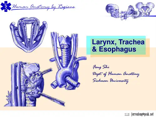
▪ Relations of Cervical Trachea & Esophagus
The General Arrangement of Neck & Its Transversal Compartments
▪ visceral compartment ▪ vascular
compartment ▪ vertebral
lig.
Laryngeal Ventricle
▪ the mucosa overlying vestibular & vocal ligaments become the vestibular & vocal folds
Rima裂 Vestibuli
Rima Glottidis
Laryngeal Joints
▪ recurrent laryngeal n. & cervical sympathetic trunks
CLINICAL NOTE
Tracheoesophageal Fistula
Case
当John完成了第一次解剖学考试后,父亲决定带他去吃牛排以示庆祝。 在喝了一点酒后,他注意到父亲说话有些含糊不清,进食速度加快。过 了一会儿,他发现父亲脸色突然改变,然后突然倒在地板上。开始他怀 疑父亲可能是休克、心脏病或癫痫发作。但他还发现父亲脉搏很强,脸 色开始发青。这时他意识到可能是窒息。他将父亲的嘴张开,看到一大 块牛排卡在喉的后部。他将示指深入父亲口中,试图取出牛排,但未成 功。然后他将父亲翻到俯卧位,实施海姆利克操作法。
Innervation of the Larynx
▪ recurrent laryngeal n.: - motor: all intrinsic mm. except cricothyroid m. - sensory: the laryngeal cavity below the level of the vocal folds
The General Arrangement of Neck & Its Transversal Compartments
▪ visceral compartment ▪ vascular
compartment ▪ vertebral
lig.
Laryngeal Ventricle
▪ the mucosa overlying vestibular & vocal ligaments become the vestibular & vocal folds
Rima裂 Vestibuli
Rima Glottidis
Laryngeal Joints
▪ recurrent laryngeal n. & cervical sympathetic trunks
CLINICAL NOTE
Tracheoesophageal Fistula
Case
当John完成了第一次解剖学考试后,父亲决定带他去吃牛排以示庆祝。 在喝了一点酒后,他注意到父亲说话有些含糊不清,进食速度加快。过 了一会儿,他发现父亲脸色突然改变,然后突然倒在地板上。开始他怀 疑父亲可能是休克、心脏病或癫痫发作。但他还发现父亲脉搏很强,脸 色开始发青。这时他意识到可能是窒息。他将父亲的嘴张开,看到一大 块牛排卡在喉的后部。他将示指深入父亲口中,试图取出牛排,但未成 功。然后他将父亲翻到俯卧位,实施海姆利克操作法。
Innervation of the Larynx
▪ recurrent laryngeal n.: - motor: all intrinsic mm. except cricothyroid m. - sensory: the laryngeal cavity below the level of the vocal folds
面神经(Facial nerve)ppt课件
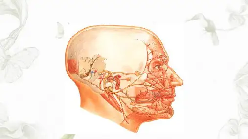
(2)在下颌下缘的平面从后到前越过面后V、下颌角, 面前V的浅面。
(3)与颌外A的关系:单支,经A浅面或深面;多支: 分别经A浅面或深面。转向上至三角肌和下唇方肌,支 配此二肌。
(4)1/5的人越面后V至颌下区。具体位置以颌外A和咬 肌前下角为标志。在标志后,行在下颌支下缘下1cm, 标志处平下颌下缘,其前方行于下颌下缘上。
当一侧上位N元 发生病变时:对侧睑 裂以下表情肌瘫痪, 表现为鼻唇沟变浅, 不能上提口角,食物 易留于口腔前庭,但 肌不萎缩,额纹存在。
当下位N元表现 病变时:同侧全部表 情肌瘫痪,睑不能闭 合,不能提眉 、额 纹消失、鼻唇沟消失、 不能上提口角、若久 治不愈,肌可萎缩。
面神经(Facial nerve)
→向下→茎乳
②
孔→向前穿过
腮腺,呈扇形
③
分布。
④
⑤
面神经(Facial nerve)
三、分段(以茎乳孔为界分管段和颅外段)
(一)面N管段的分支 1、岩浅大N:主要含副交感纤
维,面N外膝→蝶腭N节→泪 腺,鼻,腭粘膜腺。 2、镫骨N:支配镫骨肌。 3、鼓索:在茎乳孔上方 6mm处 自面N发出,进入鼓室、穿岩 鼓裂→颞下窝加入舌N。
(5)颌下手术切口,一般应在下颌下缘1.5cm-2cm处, 并应切开颈深筋膜浅层。
(6)损伤后,口角歪斜。
面神经(Facial nerve)
5、颈支:
(1)1-2支:穿腮腺
下端,行于下颌缘支
后方,二者少交通。
①
(2)在颈阔肌深面、
②
下颌角与胸锁孔突肌
之间向前下至颌下三 角,布于颈阔肌。
③
④
(3)发出分支与颈皮
即:颞支、颧支、
颊支、下颌缘支、
(3)与颌外A的关系:单支,经A浅面或深面;多支: 分别经A浅面或深面。转向上至三角肌和下唇方肌,支 配此二肌。
(4)1/5的人越面后V至颌下区。具体位置以颌外A和咬 肌前下角为标志。在标志后,行在下颌支下缘下1cm, 标志处平下颌下缘,其前方行于下颌下缘上。
当一侧上位N元 发生病变时:对侧睑 裂以下表情肌瘫痪, 表现为鼻唇沟变浅, 不能上提口角,食物 易留于口腔前庭,但 肌不萎缩,额纹存在。
当下位N元表现 病变时:同侧全部表 情肌瘫痪,睑不能闭 合,不能提眉 、额 纹消失、鼻唇沟消失、 不能上提口角、若久 治不愈,肌可萎缩。
面神经(Facial nerve)
→向下→茎乳
②
孔→向前穿过
腮腺,呈扇形
③
分布。
④
⑤
面神经(Facial nerve)
三、分段(以茎乳孔为界分管段和颅外段)
(一)面N管段的分支 1、岩浅大N:主要含副交感纤
维,面N外膝→蝶腭N节→泪 腺,鼻,腭粘膜腺。 2、镫骨N:支配镫骨肌。 3、鼓索:在茎乳孔上方 6mm处 自面N发出,进入鼓室、穿岩 鼓裂→颞下窝加入舌N。
(5)颌下手术切口,一般应在下颌下缘1.5cm-2cm处, 并应切开颈深筋膜浅层。
(6)损伤后,口角歪斜。
面神经(Facial nerve)
5、颈支:
(1)1-2支:穿腮腺
下端,行于下颌缘支
后方,二者少交通。
①
(2)在颈阔肌深面、
②
下颌角与胸锁孔突肌
之间向前下至颌下三 角,布于颈阔肌。
③
④
(3)发出分支与颈皮
即:颞支、颧支、
颊支、下颌缘支、
神经解剖学英文课件-甲状腺解剖英文课件

Isthmus lies in front 2nd to 4th tracheal rings
Pyramid lobe is usually to left of median plane
Parathyroid glands
Parathyroid glands & thyroid gland
Thyroid –arteries
esophagus, external & recurrent laryngeal nerves ❖ Posteriorly: parathyroid gland
Thyroid ---capsules
false thyroid capsule True capsule
Thyroid –external feature
Thyroid –arteries
❖ Inferior thyroid artery : branch of the thyrocervical trunk, posterior to the carotid sheath to reach the posterior aspect of the gland.
Thyroid gland & Root of neck
Thyroid gland
Outline 1. Location & superficial feature 2. Blood supply 3. Venous drainage 4. nerves 5. Relationship of its vessels and arteries
Thyroid Ima Artery
Superior, middle, & inferior thyroid v.
Pyramid lobe is usually to left of median plane
Parathyroid glands
Parathyroid glands & thyroid gland
Thyroid –arteries
esophagus, external & recurrent laryngeal nerves ❖ Posteriorly: parathyroid gland
Thyroid ---capsules
false thyroid capsule True capsule
Thyroid –external feature
Thyroid –arteries
❖ Inferior thyroid artery : branch of the thyrocervical trunk, posterior to the carotid sheath to reach the posterior aspect of the gland.
Thyroid gland & Root of neck
Thyroid gland
Outline 1. Location & superficial feature 2. Blood supply 3. Venous drainage 4. nerves 5. Relationship of its vessels and arteries
Thyroid Ima Artery
Superior, middle, & inferior thyroid v.
7head局部解剖英文课件头部
Retromandibular v.
• Formed by union of superficial temporal and maxillary veins, • Divides into two branches: —Ant. branch unites with facial v. , —Pos. branch joins pos. auricular v. to become external jugular v.
Division of head
Head is divided into cranium and face by line through:
• superior border of orbit, • superior border of zygomatic arch, • superior border of external acoustic pore, • mastoid process
Head
Boundaries of head and neck
Line between:
• • • • • inferior border of mandible, angle of mandible, apex of mastoid process, superior nuchal line, external occipital protuberance
2 parts: are divided by postle or facial n. plexus passing through thyroid gland Superficial part: triangular in shape, lies below and in front of the external acoustic meatus, and partially covers the masseter. Deep part: lies deep to medial pterygoid .
神经解剖学英文课件-面部和头皮解剖英语课件
i muscle
上皱眉肌
降眉间肌
Orbicularis oculi muscle
frontalis
Muscles of the mouth
• orbicularis oris m. • buccinator m.
• Depressor anguli oris m. • Zygomatic major/minor m. • Levator labii superioris m.
Vessels of face
The lymphatic vessels of the face
• Lymph from the deep parotid nodes drains into deep cervical lymph nodes.
•Lymph from the upper lip and lateral parts of the lower lip drain into the submandibular lymph nodes
The retromandibular vein
• the union of the superificial temporal vein and maxillary veins, and descends within parotid gland
Facial vein
• begins at the angular vein
cavernous plexus
②Deep facial v→ pteygoid plexus→cavernous sinus
• deep facial vein which links facial vein to the pterygoid plexus, so, appearing “danger area of the face”— from root of nose to angles of the mouth
上皱眉肌
降眉间肌
Orbicularis oculi muscle
frontalis
Muscles of the mouth
• orbicularis oris m. • buccinator m.
• Depressor anguli oris m. • Zygomatic major/minor m. • Levator labii superioris m.
Vessels of face
The lymphatic vessels of the face
• Lymph from the deep parotid nodes drains into deep cervical lymph nodes.
•Lymph from the upper lip and lateral parts of the lower lip drain into the submandibular lymph nodes
The retromandibular vein
• the union of the superificial temporal vein and maxillary veins, and descends within parotid gland
Facial vein
• begins at the angular vein
cavernous plexus
②Deep facial v→ pteygoid plexus→cavernous sinus
• deep facial vein which links facial vein to the pterygoid plexus, so, appearing “danger area of the face”— from root of nose to angles of the mouth
人体解剖学面部和头皮faceandscalp

Superficial temporal vein
• It drains the forehead and scalp and receives tributaries from the veins of the temple and face. Near the auricle, the superficial temporal vein enters the parotid gland.
Vessels of face
Vessels of face
The lymphatic vessels of the face
• Lymph from the deep parotid nodes drains into deep cervical lymph nodes.
indirectly to the internal
jugular vein)
Feature of veins
• no valves • communicating with cavernous sinus by the 2 routes: ① Facial V →angular V →ophthalmic v→
Facial a. & superficial temporal a.
Transverse facial artery
It arises from the superficial temporal artery within the parotid gland and crosses the face superficially to the masseter. It divides into numerous branches that supply the parotid gland and duct, the masseter, and skin of the face.
神经解剖学英文课件-头骨解剖英文课件
Fractures of the Calvaria
incisive fossa greater & lesser palatine foramina
• Mandibular fossae • Occipital condyles • Jugular foramen • Carotid canal • Jugular foramen. • Stylomastoid foramen
Internal Aspect of Base of Skull
is formed by frontal bone anteriorly, ethmoid bone in middle, body & lesser wings of sphenoid posteriorly. Its greater part is formed by ridged orbital plates of frontal bone, which support frontal lobes of brain and form roofs of orbits.
foramen spinosum • sulcus for ICA
is formed largely by occipital bone. Broad grooves in this fossa are formed by transverse and sigmoid sinuses.
Posterior cranial fossa
Pterion
Infratemporal fossa
Posterior aspect of skull
External occipital protuberance Superior nuchal line
神经解剖学英文课件-面部和头皮英文课件
Orbicularis oculi muscle
Orbicularis oculi muscle
frontalis
Muscles of the mouth
• orbicularis oris m. • buccinator m.
• Depressor anguli oris m. • Zygomatic major/minor m. • Levator labii superioris m.
The facial artery sends branches to the upper and lower lips, the side of the nose, and terminates as the angular artery at the medial canthus.
Superficial temporal artery
Superficial temporal vein
• It drains the forehead and scalp and receives tributaries from the veins of the temple and face. Near the auricle, the superficial temporal vein enters the parotid gland.
Muscles of the mouth
Arteries of the face
Branches of ECA & ICA
Facial artery
Arises from the external carotid artery, runs upwards and forwards, turns round the inferior border of mandible. It appears at just anterior to the masseter muscle.
最新系统解剖英文版神经系统脊髓Spinalcord精品课件
第十六页,共61页。
★脊髓(jǐ suǐ)圆锥Conus medullaris-the lower pyramid end of spinal cord
★终丝Filum terminale-a delicate filament, about 20 cm in length, prolonged downward from the apex of the conus medullaris to the back of the first segment of the coccyx, continuous above with that of the pia mater.
to Vertebral Numbers
Spinal segments
Vertebral levels (spines)
C1~C4 C5~T4
Lie opposite the corresponding vertebrae
One lower in number than corresponding vertebrae
Brain stem脑干
第三页,共61页。
Major Divisions of the Peripheral Nervous System
脑神经(shénjīng)Cranial nerves (12 pairs) 脊神经(shénjīng)Spinal nerves (31 pairs) 内脏神经(shénjīng)Visceral nerves Visceral sensory nerve Visceral motor nerve Sympathetic part 交感神经(shénjīng) Parasympathetic part 副交感神经
第十一页,共61页。
★脊髓(jǐ suǐ)圆锥Conus medullaris-the lower pyramid end of spinal cord
★终丝Filum terminale-a delicate filament, about 20 cm in length, prolonged downward from the apex of the conus medullaris to the back of the first segment of the coccyx, continuous above with that of the pia mater.
to Vertebral Numbers
Spinal segments
Vertebral levels (spines)
C1~C4 C5~T4
Lie opposite the corresponding vertebrae
One lower in number than corresponding vertebrae
Brain stem脑干
第三页,共61页。
Major Divisions of the Peripheral Nervous System
脑神经(shénjīng)Cranial nerves (12 pairs) 脊神经(shénjīng)Spinal nerves (31 pairs) 内脏神经(shénjīng)Visceral nerves Visceral sensory nerve Visceral motor nerve Sympathetic part 交感神经(shénjīng) Parasympathetic part 副交感神经
第十一页,共61页。
神经解剖学英文课件-头骨解剖英文课件
Anterior cranial fossa
➢ Mental foramen is inferior to 2nd premolar tooth.
Fractures of the Maxillae and Associated Bones
Lateral Aspect of Skull
• Zygomatic arch • temporal & infratemporal regions • Temporal fossa • Mastoid process & styloid process
Inferior Aspect of Skull
Bowl-shaped floor of cranial cavity has 3 cranial fossae. Anterior cranial fossa is at the highest level, & posterior cranial fossa is at the lowest level.
skull
Cranium skeleton: unpaired: frontal, occipital, ethmoid & sphenoid bones paired: parietal, temporal bones
Facial skeleton: • maxilla, mandible,
zygomatic, nasal, lacrimal, vomer, palatine bones & inferior nasal concha甲, (hyoid bones).
Internal Aspect of Base of Skull
is formed by frontal bone anteriorly, ethmoid bone in middle, body & lesser wings of sphenoid posteriorly. Its greater part is formed by ridged orbital plates of frontal bone, which support frontal lobes of brain and form roofs of orbits.
➢ Mental foramen is inferior to 2nd premolar tooth.
Fractures of the Maxillae and Associated Bones
Lateral Aspect of Skull
• Zygomatic arch • temporal & infratemporal regions • Temporal fossa • Mastoid process & styloid process
Inferior Aspect of Skull
Bowl-shaped floor of cranial cavity has 3 cranial fossae. Anterior cranial fossa is at the highest level, & posterior cranial fossa is at the lowest level.
skull
Cranium skeleton: unpaired: frontal, occipital, ethmoid & sphenoid bones paired: parietal, temporal bones
Facial skeleton: • maxilla, mandible,
zygomatic, nasal, lacrimal, vomer, palatine bones & inferior nasal concha甲, (hyoid bones).
Internal Aspect of Base of Skull
is formed by frontal bone anteriorly, ethmoid bone in middle, body & lesser wings of sphenoid posteriorly. Its greater part is formed by ridged orbital plates of frontal bone, which support frontal lobes of brain and form roofs of orbits.