软骨移植术
软骨再生的研究现状和前景

软骨再生的研究现状和前景软骨是人体中的一种重要组织,它覆盖着关节表面和气管等内腔。
软骨的特点是不含血管和神经,不能自行修复。
因此,软骨损伤的治疗一直是医学中的难题。
在过去的几十年里,随着细胞和生物材料学的发展,科学家们正不断探索软骨再生的新方法。
本文将探讨软骨再生的研究现状和前景。
一、研究现状1. 自体软骨移植过去,自体软骨移植一直是治疗软骨损伤的主要手段。
该方法的优点是可以使用患者自己的软骨,在免疫方面相对较好。
但是,由于损伤面积大、愈合时间长且损伤后容易出现二次损伤,自体软骨移植仅适用于少量软骨损伤的治疗。
2. 生物活性材料种植生物活性材料种植旨在创造一种模拟自然软骨环境的“人工软骨”来治疗软骨损伤。
该方法的优点是手术简单、愈合时间短、可实现较好的组织工程。
但是,生物活性材料种植还存在一些缺陷,例如材料与周围组织的相容性、材料的耐久性等方面还需要进一步研究和改进。
3. 细胞治疗细胞治疗是在体内或外培养细胞,以帮助软骨修复和再生。
该方法的优点是可以使用患者自己的细胞进行修复,以减少排斥等问题。
但是,细胞治疗依赖于术前和术后细胞的收集和培养,操作难度大,治疗成本高。
4. 基因治疗基因治疗是指人工合成的DNA编码人类生长因子或身体自身产生的基因从体外转移到体内,以促进生理过程或抗疾病。
该方法的优点是能在体内产生有利于软骨生长的蛋白质,具有强大的生物功能活性和跨界治疗优势,但其技术仍处于研发状态,需要进一步探索和完善。
二、研究前景随着生物技术和材料科学的迅速发展,软骨再生领域也出现了新的研究方向和方法。
1. 三维打印技术三维打印技术是一种先进的数字制造技术,可制造复杂形状的人工软骨,如人工氧化膜、人工骨和人工软骨等。
它的研究前景广阔,例如使用三维生物打印技术制造氧化膜、细胞外基质、骨基质、骨骼肌细胞和软骨细胞等活性材料,将为人类生产高质量、完全定制、精确合适的材料。
2. 基因编辑技术基因编辑技术是一种指定基因的定点突变或修剪,以产生所需的基因变异的技术。
自体骨软骨移植治疗股骨髁关节软骨缺损
维普资讯
・
4 0。 9
临床 骨 科 杂 志
Jun lfCii l r oad s 2 0 e :0 6 o ra o l c t p ei 0 7D c1 ( ) naO h c
・
临床 研 究 ・
自体 骨 软 骨 移 植 治疗 股 骨髁 关 节 软 骨 缺 损
d fc s y tma i r h b l a in w sp r r d a d MRIW h c e f ro ea in Re u t T e c ii a y - e e t.S se t e a i tt a e o me n c i o f a c e k d at p rt . s l s e o s h l c l s mp n
软骨移植手术记录模板
软骨移植手术记录模板
关节软骨具有坚韧弹性好和摩擦系数小的力学特性,这些特性与其组成成份,含量及排列结构密切相关。
它可由于撞击剪切和摩擦等外力导致损伤。
通常情况下软骨细胞不能进行有丝分裂,这导致了关节软骨的自我修复能力较差。
对于人来说,软骨缺损面积小于2 cm2为小面积缺损,2-10 cm2为中等面积缺损,大于10 cm2为大面积缺损。
小面积缺损即不能自身修复,最终会导致骨性关节炎从而致残。
关节软骨的选择临床及实验研究证明,自体骨软骨移植是--种修复软骨缺损的较为可靠、可行的方法。
由于均为活细胞,且无排斥反应,能快速结合等优点故较易成活。
但其来源受限,常另需做切口,增加患者痛苦,且造成另一部分骨软骨缺损,故临床应用受到限制。
异体骨软骨移植材料获得相对较易,大小形状不受限制,且具有生物活性与受体部分能发生生物愈合,为重建关节和肢体功能提供了条件。
软骨损伤修复新技术之软骨块胶体移植技术
软骨损伤修复新技术之软骨块胶体移植技术熟悉软骨损伤或者经历过软骨损伤的人都知道,软骨损伤的修复是一个非常困难的事情,大夫都承认是世界难题。
一旦软骨损伤,很不幸,你将付出后半生伴随这样那样不舒服的代价。
在国内,目前的软骨修复技术相信大家都有所了解,文献介绍的都是病灶清理+微骨折技术(MICROFRACTURE),自体软骨移植技术(OATS即oateochondral autograft)),软骨细胞移植技术(MACI)。
对于足踝的软骨损伤,清理+微骨折技术是一线手术选择,自提软骨移植是二线手术选择,软骨细胞移植手术据我所知目前国内还没用应用在脚踝手术上,膝盖软骨修补或许已经少量开展。
简单介绍一下这些技术。
微骨折技术属于骨髓刺激技术的一种,手术时首先把坏死软骨清理干净,露出骨床,然后骨床上钻孔,依靠骨床上渗出的血滴在软骨缺损池中形成血凝块,来修复损伤的软骨。
新形成的血凝块组织是纤维软骨。
目前的研究已经证明,微骨折技术对于损伤范围10mm以内的患者效果良好,较大损伤的效果并不理想。
微骨折后一切就都ok了吗?目前了解好像并不是如此,这些纤维软骨修复组织据国外网友反馈也就是能用5年而已。
所以,微骨折技术是一个并不完美的修复技术。
对于微骨折手术不佳的患者,一般就要用到自体软骨移植技术。
该技术是将软骨缺损处用刀具旋出一个骨柱,形成了接受坑,然后从膝盖非负重区取软骨柱(华山开展较多),或髂骨或小腿骨取一块柱状骨(北三开展较多),填充到接受坑内。
由于距骨是夹在胫骨和腓骨之间,很不幸,软骨移植手术操作时基本都需要将内踝切除(因为距骨骨软骨损伤内侧部位较多),以使距骨充分露出,而并不能在关节镜下操作。
该手术据大夫说效果很好,但缺点也显而易见:1 内踝切除致二次伤害大;2 一旦手术失败,基本没有补救机会。
因此,建议选择植骨手术需要非常慎重,至少关节镜清理或微骨折等别的技术用过无效再选择植骨。
最后说一下MACI(matrix-induced autologous chondrocyte implantation)手术,即软骨细胞移植手术。
骨软骨移植术治疗距骨骨软骨损伤

西南军医2020年9月第22卷第5期Journal of Military Surgeon in Southwest China,Vol.22,No.5,Sept.,2020距骨骨软骨损伤(osteochondral injury of the talus ,OIT )是指距骨滑车骨软骨受到局限性的损伤或破坏,引起局部的踝关节软骨发生剥脱,并累及至软骨深层下骨[1],临床常常表现为关节肿胀、负重步行疼痛明显、踝关节活动性差[2]。
2014年1月~2018年1月,我们采用软骨移植术治疗30例OIT 患者,旨在探讨自体骨软骨移植术治疗距骨骨软骨损伤的疗效,报道如下。
1材料与方法1.1病例选择标准纳入标准:①患者在术前均经X 线片检查和MRI 扫描证实为距骨骨软骨损伤;②患者及家属签署知情同意书。
排除标准:①合并全身其他部位严重骨折或多发骨折患者;②合并心、肝、肾等脏器严重疾病患者。
1.2病例资料本组患者30例,男21例,女9例,年龄18~65(31.46±6.77)岁。
受伤原因:车祸伤12例,砸伤10例,扭伤8例;损伤部位:左侧17例,右侧13例,内侧18例,外侧12例。
距骨损伤类型:创伤性骨软骨缺损20例,剥脱性骨软骨炎9例,局灶性骨关节炎1例。
损伤面积1.5~3.5(2.3±0.9)cm 2。
1.3手术方法全身麻醉。
患者仰卧位,上气囊止血带,压力设为45kPa ,计时1h 。
按损伤部位选患侧踝关节内侧或后内侧手术入路,切口长5cm ,显露内踝及踝穴。
将2枚直径2.0mm 克氏针打入内踝作为螺钉固定标记。
拔除克氏针后用摆锯截骨显露距骨,内侧损伤者在内踝尖近端2.5cm 处行斜型截骨,外侧损伤采用外踝腓骨截骨。
术中见距骨关节软骨面损伤,软骨脱落或表面不光滑。
测量软骨面的损伤范围,切除距骨内病损骨组织。
用克氏针打磨缺损区,直至缺损区渗血满意。
同侧膝关节外侧膑旁入路,切口长5cm ,显露股骨外髁软骨面内侧非负重区,用骨软骨取出器于股骨外髁软骨面内侧非负重区打入并取出1cm 深,直径0.5cm 的相邻圆柱状骨2枚。
自体软骨镶嵌移植术治疗膝关节股骨髁软骨缺损23例

・临 床 医 学 ・
自体 软 骨镶 嵌 移 植 术 治 疗 膝 关 节 股 骨髁 软 骨 缺 损 2 3例
张廷玖 , 张 东, 曾凡伟 , 庾 明, 王枰 稀
[ 摘要 ] 目的 : 评 估 自体软骨镶嵌移植术治疗膝关节股骨髁 软骨 缺损 临床疗效 。方法: 2 3例膝关节 股骨髁 软骨缺损患者均接受
自体 软骨镶嵌 移植术 , 并对其 L y s h o l m评分 、 国际软骨修复学会 ( I C R S ) 评级、 MR I 检查及 二次关节镜 复查取 活检组织 学检查 , 评估 其临床疗效 。结果: 术后 6个月 的优 秀率 与术前 的差异有统计学意义 ( P< 0 . 0 1 ) , I C R S评级较术前 明显 改善 ( P< 0 . 0 1 ) 。 3例 随访期 间出现肿胀 和疼痛 , 接受关节腔抽液及灌洗治疗恢 复 良好 。未发现骨性关节炎 、 深部 感染 、 血栓栓塞 等并发症 。 结 论: 自体镶嵌 骨移植术 治疗 股骨髁负重区局限性软骨损伤临床疗效确切 , 值得临床推广应用 。 [ 关键词 ]软骨缺损 ; 自体骨 软骨移植 ; 关节镜
[ 中国图书 资料 分类 法分类号]R o f t he mo s a i c pl a s t y wi t h a ut o l o g o u s c a r t i l a g e i n
1 3 1 4
J B e n g b u Me d C o l l , Oc t o b e r 2 0 1 3, Vo 1 . 3 8, No . 1 0
[ 文 章编 号]1 0 0 0 - 2 2 0 0 ( 2 0 1 3 ) 1 0 — 1 3 1 4 - 0 3
t h e t r e a t me n t o f t h e c a r t i l a g i n o u s l e s i o n s o f k n e e j o i n t i n 2 3 c a s e s
双踝胫距关节新鲜异体骨软骨移植术
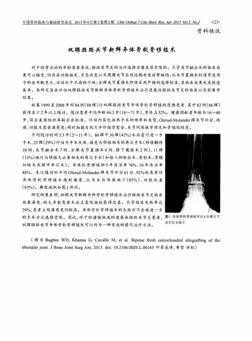
的手 术 方 式选 择 受 限 。 因此 , 对 于 经谨 慎 挑 选 的进 展性 胫 距 关 节 炎 患者 , 图 1 异 体 骨 软 骨 移 植 术 后 6年 踝 关 节 双 踩胫 距 关 节异体 骨 软骨移 植 术 可 以作 为一种 有 效的替 代 治疗 方法 。
中 国骨 科 临床 与基 础 研 究 杂 志 h o pJC / i nBa s i c Re s , A p t 2 0 1 3 V o 1 . 5 , No . 2
・ 1 2 3 ・
骨科 快 讯
双踝胫 距关节新鲜异体 骨软 骨移植 术
平 均 随访 时 间5 . 3 年( 2 - v1 1 年) 。8 6 踩 中3 6 踝( 4 2 %) 术后 需行进 一 步 手术, 2 5 踝( 2 9 %) 评 估 为手 术 失败 , 接 受 与移植 相 关的 再次 手术 ( 移植 翻 修
1 0例 、 关 节 融 合 术 7例 、 全踝 关 节 置 换 术 6例 、 膝 下 截 肢 术 2例 ) , 1 1 踝
岁, 符合 关 国组 织库 联 合 会标 准 。评 估 内容 包括 再 手术 的 频 率和 类型 , O l e r u d — Mo l a n d e r 踩 关 节 评分 , 疼
痛、 功 能及 患者 满 意度 ; 同时拍摄 X线片评 价植 骨 愈合 、 关 节 间隙狭 窄情 况和 骨塌 陷程 度 。
结果。
收集 1 9 9 9 至2 0 0 8 年间8 4 例( 8 8 踝) 行 双踝 胫 距 关 节 异体 骨软 骨 移植 的连 续 患者 , 其 中8 2 例( 8 6 踝)
自体软骨移植在鼻尖整形术中的护理体会

自体软骨移植在鼻尖整形术中的护理体会鼻尖圆钝、鼻尖低平是中国女性常见的鼻部形态,随着生活水平的提高,越来越多的爱美人士要求进行鼻尖整形术,以使面部各个器官达到和谐美观。
当前行鼻尖整形的材料很多,如膨体、自体鼻中隔软骨、肋软骨、耳廓软骨等。
我院自2008年~2009年应用自体软骨移植塑形鼻尖共35例患者,取得了较好的临床效果,现将护理体会报道如下。
1 临床资料1.1一般资料:35例鼻尖圆钝、鼻尖低平的患者初始要求行鼻尖整形术,其中女性30例,男性5例,年龄23~30岁,术前鼻前庭区皮肤均无感染病灶,所有患者近期无感冒、流鼻涕等症状,女性手术避开月经期进行。
1.2手术方法:手术在局麻下进行,采用鼻小柱“飞鸟形”切口,切开皮肤后暴露鼻翼大软骨,切除其间的脂肪、结缔组织及鼻翼大软骨外侧脚的上外侧端。
以5-0丝线水平褥式缝合鼻翼大软骨内侧脚、穹窿部,以缩小鼻小柱宽度及延长鼻小柱,移植软骨取自肋软骨者10例,来自耳甲软骨20例,鼻中隔作为供区5例。
将切取的软骨按照鼻尖低平的不同程度进行塑形、移植。
1.3 结果:术后一周拆除鼻尖缝线,所有患者鼻尖外形良好,经护理干预后没有出现切口感染现象,两例患者术后第2日鼻尖部皮肤轻度红肿,给予对症处理,红肿消退。
供软骨区无血肿、穿孔、气胸等并发症发生,切口一期愈合。
2 护理2.1术前护理:要求行鼻尖整形的患者大多是身体健康的爱美人士,对手术抱有较大的期待,甚至是些不切实际的幻想,因此,护理人员要通过耐心的术前沟通来了解患者的求美动机,特别对一些要求较高的患者应进行心理疏导,尽量消除其内心深处的明星效应,引导患者以更实际的眼光来看待自己鼻尖的缺陷,从而避免术后不必要的医患纠纷。
2.2鼻部切口的护理:切口拆线前嘱患者忌辛辣刺激食物,常规给予抗生素静点5~7天以预防切口感染。
术后24h密切观察,防止鼻部术区敷料被血性渗出物浸透,如发现应及时通知医师给予换药,取出鼻孔填塞的纱布,以碘伏消毒切口,重新填塞鼻孔,避免逆行感染。
- 1、下载文档前请自行甄别文档内容的完整性,平台不提供额外的编辑、内容补充、找答案等附加服务。
- 2、"仅部分预览"的文档,不可在线预览部分如存在完整性等问题,可反馈申请退款(可完整预览的文档不适用该条件!)。
- 3、如文档侵犯您的权益,请联系客服反馈,我们会尽快为您处理(人工客服工作时间:9:00-18:30)。
Cartilage Injuries and RestorationWhat is an articular cartilage injury?An articular cartilage injury occurs when there is damage to the joint surface. Injuries to the cartilage can be partial thickness (part of the way down to bone) or full-thickness (all the way down to bone). The problem with articular cartilage injuries is that they have very limited ability to heal.How is the articular cartilage injured?Cartilage injuries can occur from trauma, such a football tackle or twisting injury, or gradually over time. In addition, there are certain diseases, such as osteochondritis dessicans, which causes damage to an area of cartilage and bone in the knee without a definite cause. When there is significant loss of the articular cartilage, the knee is considered to have “arthritis”.How do I know my articular cartilage is injured?Injuries to the articular cartilage most typically cause pain in the knee in the area of the damage. In addition, patients can get swelling, locking, or buckling of the knee. In some cases, it can be difficult to know for sure if cartilage damage is the reason for knee pain.Do I need x-rays, MRI’s or any othe r test?A set of x-rays is usually ordered to evaluate the bones and cartilage around the knee. The x-rays are primarily used to evaluate for arthritis and severity of the articular cartilage injury of the knee joint. If the damage is small, the x-rays may appear normal. A MRI may be ordered to look for damage to the articular cartilage and rule out any other injuries to the knee. In some cases, the damage cannot be seen on the MRI, even though it is present.Is there other damage to the knee when the articular cartilage is injured?There is frequently other damage to the knee in cases of articular cartilage damage, which occur at thetime of the injury. These include ligament tears or tears of the meniscus. If surgery is needed, all of the injuries will be addressed at the time of surgery.What are the treatment options for articular cartilage injuries?Some patients with an articular cartilage injury improve with conservative treatment. The treatment includes exercises, use of anti-inflammatory medications (NSAIDs), and possibly an injection of steroid. The exercise may include a program you can do at home or formal physical therapy. Depending on the extent of the damage, some patients get better with these treatments and do not require surgery. If patients do not get better with conservative therapy, or have a large articular cartilage lesion, surgery may be necessary.How are articular cartilage injuries treated with surgery?The surgery for articular cartilage injuries depends on the extent of the problem. There are several surgical options, and which procedure is best depends on several factors. These factors include the patient’s age and activity level, the size of the lesion, and the chronicity (age) of the lesion.What are the treatment options for articular cartilage injuries?Some patients with an articular cartilage injury improve with conservative treatment. The treatment includes exercises, use of anti-inflammatory medications (NSAIDs), and possibly an injection of steroid. The exercise may include a program you can do at home or formal physical therapy. Depending on the extent of the damage, some patients get better with these treatments and do not require surgery. If patients do not get better with conservative therapy, or have a large articular cartilage lesion, surgery may be necessary.How are articular cartilage injuries treated with surgery?The surgery for articular cartilage injuries depends on the extent of the problem. There are severalsurgical options, and which procedure is best depends on several factors. These factors include the patient’s age and activity level, the size of the lesion, and the chronicity (age) of the lesion.The first step in evaluating the lesion is usually arthroscopy. The arthroscope is a fiber optic instrument (narrower than a pen) which is put into the knee joint through small incisions. A camera is attached to the arthroscope and the image is viewed on a TV monitor. The arthroscope allows me to fully evaluate the entire knee joint, including the knee cap (patella), the cartilage surfaces, the meniscus, the ligaments (ACL & PCL), and the joint lining. Small instruments ranging from 3-5 millimeters in size are inserted through additional incisions so that I can feel the joint structures for any damage, diagnose the injury, and then repair, reconstruct, or remove the damaged tissue.Once the lesion is evaluated, there are several options for treatment. These include:▪Smoothing of the lesion and removing loose edges only (debridement)▪Techniques to stimulate scar cartilage to grow into the lesion (microfracture) ▪Techniques to replace the lesion with new cartilage (osteochondral autografts, osteochondral allografts, or autologous chondrocyte implantation). Each of these techniques is briefly described below.What is debridement of the articular cartilage?Debridement is performed by using small arthroscopic instruments, such as a mechanical shaver, to smooth the cartilage edges. This is performed so that there are no loose edges to irritate the joint, and to prevent the area of damage from expanding. Debridement is primarily used for small lesions, or when severe arthritis is found that involves the entire knee joint. Many patients can do well with debridement.What is the microfracture technique?Microfracture is a technique to attempt to repair damaged articular cartilage. Small holes are poked into the bone in order to allow blood and marrow healing elements into the area of missing cartilage. This technique allows scar cartilage (fibrocartilage) to fill the area where the cartilage is missing. This technique is easy to perform and can create good results in a lot of patients. However, since the cartilage is scar cartilage, it may not be as durable as other techniques to restore cartilage defects. Microfracture can be performed during an arthroscopy, and no other incision or surgery is needed.What is an osteochondral AUTOGRAFT?An osteochondral autograft is a technique to take a small piece of cartilage and bone from one area of the knee and put it in the area that the cartilage is missing. The cartilage is taken from an area in the knee that feels minimal stress, so it is thought that patients do not notice that the piece of cartilage is missing. This technique can be very effective for small areas of missing cartilage. An osteochondral autograft can often be performed by arthroscopic techniques, but sometimes requires an open incision on the knee.What is an osteochondral ALLOGRAFT?An osteochondral allograft is a technique to take a piece of cartilage and bone from a cadaver (as opposed to your own knee) and place it in the area that the cartilage is missing. This technique is good when there is a large area of cartilage that is missing, or if there is both bone and cartilage missing (such as cases of osteochondritis dessicans). The donor allograft is testing for bacteria, hepatitis, and HIV. However, there is always a small chance of disease transmission from an allograft. An osteochondral allograft usually requires an open incision on the knee.What is Autologous chondrocyte implantation?Autologous chondrocyte implantation is a technique to restore cartilage from your own knee in the area of the cartilage injury. The technique first involves performing an arthroscopy and taking a small piece of cartilage from your knee (biopsy), which is then sent to a lab in Boston. The lab then grows more cartilage cells from the cells given in the biopsy. A second surgery is then performed (about 4 to 12 weeks later) to implant the cartilage cells. This involves an open incision on the front of the knee. A small piece of the outer layer of the bone, called the periosteum, is taken from the lower leg and is sewn in place in the area of the cartilage defect. The cartilage cells are then injected in the area of the defect, and the periosteum is sealed. In many cases, this cartilage then grows into nearly normal articular cartilage.。