抗苗勒氏管激素
抗缪勒氏管激素有关的选择题

抗缪勒氏管激素有关的选择题摘要:一、抗缪勒氏管激素的基本概念二、抗缪勒氏管激素的作用与功能三、抗缪勒氏管激素在临床应用中的重要性四、抗缪勒氏管激素检测的方法与意义五、影响抗缪勒氏管激素水平的因素六、提高抗缪勒氏管激素水平的方法七、总结与展望正文:一、抗缪勒氏管激素的基本概念抗缪勒氏管激素(Anti-Mullerian Hormone,简称AMH)是一种由卵巢内的颗粒细胞分泌的蛋白质激素。
它最早在1947年被科学家发现,被誉为“卵巢储备功能的代表性指标”。
二、抗缪勒氏管激素的作用与功能1.调节卵泡发育:抗缪勒氏管激素在女性体内主要作用于卵泡,促进其生长和发育。
2.抑制雄激素生成:抗缪勒氏管激素可抑制卵巢内细胞分泌雄激素,维持女性体内激素平衡。
3.抑制缪勒氏管发育:在胚胎发育过程中,抗缪勒氏管激素可抑制缪勒氏管的生长,确保生殖器官的正常发育。
三、抗缪勒氏管激素在临床应用中的重要性1.评估卵巢储备功能:抗缪勒氏管激素水平可用于评估女性的卵巢储备功能,帮助预测生育能力。
2.诊断多囊卵巢综合征:抗缪勒氏管激素水平降低是多囊卵巢综合征的特征之一。
3.监测辅助生殖技术疗效:在辅助生殖技术治疗过程中,监测抗缪勒氏管激素水平可评估治疗效果。
四、抗缪勒氏管激素检测的方法与意义1.检测方法:抗缪勒氏管激素水平的检测方法有酶联免疫吸附法、化学发光法等。
2.检测意义:定期检测抗缪勒氏管激素水平,有助于了解女性生殖健康状况,为生殖健康决策提供依据。
五、影响抗缪勒氏管激素水平的因素1.年龄:随着年龄的增长,女性抗缪勒氏管激素水平逐渐降低。
2.生育状况:生育过的女性抗缪勒氏管激素水平较低。
3.内分泌疾病:如多囊卵巢综合征、卵巢早衰等疾病会影响抗缪勒氏管激素水平。
六、提高抗缪勒氏管激素水平的方法1.生活方式调整:保持良好的作息、饮食,增加运动,减轻压力。
2.药物治疗:在医生指导下,使用促排卵药物或激素调节药物。
3.辅助生殖技术:如试管婴儿等辅助生殖技术可提高抗缪勒氏管激素水平。
抗谬勒式管激素偏低的原因

抗谬勒式管激素偏低的原因1. 引言大家好,今天咱们要聊一个话题,听上去有点拗口,但其实也没那么复杂。
那就是抗谬勒式管激素(AMH)偏低的问题。
别急,虽然名字听起来像是化学课上的难题,其实它跟咱们的生活息息相关,只要捋顺了这些概念,了解了它的作用,咱们就能很轻松地掌握这个问题。
是不是有点小激动?那就跟我一起来深入了解一下吧!2. AMH是什么鬼?2.1. 先说说这个AMH到底是什么。
抗谬勒式管激素听上去像个外星名词,但其实它是咱们身体里一位重要的“助手”。
它主要是由卵巢中的颗粒细胞分泌的,作用是帮助调控卵巢中的卵泡发育。
简单来说,就是它能告诉咱们卵巢的卵子储备情况。
如果AMH水平偏低,就可能意味着咱们的卵巢里“库存”不多了,卵子资源可能会出现问题。
2.2. 那么,AMH偏低又是因为什么呢?这就要从几个方面来看了。
首先,年龄是个重要因素。
咱们常说“岁月不饶人”,年纪大了,卵巢里的卵子数量自然也会减少。
这是自然规律,谁都难以抗拒。
还有就是遗传因素,家里的“基因传承”可能也会影响到AMH的水平。
如果家里的女性都容易出现卵巢功能问题,那你也可能会受影响。
3. 偏低的原因3.1. 除了年龄和遗传,生活方式也能“搅和”AMH水平。
比如,过度的压力、饮食不规律,甚至是熬夜,这些坏习惯都可能让咱们的激素水平失衡。
咱们都知道“健康是革命的本钱”,可是生活中很多人总是把健康当成“后花园”,随意糟蹋。
尤其是那些爱吃快餐、长期熬夜的人,AMH偏低的可能性就更大了。
3.2. 还有一种情况是一些特殊的医疗状况。
比如多囊卵巢综合症(PCOS)和一些自体免疫性疾病。
这些病症往往会影响卵巢的正常功能,AMH水平自然也会受到影响。
说白了,就是这些疾病让咱们的“卵巢工厂”出问题了,产出的“产品”也就不如预期了。
再加上有些药物的副作用,可能也是AMH偏低的原因之一。
4. 应对方法4.1. 知道了原因,那咱们该如何应对呢?首先,保持健康的生活方式是关键。
抗缪勒氏管激素有关的选择题
抗缪勒氏管激素有关的选择题
(实用版)
目录
1.抗缪勒氏管激素的基本概念
2.抗缪勒氏管激素的临床应用
3.抗缪勒氏管激素的影响因素
4.抗缪勒氏管激素的检查结果分析
5.抗缪勒氏管激素与卵巢储备能力的关系
正文
抗缪勒氏管激素(AMH)是一种由卵巢内的小卵泡分泌的激素,它是
评估女性卵巢储备能力的重要指标。
卵巢内的小卵泡数量越多,AMH 的浓度便越高;反之,当卵泡随着年龄及各种因素逐渐消耗,AMH 浓度也会随之降低,接近绝经期时,AMH 便渐趋于 0。
因此,AMH 的检查结果可以反映卵巢储备能力的高低。
AMH 在临床上的应用广泛,它可以用于评估女性的年龄相关生育能力,预测卵巢储备功能,诊断多囊卵巢综合症等。
与性激素六项相比,AMH 的检测数值存在个体差异性,但它对卵巢储备功能的预测价值更高。
目前认为,AMH 结合窦卵泡计数(AFC)是评价卵巢储备功能的最佳指标。
AMH 的水平受多种因素影响,包括年龄、生育状况、吸烟、饮酒、体重指数、卵巢手术、化疗、放疗等。
一般来说,随着年龄的增长,AMH 的水平会逐渐下降。
对于育龄女性来说,AMH 与年龄呈负相关。
在检查 AMH 时,如果数值小于 1.0,说明卵巢功能逐渐下降,卵巢
储备能力降低。
对于 24 岁的女性来说,如果 AMH 值偏低,意味着卵巢功能低下,需要尽早考虑结婚生育。
当然,具体的诊断和治疗还需要根据个体差异和临床表现来综合判断。
总之,抗缪勒氏管激素(AMH)是评估女性卵巢储备能力的重要指标,它可以反映卵泡数量的减少,从而预测怀孕的几率。
抗缪勒管激素临床八大应用

AMH 值会随着卵巢功能的变化呈起伏变化。
AMH 指数越高,说明卵子的库存量越大,生育能力 自然就较强。
AMH 降低时,代表卵巢正在老化,表示女性生育力 的衰退。
通过检测 AMH 可更早更准确反映年龄相关卵巢储备 功能的下降。
5. 卵巢颗粒细胞瘤:
因由于 AMH 仅由颗粒细胞表达,其表达水平由卵巢 初级卵泡和窦前卵泡的颗粒细胞决定,这说明 AMH 可以作为卵巢颗粒细胞瘤的一个标记物。
据报道 76%~93% 的卵巢颗粒细胞瘤中 AMH 表达均 为阳性,且 AMH 与卵巢颗粒细胞瘤的肿瘤大小及分 化程度具有独立相关性,并且对卵巢颗粒细胞瘤的复 发有一定预测价值。
总结
AMH 作为与一种女性生殖功能相关的激素,目前主 要用于卵巢储备功能评估、卵巢早衰、卵巢过度刺激 综合征、卵巢颗粒细胞肿瘤,辅助生殖以及某些自身 免疫性疾病等的诊治。缺乏全球化的统一诊断标准是 AMH 检测在临床应用中的一大障碍。不过,随着临 床应用的不断增多,其临床价值将获得更多的循证医 学证据,将有着更广泛的应用前景。
4. 辅助生殖技术:
AMH 水平能够预测卵巢反应性,识别有卵巢过度刺 激综合征风险的女性,可根据 AMH 数值来判断使用 促排卵药物的用量。
研究发现接受 IVF/ICSI 治疗的患者血清及卵泡液中 AMH 水平越高则受精率越高,AMH 可能成为预测受 精率的指标。
除此之外,AMH 预测 OHSS 优于年龄和 BMI,发生 OHSS 患者的基础 AMH 较正常人高 6 倍,提示 AMH
储备功能也仍然良好。
女性的生育能力与其卵巢储备功能和体内的各类 生殖激素具有密切的关系。
抗苗勒管激素测定标准

抗苗勒管激素测定标准
抗苗勒氏管激素正常值在2.0-6.8ng/ml,该激素数值出生就存在,在性成熟以后达到稳定状态,年龄增大以后数值会下降。
抗苗勒氏管激素是卵巢窦前卵泡及小窦卵泡的颗粒细胞分泌的一种糖蛋白,在体内有比较稳定的数值,并不受月经周期的影响,可以直接反映卵巢的储备能力,通过抽血检查数值在2.0-6.8ngml 属于标准范围。
数值降低提示卵巢储备能力下降;数值升高提示多囊卵巢综合征的可能。
此外,出现数值明显下降,本身还有备孕需求,要及时就医,部分人群需要做卵泡储备。
抗缪勒氏管激素质控品产品技术要求博粹

抗缪勒氏管激素质控品产品技术要求博粹抗缪勒氏管激素(Anti-Müllerian Hormone, AMH)是一种重要的生物标志物,用于评估卵巢储备功能和女性生殖健康状况。
其测试结果对于评估女性的生殖健康状态、确定生育年龄、进行不孕不育疾病的诊断和治疗具有重要意义。
1.纯度要求:抗AMH质控品的纯度要求高,必须纯净且不含其他有可能对测试结果造成干扰的杂质。
其纯度可以通过高效液相色谱(HPLC)和酶联免疫吸附测定(ELISA)等技术进行验证。
2.稳定性要求:抗AMH质控品应具有良好的稳定性,以确保其在长期储存和使用过程中的一致性和可靠性。
稳定性可以通过长期储存试验、温度敏感性试验等进行验证。
3.活性要求:抗AMH质控品应具有高度的活性,能够与AMH快速、特异地结合,并能够准确地模拟AMH在生物体内的生理功能。
其活性可以通过生物活性试验、逆向酶联免疫吸附测定等进行验证。
4.重复性要求:抗AMH质控品的制备应具备良好的重复性,确保每一批制备的质控品在不同的实验条件下具有一致的检测结果。
重复性测试可以通过同批次和不同批次的重复性试验进行验证。
5.安全性要求:抗AMH质控品应符合生物安全要求,不得存在有害成分,并且对操作人员和环境无危害。
安全性可通过毒理学试验进行验证。
总之,抗缪勒氏管激素质控品的产品技术要求包括纯度、稳定性、活性、重复性和安全性等方面。
制造商在生产过程中应严格按照相关质量标准进行操作,确保质控品的质量稳定和一致,并通过严格的质量控制措施
确保质控品的合格率和可靠性,以满足AMH检测试剂盒的准确性和可靠性要求。
抗苗勒管激素测定标准
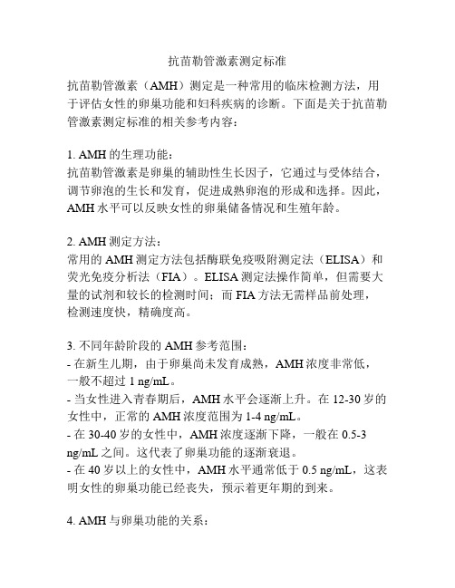
抗苗勒管激素测定标准抗苗勒管激素(AMH)测定是一种常用的临床检测方法,用于评估女性的卵巢功能和妇科疾病的诊断。
下面是关于抗苗勒管激素测定标准的相关参考内容:1. AMH的生理功能:抗苗勒管激素是卵巢的辅助性生长因子,它通过与受体结合,调节卵泡的生长和发育,促进成熟卵泡的形成和选择。
因此,AMH水平可以反映女性的卵巢储备情况和生殖年龄。
2. AMH测定方法:常用的AMH测定方法包括酶联免疫吸附测定法(ELISA)和荧光免疫分析法(FIA)。
ELISA测定法操作简单,但需要大量的试剂和较长的检测时间;而FIA方法无需样品前处理,检测速度快,精确度高。
3. 不同年龄阶段的AMH参考范围:- 在新生儿期,由于卵巢尚未发育成熟,AMH浓度非常低,一般不超过1 ng/mL。
- 当女性进入青春期后,AMH水平会逐渐上升。
在12-30岁的女性中,正常的AMH浓度范围为1-4 ng/mL。
- 在30-40岁的女性中,AMH浓度逐渐下降,一般在0.5-3ng/mL之间。
这代表了卵巢功能的逐渐衰退。
- 在40岁以上的女性中,AMH水平通常低于0.5 ng/mL,这表明女性的卵巢功能已经丧失,预示着更年期的到来。
4. AMH与卵巢功能的关系:较高的AMH水平通常与更多的卵泡储备和更好的卵巢功能相对应,而较低的AMH水平则意味着卵泡储备的减少甚至耗尽。
因此,AMH测定可以用于评估女性的生殖潜力、预测卵巢反应性、指导辅助生殖技术的治疗方案选择等。
5. AMH测定在妇科疾病的应用:- 多囊卵巢综合征(PCOS):PCOS患者的AMH水平通常较高,可用于PCOS的诊断和病情评估。
- 卵巢肿瘤:卵巢肿瘤患者的AMH水平通常降低,可以作为肿瘤的辅助诊断指标。
- 卵巢早衰:卵巢早衰患者的AMH水平非常低,可以用于早衰的早期诊断和管理。
总之,抗苗勒管激素测定标准作为一种重要的临床检测方法,可以用于评估女性的卵巢功能和妇科疾病的诊断。
抗苗勒氏管激素生理基础及临床应用

抗苗勒氏管激素生理基础及临床应用抗苗勒氏管激素生理基础及临床应用
1、引言
1.1 研究背景
1.2 研究目的
1.3 研究意义
2、苗勒氏管激素生理基础
2.1 苗勒氏管概述
2.2 激素调控
2.2.1 长期调节
2.2.2 短期调节
2.3 激素信号传导路径
2.3.1 受体信号传导
2.3.2 下游效应器
3、抗苗勒氏管激素的临床应用
3.1 激素治疗相关疾病
3.1.1 生长激素治疗
3.1.2 甲状腺激素治疗
3.1.3 垂体激素替代治疗
3.2 抗苗勒氏管激素药物开发
3.2.1 抗生长激素药物
3.2.2 抗甲状腺激素药物
3.2.3 抗垂体激素药物
4、研究方法
4.1 动物模型构建
4.2 激素测量方法
4.3 细胞培养和处理
5、结果与讨论
5.1 苗勒氏管激素的生理调节机制 5.2 抗苗勒氏管激素的临床应用
5.3 研究结果的解释与讨论
6、结论
6.1 主要研究结论总结
6.2 进一步研究的展望
附件:
附件1:研究数据表格
附件2:实验图片
:::
法律名词及注释:
1、苗勒氏管:指人体中的一种器官,通常用来指代泌乳激素及其相关调节机制。
2、抗苗勒氏管:指针对苗勒氏管激素的制剂或治疗方法。
3、激素调节:指体内激素水平在一定范围内维持稳定,并通过反馈机制来调节机体功能。
4、受体信号传导:指激素分子与细胞上的特定受体结合后,启动一系列信号传导过程。
5、下游效应器:指受体信号传导途径中的分子或细胞器,对信号进行进一步处理和传递。
- 1、下载文档前请自行甄别文档内容的完整性,平台不提供额外的编辑、内容补充、找答案等附加服务。
- 2、"仅部分预览"的文档,不可在线预览部分如存在完整性等问题,可反馈申请退款(可完整预览的文档不适用该条件!)。
- 3、如文档侵犯您的权益,请联系客服反馈,我们会尽快为您处理(人工客服工作时间:9:00-18:30)。
Iran J Reprod Med Vol. 13. No. 4. pp: 227-230, April 2015Anti-mullerian hormon level and polycystic ovariansyndrome diagnosisShahrzad Zadehmodarres1 M.D., Zahra Heidar1 M.D., Zahra Razzaghi2 Ph.D., Leili Ebrahimi3 M.D.,Kaveh Soltanzadeh3 M.D., Farhang Abed3 M.D.Introductionolycystic ovarian syndrome (PCOS),a common endocrinopathycharacterized by oligo-or anovulation, clinical or biochemical hyperandrogenmia, and polycystic ovaries on ultrasonography, affects 5-10% of women of reproductive age (1,2). Recent studies have shown that 50% of women with PCOS fulfill the criteria of metabolic syndrome and that PCOS is frequently associated with insulin resistance accompanied by compensatory hyperinsulinemia, resulting in an increased risk for the development of type 2 diabetes mellitus and cardiovascular disease (2,3). In comparison with healthy women, PCOS have higher level of anti-mullerian hormone (AMH) that is a peptide produced by the granulosa cells of follicles that is widely considered as a highly sensitive marker of ovarian reserve (3). Previous studies have suggested that AMH may play a pathogenetic role in follicular status of PCOS (4,5). Because of long term effect of PCOS and metabolic syndrome, early diagnosis of this endocrinopathy is very important. Our purpose in this study was to find any relation between AMH levels and PCOS diagnosis.Materials and methodsIn this cross sectional study, 117 women between 20-40 years old referred to Infertility clinic, Mahdieh Hospital, Tehran, Iran from 2012 to 2013 were participated in two groups. Written informed consent was obtained from all of them. The case group consisted of 60PZadehmodarres et al228 Iranian Journal of Reproductive Medicine Vol. 13. No. 4. pp: 227-230, April 2015PCOS women (based on Rotterdam criteria consensus) and the control group was 57 women with normal ovulatory state. Our study protocol was approved by Ethics Committee of Mahdieh Hospital.Inclusion criteria in case group was PCOS diagnosis, 20<age<40, and presence of both ovaries. PCOS was ascertained, using the Rotterdam consensus statements, as the presence of two of the following three criteria: PCO morphology (more than 12 follicle with size 2-9 mm or ovarian volume more than 10 ml in one ovary), clinical or biochemical hyperandrogenism (hirsutism with score ≥8 based on Freeman-Galloway scoring or testosterone >2.5 nmol/l, free testosterone ≥0.6 nmol/l), and oligomenorrhea (cycle length >35 days). Exclusion criteria were history of ovarian surgery, and induction ovulation in recent 6 month. Thyroid and adrenal function tests were normal in both groups and they did not use OCP in last month.All of participants were examined carefully at beginning and their demographic data such as age, gravity, weight, height, waist, hip circumference, and history of medical state were written in information sheet. In day 2-4 of cycle, transvaginal sonography (Honda, Japan) was performed and serum hormonal level of AMH (ELIZA, Beckman-culter, ng/ml,), luteinizing hormone (LH) (RIA, mIU/ml), follicle stimulating hormone (FSH), estradiol (E 2) (ECL, pmol/ml), (ECL ng/ml), fasting blood (mg/dl), thyroid-stimulating (mIu/l), and prolactin (PRL) measured.Statistical analysisStatistical analysis was performed with statistical package for the science (SPSS Inc, Chicago, Illinois, version 16.0. The data were analyzed using the Chi-square, fisher exact test, and Student ’s t -test. A p<0.05 was considered as significant.ResultsOur results show that the case and control groups were matched respecting the age and BMI. Mean of AMH level in group was 7.14±6.53ng/ml and in controls 3.34±3.45 ng/ml which the difference statistically significant (p=0.001). Also, level was significantly difference in groups(p=0.0001) (Table I). PCO morphology in none of controls was seen. Overall 29 (48.3%) cases and 11 (19.9%) controls have hirsutism with score >8 (p=0.001). Irregular mense in 37 (61.7%) cases and 15 (26.3%) controls were seen (p=0.001). In four (6.7%) cases and one of the controls (1.8%) hyperprolactinemia was seen (p=0.89).Differences in plasma level of LH, TSH, FBS and E 2 were not statistically significant (Table I). To determine of AMH diagnostic cut off, ROC curves were constructed that presented 3.15 with 70.37% sensitivity and 77.36% specificity and positive predictive value (PPV)=76% and NPV(negative predictive value)=71.93 in order to determinate AMH cut off level in diagnosis of PCOS (Figure 1).Table I. Demographic characteristics of case (PCOS women) and control groupsVariables Case group Control group p -value* Age (Year) 27.07 ± 4.49 28.68 ± 4.98 0.072 BMI (Kg/m 2) 29.02 ± 6.53 28.76 ± 3.41 0.389 AMH (ng/ml) 7.14 ± 6.53 3.34 ± 3.45 0.001 FSH (mIu/ml) 4.53 ± 1.62 6.88 ± 5.56 0.0001 LH (mIu/ml) 5.96 ± 2.93 5.58 ± 3.09 0.452 TSH(mIU/ml)3.03 ±6.272.73± 2.160.34FBS )mg/dl) 92.81±11.14 93.74±10.11 0.755 Testosterone*Student’s t testFigure 1. Roc curve of AMH (blue line) with reference line (green line).Accuracy of AMH in PCOS diagnosisIranian Journal of Reproductive Medicine Vol. 13. No. 4. pp: 227-230, April 2015 229DiscussionAs mentioned earlier because of long term sequel of PCOS including infertility, endometrial hyperplasia, metabolic syndrome, and cardiovascular risk factor, early identification of at risk women would be very useful. Once the diagnosis of PCOS is made, additional evaluation is suggested including a cardio metabolic risk assessment, as well as screening for mood disorder and sleep apnea, screening for diabetes mellitus and for women pursuing fertility assessment of ovulatory status (6). The present study demonstrated that there is positive correlation between AMH level and PCOS diagnosis and 3.15 nmol/ml as cut off level (with sensitivity and specificity of 70.37% and 77.36% respectively) could use for PCOS diagnosis (p=0.001). In other researches AMH>7.7 ng/ml (3) and >3.5 ng/ml (9) represented as diagnostic cutoff level for PCOS.The differences of FSH, PCOS morphology, hirsutism and irregular mense between patients and normal women were statistically significant but testosterone, PRL, FBS, E 2 and BMI differences between case and control groups were not significant. In another study there was correlation between PCO morphology and AMH level in regular cycle adolescent but in our research and Eilertsen , PCOS morphology between case and controls was significant different (7,8). There is some debate about value of sonographic finding in PCOS diagnosis Vise versa our finding, in Villarroel study PCOM was a common finding in normal ovulatory women(8) thus there is need to more research in this field.In two studies, androgen level in PCOS patients was significantly higher, and in one study androgen level in overweigh (BMI >27) was higher but our study did not show this different race in study groups may be responsible (11-13). In some studies there was linear relation between AMH and testosterone level, hirsutism and oligomenorrhea but our study did not find such result (7, 11, 12). It needs to mention that our population study group selected from infertile women and it maybe affected our result. This is obvious that larger studies in different rational group of patients are needed to determine the accurate diagnostic cut off level of AMH.AcknowledgementsWe appreciate of Infertility ward of Mahdieh Hospital, Tehran, Iran for supporting and cooperation.Conflict of interestThe authors declare that they have no conflicts of interest.References1. Norman RJ, Dewailly D, Legro RS, Hickey TE.Polycystic ovary syndrome. Lancet 2007; 370: 685-697.2. Welt CK, Gudmundsson JA, Arason G, Adams J,Palsdottir H, Gudlaugsdottir G, et al. Characterizing discrete subsets of polycystic ovary syndrome as defined by the Rotterdam criteria: the impact of weight on phenotype and metabolic features. J Clin Endocrinol Metab 2006; 91: 4842-4848.3. Azziz R, Carmina E, Dewailly D, Diamanti-Kandarakis E, Escobar-Morreale HF, Futterweit W, et al. Position statement: criteria for defining polycystic ovary syndrome as a predominantly hyperandrogenic syndrome: an Androgen Excess Society guideline. J Clin Endocrinol Metab 2006; 91: 4237-4245.4. Weenen C, Laven JS, Von Bergh AR, Cranfield M,Groome NP, Visser JA, et al. Anti-Mullerian hormone expression pattern in the human ovary: potential implications for initial and cyclic follicle recruitment. Mol Hum Reprod 2004; 10: 77-83.5. Pigny P, Merlen E, Robert Y, Cortet-Rudelli C,Decanter C, Jonard S, et al. Elevated serum level of anti-Mullerian hormone in patients with polycystic ovary syndrome: relationship to the ovarian follicle excess and to the follicular arrest. J Clin Endocrinol Metab 2003; 88: 5957-5962.6. Wild RA, Carmina E, Diamanti-Kandarakis E.Assessment of cardiovascular risk and prevention of cardiovascular disease in women with the polycystic ovary syndrome: a consensus statement by the Androgen Exess and polycystic ovary syndrome Society. J Clin Endocrinol Metab 2010; 95: 2038. 7. Eilertsen TB, Vanky E, Carlsen SM. Anti-Mullerianhormone in the diagnosis of polycystic ovary syndrome: can morphologic description be replaced? Hum Reprod 2012; 27: 2494-2502.8. Villarroel C, MerinoPM, Lo´pez P, Eyzaguirre FC.Polycystic ovarian morphology in adolescents with regular menstrual cycles is associated with elevated anti-Mullerian hormone. Hum Reprod 2011; 26: 2861-2868.9. Chao KC, Ho CH, Shyong WY, Huang CY, Tsai SC,Cheng HY, et al. Anti mullerian hormone serum level as a predictive marker of ovarian function in Taiwanese women. J Chin Med Assoc 2012; 75: 70-74.10. Carmina E, Campagna AM, Mansuet P, Vitale G,Kort D, Lobo R. Dose the level of serum anti mullerian hormone predict ovulatory function inZadehmodarres et al230 Iranian Journal of Reproductive Medicine Vol. 13. No. 4. pp: 227-230, April 2015women with PCOS with aging. Fertil Steril 2012; 98: 1043-1046.11. Woo HY, Kim KH, Rhee EJ. Differeces of theassociation of anti-Mullerian hormone with clinical or biochemical characteristics between women with and without polycystic ovary syndrome. Endocrine J 2012; 59: 781-790.12. Pingy P, Jonard S, Robert Y, Dewailly D. Serum anti- Mullerian hormone as a surrogate for antral follicle count for definition of the polycystic ovary syndrome. J Clin Endocrinol Metab 2006; 91: 941-944.13. Swellam M, Khaial A, Mosa T, El-Baz H. Anti-Mullerian and androgens hormones in women66 with polycystic ovary syndrome undergoing IVF/ICSI. Iran J Reprod Med 2013; 11: 883-890.。