鼻咽癌不同方式放疗疗效分析
鼻咽癌的综合治疗方案及疗效分析

鼻咽癌的综合治疗方案及疗效分析鼻咽癌是一种常见的头颈部恶性肿瘤,其发病率在亚洲地区尤为突出。
目前,虽然鼻咽癌的治疗手段日趋完善,但其治愈率依然较低。
为此,针对鼻咽癌的综合治疗方案日渐受到关注。
本文将从手术治疗、放疗、化疗及生物治疗等方面综合分析鼻咽癌的综合治疗方案及疗效,并探讨鼻咽癌未来治疗发展的方向。
一、手术治疗手术治疗是鼻咽癌治疗的重要手段之一。
其适用于早期鼻咽癌以及局部进展的鼻咽癌。
目前,采用显微手术的方式进行手术治疗已经成为常用方法。
显微手术的优点在于缩小了手术创面,减少创面感染的风险以及促进病人的康复。
对于早期鼻咽癌,采用显微手术可以达到较好的治愈效果;而对于其他阶段的鼻咽癌,手术治疗则需要与放疗或化疗结合,以达到更好的疗效。
二、放疗放疗是鼻咽癌治疗的核心手段之一,且常与手术或化疗联合使用。
目前,放疗治疗主要采用三维适形放疗和强度调制放疗。
三维适形放疗具有放疗精度高、副作用小等优点,且节约时间,是一种非常有效的治疗手段。
强度调制放疗可根据患者的病变情况进行放射剂量的非均一分配。
其优点在于可使肿瘤灶部位得到更高剂量的放射,同时保证正常组织得到较低剂量的放射。
放疗优点在于不需要手术,对于已经转移的鼻咽癌患者较为适用。
但放疗可导致口干、咽喉疼痛、面部水肿、乏力、口腔感染等副作用,因此在放疗治疗时也要注意患者的生活质量。
三、化疗化疗是鼻咽癌常用的综合治疗手段之一,特别适用于中晚期鼻咽癌的治疗。
化疗可通过抑制癌细胞的DNA合成、两性激素受体、细胞周期和DNA修复机制等途径发挥作用。
化疗手段一般包括使用单一药物或联合使用多种药物。
单一药物使用化疗的效果相对较差,通常采用多药物联合治疗以达到较好的疗效。
临床常用化疗方案为TP(多柿原代环磷酰胺、紫杉醇)、PF(南瓜胡萝卜素、氟脲嘧啶)等。
四、生物治疗生物治疗是近年来的新兴治疗手段,其可通过调节免疫系统、促进细胞凋亡、靶向药物等方式发挥疗效。
目前,免疫治疗(针对PD-1、PD-L1、CTLA4等抑制剂)被认为是一种可将机体免疫力调节至一定程度上来达到治疗效果的重要手段。
鼻咽癌调强放射治疗51例临床疗效分析

通讯 作者 :周同 冲 邮箱 :y d l 7 2 iae m g al 0 2 @sn .o 7 1
2 9 92
国际医药卫 生导报 2 1 0 0年 第 1 6卷 第 2 4期
I MHGN,De e e 0 0,V 11 N .4 cmb r2 1 o.6 o2
日.5 / ,G V分 次剂量为 22G ,其处方剂量 为 7 - 48G /2~3 次 周 T . y 04~7 . y3 4次;C V1 次剂量 T 分 为 1 v 处 方剂量为 6 .~6 .G /4 6 .G , 8 1 2 48 y3 ~3 次;C V 分次剂量 为2G , T2 y 处方剂量 为 5 0~5 y 4G /
( 稿 日期 :2 1 — — 3 收 001 0) 0
( 任 校 对 :彭 鹏 旭 ) 责
鼻 咽癌调 强放 射治疗 5 例 临床疗效 分 析 1
杜莉莉 周同冲 石 兴源 刘源 宋先璐 廖 志伟
【 摘要 】 目的
观察 鼻咽癌调强放 射治疗 的急性 毒副反应 和近期疗效 ,并 于三维适形 放
6 v3 4G /0~3 次 。C V 处方剂量 5 5 y2 ~2 次 , 2 T2 0 4G /5 7 有残 留者适当缩野补量 。采用放 化综合 治疗 , 结合患者分期 情况给予顺铂 + 氟腺嘧 啶方 案化疗 ,中位 随访 时间 1 个月 。结果 7 D H分 V
压 、血氧饱 和度监测 ,而且必须 有专职麻醉师协 械 以满 足临床需求 。另外 ,部分 患者检查时会 出 助完 成 ,不 但耗时 ,而且 费用较 大 ,而经鼻 电子 现鼻衄 、鼻痛 ,本组 共出现 9 ,但 经相应处理 例 胃镜 检查 时从 鼻 孔插 入 ,可 不 接触 舌 根就 到达 后 可缓 解 ,并 能 完成 检查 。
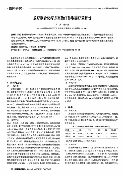
放疗联合化疗方案治疗鼻咽癌疗效评价

5% ; 0 恶化( D)鼻咽及 颈部肿瘤增大 >2 %或 出现新病变。远 P : 5
殊性和鼻咽癌细胞的生物学特点 , 前治疗 以放疗为 主, 5 目 但 年
生存率仅在 3 _% 一5 .%。 63 3 9 失败 的主要原 因是局部复发和远处 转移 , 为了提高鼻 咽癌的疗效 , 人们不断努 力 , 采取 了许多措施 。 2 0 年 1 ~2 0 01 月 0 4年 1 月我科采用放疗联合 P F方案与单纯放
两 组 年 龄 分 布 无 差 异 , 性 别 构 成 有 统 计 学 差 异 ( : .04 但 x =5 5 , 7
或出现新病灶 。毒副反应按 WH O的标准评价 , 统计学方法采用
x0 验 。 检
P . ) <0 5 。所有病例均经鼻咽部活检确诊 , 0 病理类型 : 单放组 : 低
分化鳞 癌 3 7例 , 其他 3例 ; 放化 组 : 低分 化鳞 癌 4 4例 , 他 1 其
例 ;两组间无统计学差异 ( . 3 P . ) x =40 , >O0 。临床分期 按 6 5 19 92年福州分期 : 单放组 : Ⅱ期 1 , 5例 ; 化组 : 5例 Ⅲ2 放 Ⅱ期 2 3 例 , 2例。两组问无统计学差异 ( . 3 P . ) Ⅲ2 x =43 , >00 。 z 7 5
联合 P F方案 治疗 。结果 放疗联合 P F方案治疗组有效率( R P )00 13 5 C + R 8. %,、 、 年生存率为 10 7 .%、8 % ; 0 %、78 6 . 单纯放 9 疗组有效率( R P 5 . 13 5年生存率为 10 7 . 、7 %。结论 放疗联合 P C + R)25 、 、 %, 0 %、5 % 5 . O 5 F化疗方案治疗鼻咽癌 比单纯放疗
复发或转移性鼻咽癌PD-1单抗耐药后联合姑息放疗的疗效分析

复发或转移性鼻咽癌PD-1单抗耐药后联合姑息放疗的疗效分析摘要:目的:本文旨在探究针对复发或转移性鼻咽癌PD-1单抗耐药的姑息放疗联合方案的疗效以及其安全性。
方法:选取2016年1月至2019年12月期间上海市第一人民医院拟定接受PD-1单抗治疗的21例复发或转移性鼻咽癌患者,其中15例PD-1单抗耐药。
均进行姑息放疗治疗,且给予口服保肝药物治疗。
结果:全部21例患者均完整随访,中位随访时间为15个月。
15例PD-1单抗耐药患者中,10例患者经3个月姑息放疗后,局部病变得到缓解,其中9例病灶完全消失;而5例患者局部病变得到部分缓解。
其中治疗后总生存时间(OS)中位数为13个月,无进展生存时间(PFS)中位数为4个月。
并未发生严重不良事件。
结论:姑息放疗联合口服保肝药物治疗可对PD-1单抗耐药的复发或转移性鼻咽癌患者进行有效的姑息治疗,尽可能地控制肿瘤局部病变,延缓肿瘤的进展,提高患者的生存质量和生存期。
该方案安全性较高,值得广泛推广应用。
关键词:鼻咽癌;姑息放疗;PD-1单抗耐药;保肝药物;生存期Abstract: Objective: The purpose of this paper is to explore the efficacy and safety of palliative radiotherapy combined with liver-protecting drugs for patients with relapsed or metastatic nasopharyngeal carcinoma with PD-1 monoclonal antibody resistance.Methods: In this study, 21 patients with relapsed or metastatic nasopharyngeal carcinoma who were scheduled to receive PD-1 monoclonal antibody treatment from January 2016 to December 2019 were selected, of which 15 patients were resistant to PD-1 monoclonal antibody. All patients had palliative radiotherapy and oralliver-protecting drugs treatment.Results: All 21 patients were completely followed up, with a median follow-up time of 15 months. Among the15 patients resistant to PD-1 monoclonal antibody, 10 patients had local lesions relieved after 3 months of palliative radiotherapy treatment, of which 9patients' lesions disappeared completely; while the local lesions of the other 5 patients were partially relieved. The median overall survival time (OS) after treatment was 13 months, and the median progression-free survival time (PFS) was 4 months. No serious adverse events occurred.Conclusion: Palliative radiotherapy combined with oralliver-protecting drugs can effectively palliate for patients with relapsed or metastatic nasopharyngeal carcinoma with PD-1 monoclonal antibody resistance, control tumor lesions as much as possible, delay tumor progression, and improve patients' quality of life and survival time. This scheme has higher safety and is worth promoting.Keywords: nasopharyngeal carcinoma; palliative radiotherapy; PD-1 monoclonal antibody resistance; liver-protecting drugs; survival timNasopharyngeal carcinoma (NPC) is a common malignant tumor in southern China and Southeast Asia. Although radiotherapy and chemotherapy have been proven effective in the treatment of NPC, relapse and metastasis are still common. The use of PD-1 monoclonal antibody has shown promising results in improving the survival rate of patients with advanced NPC, but resistance to the treatment can develop.To address this issue, a combination of palliative radiotherapy and liver-protecting drugs has been proposed as an effective solution. Palliative radiotherapy can provide symptom relief and improve the quality of life for patients with advanced NPC. At the same time, liver-protecting drugs can preventliver damage caused by radiotherapy and chemotherapy, and improve the overall tolerance of the patients to the treatment.In clinical practice, the combination therapy has been shown to effectively control tumor lesions, delay tumor progression, and improve the survival time of patients with relapsed or metastatic NPC with PD-1 monoclonal antibody resistance. Moreover, the scheme has been demonstrated to have relatively low toxicity and is well-tolerated by patients.In conclusion, the combination of palliative radiotherapy and liver-protecting drugs is a safe and effective treatment option for patients with advanced NPC who have developed resistance to PD-1 monoclonal antibody treatment. It is a valuable approach that should be further promoted and researched to improve the outcomes and quality of life of NPC patientsFurthermore, it is important to note that this treatment approach can also benefit patients with other types of tumors who have developed resistance to immunotherapy. The use of liver-protecting drugs can help prevent the potential liver damage caused by radiation, while the radiation itself can enhance the therapeutic effect of the drugs. As such, thisinnovative treatment approach has the potential to provide new hope for cancer patients who have exhausted traditional treatment options.However, there is still much to be learned about the optimal application and administration of this treatment approach. For example, the appropriate dosage and timing of the liver-protecting drugs and radiation therapy may vary depending on the patient's individual characteristics and medical history. Additionally, more research is needed to determine the long-term safety and efficacy of the treatment, particularly in patients with advanced or metastatic disease.Overall, the combination of palliative radiotherapyand liver-protecting drugs represents a promising new approach to treating cancer patients who have developed resistance to immunotherapy. By leveraging the strengths of both therapies, this approach can improve tumor control, reduce toxicity, and contribute to improved long-term outcomes and quality of life for patients. As research in this area continues to expand, we can anticipate further progress in the development of more effective and personalized cancer treatmentsIn recent years, immunotherapy has emerged as a powerful new approach to treating cancer. By harnessing the body's own immune system to fight cancer cells, immunotherapy has shown remarkable success in treating a range of cancer types. However, despite its promise, not all patients respond to this type of therapy, and resistance can develop over time. In order to overcome the limitations of immunotherapy, researchers have been exploring new combination therapies that can enhance its efficacy and reduce toxicity.One such approach is the use of radiotherapy in combination with immunotherapy. Radiotherapy, which uses high-energy radiation to kill cancer cells and shrink tumors, has traditionally been used as a standalone treatment or in combination with chemotherapy. However, recent research has shown that it can also enhance the immune response to cancer by stimulating the release of antigens and promoting the infiltration of immune cells into tumors.When combined with immunotherapy, radiotherapy can help to overcome resistance by creating a more favorable environment for immune cells to attack cancer cells. In addition, radiotherapy can also alter the expression of immune checkpoint proteins, whichare proteins that act as "off" switches for the immune system. By blocking the activity of these proteins, immunotherapy can become more effective in targeting and killing cancer cells.Another promising approach to overcoming resistance to immunotherapy is the use of liver-protecting drugs. These drugs work by reducing the toxicity of immunotherapy drugs on the liver, which can oftenlimit the amount of immunotherapy that can be given to a patient. By reducing the risk of liver damage,liver-protecting drugs can allow for higher doses of immunotherapy to be administered, which in turn can lead to better outcomes.Combining radiotherapy and liver-protecting drugs with immunotherapy is a promising new approach to cancer treatment. By combining the strengths of different therapies, this approach can enhance tumor control, reduce toxicity, and improve long-term outcomes for patients. However, more research is needed in order to determine the optimal combination of therapies and dosing regimens for individual patients.As research in this area continues to expand, we can look forward to the development of more effective and personalized cancer treatments. Through a betterunderstanding of the immune response to cancer and the mechanisms of resistance, researchers can identify new targets for therapy and develop new approaches to overcome the limitations of current treatments. In the years ahead, we can anticipate significant progress in the fight against cancer, and the development of new therapies that can offer hope and improved quality of life for patientsIn conclusion, cancer immunotherapy has emerged as a promising approach to treat various types of cancer by boosting the immune system's ability to recognize and eliminate cancer cells. However, there are still limitations and challenges associated with this approach, including resistance mechanisms and toxicity. Nevertheless, ongoing research and clinical trials in immunotherapy hold great promise for developing more effective and personalized cancer treatments in the future. Overall, the field of cancer immunotherapy is rapidly evolving, and it holds significant potentialfor improving cancer outcomes and patients' quality of life。
鼻咽癌精确放疗与普通放疗的区别

鼻咽癌是一种较为常见的恶性肿瘤,如果鼻咽癌得不到及时有效的治疗,可能会直接危及患者的生命,所以在出现鼻咽癌后需要积极的治疗。
对于鼻咽癌的治疗,目前西医经常使用的手段就有放疗,许多人对于放疗的认识还局限于影视剧中的一些情节或者是日常细碎、凌乱的知识碎片,自从在临床医学的使用中来,也经历了一定发展阶段,开始出现了精准放疗。
那么鼻咽癌精确放疗与普通放疗的区别是什么呢?普通的放疗一般就是指常规二维放疗,操作简单,费用较低,但是靶区的剂量分布较差,对关键组织和器官的保护较差,副作用相对较大。
除了常规二维放疗,还有一些其他放疗,其中就包括三维适形放疗,是一种相对高精度的放疗。
利用CT图像重建肿瘤和正常组织,通过不同方向设置一系列与病灶形状一致的适形照射野,尽量使高剂量区分布与靶区一致,周围正常组织剂量降低。
而调强放疗又是三维适形放疗的高级形式,不但要求射野的形态与靶区一致,而且射线束的剂量强度能够按要求进行调整,不仅能使靶区接受较高剂量和均匀剂量照射,而且能降低周围正常组织的照射剂量。
通常可以增加肿瘤照射剂量,提高疗效;降低毒副作用;使某些常规不能治疗的患者得到治疗(多靶点,邻近重要器官);图像引导放疗是一种四维放疗技术,它是在三维放疗技术的基础上加入了时间因素,考虑到了肿瘤及正常器官在治疗过程中的运动和分次治疗间的位移误差,在治疗前和中利用影像设备对肿瘤及正常器官进行实时的监控,做到更高层次上的精确放疗。
说了这么多的专业术语,可能还是会有患者听起来云里雾里,通俗点讲就是,因为放疗是利用不同能量的放射线照射肿瘤部位,以期杀死癌细胞的治疗手段,为了保证对癌细胞足够的杀伤力,不得不扩大照射的范围来杀死周围正常组织可能存在的癌细胞。
这样却会给正常的组织和体细胞带来一些不必要的伤害,这也是引起放疗毒副作用的原因。
而精准放疗则是能够更加精确地划分病灶和正常组织的界限,从而更好的确定放射线照射的区域,因此,会减少普通放疗带来的毒副作用,减少一些对正常组织的伤害。
不同放疗方式对中晚期鼻咽鳞癌患者唾液腺功能损伤的临床研究

不同放疗方式对中晚期鼻咽鳞癌患者唾液腺功能损伤的临床研究目的鼻咽癌放疗后唾液减少导致口干仍然是一大难题,而唾液的成分中有60-65%由腮腺分泌组成。
本文主要对中晚期鼻咽鳞癌患者放疗中应用不同放疗方式对腮腺功能的损伤状况进行比较。
方法回顾性分析98例接受中晚期鼻咽鳞癌放疗患者,根据放疗方式不同分为:CF组,面颈联合野常规分割放疗,共34例;LCAF组,面颈联合野后程加速超分割放疗,共32例;IMRT组,强调适形放疗,共32例。
比较三组患者的急性放射性腮腺炎发生率、静止性腮腺分泌率以及唾液中淀粉酶、溶菌酶水平的差异。
结果IMRT组的急性放射性腮腺炎发生率以及静止性腮腺分泌率小于其他两组(P<0.05),而唾液中淀粉酶、溶菌酶水平大于其他两组(P<0.05)。
结论在中晚期鼻咽癌鳞癌患者面颈联合野放疗中应用强调适形放疗方式对腮腺功能的损伤最小。
标签:鼻咽癌;放射治疗;腮腺;放射损伤中晚期鼻咽鳞癌患者首先治疗方法即是面颈联合野放疗,但由于鼻咽部的解剖位置及其周围淋巴结分布的特殊性,其射野设计复杂[1]。
既要满足肿瘤致死计量的要求,又要考虑保护脊髓、脑干、晶状体、视神经等重要器官,而在放疗过程中腮腺被包含在放疗野中,因此腮腺的放射损伤及其各种继发病变极为常见,且多年来一直没有很好的治疗方法[2]。
为较好地解决这一问题,近年来设计多种放疗方式,现将我院接受面颈联合野放疗的中晚期鼻咽癌患者腮腺功能的受损情况分析如下。
1 临床资料与方法1.1 研究对象选取2007年1月至2011年10月我院治疗接受中晚期鼻咽鳞癌面颈联合野放疗患者98例作为研究对象进行回顾性研究,其中男65例,女33例,平均年龄(40.2±7.4)岁。
所有患者均经鼻咽活检病理证实,其中低分化鳞癌72例,未分化鳞癌26例。
根据”1992年福州会议推荐的分期标准,其中Ⅲ期61例,Ⅳ期37例。
所有患者均为首次接受放射治疗,并且既往无慢性腮腺炎病史。
鼻咽癌调强放疗和常规放疗效果的比较分析
足够浓度 的对 比剂在 扫描 时 间内持 续通过胸腹 主动脉是保 证胸腹 主动 脉C T A成功的基础 。胸腹主 动脉C T A 扫描范 围广 ,扫描 时间长 ,
不能忽 略扫描时 间这一 影响 因素 。扫描时 间必须 短于对 比剂 峰值持续 时 间。主动脉血管 峰值 的强化需 要依 靠足够液体 量来维持 ,对 比剂在
i s c h e mi a [ J ] . J N e u r o I ma g i n g , 2 0 0 8 , 1 8 ( 1 ) : 4 6 - 4 9 .
表1 胸腹 主 动脉 及其 分支显 示 的主观 阅 片结 果
表 2 两组病 例主 动脉 弓、T 6 水平 胸主 动脉 、L 3 水平腹 主 动脉C T
值
达 目标血管 的剂量 少 ,血管显示 淡 ;延迟 时间太长 ,目标 血管周 围脏 器和静 脉均显影 ,背景对 比度差 ,达不到诊 断要求 。三组 患者的扫描
低 剂量 组 2 5 8 . 7 1 士5 9 . 7 4 2 8 5 . 2 8 -6 4 3 . 3 7 2 8 0 . 8 1 -1 4 3 6 . 5 2 2 6 6 . 6 6±3 2 . 4 1
P 0 . 0 3 7 0 . 0 4 2 0 . 0 3 3 0. 0 3 1
延迟 采用智能触发扫描 ,当被检 测层 面兴趣区c T 值 达到设定阈值时启 动扫描 ,4 s 后 开始扫描 。实时监控触 发扫描 的优 势是可 以直 接观察成
组别
常规 组
主动脉 弓
鼻咽癌的放疗技术VMAT与TOMO的应用与优势
鼻咽癌的放疗技术VMAT与TOMO的应用与优势鼻咽癌是指起源于鼻咽的恶性肿瘤,常见于华南地区。
由于鼻咽癌的位置特殊,容易侵犯周围重要组织,如视神经、颈动脉等,因此放疗是鼻咽癌治疗的关键环节之一。
随着医学技术的发展,越来越多的放疗技术被引入鼻咽癌的治疗中,其中VMAT和TOMO两种技术备受关注。
本文将重点介绍鼻咽癌放疗技术VMAT与TOMO的应用与优势。
一、鼻咽癌放疗技术概述放疗作为鼻咽癌的主要治疗方式之一,旨在通过高能射线照射瘤体,达到杀死肿瘤细胞的目的。
传统的放疗技术包括立体定向放疗(3DCRT)和强度调控放疗(IMRT),它们都有一定的局限性,如辐射剂量分布不均匀、副作用较大等。
而VMAT和TOMO作为新型放疗技术,能够更好地解决这些问题。
二、VMAT的应用与优势VMAT(Volumetric Modulated Arc Therapy)是一种以强度调控放疗为基础的技术,它通过改变射束的强度和辐射源的运动轨迹,实现对瘤体的精确照射。
相比传统放疗技术,VMAT有以下应用与优势:1. 高精度的照射:VMAT技术具有更高的精度,可以更准确地照射靶区,减少对周围正常组织的损伤。
2. 快速的治疗时间:VMAT技术通过优化照射计划,缩短了整个治疗过程的时间,减轻了患者的负担。
3. 适应不规则瘤体:VMAT技术适用于不规则形状的瘤体,可以更好地覆盖瘤体,避免辐射漏斗现象。
4. 副作用减少:由于VMAT技术的精确性和准确性,辐射副作用得到有效的控制,可以降低患者的不良反应。
三、TOMO的应用与优势TOMO(Tomotherapy)是一种立体定向放疗的改进技术,它采用旋转调强技术来实现放疗。
TOMO的应用与优势包括:1. 高剂量精确递送:TOMO技术采用螺旋扫描方式逐层递送辐射,具有非常高的剂量精确性,可有效抑制肿瘤生长。
2. 整体调控:TOMO技术可以根据患者的病情和肿瘤特点,制定个体化的治疗方案,实现对肿瘤的全面调控。
三维适形放疗同步化疗治疗局部中晚期鼻咽癌临床疗效分析
三维适形放疗同步化疗治疗局部中晚期鼻咽癌临床疗效分析【摘要】目的:探讨三维适形放疗加tp方案同步化疗的疗效和副作用,为制定合理的治疗措施提供依据。
方法:本院70例经病理确诊的局部中晚期鼻咽癌初诊患者纳入本研究,随机分治疗组和对照组,各35例。
治疗组先予常规定位放疗50gy,再适形放疗至根治剂量,在放疗期间行tp方案化疗;对照组采用单纯常规放疗,放疗剂量同治疗组。
结果:经治疗,两组比较均有显著差异﹙p0.05﹚。
1.2放疗方法两组患者开始均采用常规放疗,设面颈联合野加下半颈切线野,予dt36gy后缩野避脊髓,加双侧耳后电子线野补量到50gy后对照组患者改双侧耳颞野推量至70~76gy,肿大淋巴结推量至60~65 gy,淋巴结残留明显者局部再推量6~10 gy;治疗组患者则采用三维适形放疗技术,ct模拟定位,从不同方向设野,尽量避开腮腺、口腔粘膜等组织,推量到根治剂量70~76gy。
1.3化疗方法在放疗的第1、4、7周采用tp方案同期化疗,共三个疗程。
顺铂﹙ddp,cisplatin﹚20mg/m2,连用4天;多西紫杉醇﹙docetaxel,txt﹚50mg/m2,化疗第一天用,每次化疗前复查肝、肾功能,每周检查血常规1~2次。
化疗期间常规使用止吐药物,酌情选用基因重组人粒细胞集落刺激因子﹙rhg-csf﹚或粒-单核细胞集落刺激因子﹙rhgm-csf﹚以及适当抗炎、加强营养支持等治疗。
1.4 观察指标及随访治疗中主要观察皮肤反应、口腔黏膜反应、骨髓抑制情况、胃肠道反应等。
急性反应和晚期损伤参照rtog或eortc标准。
治疗结束后1年内,每3个月复查1次,1年后则间隔6个月复查1次。
复查时常规检查胸片、腹部彩超和鼻咽+全颈ct增强扫描、鼻咽镜。
有临床症状者行头颅ct或mri检查及全身骨骼ect扫描。
全组病例随访至2013年8月,随访率100%。
1.5 统计方法生存期从治疗开始至死亡或末次随诊时间计算,局部控制率从肿瘤消退时开始计算.用spss14.0 统计软件包的kaplan-meier法计算两组生存率和局部控制率,并用log-rank进行显著性检验.用t检验比较两组出现远处转移的平均时间.2 结果2.1 近期疗效肿瘤量dt50gy时治疗组鼻咽肿瘤消退率﹙cr﹚、颈部淋巴结消退率﹙cr﹚分别为77.1%、80.0%;对照组为48.6%、51.4%,﹙p0.05﹚。
利用DVH图比较鼻咽癌两种放疗方法对正常组织受量的影响
利用DVH图比较鼻咽癌两种放疗方法对正常组织受量的影响【关键词】鼻咽肿瘤DVH comparison of threedimensional conformal and conventional radiotherapies in normal tissues of patients with nasopharyngeal carcinoma【Abstract】 AIM: To investigate the effect of threedimensional conformal radiotherapies (3DCRT) and conventional radiotherapies on normal tissues in patients with nasopharyngeal carcinoma by comparing the dose distribution in normal tissues so as to pick out the better method. METHODS: Forty cases of nasopharyngeal carcinoma (18 cases at stage T1 and 22 at stage T2, according to 92 Fuzhou staging) underwent conventional radiotherapy and 3DCRT respectively. Treatment planning system (TPS) was used to mark the dose distribution and quantity in normal tissues (bilateral lens, optic nerves, brain stem, spinal cord, parotid glands and temporomandibular joints) and the data obtained were analyzed with dosevolume histogram (DVH). The major fields in conventional radiotherapy were bilateral neckconjoined horizontal field and preauricular field, the prenasal. The tophead fields were respectively added in the first and second phases in 3DCRT and 3 to 5 conplane or nonconplane fields were designed in the third phase. The prescribed dose of the two groups was 70Gy respectively. RESULTS: 3DCRT had satisfactory dose coverage of target volume of nasopharyngeal carcinoma compared with conventional radiotherapy. 3DCRT plans spared more parotid glands and twoside lens than conventional treatment(P<0.05), the conventional treatment spared a little more brain stem and bilateral optic nerves than 3DCRT(P<0.05) and the received dose in other organs was similar in the two plans(P>0.05). CONCLUSION: 3DCRT not only satisfies the dose coverage of target volume, especially in subclinical lesion region, but also spares more normal tissues compared with conventional radiotherapy.【Keywords】 nasopharyngeal neoplasms; radiotherapy; dosevolume histogram【摘要】目的:两种鼻咽癌放疗方法对比,应用剂量体积直方图(DVH)对正常组织的受量进行分析,确定最佳治疗方案. 方法:鼻咽癌40例,按92福州分期法确定为T1期18例和T2期22例,分别采用常规放疗法和三维适形放疗(3DCRT)法,通过治疗计划系统(TPS)进行布野、给量、优化并计算,最后用DVH图对正常组织(两侧晶体、视神经、脑干、脊髓、腮腺及颞颌关节)进行受量分析,两种方法均分三阶段进行. 常规放疗法以两侧水平面颈联合野和耳前野为主(2野照射),3DCRT法第一阶段加设鼻前野(3野照射),第二阶段加设一头顶野(3野照射),第三阶段设3~5个共面或非共面野. 两种放疗方法总剂量DT 70 Gy. 结果:三维适形法脑干、两侧视神经受量略高于常规方法,脊髓剂量相仿,而双侧晶体及腮腺受量明显低于常规方法(P<0.05). 结论:用3DCRT治疗鼻咽癌靶区适合度更好,能更好地保护正常组织或器官.【关键词】鼻咽肿瘤;放射疗法;剂量体积直方图0引言鼻咽癌的首选治疗手段为放射治疗,但正常组织或器官的损伤较重. 我们对T1T2期鼻咽癌进行三维适形放疗(threedimensional conformal radiotherapies, 3DCRT)的同时用治疗计划系统(treatment planning system,TPS)进行常规方法布野,剂量计算,剂量体积直方图(dosevolume histogram,DVH)分析两种方法治疗后的正常组织受量变化.1对象和方法1.1对象200307/200406收治鼻咽癌患者40(男32,女8)例,按福州分期法(1992),确定为T1T2期,其中T1期18例,T2期22例,年龄28~68岁. 仪器采用以色列进口双螺旋CT(包括可移动三维激光定位系统一套),美国CMS进口治疗计划系统,美国瓦里安公司进口600C/D直线加速器.1.2方法患者仰卧于治疗床上,进行体模制作,用头颈肩固定,同时在体模上进行皮肤标记,确定参考点,然后进行CT扫描,范围从头顶至胸骨切迹,病灶区层厚2.5 mm,其余为5 mm,通过网络将CT图像传输至治疗计划系统,进行三维重建,确定靶区及重要器官,在BEV窗口下进行布野、设定剂量,通过优化最后确定治疗方案,两种方法均分三阶段进行. 常规放疗方案:设左右两侧对穿照射,第一阶段以CTV为中心,设面颈联合野+颈切野(DT 36 Gy/2.0 Gy/3+W),第二阶段复查CT根据肿瘤消退情况,确定CTV,避开脊髓,颈部改电子线,DT 20 Gy/2.0 Gy/2 W,第三阶段:再查CT,针对鼻咽部肿瘤大小缩野加量,DT 14 Gy/2.0 Gy/1+W.3DCRT方案:第一阶段以CTV为中心,设面颈联合野+鼻前野+颈切野,DT 36Gy/2.0 Gy/3+W,第二阶段复查CT确定新的CTV,避开脊髓,颈部改用电子线,设两水平野+头顶野,DT 20 Gy/2.0 Gy/2 W,第三阶段再查CT,以鼻咽部肿瘤为中心,设非共面4野照射,DT 14 Gy/2.0 Gy/1+W.正常组织均于定位CT图像上在影像科医师指导下确定,保持其全程容积不变并评价. 脑干、脊髓分别采用D5(脑干5%体积的受照剂量)及D1cc(脊髓1 cm3体积的受照剂量),左右视神经采用D5(5%体积的受照剂量),晶体、腮腺及颞颌关节均采用D33(33%的体积受照射剂量)进行评价.统计学处理:两组数据用x±s表示,配对t检验分析.2结果所有患者均接受3DCRT治疗,都较好地耐受急性放射毒性,完成全程治疗,其中黏膜反应(1级9例、2级26例、3级5例),唾液腺反应(1级15例、2级22例、3级3例);另外有1例患者出现脑炎,经对症处理好转. 两组患者的正常组织受量均在可耐受范围内,适形放疗组的颞颌关节、晶体及腮腺受照剂量明显低于常规放疗组(P<0.05,Tab 1).表1正常组织不同评价标准下平均受量比较(略)3讨论鼻咽癌多发于我国南方沿海地区,放射治疗是鼻咽癌的主要手段,鼻咽癌放疗5 a生存率达55%~62%,常规放疗以面颈联合野加颈部治疗或颈防为主,由于患者的体位固定不良和采用矩行野照射,使得相当部分正常组织受到不必要的照射,增加了放射损伤的发生[1],但放疗后患者常出现口干,张口困难,视力模糊、颈部僵硬等后遗症,严重影响生存质量,国内许多学者就如何提高生存质量在放疗技术上做了大量研究[2,3],我们研究了40例鼻咽癌T1T2I期患者,采用3DCRT技术,分三阶段布野推量,同时与常规放疗方法进行比较发现:三维适形法脑干、两侧视神经受量略高于常规方法,脊髓剂量相仿,而双侧晶体及腮腺受量明显低于常规方法(P<0.05). 从理论上可大大减轻患者的正常组织的受量,从而减少患者视力模糊、口干等症状,因此提高患者生存质量.常规的放疗技术,国内多数采用两侧水平对穿野为主的面颈联合野加缩野后双侧水平野的等中心技术,这种方法的缺点是双侧腮腺、两侧颞颌关节损伤较大[4],由于常规X线模拟定位机,只能从单个平面进行模拟定位,不能跟踪肿瘤大小、位置及侵犯范围,因此,照射野难以准确地包括整个靶体积,导致咽后剂量不足[5],以致局部控制率降低,而3DCRT技术,采用分阶段设野方案,第二阶段以避开脊髓为主,第三阶段针对瘤床采用3~5共面或非共面照射野推量,而且定位体位与治疗体位保持一致,通过TPS进行虚拟模拟,能够准确地定位肿瘤靶区,侵犯范围及与周围正常组织器官的关系. 这样既利于准确定位肿瘤,又利于保护正常组织或器官,但鼻前野的设置,有可能加重了患者的急性口腔黏膜反应. 我们在第二阶段加设了一个头顶野,DT 8~10 Gy,患者的腮腺、颞颌关节受量明显减少,但有1例发生脑炎,占2.5%,说明脑炎发生虽不能完全排除放疗引起的,但应引起足够重视.采用3DCRT脑干和视神经受量增加,可能与加设头顶野有关,从BEV设野窗口下可见,靶区包绕脑干和视神经部分体积,因此引起受量增加. 但是,脑干和视神经的正常耐受剂量为V3/3 50 Gy,采用3DCRT法,两者受量均明显低于此范围.我们认为这一剂量是安全的,同时能很好地保护了双侧晶体和腮腺及两侧颞颌关节,提高患者的生存质量.参考文献[1]肖红,庞学利.应用手动多叶光栏进行鼻咽癌放射治疗78例临床观察[J]. 第四军医大学学报,2002;23(12):1137-1139.Xiao H, Pang XL. Preliminary clinical study of the treatment of 78 cases of nasopharyngeal carcinoma by using manually controlled MLC[J]. J Fourth Mil Med Univ, 2002;23(12):1137-1139.[2]罗伟,邓小武,卢泰祥.早期鼻咽癌三维适形、常规和传统外照射计划的剂量学评价[J]. 癌症,2004;23(5):605-608.Luo W, Deng XW, Lu TX. Dosimetric evaluation for threedimensional conformal,conventional, and traditional radiotherapy plans for patients with early nasopharyngeal carcinoma[J]. Chin J Cancer,2004;23(5):605-608.[3] Chau RM, Teo PM, Choi PH,et al. Threedimensional dosimetric evaluation of a conventional radiotherapy technique for treatment of nasopharyngeal carcinoma[J]. Radiother Oncol,2001;58(2):143-153.[4]谢良喜,洪红光,李德锐.应用适形放疗技术减少鼻咽癌患者腮腺功能损伤的物理剂量学研究[J].海南医学院学报,2004;10(1):154-158.Xie LX,Hong HG, Li DR. A physical study of dosiology noparotid sparing with 3dimensional conformal radiation therapy for N0 stage nasopharyngeal cancer[J].J Hainan Med Coll,2004;10(1):154-158.[5]吴德华,陈龙华.CT引导下鼻咽癌放疗后咽旁间隙经皮穿刺切割活组织检查的临床应用[J]. 第四军医大学学报,2003;24(12):1130-1132.。
- 1、下载文档前请自行甄别文档内容的完整性,平台不提供额外的编辑、内容补充、找答案等附加服务。
- 2、"仅部分预览"的文档,不可在线预览部分如存在完整性等问题,可反馈申请退款(可完整预览的文档不适用该条件!)。
- 3、如文档侵犯您的权益,请联系客服反馈,我们会尽快为您处理(人工客服工作时间:9:00-18:30)。
公 I 随 TM I 普遍使用, 认[ 着C, 的 ] 。 R 放疗设备不
断更新, 放疗新技术如立体定位放疗、 适形、 强调
适 在临 迅速推广[1 本文 形等 床 [ 。 对我院17 2 . 3 9年 9
I 一 02 I 月 20 年 月采用不同放疗方式治疗的 20 2 例 NC P 患者予以回顾性分析。
e ei y hr rc wr s icl ngf n P> . . oe r t o l tn s rp t l Te d en s til iiia 《 00) Hwv , c pci re o s c e. i ie e e ts a sni t v f e a t y c 5 e h mi o a f e a t ERB p 3CT p o is l etn e R gu P< . ) Cnuo Te BI gu a DR gu wr ol o r tsoCT p( 00 . ocsn h r n o d o r e b uy h h f r e v w a o o 5 li
摘 要:目 的 分析比较鼻咽癌不同放疗方式的疗效。方法 回顾性分析 1 7 1 20 年 1 9 年 月一 02 9 月20 2 例弃咽癌初治患者临床资料。分别采用常规外照射放疗、 体外照射联合腔内 放疗、 三维适形放疗
治疗。常 规外照射放疗患 2 接受7一8 的 放疗; 者7例, 0 7 仰 根治 体外照射联合腔内 放疗9例, 2 外照射6 0 -5 咽旁间隙 6 伪( 浸润者予以6-0 , 放疗总剂量为8 2 街, 1 3 于1 3 5 7 衍) 腔内 - 2 分 - 次, - 周完成; 三 维适形放疗5例, 6 外照射4 G 后加三维适形放疗, 0 y 剂量3一0 统计分析各组3 0 4 卿。 年生存率、年局 3
p 《BI)oeeab m ioc bewh eiac f i rii try DR ) e y ER ,r rl r a n id 3i no l o a aa n a ( CT rp - B xn e a t o n i d sn o r l t h p 3 t a di m t m nn d o e ec s
2表 3 、 0
表 2 不同放疗方案患者 3 年生存率 %
病理诊断
鳞癌 腺癌 未分化癌 67
78
5 1 3 2
分期
I I m W
单纯外照射
2 1 3 4 3 5 2 9 3 3 1 7 4 1 S 2 1 I 1 6
t r et ees a pan ac coa t 场d ent eor ih a a a o e a Te - h a uc c on oh g l im tad i r t s d tr y s q l h c e p i f t f r e a n r e f s y r e fe y f o e p r l t . o p a e m u m
表 1 各组病人病理诊断、 临床分期及治疗分组
治疗分组
21 三种放疗的3 . 年生存率、 年局部控制率 3 单纯外照射放疗组的 3 年生存率、 年局部控制 3 率为 8. 7. 体外照射联合腔内放疗组分 1 %, 8 9 7 %; 别为 8.%, . 三维适形放疗组为 7.%, 09 8 0 4 %; 86 8. 各组之间3 04 %, 年生存率、 年局部控制率差 3 异无显著性。本文因I 期患者仅 5 故而其生 例, 存率有统计学偏差。无论采用那种方法放疗, 患 者的生存率都将随临床分期的 增加而下降。见表
雾 单夕射 馨簧疗三维适形放疗 揭 纯照 联4霆 卜 F
I
I m W 的 8l
te . lt c e, wr ad h T 9 wh RB 5 wh C T Te y r p t il O a h a s 7 e t t w C , i E I ad i 3 R . 3 e c le vy f e l s 2 r e i R 4 B ee t t n 6 D t h 一 a o e m c tl s 3 y r a sv area c pci reid e ngus ccle a c pr o r re, e o rl il s o lao as i r t p wr u t n o ad no a t 一 a v l u v a n m itn n e r e r t d t f o e a ad m e e l d wh h e Rs t h 3 y r p tc tl s n 3 y r a s vare oC T u, i e o r e l Te e c le r re, 一 e o rl il s R g p t a t . us c h 一 a o e o o a ad a v l u v a f r m n t e r t o wr8.%, ,%, he e B I g u wr8.%, .%,n 7.%, .% 3C T u e 9 7 8 wi t soE RB p 9 8 0 ad 6 8 4 f D R g p e 1 7 l h f o o r e 0 e 4 8 0 o r r o
少了放疗并发症的发生。
关键词: 弃咽癌; 常规放疗; 体外照射联合腔内 放疗; 三维适形放疗 中图分类号:796 R3. 3 文献标识码: A 文章编号: 7 一 4420) - 29 0 1 2 74( 60 02一 3 6 0 2
C m ai n T e pu c e s a p o pro o hr et Ef to N s s f a i fc f o T et b Df rn T ps R d teay r e y f et e o aihrp a d ie y f o
卷第 2 期
3 N . 4 o 2
南华大学学报・ 医学版
J r l aha i rt M d a Ei n on o Nnu U v sy il t ) u a f n e i( e c di o
20 年 4月 06
20 06
鼻咽癌不同方式放疗疗效分析
熊洁萍, 立, 伟 杨 李
( 南华大学 第一附属医院, 湖南 衡阳41 1 2 0) 0
鼻咽 n p rgl n , 是我国 o an accoa P) 癌( h e aim NC s a y r 最 见的头颈部肿瘤[, 南各省高发。 P 常 I 以中 ] NC
以 低分化鳞癌为主, 恶性程度高, 局部解剖结构复 杂, 易沿淋巴转移侵犯临近重要器官。NC P 对放 疗敏感, 放疗作为 NC唯一的根治手段早已得到 P
r ii ad utn nra ts s ae a ao n r co o o m iu dm g. d tn e i f l e a d s r ihr y et a ba r ii c b e a o e p ; x r l m d tn i d d t a e n e a ao o n m K y rs ns hr g l i m ; e w d: a pa ne c c o a o o y a an r wh a va cy e p; d esn f m r ii t r y i ir a t b ht r y 3 ni c o a a ao h a t n c ir r h a t y a i o o r l tn p m n d e
部控制率及并发症发生率, 并比较其差异。 结果 常规放疗组 3 年生存率、 年局部控制率为 8.%, 3 19 7.%; 78 体外照射联合腔内 放疗组及三维适形放疗组分别为 8.%, .%及 7 .%, .%, 09 8 0 4 86 8 4 组间比较 0 差异无统计学意义( 00)体外照射联合腔内 尸> 5; . 放疗组及三维适形放疗组的各种并发症发生率明显 低于常规放疗组( < . )而前两组间无明显差异( > 5。结论 三种方式放疗治疗弃咽癌疗效 P 0 , 05 P . ) 00 无明显差异, 但腔内 放疗、 三维适形放疗相对外照射剂量降低, 减轻了 正常组织器官的放射损伤, 从而减
2 结
果
1 资料与方法
11 临床资料 1 7 I 一 02 1 . 9 年 月 20 年 月确诊 9 并在我院初次治疗的鼻咽癌病人共 20 其中 2 例, 男 1 例、 6 年龄 1 ̄ 9 平均4.岁。 6 女5 例, 4 4 7 岁, 26 按福州9 分期法, 、 , I期病例数分别为 2 I I M, V 54, 和8例, 1 , 8 1 9 5 见表 。首程接受放射治疗。
XO G 一 i , G LW i IN J p g Y N 1, e i n A 1 I e (hFs f ieHst oNnu Uiry Hn a ,u n 01 Ci ) T i Ad t oil a a vs , g n Hn 41 , h a e t ad a f h nei e y g a 2 0 r f p t n A sat b cv T c a t h a uc c on oh g l im tad i r t Oj te o pr tr et ees a pane c coa t b d en btc二 e r i o e h ep i f t f r a a n r e y e m e s y r e f teor iha . t d lil o20 eons hr g l im dg s a o i t y s aoep Me os ic dt f c s apan accoa n e c rn o p f try d h Cna a a 2 a f s o ye a n r i o d d g a c t 1 2 hu en Rg ao fm ur 97 a a 20 wra le rrp te . ptn h 9 Fz M tg u tn J a 1 tJ u 2 y d s cvy Te et e u o ei e li r a y 9 o r 0 e n z eo eil h a s 9 o n ny e a t i wrtad c v tn ri ep(R ) eeab m ii c be wh a va bc tr e t wh eia ao ryCT,xrl r ao o id irat hha e r e i o no l t a e t n dh tn e a tn n i n c i r ye- a d m t t y r a
