4. Utilization of MSCs for Repairing Cardiomyocytes
计算机各种错误信息翻译

计算机各种错误信息翻译电脑各种错误信息的中文意思(绝对有用!)一、BIOS中的提示信息提示信息说明Drive A error 驱动器A错误System halt 系统挂起Keyboard controller error 键盘控制器错误Keyboard error or no keyboard present 键盘错误或者键盘不存在BIOS ROM checksum error BIOS ROM 校验错误Single hardisk cable fail 当硬盘使用Cable选项时硬盘安装位置不正确FDD Controller Failure BIOS 软盘控制器错误HDD Controller Failure BIOS 硬盘控制器错误Driver Error 驱动器错误Cache Memory Bad, Do not Enable Cache 高速缓存Cache损坏,不能使用Error: Unable to control A20 line 错误提示:不能使用A20地址控制线Memory write/Read failure 内存读写失败Memory allocation error 内存定位错误CMOS Battery state Low CMOS没电了Keyboard interface error 键盘接口错误Hard disk drive failure 加载硬盘失败Hard disk not present 硬盘不存在Floppy disk(s) fail (40) 软盘驱动器加载失败,一般是数据线插反,电源线没有插接,CMOS 内部软驱设置错误CMOS checksum error-efaults loaded. CMOS校验错误,装入缺省(默认)设置二、BIOS刷新失败后,Bootblock启动时出现的提示信息提示信息说明Detecting floppy drive A media... 检测软驱A的格式Drive media is : 1.44Mb1.2Mb 720Kb 360K 驱动器格式是1.44Mb、12Mb、720kb、360kb的一种DISK BOOT FAILURE, INSERT SYSTEM DISK AND PRESS ENTER 磁盘引导失败,插入系统盘后按任意键继续三、MBR主引导区提示信息提示信息说明Invalid partition table 无效的分区表Error loading operating sy stem 不能装入引导系统Missing operating system 系统引导文件丢失说明:如果在计算机启动过程中,在硬件配置清单下方(也就时在平时正常启动时出现Starting Windows 98…的地方)出现不可识别字符,此时可判断硬盘分区表损坏。
SDF-1/_CXCR4_信号轴在MSCs_修复损伤组织中作用的研究进展

第43卷㊀第2期2024年㊀4月北京生物医学工程BeijingBiomedicalEngineeringVol 43㊀No 2April㊀2024㊃综㊀述㊃基金项目:重庆市自然科学基金(2009bb5040)资助作者单位:1㊀重庆市第六人民医院(重庆㊀400060)2㊀重庆市红十字会医院(江北区人民医院)(重庆㊀400020)通信作者:宋关君,副主任医师㊂E⁃mail:song9973@126 comSDF⁃1/CXCR4信号轴在MSCs修复损伤组织中作用的研究进展杨凌霄1㊀宋关君2摘㊀要㊀骨髓间充质干细胞(mesenchymalstemcells,MSCs)具有自我更新和多向分化潜能,在损伤组织修复中起着重要作用㊂基质细胞衍生因子-1(stromalcell⁃derivedfactor⁃1,SDF⁃1)/CXC趋化因子受体4(CXCchemokinereceptor4,CXCR4)信号轴是由SDF⁃1与其受体CXCR4相互作用构成的耦联分子对,能够进行细胞间信号转导㊁诱导细胞的定向迁移,参与细胞的多种生物学过程㊂研究证实,SDF⁃1/CXCR4信号轴在MSCs参与心肌缺血㊁肾脏病变㊁骨组织损伤等损伤组织修复过程中有重要的促趋化和增殖的作用㊂本文简要介绍了SDF⁃1和CXCR4的分子结构,重点阐述了SDF⁃1/CXCR4信号轴在MSCs参与相关损伤组织修复中的作用,归纳总结了该领域的研究进展,并展望了该领域未来的发展方向,为深入理解SDF⁃1/CXCR4信号轴及其在MSCs参与组织损伤修复过程中的作用提供理论基础,也为临床上更好地将MSCs应用于损伤组织修复提供参考㊂关键词㊀基质细胞衍生因子-1;CXC趋化因子受体4;间充质干细胞;组织损伤;组织修复DOI:10 3969/j.issn.1002-3208 2024 02 014.中图分类号㊀R318㊀㊀文献标志码㊀A㊀㊀文章编号㊀1002-3208(2024)02-0205-06本文著录格式㊀杨凌霄,宋关君.SDF⁃1/CXCR4信号轴在MSCs修复损伤组织中作用的研究进展[J].北京生物医学工程,2024,43(2):205-210.YANGLingxiao,SONGGuanjun.ResearchprogressontheroleofSDF⁃1/CXCR4signalaxisinMSCsrepairinginjuredtissues[J].BeijingBiomedicalEngineering,2024,43(2):205-210.ResearchprogressontheroleofSDF⁃1/CXCR4signalaxisinMSCsrepairinginjuredtissuesYANGLingxiao1,SONGGuanjun21㊀TheSixthPeople sHospitalofChongqing,Chongqing㊀400060;2㊀TheRedCrossHospitalofChongqing(JiangbeiDistrictPeople sHospitalofChongqing),Chongqing㊀400020Correspondingauthor:SONGGuanjun(E⁃mail:song9973@126 com)ʌAbstractɔ㊀Bonemarrow⁃derivedmesenchymalstemcells(MSCs)haveaself⁃renewalcapacityandmultilineagedifferentiationpotential,andplayanimportantroleintherepairofinjuredtissue.Stromalcell⁃derivedfactor⁃1(SDF⁃1)/CXCchemokinereceptor4(CXCR4)signalaxisisacoupledmolecularpairformedbytheinteractionbetweenSDF⁃1andCXCR4,whichcancarryoutsignaltransduction,inducecellmigration,andparticipateinavarietyofbiologicalprocessesofcells.StudieshaveconfirmedthatSDF⁃1/CXCR4signalaxisplaysapivotalroleinpromotingchemotaxisandproliferationinMSCs⁃mediatedtissuerepairofmyocardialischemia,kidneydisease,andbonetissueinjuryandsoon.ThisreviewpaperbrieflyintroducesthemolecularstructureofSDF⁃1andCXCR4,thendiscussestheroleofSDF⁃1/CXCR4signalaxisinMSCs⁃mediatedrepairofrelatedinjuredtissue.Finally,wesummarizetheresearchprogressandprospectthefuturedevelopmentdirectionsinthisfield.ThisreviewprovidesatheoreticalbasisforbetterunderstandingofSDF⁃1/CXCR4axisanditsroleinMSCs⁃mediatedtissuerepair,andbringsareferenceforbetterapplicationofMSCsintissuerepairinclinic.ʌKeywordsɔ㊀stromalcell⁃derivedfactor⁃1;CXCchemokinereceptor4;mesenchymalstemcell;tissueinjury;tissuerepair0㊀引言骨髓间充质干细胞(mesenchymalstemcells,MSCs)是一类多能成体干细胞,在特定环境条件下可分化为成骨细胞㊁软骨细胞㊁脂肪细胞等多种细胞㊂除具有易于分离获取㊁体外增殖能力强㊁不涉及伦理㊁低免疫原性等特点外,MSCs还具有趋化㊁迁移特性,在损伤组织的修复中起着重要作用[1]㊂基质细胞衍生因子-1(stromalcell⁃derivedfactor⁃1,SDF⁃1)主要由骨髓基质细胞和不成熟的成骨细胞分泌,是一种对免疫细胞有趋化作用且相对分子量较小的趋化因子蛋白㊂SDF⁃1又叫前B细胞生长刺激因子(pre⁃B⁃cellgrowthstimulatingfactor,PBSF),在分类上归为趋化因子CXC亚组,系统命名为CXCL12(CXCchemokineligand12),有SDF⁃1α和SDF⁃1β两个异构体,其N-末端是绑定和激活趋化受体的主要功能区,具有7个耦合到G蛋白上的跨膜结构域[2]㊂CXC趋化因子受体4(CXCchemokinereceptor4,CXCR4)属于一种G蛋白耦联受体,是目前人们了解最清楚的SDF⁃1主要受体,包括7个跨膜螺旋,由352个氨基酸组成㊂激活后的SDF⁃1/CXCR4信号能够诱导细胞的定向迁移或参与细胞的多种生物学过程,如血管生成㊁造血作用㊁免疫应答㊁炎症响应㊁癌症转移等[3]㊂越来越多的研究发现,SDF⁃1/CXCR4轴在组织损伤及修复中起着重要的作用㊂本文主要介绍SDF⁃1/CXCR4轴在MSCs参与损伤组织修复中作用的相关研究进展㊂1㊀在MSCs参与心肌梗死修复中的作用心肌梗死(myocardialinfarction,MI)导致的心脏功能失调是当今人类面临的重大健康问题之一,主要表现为长期的肌肉损伤㊁瘢痕形成㊁心脏功能衰退和冠状动脉瞬时堵塞㊂由SDF⁃1参与的基于MSCs的细胞疗法是治疗MI的潜在手段之一[4]㊂在对MI模型的研究中,Tang等[5]发现SDF⁃1α修饰后的MSCs能够提高成活率并且促进MSCs表达SDF⁃1㊁血管内皮生长因子(vascularendothelialgrowthfactor,VEGF),进而激活抗凋亡激酶ERK和AKT信号通路㊂SDF⁃1α修饰后的MSCs移植后具有心肌细胞的表型特征(如表达肌钙蛋白T)和内皮细胞的表型特征(如表达CD31)[6]㊂Zhang等[7]发现MSCs分泌的SDF⁃1能够有效地阻止由于组织部位的缺血导致的心肌细胞死亡,并能够使受损心肌处的胶原I(collagenI,ColI)㊁胶原III(collagenIII,ColIII)和基质金属蛋白酶2(metalloprotease2,MMP2)㊁基质金属蛋白酶9(metalloprotease9,MMP9)㊁转化生长因子β(transforminggrowthfactor⁃β,TGF⁃β)表达降低㊂Zhuang等[8]将SDF⁃1注入兔MI模型中,发现不但MSCs向受伤心肌处的迁移增加,而且受损处的新血管形成能力明显提高㊂采用SDF⁃1处理MSCs后再移植,都呈现不同程度的左心室壁厚度增加㊁梗死面积减少㊁毛细血管和小动脉数量增加㊁心室扩张减小等心脏功能改善的现象㊂有研究发现心肌中SDF⁃1的表达只在MI的早期阶段出现㊂将MSCs注射到缺血心肌处后的4d内能够起到改善心肌的效果,而在注射后的8d和16d观察这种积极的作用消失,与此同时心肌中SDF⁃1的表达也很低㊂最近的研究也证实,SDF⁃1/CXCR4介导的干细胞动员参与了电针对心肌梗死小鼠的心脏保护作用[9]㊂这些结果提示,SDF⁃1是募集MSCs的关键作用因子㊂同时,SDF⁃1在MI的早期阶段表达也提示,在应用MSCs进行MI治疗中,对患者进行MSCs治疗的最佳时间也是一个不容忽视的问题㊂总的来看,SDF⁃1/CXCR4信号轴能促进MSCs向MI部位定向迁移,迁移到损伤部位的MSCs能阻止心肌细胞凋亡,促进血管生成,对MI㊃602㊃北京生物医学工程㊀㊀㊀㊀㊀㊀㊀㊀㊀㊀㊀㊀㊀㊀㊀㊀㊀㊀㊀第43卷导致的心脏损伤组织表现出良好的修复作用,但由于SDF⁃1在MI中的表达呈现出时效性,因此,在临床上应用MSCs进行MI患者治疗中如何确定MSCs治疗的最佳时间以取得更好的疗效还需进一步探究㊂2㊀在MSCs参与肾脏疾病修复中的作用新近的研究发现,MSCs可能通过其旁分泌和自分泌的机制实现对肾脏疾病的修复,包括促有丝分裂㊁抗凋亡㊁抗炎㊁抗纤维化和促血管生成等作用实现,而在此过程中MSCs的分化效果却并不十分明显[10]㊂SDF⁃1能够增强低氧预处理(hypoxicpreconditioning,HP)后的MSCs对肾脏疾病的治疗作用,包括促进MSCs分泌SDF⁃1和其受体CXCR4㊁CXCR7[11]㊂其中,SDF⁃1/CXCR4提高MSCs的趋化性,而SDF⁃1/CXCR7增加迁移后MSCs的成活数量㊂通过建立肾脏疾病模型,Tögel等[12]发现SDF⁃1对高表达CXCR4受体的细胞起到重要的募集和归巢作用㊂SDF⁃1对肾脏缺血的这种响应是受低氧条件中调节细胞反应的主要转录因子HIF⁃1(hypoxia⁃induciblefactor⁃1)所调节㊂SDF⁃1还能够显著提高MSCs对其他细胞因子的旁分泌作用,比如:诱导血管内皮生长因子(vascularendothelialgrowthfactor,VEGF)㊁碱性成纤维细胞生长因子(basic⁃fibroblastgrowthfactor,b⁃FGF)㊁胰岛素样生长因子1(insulin⁃likegrowthfactor,IGF⁃1)㊁肝细胞生长因子(hepatocytegrowthfactor,HGF)等的分泌㊂另外,SDF⁃1也能诱导T细胞的排斥反应,从而呈现出在受损组织处的抗炎症反应㊂也有研究发现缺血肾脏处自身表达SDF⁃1也在一定程度上增加了MSCs向其部位的迁移㊁粘附功能,促进了MSCs对肾脏损伤的修复作用[13]㊂MSCs定向迁移到损伤部位后,主要以旁分泌和定向分化两种机制实现对损伤组织的修复作用[1]㊂在MSCs参与肾脏损伤组织修复研究中,发现SDF⁃1/CXCR4能提高MSCs的趋化性,促进其旁分泌作用,进而展现出促肾脏细胞增殖㊁促血管生成㊁抗凋亡㊁抗炎㊁抗纤维化等系列修复作用,但在该修复过程中MSCs的定向分化作用并不明显[10],其原因值得深入探讨㊂在该过程中若能同时发挥MSCs的旁分泌功能和定向分化两种作用,应该会收到更好的修复效果㊂3㊀在MSCs参与骨组织损伤修复中的作用在骨组织工程和骨组织损伤修复领域,提高MSCs向受损组织处的定向募集和归巢能力是一种有效的方法[14]㊂SDF⁃1能够刺激MSCs向异位植入位点的迁移㊂对骨形成蛋白2(bonemorphogeneticprotein2,BMP2)诱导的MSCs向成骨细胞分化的调节作用也是学者关注的关键问题之一[15]㊂Kitaori等[16]的研究发现,在骨修复的初期,骨移植处的SDF⁃1表达水平增高,进而SDF⁃1通过与其受体CXCR4之间的相互作用招募MSCs到达受伤位点,从而加速新骨形成㊂而在SDF⁃1诱导MSCs向骨细胞分化方面,有实验研究显示,阻断SDF⁃1/CXCR4信号显著降低BMP2诱导的MSCs成骨分化中前成骨细胞标志物碱性磷酸酶(alkalinephosphatase,ALP)的活性和成熟成骨细胞标志物骨钙蛋白(osteocalcin,OCN)的合成[17]㊂其次,在MSCs成骨分化过程中,破坏SDF⁃1信号会损害受伤位点处的骨结节矿化㊂阻断SDF⁃1信号也抑制BMP2诱导的MSCs成骨分化的两个关键因子Runx2(runt⁃relatedtranscriptionfactor⁃2)和Osterix(Osx)的早期表达[18]㊂进一步的研究发现,这种影响主要是通过SDF⁃1/CXCR4轴对细胞内的Smad和ERK的活性调节来实现的[19]㊂此外也有研究发现,在含BMP2的植入物中添加SDF⁃1,可以提高从骨髓中募集骨祖细胞的效率,增加BMP2诱导的异位骨的形成[20]㊂4㊀在MSCs参与脑损伤修复中的作用将MSCs移植到中枢神经系统紊乱的动物模型(如脑卒中)中,MSCs可以向中枢神经受损处募集㊁迁移,并且能够提高神经细胞特异性蛋白的表达,进而提高局部神经系统的功能[21]㊂Kortesidis等[22]深入探究了其分子机制,发现移植后的MSCs通过自分泌和旁分泌的方式上调SDF⁃1及其受体CXCR4的表达,促进自身的增殖和存活㊂Shichinohe等[23]首次直接通过体内CXCR4敲除的小鼠动物模型实验,发现脑卒中区域能够激活星形胶质细胞分泌SDF⁃1,SDF⁃1与MSCs上表达的CXCR4作用,诱导MSCs向卒中处的迁移㊂迁移后的MSCs又通过自身表达的SDF⁃1促进其本身在宿㊃702㊃第2期㊀㊀㊀㊀㊀㊀杨凌霄,等:SDF⁃1/CXCR4信号轴在MSCs修复损伤组织中作用的研究进展主大脑处的增殖和成活,通过调动体内的相关修复机制,最终参与神经系统功能的恢复㊂该研究结果揭示了SDF⁃1/CXCR4对移植后MSCs存活和增殖的作用机制㊂Wang等[24]的研究也发现,SDF⁃1α和其受体CXCR4在诱导干细胞向受伤组织处的迁移中发挥的积极作用,并且通过绿色荧光蛋白(greenfluorescenceprotein,GFP)标记的MSCs发现,在脑卒中损伤中,MSCs的迁移是沿着嗅神经-丘脑和海马-皮质路线这一轨迹进行的㊂在受伤脑组织中,SDF⁃1/CXCR4能够诱导MSCs的募集和迁移㊁粘附以及调节造血作用等[25]㊂同时,由于很多白细胞能够表达CXCR4受体,所以SDF⁃1也表现出了抗炎的潜在作用,即SDF⁃1能够调动脑卒中处的固有免疫反应[26]㊂Bakondi等[27]还发现大脑初级神经元中存在以SDF⁃1为基础的生存信号,以保护神经前体细胞免受缺氧造成脑部损伤引起的细胞凋亡,证明SDF⁃1具有抗凋亡的作用㊂近年来发现,SDF⁃1的另一受体CXCR7在这一过程中也发挥重要的作用[28],但对其分子机制尚缺乏深入认识㊂因此,CXCR4和CXCR7两种受体在该过程中的作用方式(独立或协同)以及贡献大小等问题都需要进一步明确㊂5㊀在MSCs参与肿瘤微环境重塑中的作用正常组织发生恶变可被视为一种特殊的组织损伤,炎性微环境是肿瘤组织的重要特征之一㊂肿瘤组织能募集MSCs参与肿瘤微环境的重塑,并对肿瘤细胞的生物学行为产生重要影响㊂肿瘤细胞与MSCs之间的交互对话及相互影响成为近年来肿瘤领域的研究热点,但是,目前人们对于MSCs如何参与肿瘤微环境的重塑以及MSCs如何影响肿瘤细胞的生物学行为还缺乏系统认识㊂有研究发现,迁移到肿瘤组织的MSCs对肿瘤细胞的增殖起抑制作用㊂Lu等[29]将小鼠骨髓来源MSCs与小鼠肝癌细胞系㊁淋巴瘤及大鼠胰岛瘤细胞系共培养,发现MSCs对鼠瘤的生长起抑制作用,并且抑制效果与MSCs的量成正比㊂Khakoo等[30]也发现MSCs对卡波西肉瘤的抑制是剂量相关的,提示MSCs对肿瘤细胞的抑制行为可能呈现出剂量依赖关系㊂皮下注射MSCs到黑色素瘤鼠体内发现肿瘤细胞凋亡明显增加,其生长也受到明显抑制[31]㊂多种细胞因子或趋化因子能促进MSCs向肿瘤组织迁移㊂研究发现,MSCs与肿瘤细胞(或其条件培养基)共培养时,MSCs能高表达SDF⁃1,诱导MSCs向肿瘤细胞迁移[32]㊂相关研究进一步探讨了后续信号的传递,发现SDF⁃1激活了信号通路JAK2/STAT3和MAPK,进而活化下游信号PAX(paxillin)和NF⁃kB,导致细胞骨架的重排和细胞迁移行为的变化[33]㊂SDF⁃1/CXCR4在诱导MSCs对急性髓性白血病(acutemyeloidlekemia,AML)的修复中也具有重要作用[34]㊂研究发现,AML患者的外周血中SDF⁃1的分泌量有所下降,对MSCs的迁移效率带来不利影响,但SDF⁃1的这种不足可以在外源加入MSCs之后得到明显改善[35]㊂在MSCs参与肿瘤微环境的重塑中,也有研究发现MSCs促进了多种类型肿瘤细胞的增殖㊁侵袭和转移[36-37],或者促进肿瘤血管形成[38],提示MSCs对肿瘤细胞的生物学行为呈现双向影响㊂SDF⁃1/CXCR4轴在肿瘤的侵袭转移中发挥了重要作用,对其有效干预可能成为肿瘤治疗的新靶点㊂但是,由于MSCs对肿瘤细胞的生物学行为呈现出双向影响效应,因此如果要应用MSCs进行肿瘤患者损伤组织的修复,应该特别警惕MSCs在肿瘤微环境重塑中的负面作用㊂将来的研究工作需进一步深入探究MSCs对肿瘤组织的作用并揭示其分子机制,这样不仅能更好地认识MSCs重塑肿瘤微环境后,肿瘤细胞生物学行为的变化特征,而且能为将MSCs发展成为安全有效的抗肿瘤和损伤组织修复工具提供理论指导㊂6㊀结语SDF⁃1及其受体CXCR4构成的SDF⁃1/CXCR4轴对细胞的多种生物学行为起着重要调控作用㊂近年来,越来越多的研究证实了SDF⁃1/CXCR4轴在MSCs对损伤组织进行修复过程中所扮演的重要角色㊂本文主要总结了MSCs在参与心肌梗死㊁肾脏疾病㊁骨组织损伤㊁脑损伤修复以及肿瘤微环境重塑中的主要生物学效应以及SDF⁃1/CXCR4信号轴在该过程中的关键信号介导作用(表1)㊂尽管人们在该领域的研究已取得了不少成果,但目前人们对于SDF⁃1/CXCR4轴参与MSCs介导的损伤组织修复的详细分子机制还缺乏系统㊁深入的认识㊂另一㊃802㊃北京生物医学工程㊀㊀㊀㊀㊀㊀㊀㊀㊀㊀㊀㊀㊀㊀㊀㊀㊀㊀㊀第43卷方面,近年的研究发现CXCR7是SDF⁃1的另一受体㊂对于SDF⁃1/CXCR7在MSCs参与的损伤组织修复中的作用以及CXCR4与CXCR7之间的关系,有许多工作尚需进一步深入探索㊂随着国内外学者对SDF⁃1/CXCR4和SDF⁃1/CXCR7影响MSCs增殖㊁迁移㊁分化等生物学行为研究的不断深入,SDF⁃1/CXCR4和SDF⁃1/CXCR7参与MSCs进行组织修复的分子机制及相关信号调控网络将被逐步阐明,这对更好地将MSCs应用于损伤组织修复和再生医学具有重要意义㊂表1㊀MSCs在不同损伤组织修复中的生物学效应Table1㊀ThebiologicaleffectsofMSCsintherepairofdifferentdamagedtissues损伤组织类型主要生物学效应参考文献心肌梗死SDF⁃1/CXCR4促进MSCs定向迁移;MSCs阻止心肌细胞凋亡,促进血管生成[5-8]肾脏组织损伤SDF⁃1/CXCR4提高MSCs趋化性,促进其旁分泌作用;MSCs促肾脏细胞增殖㊁抗凋亡㊁抗炎㊁抗纤维化和促血管生成[10-12]骨组织损伤SDF⁃1/CXCR4增强MSCs的募集和归巢;诱导MSCs的成骨分化,加速新骨形成[15-20]脑组织损伤MSCs上调SDF⁃1和CXCR4表达;诱导MSCs的迁移㊁粘附;调节脑卒中组织的免疫反应和造血作用[21-27]肿瘤微环境重塑SDF⁃1/CXCR4促进MSCs向肿瘤组织迁移;MSCs对肿瘤细胞增殖㊁侵袭和转移起抑制或促进作用,对肿瘤细胞生物学行为的影响呈现双向效应[29-38]参考文献[1]㊀FuX,LiuG,HalimA,etal.Mesenchymalstemcellmigrationandtissuerepair[J].Cells,2019,8(8):784.[2]㊀SadriF,RezaeiZ,FereidouniM.ThesignificanceoftheSDF⁃1/CXCR4signalingpathwayinthenormaldevelopment[J].MolecularBiologyReports,2022,49(4):3307-3320.[3]㊀LingL,HouJ,LiuD,etal.ImportantroleoftheSDF⁃1/CXCR4axisinthehomingofsystemicallytransplantedhumanamnion⁃derivedmesenchymalstemcells(hAD⁃MSCs)toovariesinratswithchemotherapy⁃inducedprematureovarianinsufficiency(POI)[J].StemCellResearch&Therapy,2022,13(1):79.[4]㊀FreitasC,WangX,GeY,etal.Comparisonoftroponinelevation,priormyocardialinfarction,andchestpaininacuteischemicheartfailure[J].CJCOpen,2020,2(3):135-144.[5]㊀TangJ,WangJ,GuoL,etal.Mesenchymalstemcellsmodifiedwithstromalcell⁃derivedfactor1αimprovecardiacremodelingviaparacrineactivationofhepatocytegrowthfactorinaratmodelofmyocardialinfarction[J].MoleculesandCells,2010,29(1):9-19.[6]㊀JiangQ,HuangK,LuF,etal.ModifyingstrategiesforSDF⁃1/CXCR4interactionduringmesenchymalstemcelltransplantation[J].GeneralThoracicandCardiovascularSurgery,2022,70(1):1-10.[7]㊀ZhangM,MalN,KiedrowskiM,etal.SDF⁃1expressionbymesenchymalstemcellsresultsintrophicsupportofcardiacmyocytesaftermyocardialinfarction[J].FASEBJournal,2007,21(12):3197-3207.[8]㊀ZhuangY,ChenX,XuM,etal.Chemokinestromalcell⁃derivedfactor1/CXCL12increaseshomingofmesenchymalstemcellstoinjuredmyocardiumandneovascularizationfollowingmyocardialinfarction[J].ChineseMedicalJournal,2009,122(2):183-187.[9]㊀ZhaoTT,LiuJJ,ZhuJ,etal.SDF⁃1/CXCR4⁃mediatedstemcellmobilizationinvolvedincardioprotectiveeffectsofelectroacupunctureonmousewithmyocardialinfarction[J].OxidativeMedicineandCellularLongevity,2022,2022:4455183.[10]㊀Sierra⁃ParragaJM,MerinoA,EijkenM,etal.Reparativeeffectofmesenchymalstromalcellsonendothelialcellsafterhypoxicandinflammatoryinjury[J].StemCellResearch&Therapy,2020,11(1):352.[11]㊀LiuH,LiuS,LiY,etal.TheroleofSDF⁃1⁃CXCR4/CXCR7axisinthetherapeuticeffectsofhypoxia⁃preconditionedmesenchymalstemcellsforrenalischemia/reperfusioninjury[J].PLoSOne,2012,7(4):e34608.[12]㊀TögelF,IsaacJ,HuZ,etal.RenalSDF⁃1signalsmobilizationandhomingofCXCR4⁃positivecellstothekidneyafterischemicinjury[J].KidneyInternational,2005,67(5):1772-1784.[13]㊀KameishiS,DunnCM,OkaM,etal.Rapidandeffectivepreparationofclonalbonemarrow⁃derivedmesenchymalstem/stromalcellsheetstoreducerenalfibrosis[J].ScientificReports,2023,13(1):4421.[14]㊀SunX,LiX,QiH,etal.MiR⁃21nanocapsulespromoteearlybonerepairofosteoporoticfracturesbystimulatingtheosteogenicdifferentiationofbonemarrowmesenchymalstemcells[J].JournalofOrthopaedicTranslation,2020,24:76-87.[15]㊀StokovicN,IvanjkoN,MaticicD,etal.Bonemorphogeneticproteins,carriers,andanimalmodelsinthedevelopmentofnovelboneregenerativetherapies[J].Materials(Basel,Switzerland),2021,14(13):3513.[16]㊀KitaoriT,ItoH,SchwarzEM,etal.Stromalcell⁃derivedfactor1/CXCR4signalingiscriticalfortherecruitmentofmesenchymalstemcellstothefracturesiteduringskeletalrepairinamousemodel[J].ArthritisandRheumatism,2009,60(3):813-823.[17]㊀LiJ,ChenH,ZhangD,etal.Theroleofstromalcell⁃derived㊃902㊃第2期㊀㊀㊀㊀㊀㊀杨凌霄,等:SDF⁃1/CXCR4信号轴在MSCs修复损伤组织中作用的研究进展factor1oncartilagedevelopmentanddisease[J].OsteoarthritisCartilage,2021,29(3):313-322.[18]㊀YangJ,LiY,LiuY,etal.RoleoftheSDF⁃1/CXCR4signalingpathwayincartilageandsubchondralboneintemporomandibularjointosteoarthritisinducedbyoverloadedfunctionalorthopedicsinrats[J].JournalofOrthopaedicSurgeryandResearch,2020,15(1):330.[19]㊀VerheijenN,SuttorpCM,vanRhedenREM,etal.CXCL12-CXCR4interplayfacilitatespalatalosteogenesisinmice[J].FrontiersinCellandDevelopmentalBiology,2020,8:771.[20]㊀LauerA,WolfP,MehlerD,etal.BiofabricationofSDF⁃1functionalized3D⁃printedcell⁃freescaffoldsforbonetissueregeneration[J].InternationalJournalofMolecularSciences,2020,21(6):2175.[21]㊀PirzadJahromiG,PShabanzadehA,MokhtariHashtjiniM,etal.Bonemarrow⁃derivedmesenchymalstemcellandsimvastatintreatmentleadstoimprovedfunctionalrecoveryandmodifiedc⁃Fosexpressionlevelsinthebrainfollowingischemicstroke[J].IranianJournalofBasicMedicalSciences,2018,21(10):1004-1012.[22]㊀KortesidisA,ZannettinoA,IsenmannS,etal.Stromal⁃derivedfactor⁃1promotesthegrowth,survival,anddevelopmentofhumanbonemarrowstromalstemcells[J].Blood,2005,105(10):3793-3801.[23]㊀ShichinoheH,KurodaS,YanoS,etal.RoleofSDF⁃1/CXCR4systeminsurvivalandmigrationofbonemarrowstromalcellsaftertransplantationintomicecerebralinfarct[J].BrainResearch,2007,1183:138-147.[24]㊀WangY,DengY,ZhouGQ.SDF⁃1alpha/CXCR4⁃mediatedmigrationofsystemicallytransplantedbonemarrowstromalcellstowardsischemicbrainlesioninaratmodel[J].BrainResearch,2008,1195:104-112.[25]㊀FelkerS,ShresthaA,BaileyJ,etal.DifferentialCXCR4expressiononhematopoieticprogenitorcellsversusstemcellsdirectshomingandengraftment[J].JCIInsight,2022,7(9):e151847.[26]㊀MichalettosG,RuscherK.Crosstalkbetweengabaergicneurotransmissionandinflammatorycascadesinthepost⁃ischemicbrain:relevanceforstrokerecovery[J].FrontiersinCellularNeuroscience,2022,16:807911.[27]㊀BakondiB,ShimadaIS,PetersonBM,etal.SDF⁃1ɑsecretedbyhumanCD133⁃derivedmultipotentstromalcellspromotesneuralprogenitorcellsurvivalthroughCXCR7[J].StemCellsandDevelopment,2011,20(6):1021-1029.[28]㊀DongBC,LiMX,WangXY,etal.EffectsofCXCR7⁃neutralizingantibodyonneurogenesisinthehippocampaldentategyrusandcognitivefunctioninthechronicphaseofcerebralischemia[J].NeuralRegenerationResearch,2020,15(6):1079-1085.[29]㊀LuYR,YuanY,WangXJ,etal.Thegrowthinhibitoryeffectofmesenchymalstemcellsontumorcellsinvitroandinvivo[J].CancerBiology&Therapy,2008,7(2):245-251.[30]㊀KhakooAY,PatiS,AndersonSA,etal.HumanmesenchymalstemcellsexertpotentantitumorigeniceffectsinamodelofKaposi ssarcoma[J].JournalofExperimentalMedicine,2006,203(5):1235-1247.[31]㊀RautiainenS,LaaksonenT,KoivuniemiR.Angiogeniceffectsandcrosstalkofadipose⁃derivedmesenchymalstem/stromalcellsandtheirextracellularvesicleswithendothelialcells[J].InternationalJournalofMolecularSciences,2021,22(19):10890.[32]㊀MaM,YeJY,DengR,etal.MesenchymalstromalcellsmayenhancemetastasisofneuroblastomaviaSDF⁃1/CXCR4andSDF⁃1/CXCR7signaling[J].CancerLetters,2011,312(1):1-10.[33]㊀GaoH,PriebeW,GlodJ,etal.Activationofsignaltransducersandactivatorsoftranscription3andfocaladhesionkinasebystromalcell⁃derivedfactor1isrequiredformigrationofhumanmesenchymalstemcellsinresponsetotumorcell⁃conditionedmedium[J].StemCells,2009,27(4):857-865.[34]㊀Cuesta⁃GomezN,GrahamGJ,CampbellJDM.Chemokinesandtheirreceptors:predictorsofthetherapeuticpotentialofmesenchymalstromalcells[J].JournalofTranslationalMedicine,2021,19(1):156.[35]㊀LadikouEE,ChevassutT,PepperCJ,etal.DissectingtheroleoftheCXCL12/CXCR4axisinacutemyeloidleukaemia[J].BritishJournalofHaematology,2020,189(5):815-825.[36]㊀HillBS,SarnellaA,D AvinoG,etal.Recruitmentofstromalcellsintotumourmicroenvironmentpromotethemetastaticspreadofbreastcancer[J].SeminarinCancerBiology,2020,60:202-213.[37]㊀CeccarigliaS,CargnoniA,SiliniAR,etal.Autophagy:apotentialkeycontributortothetherapeuticactionofmesenchymalstemcells[J].Autophagy,2020,16(1):28-37.[38]㊀SirithammajakS,ManochantrS,TantrawatpanC,etal.Humanmesenchymalstemcellsderivedfromtheplacentaandchorionsuppresstheproliferationwhileenhancingthemigrationofhumanbreastcancercells[J].StemCellsInternational,2022,2022:4020845.(2023-06-29收稿,2023-09-07修回)㊃012㊃北京生物医学工程㊀㊀㊀㊀㊀㊀㊀㊀㊀㊀㊀㊀㊀㊀㊀㊀㊀㊀㊀第43卷。
MacOS终端中的文件系统修复命令

MacOS终端中的文件系统修复命令在MacOS操作系统下,终端提供了一些文件系统修复命令,用于修复文件系统中的错误或恢复丢失的数据。
这些命令可以帮助用户解决一些常见的问题,比如文件损坏、磁盘错误等。
本文将介绍几条常用的文件系统修复命令,并提供相应示例。
1. fsck命令fsck命令是一个文件系统检查和修复工具,用于扫描并修复文件系统中的错误。
该命令可以在终端中使用,语法为:```shellfsck [options] device```其中,device为要修复的文件系统的设备或卷标。
示例:```shellfsck /dev/disk0s2```该命令将对/dev/disk0s2文件系统进行检查和修复。
2. diskutil命令diskutil命令是MacOS中用于处理磁盘和卷的命令行工具。
该命令提供了一系列有用的功能,包括文件系统修复。
使用diskutil修复文件系统的语法如下:```shelldiskutil repairVolume device```其中,device为要修复的设备或卷标。
示例:```shelldiskutil repairVolume /dev/disk0s2```该命令将修复/dev/disk0s2文件系统。
3. hfs.util命令hfs.util命令是MacOS中用于修复HFS+文件系统错误的命令行工具。
该命令可以用于修复文件系统结构、重建目录等。
使用hfs.util修复文件系统的语法如下:```shellhfs.util -Rv device```其中,device为要修复的设备。
示例:```shellhfs.util -Rv /dev/disk0s2```该命令将对/dev/disk0s2文件系统进行修复。
4. DiskWarrior工具除了系统自带的修复命令,MacOS上还有一些第三方工具可以用于修复文件系统。
其中一个知名的工具是DiskWarrior,它可以修复文件系统中的各种问题,并恢复丢失的数据。
MSCS故障解决方案
MSCS故障解决方案错误现象系统启动后,发现集群服务无法正常启动,集群管理器也无法正常打开,在任一节点,打开资源管理器,都无法看到共享磁盘柜,相应安装在共享磁盘柜上的软件也就无法正常启动和使用。
打开事件日志,可以发现以下错误:Description: The log file Q:\MSCS\quolog.log was found to be corrupt. An attempt will be made to reset it, or you should use the Cluster Administrator utility to adjust the maximum size.> 或者检查点chkXXX.tmp文件不能读取,或者文件已坏。
除以上比较明显描述的错误外,事件日志中还可以发现以下记录:Event ID:1067;Event ID: 1148解决方法从事件日志分析,quorum.log 文件出现了错误,导致了系统集群服务不能正常启动,解决问题的关键在于能够把quorum.log文件恢复到一个初始化状态,从而可以使系统集群服务正常启动。
解决步骤1.通过-ResetQuorumLog参数来启动Cluster,以达到初始化quorum.log的目的。
1)打开控制面板,双击“服务”(Service)图标;2)在服务管理器中找到Cluster service,右键点击“属性”(properties);3)在启动变量(Start Parameters)空白框中写入-ResetQuorumLog,启动Cluster服务。
2.如果以上方法无法启动Cluster,则通过-NoQuorumLogging启动Cluster。
步骤1不能正常启动Cluster服务,说明需要完全重建检查点和quorumlog文件,可以按以下步骤实施:1)打开控制面板,点击“服务”(Service)图标;2)在服务管理器中找到Cluster service,右键点击“属性”(properties);3)在启动变量(Start Parameters)空白框中写入-NoQuorumLogging,启动Cluster服务。
电脑常见问题,英文翻译
电脑常见问题解决方法本贴是从网上找到的关于计算机的一些问题,在此供各位参考。
一:机子越来越慢的原因有很多人,都说自已的计算机如何慢、如何慢,其实令自已的计算机慢的原因,如果将所有原因算出来,简直多的是!不过我会在这里简介一下令计算机慢的主因!1、在开机时加载太多程序2、桌面上开启桌布3、没有定期清理硬磁盘和重组硬磁盘以下的,我就会很详细地说明一下如何加速计算机和令计算机慢的病征!解决七大常见影响计算机表现的原凶当计算机忽然慢下来时,你可能会怀疑自己是否做错了甚么而引致损害了计算机?其实不必太介怀,因为一个小小改变,都可能会影响到计算机运作表现,而且在使用计算机的过程中,无可避免地都会引起各种些微的改变。
任何问题总有方法可以解决的,就让我细细道出七大常见影响计算机表现的原因,解决了它们之后,便可以安心享用计算机的高性能。
4、删除常驻程序何谓常驻程序呢?常驻程序就是在开机时加载的程序,而那些程序就叫做常驻程序。
常驻程序不但拖慢开机时的速度,而且更快地消耗计算机资源以及内存,但你可能会问:那些程序全都有用的耶!那我可以答你的就是:你想要那些有用的程序来牺牲速度,还是不要那些程序来回复速度呢?自己想一想吧!一般来说,如果想删除常驻程序,可去"启动"清单中删除,但如果想详细些,例如是icq、popkiller 之类的软件,是不能在"启动"清单中删除的,要去"附属应用程序",然后去 "系统工具",再去"系统信息",进去后,按上方工具列的"工具",再按"系统组态编辑程序",进去后,在"启动"的对话框中,就会详细列出在启动电脑时加载的常驻程序了!5、桌面上不要摆放桌布和关闭activedesktop不知大家有否留意到,我们平时一直摆放在桌面的桌布,其实是很浪费计算机资源的!不但如此,而且还拖慢计算机在执行应用程序时的速度!本想美化桌面,但又拖慢计算机的速度,在这时,你是否会有一种"不知怎样"的感觉呢?还有一点,不知大家有否试过,就是当开启桌布时,每逢关闭一个放到最大的窗口时,窗口总是会由上而下、慢慢、慢慢地落,如果有这种情况出现,你必须关闭桌布!方法是:在桌面上按鼠标右键,再按内容,然后在"背景"的对话框中,选"无",建议在"外观"的对话框中,在桌面预设的青绿色,改为黑色......至于关闭activedesktop,即是叫你关闭从桌面上的web画面,例如在桌面上按鼠标右键,再按内容,然后在"背景"的对话框中,有一幅桌布,名为windows98,那副就是web 画面了!所以千万不要开启。
msck repair table参数

任务名称:msck repair table参数一、什么是msck repair table参数在讨论msck repair table参数之前,我们首先要了解一下msck repair table的基本概念。
msck repair table是一种Hive命令,用于修复分区表中的元数据。
在Hive中,分区表是指根据数据的某个列值进行分区的表。
例如,我们可以根据日期、地区等维度对数据进行分区。
分区表的一个重要特点是数据存储在不同的分区目录下,这种方式可以提高查询效率,减少IO开销。
然而,当我们向分区表中插入数据时,Hive并不会自动创建新的分区目录,也不会自动更新分区元数据信息。
这就导致了一个问题:分区表中的分区目录和分区元数据可能会不一致。
msck repair table参数就是用来解决这个问题的。
它的作用是扫描分区表的数据目录,检测是否存在未在分区元数据中注册的分区,并将这些分区添加到分区元数据中。
通过执行msck repair table参数,我们可以保证分区表的分区目录和分区元数据的一致性,避免数据错误的发生。
二、为什么需要使用msck repair table参数为什么需要使用msck repair table参数呢?这是因为分区表的分区目录和分区元数据之间可能会不同步。
下面列举了几种可能导致不同步的情况:1. 手动添加或删除分区当我们手动添加或删除分区时(例如通过Hive命令ALTER TABLE ADD/DROP PARTITION),分区目录和分区元数据就会不同步。
这时,就需要使用msck repair table参数来修复分区表。
2. 直接在Hadoop上添加或删除分区目录有时候,我们可能会直接在Hadoop上添加或删除分区目录。
这样一来,分区目录和分区元数据也就不一致了。
使用msck repair table参数可以解决这个问题,将分区目录和分区元数据重新同步。
3. 元数据丢失在某些情况下,分区表的元数据可能会丢失。
waters质谱masslynx软件使用说明
Copyright Notice
Micromass UK Limited believes that the information in this publication is accurate. However the information is subject to change without notice and should not be construed as a contractual undertaking by Micromass UK Limited. Despite the care that has been given to the preparation of this publication, Micromass UK Limited accepts no responsibility for any loss or any other matter that may arise from any error or inaccuracy that may inadvertently have been included. Copyright 1993-2002 Micromass Ltd. All Rights Reserved. No part of this publication may be copied without the express written permission of Micromass UK Limited.
Page ii
MassLynx NT Users Guide
Contents
MassLynx NT User’s Guide............................................................................
ANSYS错误提示及其含义
1 在 Ansys 中出现“ Shape testing revealed that 450 of the 1500 new or modifiedelements violate shape warning limits.”,是什么原由造成的呢?单元网格质量不够好,尽量用规则化网格,或许再较为精密一点。
2 在 Ansys 中,用 Area Fillet对两空间曲面进行倒角时出现以下错误:Area 6 offset could not fully converge to offset distance 10. Maximum error between the two surfacesis 1% of offset distance.请问这是什么错误?怎么解决?此中一个是圆柱接收表面,一个是碟形封头表面。
ansys 的布尔操作能力比较弱。
假如必定要在ansys 里面做的话,那么你试一试看先对线进行倒角,而后由倒角后的线形成倒角的面。
建议最好用UG、PRO/E 这种软件生成实体模型然后导入到 ansys 。
3 在 Ansys 中,出现错误“There are 21 small equation solver pivot terms。
”,能否是在成立接触contact时出现的错误?不是成立接触对的错误,一般是单元形状质量太差(比若有靠近零度的锐角或许靠近180度的钝角)造成small equation solver pivot terms4 在 Ansys 中,出现警示“SOLID45 wedges are recommended only in regions ofrelatively low stress gradients.”,是什么意思?" 这不过一个警示,它告诉你:介绍SOLID45单元只用在应力梯度较低的地区。
它不过告诉你注意这个问题,假如应力梯度较高,则可能计算结果不可以信。
Windows运行chkdsk磁盘修复工具命令参数详解
Windows运⾏chkdsk磁盘修复⼯具命令参数详解chkdsk是Windows系统⾃带的磁盘修复⼯具,通常在电脑⾮正常关机之后再开机,系统就会⾃动调⽤chkdsk⼯具进⾏磁盘扫描和修复。
同时,我们也可以在Windows系统中打开命令提⽰符,⼿动运⾏chkdsk⼯具进⾏检测和修复。
到了最新的Win10系统,微软⾮但没有抛弃这个经典的⼯具,⽽且还沿⽤了Windows8时代新增的⼏个命令参数,可以满⾜更丰富的需要,实现更强⼤的功能。
下⾯MS酋长就与⼤家分享⼀下chkdsk命令的参数及功能详解:chkdsk命令 + 参数的格式:CHKDSK [volume[[path]filename]]] [/F] [/V] [/R] [/X] [/I] [/C] [/L[:size]] [/B] [/scan] [/spotfix]经典的chkdsk命令参数:volume 指定驱动器号(后⾯跟⼀个冒号),装⼊点或卷名。
filename 仅 FAT/FAT32: 指定要检查碎⽚的⽂件。
/F 修复磁盘上的错误。
/V 在 FAT/FAT32 上: 显⽰磁盘上每个⽂件的完整路径和名称。
在 NTFS 上: 显⽰清理消息(如果有)。
/R 查找坏扇区并恢复可读信息(未指定 /scan 时,隐含 /F)。
/L:size 仅 NTFS: 将⽇志⽂件⼤⼩更改为指定的 KB 数。
如果未指定⼤⼩,则显⽰当前⼤⼩。
/X 如果必要,则先强制卸除卷。
该卷的所有打开的句柄都将⽆效(隐含 /F)。
/I 仅 NTFS: 对索引项进⾏强度较⼩的检查。
/C 仅 NTFS: 跳过⽂件夹结构内的循环检查。
/B 仅 NTFS: 重新评估该卷上的坏群集(隐含 /R)Win8/Win10新增的chkdsk命令参数:/scan 仅 NTFS: 在卷上运⾏联机扫描/forceofflinefix 仅 NTFS: (必须与 "/scan" ⼀起使⽤)跳过所有联机修复;找到的所有故障都排队等待脱机修复(即 "chkdsk /spotfix")。
c盘无法清理碎片_获取管理员权限
出现问题:C盘无法进碎片整理问题提示:Check file system on C: The type of the file system is NTFS. Volume label is OS. The volume is clean. Cannot open volume for direct access. Autochk cannot run due to an error caused by a recently installed software package. Use the system restore feature from the control panel to restore the system to a point prior to the recent software package installation. Unspecified error occurred (766f6c756d652e63 3f1)解决方法1首先检查是否安装了云端如果是请卸载云端貌似存在兼容性问题在运行磁盘碎片检查的时候查看不到C盘盘符chkdsk也找不到C盘或者如果不想放弃云端的话解决方法2开机按住F8 选择修复计算机命令行模式输入set windir查看系统盘位置输入chkdsk 你刚才查到的系统盘符/r /f等待修复然后重启问题解决解决方法3:右键单击你要整理的磁盘盘符/属性/工具/在“查错”栏点击“开始检查”按钮,弹出窗口,勾选“自动修复系统文件错误”,点击“开始”,弹出窗口,点击“是(Y)”,然后重启电脑,启动过程中会自动检测并修复磁盘错误,此时要耐心等待,检测修复完毕并启动到桌面后,再执行磁盘碎片整理即可(检测后开机才能对磁盘进行修复)。
推荐1和3方法一、“开始”——“运行”——“cmd”输入“net user”,即可看到当前PC上的所有账户和当前状态若要激活或禁用某个用户,按照如下规则:net user [username] /active: yes[/no]如要激活Administrator 账户(系统自带但已禁用):net user administrator /active:yes 回车即可禁用:net user administrator /active:no 回车即可注销后,就完成了!方法二:为Windows7的右键菜单添加取得所有权的菜单:具体实现的方法不难,将以下内容另存为文本文件,然后修改该文件的扩展名为.reg ,双击导入注册表即可。
- 1、下载文档前请自行甄别文档内容的完整性,平台不提供额外的编辑、内容补充、找答案等附加服务。
- 2、"仅部分预览"的文档,不可在线预览部分如存在完整性等问题,可反馈申请退款(可完整预览的文档不适用该条件!)。
- 3、如文档侵犯您的权益,请联系客服反馈,我们会尽快为您处理(人工客服工作时间:9:00-18:30)。
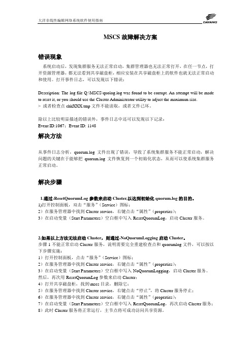
4Utilization of MSCs for Repairing CardiomyocytesXiaojie Xie1,Qiyuan Xu21,2Second Affiliated Hospital,Zhejiang University College of Medicine, Hangzhou,ChinaE-mail:1xiaojiexie@2aceline qiy@Abstract:Heart disease including myocardial infarction and ischemia is as-sociated with the irreversible loss of cardiomyocytes and vasculature,both via apoptosis or necrosis.However,the native capacity for the renewal and repair of myocardial tissue is inadequate as have been current therapeutic measures to prevent left ventricular remodeling and heart failure.Cell transplantation has emerged as a potentially viable therapeutic approach to directly repop-ulate and repair the damaged myocardium.A detailed analysis and a vision for future progress in MSCs applications,both in myocardial infarction and cardiomyopathy are presented in this review,highlighting research cardiology. Cardiomyocytes come from the splanchnic mesoderms mesenchymal cells.In the embryonic development period,the mesenchymal cells differentiate to myoblast cells,and on to the mature myocytes.It has not yet been clarified whether cardiac stem cells(CSCs)exist in the adult heart.Investigators in several laboratories concur with the notion that the heart contains a compart-ment of undifferentiated cells with the characteristics of stem cells(Hierlihy et al,2002;Urbanek,2003,2005).However,the actual number of CSCs re-mains controversial.Reports in mice(Matsuura et al,2004),rats(Beltrami et60Mesenchymal Stem Cells for Heart:From Bench to Bedsideal,2003),dogs(Linke et al,2005),and humans(Hierlihy et al,2002)indicate that there is1stem cell per8,000to20,000myocytes,or32,000to80,000 cardiac cells.A new conceptual framework of the heart has emerged and it is now viewed as a self-renewing organ in which myocyte regeneration occurs through-out the organism’s lifespan(Goldman and Wurzel,2001).The adult heart has a sub-population of myocytes that are not terminally differentiated;these myocytes evidently can reenter the cell cycle and undergo nuclear mitotic division soon after infarction.Although CSCs are efficient in preserving or-gan homeostasis and cell turnover,the decompensated heart is character-ized by a loss of myocytes and vascular structures.These factors cannot be counteracted by the activation and differentiation of CSCs,which undergo progressive replicative senescence,leading to a dramatic reduction of the stem cell compartment.Cardiac aging and chronic heart failure can occur, resulting in myocardial apoptosis and necrosis.This limited proliferation and self-renewal cannot compensate for heart injury,leading to replacement of cardiomyocytes byfibroblasts and consequent formation offibrosis.Due to scar-and ischemia-related post-infarction events,clinical manifestations are enormous and heterogeneous.The damaged left ventricle undergoes progres-sive“remodeling”and chamber dilation.These events reflect an apparent lack of effective intrinsic mechanisms for myocardial repair and regenera-tion.Unless deep(and still unknown)modifications are introduced in the area proximate to the damage to force the proliferation of resident cardiac progenitor cells,all restorative therapies must consider the use of exogenous multipotent stem cells capable to differentiate,at least,into cardiomyocytes. From this point of view,bone marrow-located stem cells have been consid-ered to display the required biologic properties for a cell therapy approach to treat patients with myocardial infarction.In addition to the use of bone marrow-derived hematopoietic precursor cells,cellular,molecular and preclinical data have shown that bone marrow-derived MSCs represent a suitable cell archetype for regenerative purposes after myocardial infarction.Under proper stimulation,MSCs can be induced to differentiate into myocytes,endothelium and smooth muscle cells in the infarcted heart(Kajstura et al,2005),revealing a high degree of plasticity. MSCs isolated from several human sources,including bone marrow and pe-ripheral and umbilical cord blood,exhibit a high ex vivo expansion capacity. This property has been used to assess the biologic properties of MSCs to per-form transfection with viral vectors(Conget and Minguell,2000;Partridge, 2002)and initiate studies toward the use of MSCs in clinical strategies(Hor-witz et al,1999;Barry and Murphy,2004).The promising therapeutic effect of MSCs relies on their capacity to engraft and survive long term in distinctive target tissue.Chapter4Utilization of MSCs for Repairing Cardiomyocytes61 In this review we will analyze the experimental evidence that warrants the utilization of MSCs in the treatment of myocardial infarction and chronic heat failure.4.1Application of MSCs on Myocardial Infarction Myocardial Infarction(MI),over95%of which is caused by atherosclerosis of the coronary artery,is one of the cardiovascular emergencies with high disability and mortality.Atherosclerotic plaques lead to intraluminal sclero-sis and a slowing down of bloodflow,contributing to cardiac ischemia and necrosis over time.This is a serious and persistent process.The complications that can occur in patients with extensive MI,such as malignant arrhythmia, heart failure,formation of ventricular aneurysm,and sudden cardiac death, are life-threatening,but can also disrupt the quality of life.Medications,per-cutaneous coronary intervention(PCI)and coronary artery bypass grafting (CABG)are involved in treating patients with MI,failing to induce functional cardiomyogensis,but scar formation.Evidences concerning bone marrow stem cells repairing the infarcted my-ocardium has accumulated based on a multitude of animal studies.Thefirst reports from Makino and colleagues(Makino et al,1999)in1999had demon-strated that bone marrow stem cells could in vitro differentiate into cardiomy-ocytes induced by5-azacytidine.In vivo evidence of transdifferentiation from Orlic studies in2001(Orlic et al,2001)showed that bone marrow stem cells transplanted into the infarcted myocardium can differentiate into cardiomy-ocates and consequently improve cardiac function(Fig.4.1).MSCs have also been shown as suitable candidates for MI repair.One of the famous studies in China is from Ge JB(Ma et al,2004;Zhang et al,2005),who estab-lished MI models in swine and Sprague-Dawley(SD)rats and injected MSCs transcoronarily or intravenously.As a result,MSC transplantation could im-prove the cardiac function,mediated by promoting cardiomyogenesis and neoangiogeneis in the ischemic borderline of infarcted myocardium.The in-creases in the ejection fraction(EF)by echocardiogram were correlated with the baseline.The worse the baseline of the ejection fraction in MI models, the better the therapeutic effect of MSCs transplantation.We have been focusing on the therapeutic effects and safety of MSC trans-plantation in animal models as well as patients suffering from MI or chronic heat failure.We have established MI models in New Zealand rabbits(Wang et al,2004,2005),SD rats(Chen et al,2006;Wang,2006)and mice(Hu et al,2007,2008)by ligation of the left anterior descending artery respectively. Allogenic bone marrow-derived MSCs were isolated,expanded in vitro and analyzed byflow cytometry before transplantation.MSCs at Passage3to 10were ready for transplantation and trypsinized from the plates,washed twice with sterile phosphate buffer solution(PBS),centrifuged and suspended in PBS or saline.MSCs suspension was injected intramyocardially into the62Mesenchymal Stem Cells for Heart:From Bench to BedsideFig.4.1.Markers of differentiating cardiac cells.(A-F)Labeling of CM by nestin (A,yellow),desmin(B,red),and connexin43(C,green);redfluorescence=cardiac myosin(A and C).(D and E)Yellow-greenfluorescence reflects labeling of EC byflk-1(arrows,D)and VE-cadherin(arrows,E);redfluorescence=factor VIII in EC(D and E).(F)Greenfluorescence labeling of SMC cytoplasms byflk-1; endothelial lining is also labeled byflk-1;redfluorescence=α-smooth muscle actin; bluefluorescence=propidium iodide(PI)labeling of nuclei.(A and E,×1,200;B and F×800;C,×1,400;D,×1,800.)infarcted region and the ischemic borderline respectively.Morphological and pathophysiological parameters,including cardiac function,ventricular remod-eling and the possible mechanisms,were evaluated in the engrafted heart and the controls.Consequently,compared with those in the controls,MSCs trans-plantation could significantly increase ejection fraction(EF)and fractional shortening(FS),decrease left ventricular internal diameter at end-diastole (LVIDd)and relieve ventricular remodeling.The therapeutic effects could also be achieved by autologous and heterogenous MSCs transplantation.Fur-ther pathological examinations showed that injected MSCs were localized in the injection sites and transdifferentiated into cardiomyocyte-like cells,en-dothelial cells and smooth muscle cells,thus reducing the infarcted exten-sion and improving cardiac contractility by the survived myocytes(Wang et al,2004,2005,2006;Chen et al,2006).Our in vitro studies indicated that MSCs under hypoxic conditions could not only significantly synthesizeChapter4Utilization of MSCs for Repairing Cardiomyocytes63 and up-regulate a multitude of growth factors,including hypoxia induced factors-1alpha(HIF-1α),vascular endothelial growth factors(VEGF),ery-thropoietin(EPO)and its receptor,angiotensin I,Flk-1,etc,but promote anti-apoptotic gene expression,such as Bcl-xl,Bcl-2,caspase3.Hypoxia conditioned MSCs could enhance neoangiogenesis,alleviate apoptosis and attenuate ventricular remodeling in MI animals(Hu et al,2008;Wang et al, 2008).The anti-apoptosis by MSCs therapy was associated with inactivation of voltage-dependent potassium channels in MSCs(Wangs et al,2008).Post-infarcted ventricular remodeling attenuated by MSCs transplantation might be mediated by down-regulating the expressions of matrix metalloproteinases (MMP-2,MMP-9)and reducingfibrosis(Wang et al,2006).Endothelial progenitor cells(EPCs)are also ideal candidates for cardiac celloplasty.Human EPCs isolated from adult periperal blood were labelled and injected intravenously into athymic rats3-hour post-MI(Kawamoto et al,2001).EPCs immigrated into the infarcted myocardium and participated in neoangiogenesis.EPCs transplantation contributed to the improvement of cardiac function,a significant increase of capillary density in the ischemic region,and diminution of the scar area in the infarcted myocardium.Specific human endothelial markers could also be detected in these engrafted cells.In clinical trials,EPC therapy could increase left ventricular ejection fraction (LVEF)and improve the survival of infarcted cardiomyocytes in patients with MI compared with those in the control groups.The therapeutic effects of MSCs transplantation may be achieved by reconstitution of the blood supply in the infarcted myocardium(Assmus et al,2002;Liu et al,2005).In summary,bone marrow cell transplantation to treat patients with MI or ischemic cardiomyopathy is safe,feasible and effective,especially with MSCs.The mechanisms of the therapeutic effects using cell transplantation might include not only stem cell transdifferentiation,but neoangiogenesis, anti-apoptosis,paracrine and so forth.(1)It has been demonstrated that transplanted bone marrow-derived MSCscould transdifferentiate into cardiomyocytes-like cells,endothelium and smooth muscle cells in the infarcted myocardium(Makino et al,1999;Or-lic et al,2001;Wang,2004,2005;Kajstura et al,2005).However,there are inconsistent reports that some investigators regard the transplanted cells as cell fusion with the host cardiomyocytes rather than transdiffer-entiation(Alvarez-Dolado et al,2003;Terada et al,2002).(2)Evidence showed that bone marrow-derived MSCs can secrete a plethoraof growth factors,including basicfibroblast growth factor(bFGF),vas-cular endothelial growth factor(VEGF),interleukin-1beta(IL-1β),and tumor necrosis factor-2alpha(TNF-2α),etc.The paracrine effects of MSCs might contribute to neoangiogenesis and anti-apoptosis in the is-chemic myocardium,improve myocardial perfusion,especially for the hi-bernating and stunned cardiomyocytes,which may limit the extension of64Mesenchymal Stem Cells for Heart:From Bench to Bedside the infarcted area and protect it from ischemic injury(Hu et al,2008;Kamihata et al,2001).(3)Another possible mechanism is that bone marrow-derived MSCs therapycould attenuate the compensatory activation of sympathetic nerves after MI,activate cardiac vagus nerve system and keep the balance of cardiac autonomic nerve regulation.MSCs transplantation might relieve the re-modeling of cardiac nerves in the non-infarcted region as well(Ding et al, 2006),promoting the sprouting of cardiac nerve terminals,and thus im-prove the neurotrophy and neuroregulation in the infarcted myocardium (Pak et al,2003).(4)In addition,bone marrow-derived MSCs could express the proteins in-volved in the intercellular gap junction,including connexin40,connexin 43and connexin45in the infarcted region.The transplanted cells could establish the intercellular electric-mechanic coupling with the host car-diomyocytes,contributing to host heart contraction and the compliance of infarcted myocardium,significantly improving the post-infarctional cardiac systolic and diastolic function(Lian et al,2002;Pijnappels et al, 2006;Mills et al,2007).Structural and functional integration of injected cells with host myocardium is crucial to achieve a therapeutic effect.There has been increasing concern about whether patients suffering from MI can benefit persistently from cell therapy.Not only the type and the num-ber of transplanted cells can affect the therapeutic effect,but also the path-way and the number of transplantations.The promising therapeutic effect of MSCs relies on their convenience,in both isolation and in vitro expan-sion,as well as their ability for multipotent transdifferentiation,transfection and expression of target genes.It indicated that cell transplantation shows a dose-dependent correlation with the improvement of cardiac function.How-ever,only a small part of transplanted cells can migrate and survive in the infarcted area and the ischemic margin of MI animal models.The efficiency of cell therapy is still challenging in its clinical application.It had been demonstrated that an appropriate ratio of transplanted cells to host cardiomyocytes contributes to the elicitation of therapeutic effects (Chang et al,2006).When human MSCs in vitro are co-cultured with neona-tal cardiomyocytes at a ratio of1:9or1:4,reentrant arrhythmias could be induced in86%of the cultured system.MSCs co-cultured with neonatal car-diomyocytes at a ratio of1:99,do not lead to the decrease in conduction velocity and reentrant arrhythmias.The number of transplanted MSCs is crucial to cardiac electrophysiological heterogenicity and arrhythmogenicity after cell transplantation.Transcoronary injection of MSCs several times to treat MI in swine models is a feasible and safe pathway for cell therapy,in-creasing transplantation efficiency(Poh et al,2007).It is consistent with the results from our studies that duplicate transplantation of MSCs leads to a higher increase in cardiac function than a bolus cell injection.Chapter4Utilization of MSCs for Repairing Cardiomyocytes65 We are still focusing on the optimal time points for cell transplantation in MI animal models.Consistent with the results from Lim and colleagues that a6%increase in the cardiac function was achieved by MSCs trans-plantation at the early stage of acute myocardial infarction(AMI)in swine models(Lim et al,2006),our studies showed that2weeks after AMI was an optimal time point to upregulate the impaired cardiac function for MSCs transplantation in rat models(Hu et al,2007),because at the2-week time-window inflammation was attenuated andfibrosis remained unformed(Ma et al,2005;Bartanek et al,2006).Optimal delivery time of bone marrow-derived stem cells(BMC)is5to6days after AMI in a REPAIR-AMI trial (Erbs et al,2007),while no benefits can be achieved within24hours post-MI (Janssens et al,2006).Another group in China,Ge JB and colleagues,had demonstrated that both24hours and3-7days post-infarction were available windows for BMC transcoronary delivery of percutaneous coronary interven-tion(PCI)in patients with AMI(Huang et al,2006).Consequently,many more studies and clinical trials are needed to prove the therapeutic effects of bone marrow-derived stem cells.4.2Application of MSCs on Cardiomyopathy and Chronic Heart FailureTransplantation of a bone marrow-derived stem cell,traditionally used to post-MI,is a promising therapy now being introduced to treat patients with cardiomyopathy and chronic heart failure.Results from animal studies have been carried out to ascertain the therapeutic effects.Because of diffused my-ocardial damage,dilated cardiomyopathy(DCM)is one of the common causes of chronic heart failure.Since evidence shows that patients with MI can ben-efit from MSCs transplantation due to its multipotent transdifferentiation and paracrine properties,some investigators are attempting to inject MSCs in DCM animal models.In China,Li and colleagues(Li GC et al,2004;Li WQ et al,2005)had es-tablished DCM rabbit models induced by intravenous injection of doxorubicin hydrochloride for8weeks.Three weeks later,bone marrow-derived MSCs had been expanded in vitro to1×106and intramyocardially injected into the an-terior wall of the left ventrium at4sites.Cardiac function was significantly increased after4weeks in rabbits with allogenic MSCs therapy compared with those of in the controls.The transplanted MSCs were found to prolifer-ate and transdifferentiate into cardiomyocyte-like cells and endothelial cells. Neurohumoral regulation was also involved in the therapeutic effect of MSCs transplantation.A consistent result had been achieved from Zhang’s stud-ies showing that DCM rabbits could also benefit from MSCs transplantation (Zhang et al,2007).Nagaya and colleagues(Nagaya et al,2005)had also showed that injection of MSCs into the left ventricle of DCM rats could im-prove left ventricular ejection fraction(LVEF)infive weeks mediated by pro-66Mesenchymal Stem Cells for Heart:From Bench to Bedsidemoting cardiomyogensis and attenuatingfibrosis.The therapeutic effects of MSCs transplantation include not only cell transdifferentiation potential,but paracrine characteristics,producing a series of pro-angiogenic,anti-apoptotic and mitogenic factors.Chronic ischemic cardiomyopathy(ICM)is a subtype of chronic ischemic coronary disease with a high morbidity for chronic heart failure.The patho-logical manifestation is that of a persistent myocardial ischemia caused by diffused atherosclerosis in coronary artery,resulting in myocardial necrosis andfibrosis with an amount of hibernating and stunning cardiomyocytes. Zhu SG and collaborators had attempted bone marrow-derived cell trans-plantation in ICM animals(Zhu et al,2006).They had established ICM swine models by placing an Ameroid circle at the onset of the circumflex branch of the left coronary to reduce bloodflow for four weeks.Autologous bone marrow mononuclear cells(BMCs)were isolated in vitro and then injected intracoronary.After4weeks,an echocardiogram was performed to evaluate LVEF,and immunohistochemistry was used to examine capillary density.As a result,LVEF was significantly elevated and neoangioenesis was detected in ICM rats with BMCs transplantation compared to those of the controls. Their results indicated that BMC therapy might improve cardiac function of ICM patients by promoting neoangiogenesis and collateral circulation.Diabetes mellitus(DM)is a common endocrine and metabolism disease with high morbidity and mortality.Patients with DM often suffer from some cardiovascular complications or comorbidities,including coronary heart dis-ease(CHD),diabetic cardiomyopathy,strokes,etc.,which can impair the quality of life.A successful clinical report from Brehm and Strauer showed that a DM patient complicated with extensive AMI due to complete oc-clusion of the left anterior descending could benefit from autologous BMCs transplantation(Brehm and Strauer,2007).The implanted cells successfully reconstructed the damaged myocardium and blood vessels,taking on a car-diac contractile function.They assumed that myocardial repair was associ-ated with stem cell transplantation and/or paracrine cytokine.Intracoronary stem cell transplantation may reduce the mortality of otherwise treatment-resistant cardiogenic shock.Ma and coworkers had compared the therapeutic effects of bone marrow and cord blood-derived CD133+cells in diabetic car-diomyopathy animals(Ma et al,2006).They had isolated and prepared both kinds of stem cells,and then injected intramyocardially into NOD/SCID mice with diabetic cardiomyopathy.Both types of CD133+cells could pro-mote noeangiogenesis and elevate survival,but only bone marrow-derived cells could improve cardiac contractility.We tried applying bone marrow-derived MSCs to diabetic cardiomyopa-thy animal models(Li and Wang,2008;Zhang et al,2008).We established dilated cardiomyopathy models in SD rats after four months of a bolus in-traperitoneal injection of streptozotocin.Rat bone marrow-derived MSCs were isolated and expanded in vitro.5×106of MSCs with/without anoxicChapter4Utilization of MSCs for Repairing Cardiomyocytes67 preconditioning were injected intramyocardially atfive sites,the right ven-tricular free wall,the basal and midanterior wall,the lateral wall and the posterior wall.Two weeks after transplantation,MSCs,especially anoxic pre-conditioned MSCs,significantly increased fractional shortening(FS)of the diabetic heart.Anoxic preconditioned MSCs increased the capillary density of diabetic myocardium and attenuated myocardialfibrosis mediated by in-creasing the activity of matrix metalloproteinase-2(MMP-2)and inhibiting the transforming growth factorβ1(TGF-β1),respectively.Anoxic precon-ditioned MSCs significantly elicited anti-apoptotosis in DCM rats,possibly mediated by upregulation of Bcl-2/Bax ratio and the inhibition caspase-3 expression and activation.The results indicate that intramyocardial trans-plantation of MSCs have a protective effect on diabetic cardiomyopathy and anoxic preconditioning can enhance this protective effect,possibly through an anti-apoptotosis of diabetic cardiomyopathy and attenuation of cardiac remodeling.4.3ConclusionIn summary,bone marrow-derived stem cells are ideal candidates for cardiac celloplasty for patients with chronic heart failure and unable to receive other treatments.A multitude of animal experiments and clinical trials on stem cell transplantation are required to evaluate the therapeutic effects and their mechanisms in chronic heart failure.A cautious attitude is required concern-ing the results of these studies.Inconsistent results from some clinical trials have raised some questions that must be solved before clinical application. Some of these questions are as follows:What kind of patients are available for stem cell transplantation?What is the best candidate for cell therapy?When is the optimal time point for transplantation,and what is the mechanism of the therapeutic effects on cardiac function and remodeling by cell transplan-tation?Little evidence has been gathered on cardiomyogenesis using clinical stem cell therapy,raising more questions concerning this morbidity.How can stem cells survive and elicit biological effects in the damaged myocardium, and what percentage is necessary to generate this effect?How can we trace the biological properties of transplanted cells and evaluate their function?Is there any potential for arrhythmias or adverse effects from this cell therapy? It is critical to answer these questions before we apply stem cell transplanta-tion to patients with heart disease.Concurrent procedures for the isolation and identification of stem cells are also crucial to objectively assess the thera-peutic effect of stem cell therapy.Randomized,placebo-control clinical trials are required to further advance the application of stem cell transplantation.68Mesenchymal Stem Cells for Heart:From Bench to BedsideReferencesAlvarez-Dolado M,Pardal R,Garcia-Verdugo JM,Fike JR,Lee HO,Pfeffer K,Lois C,Morrison SJ,Alvarez-Buylla A(2003)Fusion of bone-marrow-derived cells with Purkinje neurons,cardiomyocytes and hepatocytes.Nature, 425(6961):968-973Assmus B,Sch¨a chinger V,Teupe C,Britten M,Lehmann R,Dbert N,Grnwald F,Aicher A,Urbich C,Martin H,Hoelzer D,Dimmeler S,Zeiher AM(2002) Transplantation of progenitor cells and regeneration enhancement in acute my-ocardial infarction.Circulation,106(24):3009-3012Barry FP,Murphy JM(2004)Mesenchymal stem cells:clinical application and biological properties.Int J Biochem Cell Biol,36(4):568-584Bartunek J,Wijns W,Heyndrickx GR,Vanderheyden M(2006)Timing of in-tracoronary bone-marrow-derived stem cell transplantation after ST-elevation myocardial infarction.Nat Clin Pract Cardiovasc Med,Suppl1:S52-56 Beltrami AP,Barlucchi L,Torella D,Baker M,Limana F,Chimenti S,Kasahara H,Rota M,Musso E,Urbanek K,Leri A,Kajstura J,Nadal-Ginard B,Anversa P(2003)Adult cardiac stem cells are multipotent and support myocardial regeneration.Cell,114(6):763-766Brehm M,Strauer BE(2007)Successful therapy of patients in therapy-resistant cardiogenic shock with intracoronary,autologous bone marrow stem cell trans-plantation.Dtsch Med Wochenschr,132(38):1944-1948Chang MG,Tung L,Sekar RB,Chang CY,Cysyk J,Dong PH,Marban E,Abra-ham MR(2006)Proarrhythmic potential of mesenchymal stem cell transplan-tation revealed in an in vitro coculture model.Circulation,113(15):1832-1841 Chen J,Wang JA,Luo RH,Hu XY,Xie XJ,Li JH,He AN,Sun Y(2006) Effects of mesenchymal stem cells transplantation on post-infarction ventricular remodeling in rats.Chin J Emer Med,15(4):310-314Conget PA,Minguell JJ(2000)Adenoviral-mediated gene transfer into ex vivo expanded human bone marrow mesenchymal progenitor cells.Exp Hematol, 28:382-390Ding CD,Cao KJ,Shan QJ,Zou JG,Chen ML,Jin Y(2006)Mechanism of myeloid mesenchymal stem cell transplantation in cardiac autonomic nervous function after myocardial infarction.Chin J Clin Reh,13(10):24-28Erbs S,Linke A,Sch¨a chinger V,Assmus B,Thiele H,Diederich KW,Hoffmann C,Dimmeler S,Tonn T,Hambrecht R,Zeiher AM,Schuler G(2007)Restora-tion of microvascular function in the infarct-related artery by intracoronary transplantation of bone marrow progenitor cells in patients with acute myocar-dial infarction:the Doppler Substudy of the Reinfusion of Enriched Progenitor Cells and Infarct Remodeling in Acute Myocardial Infarction(REPAIR-AMI) trial.Circulation,116(4):366-374Goldman BI,Wurzel J(2001)Evidence that human cardiac myocytes divide after myocardial infarction.N Engl J Med,344(15):1750-1757Hierlihy AM,Seale P,Lobe CG,Rudnicki MA,Megeney LA(2002)The post-natal heart contains a myocardial stem cell population.FEBS lett,530(1-3):239-243Horwitz EM,Prockop DJ,Fitzpatrick LA,Koo WW,Gordon PL,Neel M,Suss-man M,Orchard P,Marx JC,Pyeritz RE,Brenner MK(1999)Transplantability and therapeutic effects of bone marrow-derived mesenchymal cells in children with osteogenesis imperfecta.Nat Med,5(3):309-313Hu X,Wang J,Chen J,Luo R,He A,Xie X,Li J(2007)Optimal temporal delivery of bone marrow mesenchymal stem cells in rats with myocardial infarction.Eur J Cardiothorac Surg,31(3):438-443Hu X,Yu SP,Fraser J,Lu Z,Ogle ME,Wang JA,Wei L(2008)Transplan-tation of hypoxia preconditioned MSC improves infarcted heart function via enhanced survival of implanted cells and angiogenesis.J Thorac Cardiothorac Surg,135(4):799-808Huang RC,Yao K,Zhou YZ,Ge L,Qian JY,Yang J,Yang S,Niu YH,Li YL, Zhang YQ,Zhang F,Xu SK,Zhang SH,Sun AJ,Ge JB(2006)Long term follow-up on emergent intracoronary autologous bone marrow mononuclear cell transplantation for acute inferior-wall myocardial infarction.Chin J National Med,86(16):1107-1110Janssens S,Dubois C,Bogaert J,Theunissen K,Deroose C,Desmet W,Kalantzi M,Herbots L,Sinnaeve P,Dens J,Maertens J,Rademakers F,Dymarkowski S, Gheysens O,Van Cleemput J,Bormans G,Nuyts J,Belmans A,Mortelmans L, Boogaerts M,Van de Werf F(2006)Autologous bone marrow-derived stem-cell transfer in patients with ST-segment elevation myocardial infarction:double-blind,randomised controlled ncet,367(9505):113-121Kajstura J,Rota M,Whang B,Cascapera S,Hosoda T,Bearzi C,Nurzynska D,Kasahara H,Zias E,Bonaf M,Nadal-Ginard B,Torella D,Nascimbene A, Quaini F,Urbanek K,Leri A,Anversa P(2005)Bone marrow cells differentiate in cardiac cell lineages after infarction independently of cell fusion.Circ Res, 96(1):127-137Kamihata H,Hiroaki M,Takashi N,Fujiyama S,Tsutsumi Y,Ozono R,Masaki H,Mori Y,Iba O,Tateishi E,Kosaki A,Shintani S,Murohara T,Imaizumi T, Iwasaka T(2001)Implantation of bone marrow mononuclear cells into ischemic myocardium enhances colateral perfusion and regional function via side supply of angioblasts,angiogenic ligands and cytokines.Circulation,104(10):1046-1052Kawamoto A,Gwon HC,Iwaguro H,Yamaguchi JI,Uchida S,Masuda H,Silver M,Ma H,Kearney M,Isner JM,Asahara T(2001)Therapeutic potential of ex vivo expanded endothelial progenitor cells for myocardial ischemia.Circula-tion,103(5):634-637Li GC,Li GS,Zhang J,Li WQ,Zhou Q(2004)The empirical study of autologous bone marrow mesenchymal stem cell transplantation for dilated cardiomyopa-thy in rabbit.zhonghua Xin Xue Guan Bing za zhi,32(12):1095-1098Li J,Zhang N,Wang J(2008)Improved antiapoptotic and antiremodeling potency of bone marrow mesenchymal stem cells by anoxic preconditioning in diabetic cardiomyopathy.J Endocrinol Invest,31(2):103-110Li WQ,Li Y,Li GC,Zhang J,Li GS(2005)Effect of autoulogous bone mes-enchymal stem cell transplantation on the heart function and plasma BNP of dilated cardiomyopathy.Chin J Emerg Med,14(7):559-562Lian F,Zhu HS,Zheng JH,et al(2002)The research of bone marrow mesenchymal stem cell transformation into myocytes in vitro.Chin J Exp Surg,19(5):454-456。