manuscript
关于投稿及询问信的写法及注意事项
我的询问信Dear Editors,We dispatched our manuscript (manuscript ID 8558192903066551) to your journal about ten weeks ago. We have not yet received a reply and am wondering whether you have reached a decision. We fear that our submission process does not meet the requirements and should be grateful if you kindly give us some information regarding the status of the manuscript.Thanks for your information.With kind regards,SCI投稿常用英语一、投稿信1. Dear Dr. Defendi ML:I am sending a manuscript entitled “” by – which I should like to submit for possible publication in the journal of - .Yours sincerely2. Dear Dr. A:Enclosed is a manuscript entitled “” by sb, which we are submitting for publication in the journal of - . We have chosen this journal because it deals with - . We believe that sth would be of interest to the journal’s readers.3. Dear Dr. A:Please find enclosed for your review an original research article, “” by sb. All authors have read and approve this version of the article, and due care has been taken to ensure the integrity of the work. No part of this paper has published or submitted elsewhere. No conflict of interest exits in the submission of this manuscript, and we have attached to this letter the signed letter granting us permission to use Figure 1 from another source.We appreciate your consideration of our manuscript, and we look forward to receiving comments from the reviewers.二、询问有无收到稿件Dear Editors,We dispatched our manuscript to your journal on 3 August 2006 but have not, as yet, receive acknowledgement of their safe arrival. We fear that may have been lost and should be grateful if you would let us know whether or not you have received them. If not, we will send our manuscript again. Thank you in advance for your help.三、询问论文审查回音Dear Editors,It is more than 12 weeks since I submitted our manuscript (No: ) for possible publication in your journal. I have not yet received a reply and am wondering whether you have reached a decision. I should appreciated your letting me know what you have decided as soon as possible.四、关于论文的总体审查意见1. This is a carefully done study and the findings are of considerable interest. A few minor revision are list below.2. This is a well-written paper containing interesting results which merit publication. For the benefit of the reader, however, a number of points need clarifying and certain statements require further justification. There are given below.3. Although these observation are interesting, they are rather limited and do not advance our knowledge of the subject sufficiently to warrant publication in PNAS. We suggest that the authors try submitting their findings to specialist journal such as –4. Although this paper is good, it would be ever better if some extra data were added.5. This manuscript is not suitable for publication in the journal of – because the main observation it describe was reported 3 years ago in a reputable journal of - .6. Please ask someone familiar with English language to help you rewrite this paper. As you will see, I have made some correction at the beginning of the paper where some syntax is not satisfactory.7. We feel that this potentially interesting study has been marred by an inability to communicate the finding correctly in English and should like to suggest that the authors seek the advice of someone with a good knowledge of English, preferable native speaker.8. The wording and style of some section, particularly those concerning HPLC, need careful editing. Attention should be paid to the wording of those parts of the Discussion of and Summary which have been underlined.9. Preliminary experiments only have been done and with exception of that summarized in Table 2, none has been repeated. This is clearly unsatisfactory, particularly when there is so much variation between assays.10. The condition of incubation are poorly defined. What is the temperature? Were antibody used?五、给编辑的回信1. In reply to the referee’s main criticism of paper, it is possible to say that –One minor point raised by the referee concerns of the extra composition of the reaction mixture in Figure 1. This has now been corrected. Further minor changes had been made on page 3, paragraph 1 (line 3-8) and 2 (line 6-11). These do not affect our interpretation of the result.2. I have read the referee’s comments very carefully and conclude that the paper has been rejected on the sole grounds that it lake toxicity data. I admit that I did not include a toxicity table in my article although perhaps I should have done. This was for the sake of brevity rather than an error or omission.3. Thank you for your letter of –and for the referee’s comments concerning our manuscript entitled “”. We have studied their comments carefully and have made correction which we hope meet with their approval.4. I enclosed a revised manuscript which includes a report of additional experiments done at the referee’s suggestion. You will see that our original findings are confirmed.5. We are sending the revised manuscript according to the comments of the reviewers. Revised portion are underlined in red.6. We found the referee’s comments most helpful and have revised the manuscript7. We are pleased to note the favorable comments of reviewers in their opening sentence.8. Thank you for your letter. I am very pleased to learn that our manuscript is acceptable for publication in Cancer Research with minor revision.9. We have therefore completed a further series of experiments, the result of which are summarized in Table 5. From this we conclude that intrinsic factor is not account.10. We deleted the relevant passage since they are not essential to the contents of the paper.11. I feel that the reviewer’s comments concerning Figures 1 and 2 result from a misinterpretation of the data.12. We would have include a non-protein inhibitor in our system, as a control, if one had been available.13. We prefer to retain the use of Table 4 for reasons that it should be clear from the new paragraph inserted at the end of the Results section.14. Although reviewer does not consider it is important to measure the temperature of the cells, we consider it essential.15. The running title has been changed to “”.16. The Materials and Methods section now includes details for measuring uptake ofisotope and assaying hexokinase.17. The concentration of HAT media (page12 paragraph 2) was incorrectly stated in the original manuscript. This has been rectified. The authors are grateful to the referees for pointing out their error.18. As suggested by both referees, a discussion of the possibility of laser action on chromosome has been included (page16, paragraph 2).19. We included a new set of photographs with better definition than those originally submitted and to which a scale has been added.20. Following the suggestion of the referees, we have redraw Figure 3 and 4.21. Two further papers, published since our original submission, have been added to the text and Reference section. These are:22. We should like to thank the referees for their helpful comments and hope that we have now produced a more balance and better account of our work. We trust that the revised manuscript is acceptable for publication.23. I greatly appreciate both your help and that of the referees concerning improvement to this paper. I hope that the revised manuscript is now suitable for publication.24. I should like to express my appreciation to you and the referees for suggesting how to improve our paper.25. I apologize for the delay in revising the manuscript. This was due to our doing an additional experiment, as suggested by referees我来说几句:1. 一般投稿以后,不要有事没有事经常写信给编辑.总体来说,这些编辑都很认真负责的.我们只用静侯佳音即可;2. 所有编辑都很忙,而且有很多人同时担任多个杂志的编辑,且大多数都是兼职而非专职,所以我们应该体谅他们,尽量不要给他们制造"麻烦";3. 不同杂志有不同的程序和周期,我们一般可以从已经发表的文章当中看到相关信息:received:***; revised:***;accepted***;所以我们应该心中有数;4. 应该注意的是:不同文章的处理周期不可能是千篇一律的,有时长一点也不足为怪;5. 一般杂志的周期应该在3个月之内,所以我觉得3个月以上的情况,应该是可以写信询问的.当然也可以再等一段时间;6. 我们可以想想,如果我们自己是编辑,接到这样的来信,收到稿件不到一个月就急着询问结果,而且天天收到很多这样的信件,我们心里会怎么想呢?而且有些时候,好象没有办法去细细与作者解释.所以我们写信也没有用,还不如不写.下面是UKchinese 战友的一个帖子,回复状态一直是Under Review,而日期有一直在变化的疑问,有参考价值. 值得注意的是:有些网上投稿系统是不显示日期的,如Scholar One...每一次编辑的处理,都会有日期的变化,而状态不变.这说明你的稿件一直在编辑的关注之中,你不用写信询问.状态一直是Under Review,而日期在变,有以下几种情况:1. 如果杂志需要2或3名审稿人,从第一个审稿人接受审稿,第二个审稿人接受审稿,第三个审稿人接受审稿, 到第一个审稿人审稿意见返回,第二个审稿人审稿意见返回,第三个审稿人审稿意见返回,这期间都是Under Review;2. 当然也有审稿人审稿接受审稿一段时间后又拒绝审稿的情况,这时编辑得寻找新的审稿人;3. 还有审稿人的审稿意见不能按照规定的时间返回编辑手里,这时编辑得催促审稿人,在得不到有效答复后,寻找新的审稿人;4. 还有审稿人的审稿意见返回编辑手里,但编辑认为缺乏水平,他会采取不信任处理,也要寻找新的审稿人;5. 还有审稿人的审稿意见返回编辑手里,但编辑认为太简单,没有足够的参考信息,他也会寻找新的审稿人;6. 两个审稿人的意见非常矛盾,这时编辑也会考虑增加审稿人...Try to avoid writing a letter to editor only regarding the review process, because it is beyond the editor's control.If you do want remind the editor for the time, you can try another methods for contacting.For example:Ms No:Dear editorI am writing regarding our above-mentioned paper, which has been submited for your consideration 3 months ago. It comes into my attention the status of our paper has been keeping on "***" for a long time, without any change. I do not know if it is because you are having difficulties in securing referees for our paper. If that is the case, please let us know & I will provide several possible reviewers in this area for your reference.Thank you for your great efforts on processing our manuscript.Sincerely。
revised manuscript范文

revised manuscript范文Revised Manuscript SampleTitle: The Impact of Social Media on Mental Health in AdolescentsAbstract:Social media has become an integral part of modern life, especially for adolescents. While it offers numerous benefits, such as fostering social connections and access to information, there is growing concern about its potential negative impact on mental health. This study aims to investigate the relationship between social media usage and mental health outcomes, including depression, anxiety, and self-esteem, among adolescents. A cross-sectional survey was conducted with a sample of 500 high school students aged 14 to 18 years. Participants completed self-report measures assessing their social media use, depressive symptoms, anxiety levels, and self-esteem. The results revealed a significant positive correlation between excessive social media use and depressive symptoms, as well as anxiety levels. Conversely, a negative correlation was found between social media use and self-esteem. These findings highlight the potential risks associated with excessive social media engagement and underscore the importance of developing strategies to promote healthy social media habits among adolescents.Introduction:Social media platforms have revolutionized the way individuals, especially adolescents, communicate, socialize, and access information. While these platforms offer numerous benefits, such as fostering social connections and providing educational resources, there is growing concern about their potential negative impact on mental health. Adolescence is a critical developmental period characterized by significant physical, emotional, and social changes, making this population particularly vulnerable to the effects of social media.[Expand on the introduction, providing background information, stating the research problem, and presenting the study's objectives.]Literature Review:[Provide a comprehensive review of relevant literature, highlighting key findings and identifying gaps in existing research.]Methods:[Describe the study design, participant recruitment process, data collection methods, and measures used to assess social media use, depressive symptoms, anxiety levels, and self-esteem.]Results:[Present the findings of the study, including descriptive statistics, correlation analyses, and any other relevant statistical tests performed.]Discussion:[Interpret and discuss the study's findings in relation to the research objectives and existing literature. Address potential limitations and suggest directions for future research.]Conclusion:[Summarize the main findings and their implications, emphasizing the study's contributions to the field and the potential practical applications of the results.]References:[List all cited sources in the appropriate citation style.]。
manuscript
Ultrasensitive and Stable Determination of Lead Ion by Phenanthroline-based Electropolymerized Film Modified Glassy Carbon ElectrodeChangyin Ji a,Peng Li a,Hongwei Ma a,Guocheng Yang b,and Ming Zhang a,*a State Key Laboratory of Supramolecular Structure and Materials,Jilin University, Changchun 130012, P. R. Chinab School of Chemistry and Life Science,Changchun University of Technology, Changchun 130012, P. R. ChinaABSTRACTA novel electro-active compound, 3,8-bis(9,9-bis(6-(9H-carbazol-9-yl)hexyl)-9H-fluoren-2-yl)-1,10-phenanthroline(TCFC) is used to modify glassy carbon electrode (GCE) for the electrochemical detection of Pb2+. Phenanthroline unit in the backbone and four electro-active alkyl-linked peripheral carbazole groups as the side chains endow TCFC both metal chelated ability and electropolymerized possibility. By the methods of chemical preconcentration-anodic stripping technique, TCFC electropolymerized film * Corresponding author. Tel: +86-431-85167507. Fax: +86-431-85193421.E-mail address: zhming@.modified GCE (TCFC/GCE) shows excellent selectivity and sensitivity to Pb2+, even in the presence of a large amount of other heavy metal ions (HMIs). The limit of detection (LOD) towards Pb2+ could be improved to 1.33 10–11 M (S/N=3) at optimum condition. Furthermore, the TCFC/GCE is stable under ambient condition (>30 days) and can be recycled used with high sensitivity, which is of great value for practicality. Keywords:Modified electrode; Square wave voltammetry; Lead ion; Electropolymerization; Carbazole; Phenanthroline.1. IntroductionThe detection of heavy metal is always an attractive and interesting topic [1]. Among the heavy metal, lead ion (Pb2+) is nondegradable and can accumulate in bones, muscles, kidney and brain, and can result in brain damage and/or mental retardation, behavioral problems and so on [2–4], therefore, the sensitive and robust detections of trace Pb2+ are exigent.The electrochemical detection of Pb2+has some attractive features, such as high selectivity and sensitivity, intrinsic simple operation, robustness and inexpensiveness [5, 6]. Additionally, chemically modified electrode (CME) has its own extraordinary superiority, which can preconcentrate trace heavy metal ions (HMIs) during the accumulation step [7]. Thus, CME has been widely used for the trace HMIs determination. Though considerable efforts have been made to develop CMEs for HMIs sensing [8–11], the stable and sensitivity CME for Pb2+ detection is still highly needed and deserves to explore.Electropolymerization (EP) method prompts the electro-active precursors to undergo the oxidative coupling reaction, resulting in direct polymerization on the glassy carbon electrode (GCE). EP has the advantages of controlling morphology and conductivity by judicious selection of the precursors and/or potentiometric conditions. The resulting crossing-linking network of the EP film is beneficial for the application in sensing field due to the possible stability and rapid diffusion of analytes. Thus, the electrodes modified by EP materials have the excellent potential to detect HMIs.In this paper, we reported a novel electro-activematerial TCFC used as the CME to detect Pb2+ [12]. The chemical structure of TCFC is shown in Scheme 1. As can be seen, 1,10-phenanthroline (PHEN) in the backbone could endow TCFC strong metal-chelated capability, which acts as a receptor for the molecular recognition of Pb2+. Meanwhile, the alkyl-linked peripheral carbazole groups in side chains, which are coupling reactive under anodic oxidation, could provide the crossing site during EP process to form the cross-linking network films. Based on this molecular design, TCFC can be coated on GCE (TCFC/GCE) by EP method and used to detect Pb2+. The TCFC/GCE exhibits high sensitivity towards Pb2+, and the LOD towards Pb2+ can be down to 1.33 10–11 M, such low LOD has ascend the best level in Pb2+ sensing publications, which can be attributed to the strong chelated ability of PHEN in TCFC and the intrinsic microstructure of EP films [13]. Furthermore, the TCFC/GCE is stable under ambient condition and can be recycled used with high sensitivity (continuous measurement for 30 days).2. Experimental Section2.1. Chemicals and apparatusAll the reagents and solvents used for the synthesis were purchased from Sigma Aldrich or Acros companies and used as received.Electrochemical measurements were carried out with CHI 660C electrochemical workstation..All aqueous solutions were prepared using Milli-Q water of 18.2 MΩ (Millipore) resistivity. The pH values were measured by pH meter (METTLER TOLEDO LE438). The 0.1 M HAc/NaAc buffer solution (pH 4.5) was prepared by adding 0.1 M sodium acetate into 0.1 M acetic acid. Pb2+ solutions were prepared by diluting the appropriate amount of Pb2+ stock solution (0.1 M).2.2. Synthesis of TCFCThe synthetic route of TCFC has been reported [12].TCFC: 1H NMR (500 MHz, DMSO): δ 9.55 (d, 2H), 8.84 (d, 2H), 8.11 (d, 2H), 8.06 (d, 8H), 8.02 (s, 2H), 7.97 (m, 4H), 7.86 (m, 2H), 7.45 (d, 8H), 7.34 (m, 12H), 7.27(m, 2H), 7.1(t, 8H), 4.23 (t, 8H), 2.1 (m, 8H), 1.52 (m, 8H), 1.03 (m, 16H), 0.50 (m, 8H). MALDI-TOF-MS (m/z): 1506.01 [M+] 1507.3.2.3. Preparation of TCFC/GCEFirstly, the GCE was polished with different grades of alumina powder (0.05, 0.3 and 1.0 μm) on nylon cloth, emery paper and chamois, respectively, rinsed with deionized water and then cleaned by ultrasonic bath for 2 min according to the literatures (unless otherwise stated).Then, cyclic voltammetry (CV) method was used to prepare EP films on GCE by using a standard one-compartment, three-electrode electrochemical cell. Titanium metal and GCE were used as the counter electrode and the working electrode, respectively. The reference electrode was Ag/Ag+nonaqueous electrode. A mixture of TCFC precursormolecule (0.5 mg·mL–1) and TBAPF6 with CH2Cl2 and CH3CN (V:V=2:3) were used as the electrolyte solution. The TCFC electro-active precursor was electropolymerized through an oxidation coupling reaction (0 to 0.85 V) to form the cross-linking network film on GCE (TCFC/GCE).Lastly, the TCFC/GCE was washed with a mixture solution of CH2Cl2and CH3CN (V:V=2:3), then the prepared TCFC/GCE was dried in a vacuum oven at 40 °C for three hours.2.4. Electrochemical detecting experimentsScheme 1. The chemical structure of TCFC and the illustration of Pb2+ detection using TCFC/GCE.Electrochemical detection was carried out in a standard one-compartment, three-electrode electrochemical cell. The TCFC/GCE, Pt wire electrode and the Ag/AgCl (3 M KCl) aqueous electrode were used as the working electrode, the counter electrode and the reference electrode, respectively. Preconcentration of Pb2+took place at an open circle condition by dipping the working electrode into HAc/NaAc buffer solution containing Pb2+under vigorous stirring for 300 s and being rinsed with deionized water for three times. Then, these electrodes were immersed in the 10 mL electrochemical cell containing 5 mL HAc/NaAc buffer solution. Multi potential steps (M-PS) were performed at a potential of –1.2 V for 30 s. After that, square wave voltammetry (SWV) scan was performed from –1.0 V to 0.4 V by using the following waveform parameters:the scan frequency was 25 Hz, the amplitude was 25 mV and the step increment was 5.0 mV (unless stated otherwise).3. Results & Discussion3.1. Electrochemical detection of Pb2+Due to the designed metal-chelated unit, PHEN in TCFC molecule, the chelated behavior of TCFC/GCE was firstly studied by CV (Fig. 1A). As can be seen, before the exposure to Pb2+, the CV of the bare TCFC/GCE was nearly a straight line. After the accumulation of Pb2+ and being rinsed with deionized water for three times to remove the adsorptive Pb2+ and other electrolyte, significant oxidation peak and reduction peak (–0.4 V and –0.65 V) appear in the CV curves, which indicated the chelation of Pb2+ to TCFC molecule [14].TCFC/GCE shows excellent selectivity to Pb2+ even in the presence of a large amount of other HMIs. Fig. 1B is the SWV curves of TCFC/GCE detecting Pb2+ (1.0×10⁻⁸ M) with disturbing metal ions of Cd2+, Cu2+ and Hg2+ (1.0×10⁻⁶ M). As can be seen, though the concentrations of disturbing metal ions were 100 times higher than that of Pb2+, an intense stripping signal of Pb2+can be clearly observed compared with other HMIs, which indicates the high selectivity of TCFC/GCE to Pb2+.Fig. 1. (A) The cyclic voltammograms of the TCFC/GCE recorded in NaAc/HAc buffer solution with and without Pb2+(4.0×10⁻⁶) M; (B) The SWV curves of TCFC/GCErecorded in NaAc/HAc buffer solution with 1.0×10⁻⁸ M Pb2+ and 1.0×10⁻⁶ M Cd2+, Cu2+ and Hg2+.To ascertain the value of LOD, further investigations using SWV was carried. Firstly, the SWV performance of the bare GCE with 2.0⨯10–10M Pb2+in NaAc/HAc buffer solution was measured, and no peak was observed. Then, the TCFC/GCE was tested. Fig. 2A shows the SWV of TCFC/GCE with different Pb2+concentration ranged from 2.0⨯10–10M to 4.4⨯10–8M. The insert shows an amplification of SWV curves with ultralow Pb2+ concentration (2.0⨯10–10 – 1.2⨯10–9 M). The corresponding calibration plot (Fig. 2B) revealed that the peak current increases linearly in the range of Pb2+ from 2.0⨯10–10M to 1.2⨯10–8 M (R2=0.9908). The final calculated LOD is 1.33⨯10–11M, which is comparable to the previously reports [15–17].Fig. 2. (A) The SWV responses of TCFC/GCE towards Pb2+ at different concentration (from 2.0⨯10–10to 4.4⨯10–8M) dissolved in HAc/NaAc buffer solution. Insert: the amplification of SWV curves of the ultralow Pb2+concentration (2.0⨯10–10 to 1.2⨯10–9 M); (B) The calibration linear curve of Fig. 2A. (Deposition potential: –1.2 V; deposition time: 300 s; quiet time: 1.0 s; frequency: 25.0 Hz; pulse amplitude: 25 mV)3.3. The stability and recycled use of TCFC/GCEThe stability of TCFC/GCE was evaluated by the repeated serial measurements each day, which was performed continuously for 30 days. To remove the chelated Pb2+ in theTCFC/GCE and gain the original SWV window between –1.0 V to 0.4 V, the TCFC/GCE was cleaned in EDTA aqueous at a M-PS potential of 0 V for 3 min with vigorous stirring, then, dried with N2 and placed in a vacuum oven at a temperature of 40 ℃ for 2 h before being exposed in air for each measurement. Fig. 3 displays the SWV responses of TCFC/GCE towards Pb2+ at a concentration of 4.4⨯10–8 M within 30 days. As can be seen that the current response of TCFC/GCE did not change considerably even after 30 days under ambient circumstances. It is demonstrated that the intrinsic characteristic of PHEN of the TCFC/GCE can repeatedly entrapped Pb2+, which is of great value for practical sensors.Fig. 3. The SWV responses of TCFC/GCE towards Pb2+ at a concentration of 4.4⨯10–8 M within 30 days.Results are presented as mean ±SD (error bar) of triplicate experiments.4. ConclusionsIn summary, an innovative material TCFC was used to modify GCE by EP method, further this TCFC/GCE was applied to detect ultratrace Pb2+with excellent performance , which might be due to the strong metal ion chelate ability of PHEN units in TCFC and the intrinsic cross-linking network of the EP films. The TCFC/GCE shows a better linear (R2=0.9908) response to Pb2+ranged from 2.0⨯10–10to 1.2⨯10–8 M with a calculated LOD of 1.33⨯10–11 M (S/N=3). Moreover, the TCFC/GCE can be stable and recycled used with high sensitivity even after 30 days. The final results suggest that TCFC may be a promising competitor in Pb2+ sensors.Corresponding Author* Tel: +86-431-85167507, fax: +86-431-85193421. E-mail address: zhming@. ACKNOWLEDGMENTWe are grateful to the financial support from “Graduate Innovation Fund of Jilin University, (Project Nos. 2014047),and National Science Foundation of China (Nos. 50973041, 21374037 and 21005008).REFERENCES[1] D.D. Runnells, T.A. Shepherd, E.E. Angino, Envi. Sci. and Tech. 26 (1992) 2316.[2]H.A. Godwin, Curr. Opin. Chem. Biol. 5 (2001) 223.[3]T. Lasantha, S.B. Viyannalage, D. Nikolay, Anal. Chem. 80 (2008) 2042.[4]H.L. Needleman, Human Lead Exposure; CRC Press: Boca Raton, FL, 1992.[5]J. Wang, Analytical electrochemistry, 2nd edition. VCH-Wiley, New York. 2000.[6] A.J. Bard, R.L. Faulkner, Electrochemical methods: fundamentals and applications,2nd edn. VCH-Wiley, New York. 1980.[7]K. Kalcher, J.M. Kauffmann, J. Wang, I. Svancara, K. Vytras, C. Neuhold, Z. Yang,Electroanalysis 7 (1995) 5.[8]I.A. Veselova, T.N. Shekhovtsova, Anal. Chim. Acta 413 (2000) 95.[9]W. Yantasee, C. Timchalk, K.K. Weitz, D.A. Moore, Y. Lin, Talanta 67 (2004) 617.[10]E. Chow, D.B. Hibbert, J.J. Gooding, Anal. Chim. Acta 543 (2005) 167.[11]X. Yi, A.A. Rowe, K.W. Plaxco, J. Am. Chem. Soc. 129 (2007) 262.[12]P. Li, C.Y. Ji, H.W. Ma, M. Zhang, Chem. Eur. J. 20 (2014)1.[13]C.W. Liu, C.C. Huang, H.T. Chang, Anal. Chem. 81 (2009) 2383.[14]S. Anandhakumar, J. Mathiyarasu, Microchim Acta 180 (2013) 1065.[15]R.X. Xu, X.Y. Yu, C. Gao, Y.J. Jiang, D.D. Han, J.H. Liu, X.J. Huang, AnalyticaChimica Acta 790 (2013) 31.[16]A. Mardegan, S. Borgo, P. Scopece, L. Moretto, S. Hočevar, P. Ugo, Electrochem.Commun. 24 (2012) 28.[17]R. Nasraoui, D. Floner, F. Geneste, Electrochem. Commun. 12 (2010) 98.。
nature catalysis 状态manuscript under consideration

nature catalysis 状态manuscript under considerationNature Catalysis是一本国际知名的学术期刊,主要涵盖了催化科学及相关领域的研究成果。
在Nature Catalysis期刊中,状态为"manuscript under consideration"意味着该稿件目前正在评估阶段,尚未确定是否适合在该期刊中发表。
在这种情况下,我们可以参考一些与催化科学相关的文献,以进一步了解该领域的研究进展和重要性。
参考内容如下:1. Stair, P. C., Hoffmann, F. M., & Barteau, M. A. (2017). Surface science and catalysis: Science protocols for understanding and developing heterogeneous catalysts. Journal of Vacuum Science & Technology A: Vacuum, Surfaces, and Films, 35(6), 060801.这篇文章介绍了表面科学和催化学的最新进展,强调了在理解和开发异质催化剂方面的重要性。
它提供了一些研究方法和实验方法,以帮助科学家更好地理解和改进催化过程。
2. Yin, C., Wang, H., Zhang, Q., & Xu, L. (2018). Recent advances in catalysts for catalytic oxidation of volatile organic compounds. Catalysis Science & Technology, 8(8), 1979-2000.这篇综述文章总结了有机挥发物催化氧化反应中的最新催化剂研究。
它介绍了各种催化剂材料的开发和改进,并对其在环境保护和空气污染控制方面的应用进行了讨论。
Manuscript_Semi_ASMC_2005
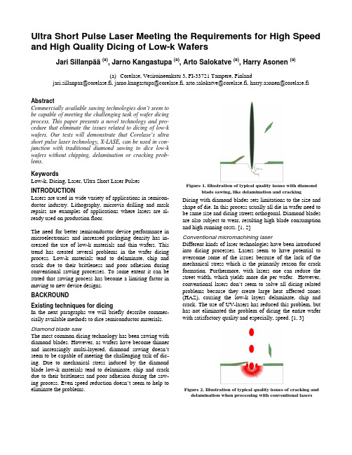
Ultra Short Pulse Laser Meeting the Requirements for High Speed and High Quality Dicing of Low-k WafersJari Sillanpää (a), Jarno Kangastupa (a), Arto Salokatve (a), Harry Asonen(a)(a)Corelase, Vesiroineenkatu 3, FI-33721 Tampere, Finlandjari.sillanpaa@corelase.fi, jarno.kangastupa@corelase.fi, arto.salokatve@corelase.fi, harry.asonen@corelase.fiAbstractCommercially available sawing technologies don’t seem to be capable of meeting the challenging task of wafer dicing process. This paper presents a novel technology and pro-cedure that eliminate the issues related to dicing of low-k wafers. Our tests will demonstrate that Corelase’s ultra short pulse laser technology, X-LASE, can be used in con-junction with traditional diamond sawing to dice low-k wafers without chipping, delamination or cracking prob-lems.KeywordsLow-k, Dicing, Laser, Ultra Short Laser Pulses INTRODUCTIONLasers are used in wide variety of applications in semicon-ductor industry. Lithography, microvia drilling and mask repairs are examples of applications where lasers are al-ready used on production floor.The need for better semiconductor device performance in microelectronics and increased packaging density has in-creased the use of low-k materials and thin wafers. This trend has created several problems in the wafer dicing process. Low-k materials tend to delaminate, chip and crack due to their brittleness and poor adhesion during conventional sawing processes. To some extent it can be stated that sawing process has become a limiting factor in moving to new device designs.BACKROUNDExisting techniques for dicingIn the next paragraphs we will briefly describe commer-cially available methods to dice semiconductor materials. Diamond blade sawThe most common dicing technology has been sawing with diamond blades. However, as wafers have become thinner and increasingly multi-layered, diamond sawing doesn’t seem to be capable of meeting the challenging task of dic-ing. Due to mechanical stress induced by the diamond blade low-k materials tend to delaminate, chip and crack due to their brittleness and poor adhesion during the saw-ing process. Even speed reduction doesn’t seem to help to eliminate the problems.Figure 1. Illustration of typical quality issues with diamond blade sawing, like delamination and crackingDicing with diamond blades sets limitations to the size and shape of die. In this process usually all die in wafer need to be same size and dicing streets orthogonal. Diamond blades are also subject to wear, resulting high blade consumption and high running costs. [1, 2] Conventional micromachining laserDifferent kinds of laser technologies have been introduced into dicing processes. Lasers seem to have potential to overcome some of the issues because of the lack of the mechanical stress which is the primarily reason for crack formation. Furthermore, with lasers one can reduce the street width, which yields more die per wafer. However, conventional lasers don’t seem to solve all dicing related problems because they create large heat affected zones (HAZ), causing the low-k layers delaminate, chip and crack. The use of UV-lasers has reduced this problem, but has not eliminated the problem of dicing the entire wafer with satisfactory quality and especially, speed. [1, 3]Figure 2. Illustration of typical quality issues of cracking and delamination when processing with conventional lasersWater jet guided laserInteresting concept has been introduced by Synova SA, Switzerland (www.synova.ch). In their technology a fo-cused laser beam is guided by a low-pressure water jet. The water jet acts as a fluid optical wave-guide. Water can re-duce the effect of HAZ and a laser can groove through low-k layers eliminating some of the issues. However, in these processes the lasers used are still conventional lasers that interact with the materials by linear absorption. This still creates HAZ, melt and debris, or inability to remove transparent materials, which can still cause problems [1, 3, 4]Diamond blade saw with laserCombinations of different technologies have been also in-troduced. Combination of laser beam and traditional dia-mond saw seem promising. In this process laser beam is used to create two grooves through low-k layers on the edges of the dicing street. Traditional diamond sawing is then used to cut between the grooves and through the wa-fer. This results better yield compared to just using dia-mond sawing. However, in these processes the lasers used are still conventional lasers, which create HAZ, melt and debris. These cause quality issues to the process. [1]Ultra short pulse laserRecently a lot of studies have been made about processing materials with ultra short laser pulses, having pulse lengths in the picosecond or femtosecond regime. In these studies it has been demonstrated that with ultra short laser pulses materials can be processed with excellent quality and high precision. This is due the fact that shortening the pulse length to ultra short regime permits a drastic reduction of the HAZ and shock affected zone (SAZ), which are the main reasons for quality issues, like delamination, cracking or chipping [2, 5, 6, 7]. However, processing speed has been a problem with these lasers. It has been demonstrated that with existing femtosecond laser systems same process-ing speed than with mechanical dicing can be achieved only when material thickness is in the range of 10µm [8]. Furthermore, ultra short pulse lasers have been complex, large, expensive systems and difficult to control, thus being unsuitable for integration with semiconductor production lines.Solution = X-LASEIn wafer dicing process low-k and thin materials tend to delaminate, chip and crack due to their brittleness and poor adhesion during conventional sawing and laser processes. Ultra short pulse laser technology has shown a promise to overcome these quality issues, but the processing speed has been a problem. At Corelase, we have developed a new laser concept with special optics and control electronics, X-LASE, which combines the processing benefits of ultra short laser pulses and with high processing speeds. The key factors and benefits of this concept are described in the picture 3. Figure 4 illustrates processing of semiconductor materials with X-LASE.Figure 3. Key factors and benefits of X-LASEFigure 4. Illustration of processing semiconductor materialwith X-LASECORELASE GROOVING EXPERIMENTS WITH ULTRA SHORT PULSE LASERCombination of laser beam and traditional diamond saw has shown a promise to overcome some of the quality is-sues and result better yields compared to just using dia-mond saw. In this process laser beam is used to cut through low-k layers and then diamond saw is used to cut through the wafer. However, the edge quality after laser grooving can be still improved which would result even better yields. Ultra short laser pulse laser could be a solution for im-proved edge quality if the processing speed is meeting the industry standard.To demonstrate improved edge quality with ultra short pulse laser Corelase conducted series of application tests. Tests were conducted in a setup where Corelase’s ultra short pulse laser, X-LASE, was integrated to robotic plat-form provided by PMJ Automec. In these tests we proc-essed series of 20µm wide and 5µm deep grooves on low-k material. Processing speeds varied from 50mm/s to 1100mm/s. After the tests quality of the grooves were in-spected with Nomarsky microscope.Our tests showed that best quality was achieved when us-ing 650mm/s processing speed. This speed is superior compared to any existing method [1, 2, 8]. Furthermore, the quality of the edges is improved when compared to published results on grooves made with conventional lasertechnology [1]. As can be seen in figure 5 there is no evi-dence of debris around the grooves even if we did not use removable protection films during the laser grooving.Figure 5. Grooves on low-k wafer made by X-LASE withexcellent edge quality. CONCLUSIONSA new laser technology has been developed to overcome quality issues in dicing of low-k wafers. Our tests demon-strate that Corelase’s ultra short pulse laser technology, X-LASE, can be used in conjunction with traditional diamond sawing to dice low-k wafers. With X-LASE we have been able to demonstrate grooving without chipping, delamina-tion or cracking problems. Furthermore, processing was done with superior processing speed, 650mm/s, compared to any existing method. Quality and cleanness of the proc-ess is excellent. Corelase believes that this technology can also be scaled to full dicing of thin wafers with superior speed and excellent dicing quality in terms of mechanical die strength and visual appearance. ACKNOWLEDGMENTSWe would like to acknowledge Jouni Suutarinen and Pekka Kettunen from PMJ Automec who provided help, support and the robot platform for the tests. REFERENCES[1]J.Vittu, D.Perrottet, J.-M.Buchilly, B.Richerzhagen,“Damage-Free Dicing Of Low-K Wafers”, FutureFAB International, Issue 17.[2]M.Schmidt, G.Eber, “The Future of Lasers in Elec-tronics”, LMF Section A – Proc.of ICALEO 2003[3]L.Herbst, J.P.Quitter, G.M.Ray, T.Kuntze,A.O.Wiessner, orkov, M.Heglin “High PeakPower Solid State Laser for Micromachining of HardMaterials”, Solid State Lasers XII – Proc. of the SPIE4968 (2003) s. 134-142[4]O.Sibailly, R.Romanowicz, L.Mayor, B.Richerzhagen,“Thin Wafer Cutting with Laser Microjet® at Infineon– A Case Study”, LMF Section D – Proc.of ICALEO2003[5]T.Abeln, J.Radtke, F.Dausinger, “High precision drill-ing with short-pulsed solid-state lasers”, Proc. Laser Microfabrication Conf. ICALEO ’99 (San Diego), P.Christensen, P.Herman, R.Patel (Eds.), LIA Vol. 88, pp. 195-203, Laser Institute of America, Orlando, FL, 2000.[6]C.Föhl, D.Breitling, K.Jasper, J.Radtke, F.Dausinger,“Precision drilling of metals and ceramics with short and ultra short pulsed solid state lasers”, Proc. SPIE 44261, Miyamoto; Y.F.Lu, K.Sugioka, J.Dubowski (Eds.) pp.104-107, Second International Symposium on Precision Microfabrication LPM 2001 (Singapore), Bellingham, WA, Intl. Soc. for Opt. Eng., 2002.[7]M.Weikert, C.Föhl, F.Dausinger, “Surface Structuringof Metals with Ultrashort Laser Pulses“, Proc. SPIE4830, I.Miyamoto, K.F.Kobayashi, K.Sugioka,R.Poprawe, H.Helvajian (Eds.) pp.501-505, Third In-ternational Symposium on Laser Precision Microfab-rication, 2003.[8] A.Ostendorf, C.Kulik, N.Bärsch, “Processing ThinSilicon with Ultrashort-pulsed laser: Creating An Al-ternative to Conventional Sawing Techniques”, LMFSection A – Proc.of ICALEO 2003。
manuscripts使用指南2019-11-19[24页]
![manuscripts使用指南2019-11-19[24页]](https://img.taocdn.com/s3/m/4c45c60076a20029bc642d93.png)
directly submit your work to thousands of different journals .How to get started?There are three ways to get started with Manuscripts.Select one of our built-in templates . Manuscripts comes with over 1,100verified templates, as well as unverified templates based on CSL styles.We are building literally thousands of them for many types of documents,including for journals across all sciences, university specific PhD theses,grant applications etc.Import an existing document. Manuscript supports multiple importformats such as MS Word, LaTeX, Markdown and many more.Create a new blank manuscript.Selecting a built-in templateChoose ‘Create new manuscript’ from the welcome screen or if do not have thewelcome screen available, go to File > New > Manuscript with Template…Figure 1:Template selector1. 2. 3.Browse or search for available templates. Manuscript-verified templates are indicated by a badge icon as shown in Figure 1. Once you have found and selected the template you need, click Choose and a manuscript template is opened for you.Figure 2:New manuscript from templateI cannot see a template for the document I would like to write, what should I do?In case you cannot find the template you are looking for, do not worry, you still have several options to get started.If you are looking to get started with a generic template that consists of an abstract, introduction, methods, results and discussion, you can choose a different journal template (e.g. Genome Biology Research Article) and modify the template to your needs. Remember that the template is only there to help you get started and to provide you with guidance. However, you are still the master of your own manuscript.If you rather get started with a blank canvas, choose Create Empty Manuscript option from the bottom of the template selector and you are all set to write your firstdocument with Manuscripts.You can contact us at support@ with a link to thejournal/template style you would like Manuscripts to support and we will do our best to add your requested template to Manuscripts.I can find a template but it does not have the ‘Manuscript verified’ badge next to it.We have added over 7,000 journal CSL styles that you can choose to get started from to create a template of your own. If you find a journal template without a badge next to the journal name it means that we have not verified the template for that journal yet.You can still choose the journal template as your starting point. When you select the template a pop up window appears where you can see and edit basic templatemetadata such as which sections it should contain, which font and font sizes will be used, and so on. Once you are ready , click Create Manuscript .Import an existing document to ManuscriptsIf you already have a draft in another document format and you would like to continue writing it with Manuscripts, you can easily import the document to Manuscripts and continue writing your draft with ease.Create a new empty manuscript from File > New > Manuscript.Go to File > Import > Content ..You can see all the supported document formats that can be imported toManuscripts by clicking ‘Options’ in the file browser window.Choose the document that you want to import to Manuscripts, click Openand the document is opened as a new manuscripts file.1.2.3. 4.Figure 3:Import an existing document to ManuscriptsHow to reorder manuscript sectionsThe manuscript outline lets you not just see and navigate an outline of your manuscript project, but also manipulate the structure of it in powerful ways. Simply drag and drop to reorder sections or paragraphs to change the flow of your document.How to use the focus mode in ManuscriptsMaintaining focus is essential to productive writing. One of the ways we help you focus in your work is the so-called focus mode. You can activate the focus mode in two ways.A. Click View > Selected Sections in the main menu.B. Click the focus button in the manuscript outline, visible when you hover on an individual section in the outline on its right edge.To toggle back to viewing the entire manuscript, simplyA. Click View > View All Sections in the main menuorB. Click the focus button againA. Focus mode in action.B. Focus mode iconFigure 4:1. The manuscript editing window when you have activated the focus mode.2.The focus mode icon, visible when you hover on a section item.How to use the gutter menusThe Manuscripts editor includes so-called "gutter helpers", designated with the blue + and o symbols in the left margin for the currently active paragraph. These symbols are buttons that lead you to shortcut actions for inserting (the+symbols) and manipulating (the o) the document contents in a way that is contextual.The gutter insertion helper (+ symbol)Figure 5:How to create a new sectionThe gutter selection helper (o symbol)The following example shows what the gutter selection helper tool presents in the case of a paragraph.Figure 6:The gutter selection helper for a paragraph.The gutter helper is entirely optional and can be toggled on or off from the Editing Preferences by choosing "Show smart gutter in the left margin when editing”.Figure 7:Smart gutter can be enabled or disabled in the Editing PreferencesExporting a selected subset of a manuscriptIt often comes handy to place material in your manuscript project which is never intended to reach readers. For instance you may want to keep…Notes regarding your research or writing progress: material that helpsyou organise your writing, but is never intended for publication or forfeedback.1.Figure 8:Sharing the current selectionNote that if you have selected your bibliography as one of the items you want to export, only the subset of it that is cited in the selected part of your manuscript is included in the bibliography.As with sharing, if you have selected your bibliography as one of the items to export, only the subset of citations in the selected part of your manuscript get included in the bibliography.Spell and grammar checking in ManuscriptsManuscripts allows you to check your spelling and grammar as you type to avoid disasters like the one shown in Figure 10. Grammar and spellchecking can be accessed from Edit > Spelling and Grammar in the main menu.Figure 9:Enable spell and grammar checkCheck spelling and grammar manuallyTo check spelling and grammar manually, choose Edit >Spelling and Grammar > Show Spelling and Grammar (⌘:).Figure 10:Check spelling and grammar manuallyTo update the spelling and grammar analysis of your document after you have made some changes, choose Edit >Spelling and Grammar > Check Document Now (⌘;).Check spelling and grammar as you typeSpelling and grammar can also be checked as you type, by toggling on Edit > Spelling and Grammar > Check Spelling While Typing. Grammatical errors are presented in the familiar green, and spelling errors in red.Figure 11:Check spelling and grammar while typingCorrect spelling automaticallyYou can even make Manuscripts correct your spelling for you by choosing Edit > Spelling and Grammar > Correct Spelling Automatically.Choosing a citation styleYou can change the manuscript's citation style in the inspector available on the right hand side of the main window by clicking the Toggle Inspector button available inthe lower right corner of the application window.This reveals the inspector which includes two tabs, second of which is the styleinspector. At the bottom it includes the Manuscript Styles inspector palette (whereyou see the keyboard focus in the screenshot below). This inspector palette is where you can change the citation style.Figure 13:Changing the citation style in the InspectorHow to change the font family and size?You can change the font family and size and edit other properties of paragraph styles in the inspector on the right hand side of the main window by clicking the Toggle Inspector button (available in the lower right corner of the application window).Figure 14:Inspector toggle button iconThis reveals the inspector which has two tabs, second of which is the style inspector that includes the Paragraph Styles palette that let you choose font sizes, etc.Figure 15:Change font and font size in the Paragraph Styles section of the Inspector How to create a figure panelThe basic workflow for adding figure panels into your Manuscripts document involves first placing a figure placeholder into your document (which you can caption at this stage), and then adding an image as a second step.Importing manuscript content that includes images is another option, i.e. File > Import > Content. This is not further discussed separately here.Creating an empty figure panelThere are three different ways to create an empty figure. Each one of them requires you to first place the text insertion cursor next to the spot where you want to insert the figure. After that, you can either:Click the figure symbol in the toolbar.Use the menu item Insert > Figure in the main menu.Use the smart gutter menu to the left of the paragraph that is in focus, before or after which you intend to add the figure. Click the blue + symbol in it, andchoose “Insert Figure”.A. Figure panel iconB.Figure 16:How to create a figure panelThe figure placeholder you create will look something like the following:Figure 17:New empty figure panelAdding images to a figure panelYou can add images to a figure panel in three ways, starting from an empty placeholder image.1.By dragging and dropping into the figure + symbol.By clicking the figure + symbol and choosing Choose File to Import…By clicking the figure + symbol and choosing amongst orphaned figuresin the manuscript in case you have previously added and then removedfigures (orphaned figure images can be removed permanently by rightclicking the figure in the list of orphaned figures, and choosing Delete ).To replace a figure, simply either:Drag an image on top of the image you wish to replace.Alternatively , click on the image and drag in the popover that opens, oragain Choose File to Import…Creating a multi-panel figureYou can create a multi-panel figure in Manuscripts simply by:Figure 18:Creating a multi-panel figureFigure file formatsManuscripts always deals with image data in a non-destructive way . For instance figure panel images all internally still store the original full sized image even if any panel members need to be scaled. Similarly , Manuscripts keep vector formattedgraphics in their original form and only rasterises them to bitmap images on export if required (see below).2.3. 1.2.Figure 19:How to create a table in ManuscriptsThe table that is created looks like this:Figure 20:New tableTo add or remove rows or columns, right-click any of the columns or rows and choose the relevant option from the context menu.Figure 21:Editing a table in ManuscriptsHow to edit the table format in ManuscriptsYou can change and edit the table format with the table styles inspector available on the right hand side of the main window by clicking the Toggle Inspector button (located in the lower right corner of the application window):A. Inspector Toggle iconB. Table Styles in the InspectorFigure 22:Edit and change table styles in the InspectorHow to create an equationManuscript includes a powerful equation editor that allows you to insert beautiful equations using LaTeX.Creating an empty equationThere are three different ways to create an empty figure. Each one of them requires you to first place the text insertion cursor next to the spot where you want to insert the figure. After that, you can either:Click the equation symbol in the toolbar.Use the menu item Insert > Equation in the main menu.Use the smart gutter menu to the left of the paragraph that is in focus, before or after which you intend to add the figure. Click on the blue + symbol in it, and choose Insert Equation.A. Equation iconB.Figure 23:Creating an equationThe equation placeholder that is inserted to the manuscript looks like this:Figure 24:Equation placeholderHow to edit an equationTo edit an equation, click on the equation so that the equation editor pops up. Now you can type or paste your equation using LaTeX, the equation will be rendered in real time. If you are not familiar yet with the LaTeX syntax for equations you can find out more here: https:///wiki/LaTeX/Mathematics#SymbolsA.B.Figure 25:Editing an equation in ManuscriptsManuscript file versioning: backups & change trackingIn short, your Manuscripts documents are versioned for backup and rollback purposes right now. It will also form the basis for change tracking features we intend to build after 1.0.that allows you to cite papers without directly requiring to interact with an externalreference manager. This is what the Manuscripts citation tool looks like:Figure 26:Using the citation tool within ManuscriptsImporting bibliography data into ManuscriptsWe support importing references from all the major reference file formats (Endnote XML, RIS, BibTeX and more). All popular reference managers can export to at least one of these formats. To insert references, either:Choose File > Open in the main menu to open a bibliography file thatyou exported from your favorite reference manager.Choose File > Import in the main menu to import bibliography data intoyour currently open manuscript.Drag a bibliography file into the Manuscripts dock icon.Configuring a citation keyboard shortcutYou can configure the keyboard shortcut to use for the internal citation tool with the option available at Preferences > Editing > Citation Shortcut :1. 2. 3.Figure 27:Configuring a citation tool shortcut in Editing Preferences Enjoy writing your next best work with ManuscriptsWe hope you really enjoy writing with Manuscripts. Visit our forums for more information and up-to-date answers to questions you may have:. Please contact us viasupport@ if you would like to talk to us or hit issues with the app. We would love to hear what you think of Manuscripts and how we can make it even better. Enjoy!The Manuscripts team。
用latex编辑期刊论文manuscript中的问题小结
用latex编辑期刊论文manuscript中的问题小结由于要发文章,期刊要求最好用latex编译,下面是我在编辑manuscript过程中遇到的小问题,当时一个小问题纠结好半天,最后通过google搜索、问latex群的好友、百度知道,各种办法,终于解决了。
可能问题很简单,但是希望受用就好。
当然,如果有错误的地方,请联系我,我随时改正过来。
如果有更好的处理方法、好的建议或者常见的问题,请提出来,大家共享。
因为我是一个latex的初学者,刚开始,就使用的basic版本,很麻烦,经常出错,error:找不到各种文件。
现编译现下载缺的各种包什么的。
所以建议下个full版本的,可以避免出错。
1.推荐网站:很多问题都可以在这个网站上搜到答案。
2.论文要求双倍行距的话:一种方法是在导言区使用语句\linespread{1.6}。
另一种方法是期刊给定的模板里可能有可选的语句。
像我这个模板,就是把原默认的\documentclass[preprint,authoryear,12pt]{...}注释掉,而选择\documentclass[authoryear,preprint,review,12pt]{....},这样也是双倍行距。
3.有些期刊,如果论文要求.tex文件有行号的话,即linenumber。
这样处理:在导言区放\usepackage{lineno},然后从有行号的地方开始加上语句\linenumbers。
4.有的期刊的论文的图都是用Fig.表示,正常默认的是Figure,所以在导言区,使用语句\renewcommand\figurename{Fig },这样出来的就是Fig. 1,什么的。
不要那个空格也可以,随意选择了。
5.参考文献,我使用的是.bib文件编辑的,可是刚开始的时候,总出错。
建议,参考文献的内容与你自己编辑的内容不一致,可以看看.bbl文件,也许就知道答案出错在哪了。
或者最后直接把.bbl的内容复制到.tex 中,这样也许能解决问题,至于在引用文献的过程中,若出现引用不了,出现?等等,那就因情况而异了。
Manuscript_formatting
FORMAT OF ARTICLES AND LETTERS.
Contributions should be double-spaced and written in English (spellings as in the Oxford English Dictionary). Contributions should be organized in the sequence: title, text, methods, references, end notes, tables, figure legends. In order to facilitate the review process, for initial submissions we encourage authors to incorporate the manuscript text and figures together in a single file (Microsoft Word or PDF, up to 30 MB in size). The figures may be inserted within the text at the appropriate positions or grouped at the end, and each figure legend should be presented together with its figure. Also, please include line numbers within the text.
Titles should not exceed 90 characters (including spaces) for Letters, or 75 characters (including spaces) for Articles. Titles should not include numbers, acronyms, abbreviations or punctuation. They should include sufficient detail for indexing purposes but be general enough for rd to appreciate what the paper is about.
manuscript的用法
manuscript的用法全文共四篇示例,供读者参考第一篇示例:手稿是指手写或打印的文本,它是作者创作的初稿,尚未进行编辑或出版处理。
手稿通常用于学术研究、文学作品、艺术创作等领域。
手稿在历史上曾是重要的载体,记录着人类的思想、智慧和传统。
手稿的使用可以追溯到古代,古代各国都有自己独特的文字和书写系统。
在古代,手稿是保存和传播知识的重要工具,在欧洲中世纪,修道院是主要的手稿复制中心,修道士们用精美的手稿记录和传扬圣经和其他文学作品。
随着印刷术的发明,手稿逐渐被印刷物所替代,但手稿在某些领域仍然具有重要的地位。
对于学术研究者来说,手稿是展示研究成果和思想的重要途径,很多学术期刊和出版社仍然接受手稿投稿。
在学术领域,撰写一篇学术手稿是一项繁重的工作,需要作者对研究领域有深入的了解,以及严谨的逻辑和清晰的表达能力。
为了确保手稿质量,作者通常会进行多次反复修改和审校,以保证内容准确、论据充分、结构合理。
除了学术研究,手稿在文学创作和艺术领域也有重要的作用。
很多作家在创作小说、诗歌或剧本时都会先手写草稿,以便更好地表达自己的想法和情感。
手稿是作家灵感的源泉,也是创作过程中不可或缺的一部分。
在艺术领域,手稿也扮演着重要的角色。
许多音乐作曲家和艺术家会先通过手写草稿来构思和设计作品,将自己的想法和创意记录下来。
手稿可以帮助艺术家更好地理清思路,找到灵感的源泉。
手稿是创作者最初的想法和表达方式,它记录着作者的心路历程和创作过程。
虽然在现代社会,电子文档和数字编辑软件已经成为主流,但手稿在某些领域仍然具有独特的价值和意义。
手稿是文化遗产的重要组成部分,也是人类文明发展的见证者。
希望手稿能够继续发扬光大,为人类创作和思想交流作出更大的贡献。
第二篇示例:Manuscript这个词源于拉丁语“manu scriptus”,意为“手写的”,通常指手稿或手抄本。
在学术界和出版行业中,manuscript是指作者已经撰写好的专业论文、书稿、杂志文章等文本,尚未经过编辑和排版处理。
Manuscript Template
---------------------------------------------------------------最新资料推荐------------------------------------------------------Manuscript TemplateJournal of Animal Ecology Author Guidelines: 1 Manuscript Style and Formatting for Standard Papers 2 3 4 5 A. Author * a , B. Author b , C. Author c , and X. Author x 6 a Department of Life Sciences, University of Somewhere, City, Country 7 b Department of Life Sciences, University of Somewhere, City, Country 8 c Department of Life Sciences, University of Somewhere, City, Country 9 x Department of Life Sciences, University of Somewhere, City, Country 10 * Corresponding author: a.author@ 11 12 13 14 Summary 15 1. This should summarise the main results and conclusions of the paper using 16 simple, factual, numbered statements. It must not exceed 350 words. 17 18 2. Summaries/abstracts are key to getting people to read your article. 19 20 3. Summaries should be understandable in isolation from your article. 21 22 4. Summaries should only have 5 points, ideally, listing; (1) what the 23 question is, (2) why it is interesting, (3) what was done in the study, (4) 24 what was found and (5) what this means. 25 26 5. Advice for optimising your Summary/Abstract (and Title) so that your 27 paper is more likely to be found in online searches is provided1 / 11at 28 /bauthor/seo.asp 29 30 31 Key-words Listed in alphabetical order, the key-words should not exceed 10 words 32 or short phrases. Please pay attention to the keywords you select: they should not 33 already appear in the title or abstract. Rather, they should be selected to draw in 34 readers from wider areas that might not otherwise pick up your paper when they are 35 using search engines. 36 37 38 Introduction 39 This should state the reason for doing the work, the nature of the hypothesis or 40 hypotheses under consideration, and should outline the essential background. 41 42 Materials and methods 43 This should provide sufficient details of the techniques to enable the work to be 44 repeated. Do not describe or refer to commonplace statistical tests in Methods but 45 allude to them briefly in Results. 46 47 Results 48 This should state the results, drawing attention in the text to important details shown 49 in tables and figures. 50 51 Discussion 52 This should point out the significance of the results in relation to the reasons for doing 53 the work, and place them in the context of other work. 54 55 Data Accessibility 56 In order to enable readers to locate archived data from papers, we require that authors 57 list the database (e.g. Dryad, figshare, GenBank etc.) and the respective---------------------------------------------------------------最新资料推荐------------------------------------------------------ accession 58 numbers or DOIs for all data from the manuscript that has been made publicly 59 available. Where data is not archived, authors need to still include a data accessibility 60 section and in it explain why data wasn’t archived (e.g. sensitive locality data for 61 endangered species). 62 63 Acknowledgements 64 In addition to acknowledging collaborators and research assistants, include relevant 65 permit numbers (including institutional animal use permits), acknowledgment of 66 funding sources, and give recognition to nature reserves or other organizations that 67 made this work possible. Do not acknowledge Editors by name. 68 69 Specifications 70 Manuscripts should be typed in double spacing with a generous margin. The paper 71 must include sequential line numbering throughout, and pages should be numbered 72 consecutively, including those containing acknowledgements, references, tables and 73 figure legends. Authors should submit the main document as a RTF or Word file. 74 Figures can be embedded or uploaded as separate files. The RTF and Word will be 75 converted to PDF (portable document format) upon upload. Reviewers will review the 76 PDF version while the Word file will remain accessible by the Editorial Office. 77 Manuscripts3 / 11must be in English, and spelling should conform to the Concise Oxford 78 Dictionary of Current English. 79 80 References 81 References in the text to work by up to three authors should be in full, e.g. (Johnson, 82 Myers James 2006). If there are more than three authors, they should always be 83 abbreviated thus: (Nilsen et al. 2009). When different groups of authors with the same 84 first author and date occur, they should be cited thus: (Jonsen, Myers James 2006a; 85 Jonsen, James Myers 2006b), then subsequently abbreviated to (Jonsenet al. 2006a, 86 b). The references in the list should be in alphabetical order with the journal name in 87 full. The format for papers, entire books, chapters in books, and PhD theses is as 88 follows. 89 90 Underwood, N. (2009) Effect of genetic variance in plant quality on the population 91 dynamics of a herbivorous insect. Journal of Animal Ecology, 78, 839847. 92 93 Jonsen, I.D., Myers, R.A. James, M.C. (2006) Robust hierarchical statespace 94 models reveal diel variation in travel rates of migrating leatherback turtles. Journal of 95 Animal Ecology, 75, 10461057. 96 97 Nilsen, E.B., Linnell, J.D.C., Odden, J. Anderson, R. (2009) Climate, season, and 98 social status modulate the functional response of an efficient stalking predator: the 99 Eurasian lynx. Journal of Animal---------------------------------------------------------------最新资料推荐------------------------------------------------------ Ecology, 78, 741751. 100 101 Otto, S.P. Day, T. (2007) A Biologist’s Guide to Mathematical Modeling in Ecology 102 and Evolution. PrincetonUniversity Press, Princeton, New Jersey, USA. 103 104 Conway. G. (2007) A Doubly Green Revolution: ecology and food production. 105 Theoretical Ecology: Principles and Applications, 3rd edn (eds R. May A. 106 McLean), pp. 158171. OxfordUniversity Press, Oxford. 107 108 Stevenson, I.R. (1994) Male-biased mortality in Soay sheep. PhD thesis, University of 109 Cambridge, Cambridge. 110 111 References should only be cited as ‘in press’ if the paper has been accepted for 112 publication. Other references should be cited as ‘unpublished’ and not included in the 113 list. Work not yet accepted for publication may be cited in the text and attributed to its 114 author as: author name (including initials), unpublished data. In press articles should 115 be uploaded with the manuscript as supplementary files. 116 We recommend the use of a tool such as EndNote or Reference Manager for reference 117 management and formatting. EndNote reference styles can be searched for here: 118 /support/enstyles.asp 119 Reference Manager reference styles can be searched for here: 120 /support/rmstyles.asp 121 122 Citations from the World5 / 11Wide Web 123 Citations from the world-wide-web are only allowed when alternative hard literature 124 sources do not exist for the cited information. Authors are asked to ensurereference list, along with 126 titles, years and authors of thehave sufficient longevity and ease of access 128 for others toscientific quality at least equal to that of peer reviewed 130 information available in learned scientific journals. 131 132 Units, symbols and abbreviations 133 Authors are requested to use the International System of Units (SI, Systme 134 International d’Units) where possible for all measurements (see Quantities, Units and 135 Symbols, 2nd edn (1975) The Royal Society, London). Note that mathematical 136 expressions should contain symbols not abbreviations. If the paper contains many 137 symbols, it is recommended that they should be defined as early in the text as possible, 138 or within a subsection of the Materials and methods section. 139 140 Scientific names 141 Give the Latin names of each species in full, together with the authority for its name, 142 at first mention in the main text. If they appear in the Summary/Abstract, use the 143---------------------------------------------------------------最新资料推荐------------------------------------------------------ common and Latin name only in the first instance, then the Latin or common name 144 thereafter. If there are many species, cite a Flora or checklist which may be consulted 145 for authorities instead of listing them in the text. Do not give authorities for species 146 cited from published references. Give priority to scientific names in the text (with 147 colloquial names in parentheses, if desired). 148 149 Makers’ names 150 When a special piece of equipment has been used it should be described so that the 151 reader can trace its specifications by writing to the manufacturer; thus: ‘Data were 152 co llected using a solid-state data logger (CR21X, CampbellScientific, Utah, USA)’. 153 154 Mathematical material 155 Mathematical expressions should be carefully represented. Suffixes and operators 156 such as d, log, ln and exp will be set in Roman type; matrices and vectors will be set 157 in bold type; other algebraic symbols will be set in italic. Make sure that there is no 158 confusion between similar characters like ‘l’ (ell) and ‘1’ (one). Also make sure that 159 expressions are spaced as you would like them to appear, and if there are several 160 equations they should be identified by eqn 1, etc. 161 162 Numbers in tables 163 Do not use an excessive number of digits7 / 11when writing a decimal number to represent 164 the mean of a set of measurements (the number of digits should reflect the precision 165 of the measurement). 166 167 Numbers in text 168 Numbers from one to nine should be spelled out except when used with units; e.g. two 169 eyes but 10 stomata and 5C. 170 171 Figures 172 The publishers would like to receive your artwork in electronic form. Please save 173 vector graphics (e.g. line artwork) in Encapsulated Postscript Format (EPS), and 174 bitmap files (e.g. half-tones) in Tagged Image File Format (TIFF). Ideally, vector 175 graphics that have been saved in a metafile (.WMF) or pict (.PCT) format should be 176 embedded within the body of the text file. Detailed information on the Wiley- 177 Blackwell digital illustration standards is available at: 178 /bauthor/illustration.asp 179 180 Figures should not be boxed (superfluous bounding axes) and tick marks must be 181 on the inside of the axes. Where possible, figures should fit on a single page in the 182 submitted paper. In a final version they will generally be reduced in size by about 50% 183 during production. Wherever possible, they should be sized to fit into a single column 184 width (c. 70mm final size). To make best use of space, you may need to rearrange 185 parts of figures (e.g. so that they appear side by side). Please---------------------------------------------------------------最新资料推荐------------------------------------------------------ ensure that symbols, 186 labels, etc. are large enough to allow reduction to a final size of c. 8 point, i.e. capital 187 letters will be about 2 mm tall. Lettering should use a sans serif font (e.g. Helvetica 188 and Arial) with capitals used for the initial letter of the first word only. Bold 189 lettering should not be used. Units of axes should appear in parentheses after the 190 axis name. Please note that line figures should be at least 600 dpi and half-tones 191 (photos) should be at least 300 dpi. 192 193 Images in the printed version of the Journal of Animal Ecology are in black and white 194 as it is the policy of the Journal of Animal Ecology for authors to pay the full cost 195 for colour paper print reproduction (currently 150 for the first figure, 50 thereafter). 196 Free colour reproduction is available for the on line version: if authors require this, 197 they should write their figure legend to accommodate both versions of the figure, and 198 indicate their colour requirements on the Colour Work Agreement Form. This form 199 should be completed in all instances where authors require colour, whether in print or 200 online. Therefore, at acceptance, please download the form and return it to the 201 Production Editor (Penny Baker, Wiley-Blackwell, John Wiley9 / 11Sons, 9600 202 Garsington Road, OxfordOX4 2DQ, UK. Please note that the ORIGINAL 203 HARDCOPY form must be returned in all instances (a faxed or scanned version 204 cannot be accepted). Please note that if you require colour content your paper cannot 205 be published until this form is received. 206 207 Figure legends 208 Legends should be grouped on a separate sheet. Furnish enough detail so that the 209 figure can be understood without reference to the text. In the full-text online edition of 210 the journal, figure legends may be truncated in abbreviated links to the full screen 211 version. Therefore, the first 100 characters of any legend should inform the reader of 212 key aspects of the figure. Figures should be referred to in the text as Fig. 1, etc. (note 213 Figs 1 and 2 with no period). 214 215 Tables 216 These should be referred to in the text as Table 1, etc. Do not present the same data in 217 both figure and table form. Each table should be on a separate page, numbered and 218 accompanied by a title at the top. 219 220 Supporting Information 221 Journal of Animal Ecology rarely publishes Appendices in the printed version. 222 However, Supporting Information that is referred to in the text may be made available 223 in the online version of the article. Guidelines for the preparation of Supporting 224 Information---------------------------------------------------------------最新资料推荐------------------------------------------------------ are available here. 225 226 For the printed version, any Appendices should be listed under ‘Supporting 227 Information’, and added after the References, with the opening s tatement: ‘The 228 following Supporting Information is available for this article online’ followed by 229 brief captions for the Appendices/Figs/Tables to be included. These should be 230 numbered Appendix S1, Fig. S1, Table S1, etc. 231 232 Any literature referred to in the Appendix or online Supporting Information should 233 also be referenced in the Appendix or online Supporting Information so that it is a 234 self-contained piece of work. This may mean duplicating references if any literature is 235 cited in both the main text and the Supporting Information. 236 237 All Supporting Information should be submitted online as part of the main manuscript. 238 Please name your online supporting files as online supporting information’ and 239 upload them with the main document. This allows the submission web site to combine 240 all the relevant files together but keep them separate when it comes to publication 241 stage. 24211 / 11。
- 1、下载文档前请自行甄别文档内容的完整性,平台不提供额外的编辑、内容补充、找答案等附加服务。
- 2、"仅部分预览"的文档,不可在线预览部分如存在完整性等问题,可反馈申请退款(可完整预览的文档不适用该条件!)。
- 3、如文档侵犯您的权益,请联系客服反馈,我们会尽快为您处理(人工客服工作时间:9:00-18:30)。
Electrochemical behavior of Dopamine in the presence of Ascorbic Acid on a reduced Au/C60 films modified glass carbon electrodePing Zhang*a, Guang-Chao Zhaoba: Anhui Wuhu Institute of Drug Control, Wuhu 241000, Chinab: School of Chemistry and Materials Science, Anhui Normal University, Wuhu 241000, ChinaAbstractThe Au/C60 nanoparticles was modified on the surface of glass carbon electrode by electroreducing the mixture of C60 and HAuCl4. SEM an AFM images displayed that the Au/C60 nanoparticles was tightly adsorbed on the surface of glass carbon electrode. After electrochemically reduced in 0.1M KOH, the surface of modified electrode was negatively charged which was proved by electrochemical characterization. The modified electrode was demonstrated to catalyze the electrochemical response of dopamine (DA) by cyclic voltammetry. The standard rate constant ( s) for the electron transfer reaction of DA at the reduced Au/C60 films was found to be 1.38 × 10⁻² cm/s. Furthermore, the separation between the voltammetric peaks of AA and DA is large, thus the simultaneous determination of AA and DA or the selective determination of DA in the presence of AA is feasible at the modified electrode. A linear response to DA and AA was obtained in the concentration range from 3.2 × 10⁻⁶ to 5.0 × 10⁻⁴ M and 8.0 × 10⁻⁶ to 5.0 × 10⁻³ M with a correlation coefficient of 0.9986 and 0.9997, respectively. The detection limit for dopamine was 2.0 × 10⁻⁷ and 5.3× 10⁻⁶ M for ascorbic acid. The proposed sensor has promising feature such as excellent sensitivity, good selectivity and high stability.Keywords: The reduced Au/C60 films modified electrode; Dopamine; Ascorbic acid; Electrocatalysis1. IntroductionDopamine (DA) is the most typical neurotransmitter and plays a significant role in the mammalian central nervous system. Extreme abnormalities of DA concentration levels may lead to several diseases such as Parkinson's disease and schizophrenia and HIV infection [1-3]. Therefore, some methods have been developed to determine the DA [4-9]. Among these, the electrochemical method attracts wide attentions due to their convenient, simple and fast analysis. However, the electrochemical determination of DA was usually affected by the presence of high concentration of ascorbic acid (AA) in the brain [10, 11], which has the almost same oxidation potential range as DA on the solid electrode [13]. So many approaches have been proposed to increase the sensitivity of the determination of DA and eliminate the interference from AA [14-17]. These methods show good selectivity and sensitivity of determination of DA. However, still there is an expanding demand for the development of stable, simple and efficient materials as sensors to determine DA with high sensitivity and selectivity.In recent years, considerable interest has been shown in the study of synthesis and properties of nanometer-scaled metal particles due to their potential applications in sensors, nanoelectronic devices and catalysts [18-24]. Those nanoparticles were prepared, mainly, through chemical reduction of aqueous solution of the chloro-metallate anion [25, 26], metal vapor synthesis routes [27, 28], or the electrochemical deposition on an inert base [19, 20, 22]. Among these, the electrochemical deposition has received much interest because of its rapidness, convenience and economy. Recently, more and more study groups selected the carbon materials as supports for deposition of metal nanoparticles for its chemical/electrochemical stability, good electrical conductance, high surface area and low surface group contents. Fullerenes, as a new form of carbon, have received numerous theoretical and experimental studies since the discovery of fullerenes in 1985 [29] and the first laboratory synthesis in 1990 [30]. Due to their nanometer size and interesting properties, fullerenes are also of great interest for many applications such as batteries [31] and chemical sensor [32]. Furthermore, high accessible surface area, unique three dimensions and high stability suggest that fullerenes are suitable support materials for deposition of metal nanoparticles. But still few reports have been published about the application of the fullerenes in this field.In this paper, we reported the feasibility of forming Au/C60 particles on a glass carbon electrode by electrochemically reducing the complexes of fullerene (C60) and Au (III). Based on the experiments, we found that the nanostructured carbon fullerene film served as a favorable support for depositing the Au nanoparticles; in addition, the Au nanoparticles incorporated onto fullerenes can promote the electrochemical activities of the fullerenes in aqueous solution. Furthermore, we investigated the electrochemical response of dopamine at this modified electrode and the results showed the proposed electrode materials exhibited an excellent electrocatalytic activity for the determination of dopamine. We also detected the effect of AA on determination of DA. The results showed the separation between the voltammetric peaks of AA and DA is large. Thus the simultaneous determination of AA and DA or the selective determination of DA in the presence of AA is feasible at the modified electrode. Based on these, an electrochemical method with high sensitivity and excellent selectivity was proposed to detect DA in the presence of AA.2. Experiment2.1 Reagents and apparatusDA and AA (sigma, USA) were used as received. The C60 with purity 99.9% was purchased from Tian-an Company (Beijing, China). All other reagents were of analytical grade. The phosphate buffer solution (PBS) was prepared from KH2PO4 and K2HPO4 (0.1M). The water used throughout was doubly distilled and all experiments were carried out under room temperature.A pHs-3C digital pH-meter (Shanghai, China) was used to measure the pH values of aqueous solutions. Electrochemical experiments were performed with CHI 660 electrochemical analyzer (CHI, USA) with a conventional three-electrode cell. The image of the surface of the modified electrode was recorded on an AJ- Atomic Force Microscopy (AFM) (China).The working electrode was a reduced Au/C60 modified GC electrode. A saturated calomel electrode (SCE) and a platinum electrode were used as the reference and the auxiliary electrode, respectively.2.2 Electrodes and preparationThe GC electrode ( =3mm) was firstly polished with an alumina slurry and washed successively with triply-distilled water and ethanol in an ultrasonic bath, then 15 cyclic scans were carried out in the potential range of 2.0 to - 2.0V (vs. SCE) in the solution of 1.0 mol/L H2SO4. 2 mg C60 was dispersed with the aid of ultrasonic agitation in 0.5 mL (1 mg / mL) HAuCl4 acetone solution for 10 minutes. The films were prepared by dropping 20 L HAuCl4/C60 acetone solution onto the surface of GC electrode and dried in air. The modified electrode was firstly cycled in 0.1 M phosphate solutions pH 7.0 in the potential range between + 1.0 ~ - 0.6 V with scan rate 100 mV/s until stable and reproducible voltammograms were obtained. Then the electrode was transferred with washing to another cell containing 0.1M KOH solutions and was cycled in the potential between + 0.8 ~ - 1.0 V with a 100 mV/ s scan rate. Then the reduced Au/C60 films modified electrode was performed. All these were done under a N2 atmosphere.3. Results and discussion3.1 The electrochemical properties of Au/C60 films modified electrode3.1.1 Electrodeposition of Au nanoparticles20 L acetone solution of C60 + HAuCl4 was dropped onto the surface of GC electrode and dried in air. Then, successive cyclic scans were carried out at the potential range of + 1.0 ~ - 0.6 V in 0.1M pH 7.0 phosphate solutions as shown in Figure 1. In the initial cycle, a cathodic peak at 0.32 V was observed, which was related to Au (III) reductive reaction (curve 1). At the followed cycle, however, the peak was shifted positively to 0.46 V while the peak current obviously decreased. After 9 cyclic scans, the cathodic peak current and potential value were trended to be constant, which indicated that the Au (III) on the surface of electrode had been reduced fully. After continuous cycles of the modified electrode in 0.1M phosphate solutions pH 7.0 at the potential range + 1.0 ~ - 0.6 V, no obvious changes of the peak current and the peak potential value were observed, which suggested the good stability of the electrode.Fig. 1 near hereFigure 2 showed typical SEM and AFM images of gold nanoparticles incorporated with fullerene films on the electrode. Inspection of the electrode at the end of the multicyclic process reveals that a black deposit is present on the electrode, which we called the Au/C60 film. Based on the literature [31] and this experiment, here the fullerene provided a good supported substrate for deposition of the Au nanoparticles.Fig. 2 near here3.1.2 Reduction of the Au/C60 filmsFigure 3 shows the electrochemical behavior of the Au/C60 films modified GC electrode and the bare Au electrode in 0.1M KOH, respectively. When the potential was scanned between + 0.8 and - 1.0V, the Au/C60 films showed two reduction peaks and an oxidation peak (Figure 3A). Compared with the bare Au electrode (Figure 3B), the Au/C60 films showed a new reduction peak at Epc2 = - 0.41 V, which is correspond to the reductive result of the C60. In addition, the peaks at Epc1 = + 0.04 V and Epo1 = + 0.49 V are associated with the characteristic redox of the Au, which is the same as the bare Au electrode. Hereafter it was reduced in 1M KOH, the Au/C60 films on the GC electrode will be referred as the reduced Au/C60 films.Fig. 3 near here3.1.3 Electrochemical characterization of electrode surfaceCyclic voltammetry of an electroactive species, such as Fe(CN)64-/3- or Co(bpy)23+/2+ (bpy=2,2'-bipyridine), is a valuable tool for testing the kinetic barrier of the interface [33]. In order to investigate the property of the electrode surface, the CV experiments were carried out in the solutions containing electroactive species Fe(CN)64-/3- and Co(bpy)23+/2+, respectively. Figure 4 showed the cyclic voltammograms obtained from 5mM of K3[Fe(CN)6] at the Au/C60 GC electrodes and the reduced Au/C60 modified electrodes in 0.1M KCl solution, respectively. At the Au/C60-modified GC electrodes, the K3[Fe(CN)6] showed a reversible redox response with E p=109 mV. However, the electron transfer kinetics at the reduced Au/C60 modified electrode was found to be sluggish. The peak-to-peak separation turned into 170 mV and the peak current obviously decreased (Fig. 4b). On the other hand, when the CV experiments were carried out in the solution containing an electroactive species Co(bpy)23+/2+, the results was different from that obtained in the Fe(CN)64-/3- solution. The peak potential obtained at a reduced Au/C60 electrode was similar to that obtained at the Au/C60-modified GC electrodes, but the peak current at the reduced Au/C60 electrode increase obviously (as shown in Fig. 5b). Above results suggested that the surface of reduced Au/C60 modified electrode would be negative charges, which resulted in repelling the negative redox couple Fe(CN)64-/3- and attracting the positive redox couple Co(bpy)23+/2+. Fig. 4 near hereFig. 5 near here3.2 Electrochemical response of DA at the reduced Au/C60 microparticle electrodeFigure 6 depicts the electrochemical behavior of DA at the bare and the reduced Au/C60 films modified GC electrode, respectively. At the bare GC electrode, the electrochemical response of DA was poor and the CV of DA demonstrated an irreversible wave with E p, the difference between the anodic peak potential (Epa) and the cathodic peak potential (Epc), 110mV (curve b). However, as shown in Fig. 6a, the reversibility of the electrode reaction of DA is improved significantly at the reduced Au/C60 modified electrode. The E p value is found to be 30 mV (curve b), which indicates faster electron transfer kinetics at the modified electrode. The formal potential (E0') of DA estimated from the average value of anodic and cathodic peak potential, (Epa+ Epc)/2, was 0.271 V (vs. SCE) and ipa / ipc was 1.41.Fig. 6 near hereWhen the electrode was transferred to the blank supporting electrolyte without DA after measuring a DA solution, it showed a weak voltammetric signal corresponding to the redox reaction of DA, which would disappear in the subsequent sweeps. This indicated DA was weakly adsorbed onto the reduced Au/C60 films and it desorbed from the films in the subsequent sweeps.The effect of scan rate on the anodic and cathodic peak currents of dopamine was investigated. The experiment results indicated that the ip was proportional to the square root of scan rate over the range of 20 ~ 200 mV/s. The linear regression equations were ipc = - 2.252 + 0.6995 1/2 and ipa = -2.7902 + 0.9325 1/2 [ip ( A); 1/2, (mV/s) 1/2], with correlation coefficients of 0.9958 and 0.9963, respectively. The results indicate that the electron transfer reaction is controlled by the diffusion DA.According to the Nicholson theory [34, 35], the electron transfer rate constant of DA at the reduced Au/C60 films, s, was calculated using the Ep value obtained at higher scan rate.Fig. 7 shows the relationship of Ep and ln ( , scan rate, mV/s). The curves approach the straight line at higher rates (6 ~ 20 V/s).Fig. 7 near hereBased on the slope and the intercept, the standard rate s can be obtained according to following equations [35]:Epa = E0' + m [0.78 + ln (D1/2/ s) - 0.5 ln m] + 0.5 m lnm = RT / [(1 - ) n F]Epc = E0' - m? [0.78 + ln (D1/2/ s) - 0.5 ln m?] + 0.5 m? lnm? = RT / n FHere the diffusion coefficient D of DA was determined by chronocoulometric method [36, 37] and calculated as 4.2×10⁻⁶ cm2 / s (25?C) under our experiment conditions. Then the rate constants ( s) are calculated to be 1.49 × 10⁻² cm / s and 1.26 × 10⁻² cm / s at the reduced Au/C60 modified electrode, respectively, and their average is 1.38 × 10⁻² cm / s, which is 6.2 times than that at the bare GC electrode, demonstrating that the reduced Au/C60 films facilitate the oxidation process of DA.The effect of solution pH on response of DA was studied in 0.1M phosphate buffer solution in the range of 3.0 ~ 8.0. It was found that the response of DA is well behaved in this range, and anodic peak potential of DA shifted negatively with the increasing of pH value. The relation between the peak potential and pH was directly investigated and a linear regression equation Epa (mV) = 551.1 - 52.63 pH; r = 0.9983 and Epc (mV) = 514.7 - 51.51 pH; r = 0.9979 were obtained. The slopes were very close to the theoretical value of -58 mV pH-1. Based on the above results and the literature [38, 39] t he uptake of electrons is accompanied by an equal number of protons.3.3 Electrochemical response of DA in the presence of AAAA coexists with DA in the extra-cellular fluid of the central nervous system, and its concentration is much higher than that of DA. It can be oxidized at a potential near to that of DA at most solid electrode, which resulted in an overlapped voltammetric response [13].To evaluate the sensitivity and selectivity of the present system for determination of DA in the presence of AA, the electrochemical behavior of the mixture of AA and DA at the reduced Au/C60 films modified electrode was studied.Fig. 8 shows the cyclic voltammograms obtained for DA and AA coexisting at bare GC electrode and the reduced Au/C60 films modified electrode, respectively. Only one broad voltammetric signal was observed at 0.33 V for the mixture AA and DA at the bare GC electrode in 0.1M phosphate buffer solution (pH 5.0) as shown in Fig. 8a. Therefore it is impossible to use the bare electrode for the voltammetric determination of DA in the presence of AA. However, the reduced Au/C60 films modified electrode can separate the mixed voltammetric signals into two well-defined voltammetric peaks at 0.11 and 0.28 V corresponding to the oxidation of AA and DA (Fig. 8b), respectively.Fig. 8 near hereTo get a better-distinguished voltammogram, the effect of pH was tested with the range 3.0 ~ 8.0 (Figure 9) and the results showed that the difference of the anodic peak potential between DA and AA, E pa (DA - AA), has a maximum value (~200 mV) at pH 5.0. So a pH 5.0 buffer solution was selected as supporting electrolyte for DA determination in the presence off AA in this experiment.Fig. 9 near here3.4 The optimized conditions of the electrode preparationThe effect of the ratio of Au to C60 in the mixture on the response of DA was investigated by using Differential Pulse Voltammetry (DPV). With increase of the ratio in the range of 0 ~ 0.15, the peak current of DA increases. When the ratio is more than 0.15, the current obviously decreases. On the other hand, when the ratio is less than 0.15 the response of DA will be obviously affected by AA. According to the experiment results, an optimal ratio of 0.15 was selected in our experiments.Some amounts of the mixture solutions were dried onto substrates respectively and produced various thickness films. The experiment results showed that there was an optimal response of DA and AA on the electrode with coverage 20 L HAuCl4 /C60 acetone solution.3.5 Simultaneous determination of DA and AAThe difference potential between the two cationic peaks of DA and AA is about 200 mV by DPV voltammograms, which is sufficient enough for the selective determination of DA or simultaneous determination of both DA and AA in a mixture.Fig. 10 exhibits the DPV voltammograms obtained for different concentrations of DA in the presence of AA at the reduced Au/C60 films modified electrode. The voltammetric peak of AA was unaltered, whereas the peak current of DA increases linearly with the increasing concentration of DA, confirming that the responses of DA and AA at the modified are independence. The detection limit of DA in the presence of 0.45 mM AA was found to be 0.83 M with the correlation coefficient of 0.998. So the selective determination of DA in the presence of AA is feasible at the modified electrode.Fig. 10 near hereFig. 11 illustrates the DPV response of the modified electrode while simultaneously varying the concentrations of both AA and DA. The calibration curves for both AA and DA were linear in the range of concentrations (8 to 100 M for DA and 30 to 350 M for AA) with correlation coefficients 0.9925 and 0.9967, respectively, which indicated that the simultaneous determination of DA and AA could be obtained.Fig. 11 near hereDA with positive charges can be attracted to the negatively charged electrode surface while AA with negative charges would be prevented to close to the surface [40]. Under this experimental condition, DA (pKa 8.9) should exist as cation while AA (pKa 4.1) should be anion [18]. Here the reduced Au/C60 modified electrode showed good sensitivity and selectivity for DA in the presence of AA. The electrostatic interaction is expected to increase the approach of cationic DA to the negative surface and therefore an enhancement in the electron transfer kinetics would be expected at the modified electrode.3.6 Amperometric determination of DA and AAFig. 12 and 13 depicted the amperonetric current-time response of the reduced Au/C60 films modified electrode for the determination of DA and AA, respectively. The response current was measured at a fixed potential in a stirring 0.1 M PBS at a pH 5.0. Fig. 12 showed the amperonetric response obtained at a potential of + 0.3 V for successive addition of 6.4 M DA to the stirring 0.1 M PBS (pH 5.0).Fig. 12 near hereThe amperonetric current for the successive addition of 36 M AA at a fixed potential of 0.1 V was shown in Fig. 13. The reduced Au/C60 films modified electrode exhibited rapid response to the changes on the concentration of AA (less than 2s).Fig. 13 near hereThe reduced Au/C60 films modified electrode showed a linear response to DA in the concentration range from 3.2 × 10⁻⁶ to 5.0 × 10⁻⁴ M with a correlation coefficient of 0.9986. For AA, the linear range was between 8.0 × 10⁻⁶ to 5.0 × 10⁻³ M and the correlation coefficient was 0.9997. The detection limit for DA was 2.0 × 10⁻⁷ and 5.3× 10⁻⁶ M. The relative standard deviation (RSD) for ten successive determinations of 5.0 × 10⁻⁵ M DA and 2 × 10⁻⁴ M AA were 1.8 and 2.3 %, respectively, indicating the good reproducibility of the present system.3.7 Analysis of mixture of AA and DA in samplesThe analytical application of the proposed system for the measurement of AA and DA was studied by analyzing the samples containing known amount of a mixture of AA and DA. Aliquots of the samples were added to the electrolyte solution and the corresponding concentrations were estimated by the calibration graphs. The results were shown in the Table 1. A good recovery observed with the present system indicates the reliability of the method for application to monitoring of AA and DA.Table 1 near hereInterference studies were also performed with other compounds. As for 5.0 × 10⁻⁵ M DA and 2.0 × 10⁻⁴ M, no interference could be observed for such compounds as: Cystine (100), Lysine (100), Cysteine (100) and valine (100), NaCl (600), KCl (600), NaBr (400), where the data in brackets were the concentration ratios.4. ConclusionsIn the present work, the reduced Au/C60 nanoparticles were prepared using an electrochemical technique and developed into a new sensor for the determination of DA. The results indicated that the reduced Au/C60 films modified electrode exhibits highly electrocatalytic activity and excellent selectivity for the response of DA in the presence of AA. Furthermore, the separation between the voltammetric peaks of AA and DA is large and thus the simultaneous determination of AA and DA or the selective determination of DA in the presence of AA is feasible at the modified electrode. Based on these, a new sensor was prepared for the determination of DA in the presence of AA. This investigation also develops the electrochemical property of fullerene films and extends the application of Au nanoparticles in aqueous solution.These attractive features of the proposed electrode suggest the promising applications, which is under progress in our laboratory.Acknowledgement. The project was supported by National Natural Science Foundation of P. R. China (No. 20271002) and Anhui Provincial Natural Science Foundation (No. 01044702).References[1] T.E. Smith, in: T.M. Devlin (Ed.), Textbook of Biochemistry with Clinical Correlation, Wiley/Liss, New York, 1992, p.929[2] R.M. Wightman, L.J. May, A.C. Micheal, Anal. Chem. 60 (1988) 769A-779A.[3] J.W. Mo, B. Ogoreve, Anal. Chem. 73 (2001) 1196-1202.[4] M. Zhu, X.M. Huang, J. Li, H.X. Shen, Anal Chim Acta 357 (1997) 261-267.[5] S.A.Wring, J.P. Hart, Analyst 117 (1992) 1215.[6] L. Virag, R.A. Whittington, J Chromatogr B 772 (2002) 267-272.[7] C.L. Guan, J. Quyang, Q.L. Liu, B.H. Liu, W.R.G. Baeyens, Talanta 50 (2000) 1197-1203.[8] H.Y. Wang, Q.S. Hui, L.X. Xu, J.G. Jiang, Y. Sun, Anal Chim Acta 497 (2003) 93-99.[9] T. Liu, M.X. Li, Q.Y. Li, Talanta 63 (2004) 1053-1059.[10] T. Zetterstrom, T. sharp, C.A. Marsden, U, Ungerstedt, J Neurochem 41 (1983) 1769-1773.[11] P. Capella, B. Ghasemzadeh, K. Mitchell, R.N. Adams, Electroanalysis 2 (1990) 175-182.[12] F. Gonon, M. Buda, R. Cespuglio, M. Jouvert, J. Pajol, Nature 286 (1980) 902-904.[13] J.M. Zen, G. Ilangovan, J.J. Jou, Anal Chem 71 (1999) 2797-2805.[14] J. Chen, C.S. Cha, J Electroanal Chem 463 (1999) 93-99.[15] M.J. Giz, B. Duong, N.J.Tao, J Electroanal Chem 465 (1999) 72-79.[16] J. Oni, T. Nyokong, Anal Chim Acta 434 (2001) 9-21.[17] L. Zhang, Y. Sun, X. Lin, Analyst 126 (2001) 1760-1769.[18] G. Schmid (Ed.), Clusters and Colloids, VCH, Weinheim, 1994.[19] M.O. Finot, G.D. Braybrook, M.T. McDermott, J. Electroanal. Chem. 466 (1999) 234-241.[20] M.O. Finot, M.T. McDermott, J. Electroanal. Chem. 488 (2000) 125-132.[21] O. Antoine, Y.Bultel, R. Durand, J. Electroanal. Chem. 499 (2001) 85-94.[22] D.Guerin, G.S. Attard, Electrochem. Commun. 3 (2001) 544-548.[23] M.S. El-Deab, T. Ohsaka, Electrochem. Commun. 4 (2002) 288-292.[24] M.S. El-Deab, T. Ohsaka, Electrochim. Acta 47 (2002) 4255-4261.[25] C.W. Chen, T. Takezako, K.Yamamoto, T. Serizawa, M. Akashi, Colloids Surfaces A: Physicochem. Eng. Aspects 169 (2000) 107-116.[26] W. Chen, W. P. Cai, C.H. Liang, L.D. Zhang, Mater. Res. Bull. 36 (2001) 335-342.[27] M. Magnusson, K. Deppet, J.O. Malm, J.O. Bovin, L. Samuelson, Nanostruct. Mater. 12 (1999) 45-48.[28] P.J. Collier, J.A. Iggo, R. Whyman, J. Mol. Catal. A: Chem. 146 (1999) 149-157.[29] H.W. Kroto, J.R. Heath, S.C. O'Brien, R.F. Curl, R.E. Smalley, Nature 318(1985) 162-163.[30] M.S. Dresselhaus, G. Dresselhaus, P.C. Eklund, Science of Fullerene and Carbon Nanotubes [M]. San Diego: Academic Press, 1996.[31] K. Vinoldgopal, M. Haria, D. Meisel, P. Kamat, Nano Lett 4 (2004) 415-418.[32] M. Csiszár, á. Szücs, M. T?lgyesi, á. Mechler, J.B. Nagy, M. Novák, J Electroanal Chem 497 (2001) 69-74.[33] H.Q. Finklea, in: A.J. Bard, I. Rubinstein (Eds.), ElectroanalyticalChemistry, Vol. 19, Marcel Dekker, New York, 1996 (Chapter 2)[34] S. Richard, Nicholson, Anal Chem 37 (1965) 1351-1355.[35] S. Richard, Nicholson, S. Irving, Anal Chem 36 (1964) 706-723.[36] F.R. Anson, Anal Chem 38 (1966) 54-59.[37] F.R. Anson, Anal Chem 36 (1964) 932-935.[38] M.D. Hawley, S.V. Tatawawadi, S. Piekarski, R.N. Adams, J. Am. Chem. Soc. 89 (1967) 447.[39] M. Zhu, X.M. Huang, J. Li, H.X. Shen, Anal. Chim. Acta 357 (1997) 261.[40] Q. Wang, D. Dong, N.Q. Li, Bioelectrochem. 54 (2001) 169-175.。