跨膜蛋白抽提试剂盒说明书_Merck 英文版
MERCK D-Tube -- TMTMTM 透析 电洗脱管 产品说明书

产品使用说明书D -Tube TM透析透析//电洗脱管电洗脱管::Mini/Midi/Maxi/Mega/D Mini/Midi/Maxi/Mega/D--Tube96TM*建议同时参考本说明书英文原文(说明书编号TB422)。
产品名称产品名称包装包装 货号货号 D-Tube™ Dialyzer Mini, 截留分子量 6–8kDa D-Tube Dialyzer Mini,截留分子量 12–14kDa 10个 10个 71504-3 71505-3 D-Tube Dialyzer Midi, 截留分子量 3.5kDa D-Tube Dialyzer Midi, 截留分子量6–8kDa 10个 10个 71506-3 71507-3 D-Tube Dialyzer Maxi, 截留分子量 3.5kDa D-Tube Dialyzer Maxi, 截留分子量6–8kDa D-Tube Dialyzer Maxi, 截留分子量12-14kDa 10个 10个 10个 71508-3 71509-3 71510-3 D-Tube96™ Dialyzer, 截留分子量6-8kDa D-Tube96 Dialyzer, 截留分子量12-14kDa1 1 71712-3 71713-3 D-Tube Electroelution Accessory Kit 1 71511-3截留分子量截留分子量体积体积 试剂盒包装试剂盒包装目录号目录号 D-Tube™ Dialyzer Mega D-Tube Dialyzer Mega D-Tube Dialyzer Mega D-Tube Dialyzer Mega 3.5kDa(蓝色) 3.5kDa(蓝色) 6-8kDa(粉色) 6-8kDa(粉色) 3-10ml 3-10ml 3-10ml 3-10ml 10个 50个 10个 50个 71739-3 71739-4 71740-3 71740-4 D-Tube Dialyzer Mega D-Tube Dialyzer Mega D-Tube Dialyzer Mega D-Tube Dialyzer Mega 3.5kDa(蓝色) 3.5kDa(蓝色) 6-8kDa(粉色) 6-8kDa(粉色) 10-15ml 10-15ml 10-15ml 10-15ml 10个 50个 10个 50个 71742-3 71742-4 71743-3 71743-4 D-Tube Dialyzer Mega D-Tube Dialyzer Mega D-Tube Dialyzer Mega D-Tube Dialyzer Mega 3.5kDa(蓝色) 3.5kDa(蓝色) 6-8kDa(粉色) 6-8kDa(粉色) 15-20m 15-20ml 15-20ml 15-20ml10个 50个 10个 50个 71745-3 71745-4 71746-3 71746-4 Floating Racks,Mega - -10个71748-3试剂盒概述试剂盒概述D-Tube™ 透析管为生物样品制备提供了极为方便、通用的操作系统。
细胞跨膜蛋白提取方法
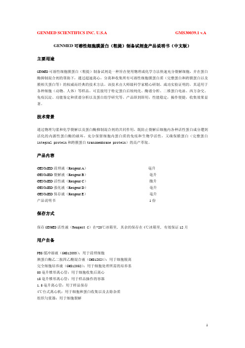
1
4
4
图片说明:用细胞跨膜蛋白提取试剂盒提取细胞样本的SDA-PAGE的电泳考染照片和Western照片。
图1泳道中1、4分别为Marker、膜蛋白样本。图2为以上样品的EGFR的WB结果照片。
试剂盒以外自备试剂和仪器
● 移液器、吸头
● 离心机及离心管
● 涡旋振荡器
● 冰箱,冰盒
使用方法: A. 细胞跨膜蛋白提取 1. 取 5-10×106 个以上细胞①,在 4℃,500g 条件下离心 2-3 分钟,小心吸取培养 基,尽可能吸干,收集细胞。 2. 用冷 PBS 洗涤细胞两次,每次洗涤后尽可能吸干上清。 3. 细胞样品中加入 200μl 冷的试剂 A,2ul 蛋白酶抑制剂混合物,2ul 磷酸酶抑制 剂混合物,高速涡旋振荡 15 秒,置冰上 10-15 分钟。 4. 再次高速涡旋振荡 5 秒,然后在 4℃,13000×g 条件下离心 5 分钟。
贝博细胞跨膜蛋白提取试剂盒是一种基于化学而非去污剂方法的高产跨膜蛋白提取 试剂盒。本试剂盒提供全套试剂,适用于从细胞和动物组织温和、高效地抽提膜/跨膜蛋白 和膜蛋白复合物。用本试剂盒抽提和富集到的蛋白包括从分子量很小到很大的蛋白,单次跨 膜至 7 次以上的蛋白,甚至一些大的蛋白复合物。提取过程简单方便,可在 1 小时内完成。 提取的膜蛋白不仅纯度高,保持天然活性,而且绝少交叉污染。提取的蛋白可用于 Western Blotting、转录活性分析、Gel shift 凝胶阻滞实验、免疫共沉淀、酶活性测定等蛋白研究。
B. 组织跨膜蛋白提取 1. 取适量组织样本剪碎,加冷 PBS,用组织匀浆器匀浆至无明显肉眼可见固体(或 用液氮研磨),冰上静置 5 分钟,小心将匀浆吸入另一预冷的干净离心管。 2. 在 4℃,500g 条件下离心 2-3 分钟,弃上清。 3. 按照细胞蛋白的提取方法的第 3 步骤往下操作即可。
GENMED可溶性细胞膜蛋白(粗提)制备试剂盒 产品说明书(中文版)
GENMED SCIENTIFICS INC. U.S.A GMS30039.1 v.A GENMED可溶性细胞膜蛋白(粗提)制备试剂盒产品说明书(中文版)主要用途GENMED可溶性细胞膜蛋白(粗提)制备试剂是一种旨在使用物理或化学方法快速充分裂解细胞,并在蛋白酶抑制混合剂的帮助下,通过超速离心,分离和收集所有可溶性细胞膜蛋白质(完整蛋白和跨膜蛋白以及膜相关蛋白等)的权威而经典的技术方法。
该技术由大师级科学家精心研制、成功实验证明的。
其适用于各种细胞(动物、人体)等样品。
可直接用于特定蛋白后续纯化、酶谱分析、二维蛋白电泳、西方杂交、免疫沉淀、功能鉴定和质谱分析以及蛋白组学研究等。
产品即到即用,性能稳定,操作便捷,收集效果显著。
技术背景通过物理匀浆和化学裂解以及蛋白酶抑制混合剂的共同作用,既防止裂解后细胞内各种活性蛋白成分遭到活化的内源性蛋白酶的破坏,充分保留细胞内蛋白质的免疫和生物学活性,又确保膜蛋白(完整蛋白integral protein和跨膜蛋白transmembrane protein)的高产萃取。
产品内容GENMED清理液(Reagent A)毫升GENMED裂解液(Reagent B)毫升GENMED活性液(Reagent C)微升GENMED强化液(Reagent D)毫升GENMED保存液(Reagent E)毫升产品说明书1份保存方式保存GENMED活性液(Reagent C)在-20℃冰箱里, 其余的保存在4℃冰箱里, 有效保证12月用户自备PBS缓冲溶液(GMS12033):用于清理细胞胰蛋白酶乙二胺四乙酸混合液(GMS12024):用于细胞脱离完全细胞培养液(GMS12052):用于细胞处理所需的培养基50毫升锥形离心管:用于细胞收集后离心15毫升锥形离心管:用于样品操作的容器1.5毫升离心管:用于样品保存4℃台式离心机:用于细胞和蛋白收集以及去除杂质组织匀浆器:用于细胞裂解实验步骤实验开始前,将试剂盒里的GENMED活性液(Reagent C)从℃的冰箱里取出,置入冰槽里冻融,然后移出毫升GENMED裂解液(Reagent B)到毫升离心管,加入微升GENMED活性液(Reagent C),充分混匀,置于冰槽里,标记为裂解工作液。
蛋白组分抽提试剂盒说明书
Overview∙Description∙References∙Product Information∙Applications∙Biological Information∙Safety Information∙Product Usage Statements∙Storage and Shipping Information∙Supplemental Information∙Prix & DisponibilitéPrix & DisponibilitéRéférence DisponibilitéConditionnement QtéPrix Quantité539790-1KIT En stockContacterle ServiceClients 1kitÀ la validationde lacommandePlusd'informations—AjouterauxfavorisAjouter au panier DescriptionOverviewFast and reproducible extraction of subcellular proteomes from mammalian cellsProteoExtract® Subcellular Proteome Extraction Kit (S-PEK) is designed for fast andreproducible extraction of subcellular proteomes from adherent and suspension-grownmammalian cells. The S-PEK takes advantage of the different solubilities of certain subcecompartments in the four selected reagents. In the case of adherent cells, the procedure isperformed directly in the tissue culture dish without the need for cell removal. Cells or theparts of the cells remain attached to the plate during sequential extraction of subcellularcompartments, until the appropriate extraction reagent is used. Thus, the early destructionthe cellular structure by enzymatic or mechanical detachment of cells from the tissue cultuplate and any mixing of different subcellular compartments is prevented. Forsuspension-grown cells, extraction starts with gentle sedimentation and washing of the celThe stepwise extraction delivers four distinct protein fractions from one sample:∙Cytosolic fraction (F1)∙Membrane/organelle protein fraction (F2)∙Nucleic protein fraction (F3)∙Cytoskeletal fraction (F4)Proteins are obtained in the native state making the S-PEK suitable for many downstreamapplications such as 1D and 2D gel electrophoresis, immunoblotting, enzyme activity assaand protein microarrays.Sample size: 3-5x10⁶or 25-50 mg tissue.CatalogueNumber539790BrandFamilyCalbiochem®Synonyms S-PEK KitFeatures and benefits ∙Stepwise extraction resulting in four distinct subcellular proteomes from one sample ∙Highly reproducible∙No ultracentrifugation steps∙Fast—needs just 2 hours with 45 minutes hands-on time∙Produces proteins suitable for functional studiesApplicationDataA:Adherent SAOS cells were extracted according to the Detailed ProtocolSubcellular Extraction of Proteins From Adherent Cells as outlined above. Imi-iv show the morphology of the cells before and after each extraction s(200-fold enlarged). The SAOS cells remained attached throughout the extracprocedure. B:An aliquot of each fraction from A were subjected to SDS-analysis (F1-F4 = fractions 1-4). The data demonstrate clear differences inprotein banding patterns among the 4 fractions. C:Aliquots of each fracfrom A were separated by SDS-PAGE and transferred to PVD membrane for blotwith the indicated antibodies. For c-Fos, the fractions were subjected timmunoprecipitation, prior to Western blotting, to enrich for any c-Fos prein each fraction. The data demonstrate that each marker protein is specificenriched within the appropriate fraction.A431 cel ls were stimulated with 0.2 µg/ml TNF-αfor the indicated times. At end of each induction period the cells were extracted as outlined in the Deta Protocol for Subcellular Extraction of Proteins from Adherent Cells. The prot from an aliquot of each fraction were separated by SDS-PAGE and transferre PVD membrane for Western blot analysis using an antibody specific for NF-The data indicate that there is measurable translocation of NF-κ B from cytoplasm to the nucleas as early as 5 min after TNF-α stimulation.*Tested on rat liver and bovine liver tissue.。
英文特生物技术(北京)有限公司-植物质膜蛋白提取试剂盒说明书
Minute TM 植物质膜蛋白提取试剂盒目录号:SM-005-P描述:植物膜蛋白占植物细胞总蛋白的很小一部分,但是在植物生理学中起着非常重要的作用。
传统的植物膜组分分离纯化方法是蔗糖密度梯度离心法和双液相法。
这些方法虽然比较有效,但是需要超高速离心和大量的起始原材料,操作过十分繁琐和费时。
为了克服植物膜组分提取中的缺点,我们特别开发了此款植物膜组分提取试剂盒。
植物组织首先通过缓冲液A中致敏,匀浆,然后通过一个特殊的离心管柱,在此过程中匀浆的组织通过柱子特有的Z字形通路后细胞膜被切割成大小相等的碎片,后续无需超高速离心,通过差速离心法和密度梯度离心法将天然的质膜组分从未破裂的细胞,细胞核,细胞浆和细胞器的混合物中分离出来。
在每次实验中仅需使用相同量的起始材料,离心力和离心时间,即可高度富集膜组分,并保证一致性良好。
整个操作过程大约1小时可以完成。
应用:试剂盒用于快速从植物组织中分离天然膜组分,可应用于SDS-PAGE,immunoblottings,ELISA,IP,膜蛋白质结构分析,2-D,酶活性测定及其他应用。
试剂盒组份(50次):1. 25ml Buffer A2. 10ml Buffer B3. 50个离心管柱4. 50个收集管5. 2根塑料研磨棒6. 组织分离粉储存:Buffer A和Buffer B 需在-20℃储存所需附加材料1XPBS涡旋震荡仪台式离心机重要产品信息1.仔细阅读整个操作说明。
将缓冲液A和缓冲液B完全解冻后摇匀,放置于冰上。
将离心管柱和接收管套管放置于冰上预冷。
2.离心机请调整成RCF/Xg模式,按照离心力设置离心机,所有离心步骤都需要在4℃室温下或者低温离心机中进行。
3.研究蛋白磷酸化,磷酸酶抑制剂应在使用前加入缓冲液A中。
蛋白酶抑制剂可以选择添加或不添加,如添加在使用前加入缓冲液A中(请按照蛋白酶或磷酸酶抑制剂母液比例,例如母液是100x,添加时按照1:100添加,1ml缓冲液A添加10ul抑制剂)。
人红细胞膜蛋白提纯分离试剂盒 说明书

人红细胞膜蛋白提纯分离试剂盒说明书好,先从“人红细胞膜蛋白提纯分离试剂盒”的说明书开始,咱们这篇内容得做得自然些,像是聊聊天一样。
首先,咱们要明白这试剂盒是干啥的。
其实很简单,说白了就是帮助咱们从血液中把那些红细胞膜上的蛋白提取出来,干啥用呢?很多实验都需要这些蛋白,比如做一些生物学研究、探究蛋白质和细胞相互作用的机制之类的。
能不能做出来,就得看你使用这款试剂盒的技巧了。
记得第一次使用这个试剂盒的时候,我特别紧张。
因为就像我去做菜一样,明明看过无数次做法,实际操作的时候总觉得心里没底。
你看,说明书上写得都挺清楚的:“将血液样本与试剂混合,轻轻摇晃均匀,离心……”一看这些步骤,心想:“这不就是做饭嘛,似乎也没什么特别的。
”结果,刚开始操作的时候,我的手有点不听使唤,试剂倒不小心洒了出来,弄得台面上都是,还得重新整理一下。
后来才发现,原来这种东西真的得细心,毕竟血液中的每一个小细胞,都是关键。
接着进入重点——提取红细胞膜蛋白。
关键步骤是离心,离心是整个过程的核心,成功与否就在这一步。
每次我都站在离心机旁边,眼睁睁看着试管里的液体被快速旋转,心里都想着:“能不能分得更清楚点?”有时候试剂盒给的离心条件如果设置得不太对,结果就很容易分不清各层,试管里的东西都混在一起了,那就得重新来过,浪费时间又心累。
不过,做几次下来,你会发现其实这些都不是事儿。
就像我现在再做,操作得已经越来越顺手了,试剂盒里的配方和步骤其实挺合理的,你只要按照它的要求走,基本上问题不大。
其实就是需要点耐心,再加上一点点细心。
因为不小心错过了哪个环节,提取出来的蛋白质量会差很多,实验的结果就不准确了,这就是“细节决定成败”的意思了。
说实话,每次看到离心后沉淀物清晰地分层,蛋白提取得一干二净的时候,心里那种成就感真是没法形容,简直是“哇塞,居然做出来了!”你会觉得所有的麻烦和小失误都不算什么,反倒成了回忆的一部分。
好了,讲了这些步骤,也别忘了这款试剂盒的优点。
GST resin产品使用说明书

这相当于50ml培养液采用2.5ml抽提试剂。如果是小规模培养,则采用约1/5培养体积的抽提试剂重悬 沉淀(例如,1.5ml培养液采用300µl抽提试剂)。如果抽提试剂过量也没有什么副作用。 选做:加蛋白酶抑制剂。BugBuster Master Mix与蛋白酶抑制剂兼容。如果目的蛋白后续要用凝血酶 (货号69671),Xa因子(货号69036)或重组肠激酶(货号69066)处理,就应该避免使用丝氨酸蛋白 酶抑制剂。尽管纯化过程可能去除活性抑制剂,建议在酶切前最好做透析或凝胶过滤。 3. 室温下将重悬的细胞液在摇板或低速搅拌器上孵育10-20分钟。 注意:孵育后获得的抽提物不是粘稠的。 4. 4℃下16,000g离心20分钟以去除不溶的细胞碎片。如果需要,沉淀可以留作“包涵体纯化”(见以 下说明)的材料。
可溶蛋白制备 采用此操作抽提到的蛋白包括来自细胞周质和细胞质的可溶蛋白。如果欲仅收集周质部分蛋白,可以参 考Novagen的TB055“PET系统操作手册”中提到的渗压休克方法或其它合适的方法。渗透休克方法中 得到的沉淀也可以用于以下操作以获得细胞质中的可溶蛋白。 1. 用经预先称重的离心管10,000g离心10分钟从液体培养体系收集细胞。如果是小规模培养(如1.5ml或
GST•Bind™ 缓冲液试剂盒
GST•Bind缓冲液试剂盒包括了从GST•Bind树脂和GST•Mag™磁性琼脂糖树脂纯化目的蛋白所需的结 合,冲洗和洗脱所需的缓冲液。试剂盒所含成分足够用于10个2.5ml柱的纯化。具体包括: • 2×100ml 10×GST Bind/Wash Buffer (43mM Na2HPO4,14.7mM KH2PO4,1.37M NaCl,27mM KCl,pH7.3) • 40ml 10×Glutathione Reconstitution Buffer (500mM Tris-HCl,pH8.0) • 1g Glutathione,Reduced,Free Acid
Thermo-Scientific-NE-PER核蛋白-胞浆蛋白抽提试剂盒
Thermo-Scientific-NE-PER核蛋白-胞浆蛋白抽提试剂盒描述Thermo Scientific NE-PER核蛋白-胞浆蛋白抽提试剂盒提供了高效的细胞裂解和抽提方法,可在两个小时之内完成胞浆蛋白与核蛋白的分离操作。
NE-PER核蛋白-胞浆蛋白抽提试剂盒的特性:• 快速—在两个小时之内获得胞核与胞浆内的可溶性蛋白组分• 可靠—NE-PER试剂盒已经被950篇以上的发表文献引用• 通用–适用于培养细胞或组织(仅适用于新鲜样本)的核蛋白抽提• 可扩展—两种规格试剂盒,满足从细胞和组织中抽提样本的应用需求• 方便—操作简单,无需超速梯度离心• 兼容性好—获得样本适合于多种下游应用,包括免疫印迹、凝胶迁移分析、蛋白分析、报告基因分析以及酶活性分析NE-PER试剂盒为用户提供了高效的核蛋白抽提方法,用户只需拥有台式微量离心机、离心管和移液器,进行简单的分步式细胞裂解及核蛋白与胞浆蛋白离心分离即可。
NE-PER试剂盒能够高效地将胞浆蛋白与核蛋白溶解和分离为不同组分,交叉污染以及基因组DNA/mRNA 的干扰均降到了最低。
一旦经过脱盐或稀释,分离后的蛋白即可用于免疫分析和蛋白相互作用实验,如迁移率分析(EMSA)、免疫共沉淀(Co-IP)和拉下实验。
分离胞核和制备核蛋白抽提物的方法很多,但大多数制备核蛋白抽提物的制备方法都很耗时,并需要机械匀浆、冻融循环、反复离心或透析,而这些都可能影响核蛋白的完整性。
NE-PER 核蛋白-胞浆蛋白抽提试剂盒使用了基于试剂的分离方案,能够分步裂解细胞并从胞浆中分离出完整的细胞核,然后再从基因组DNA与mRNA中抽提核蛋白。
这一温和的过程可在两小时之内完成,且使用培养细胞时仅需标准的桌台式离心机即可。
此外,用户还可以从细胞培养物或组织样本中分离得到具有活性的核蛋白与胞浆蛋白。
本试剂盒能够从两百万个细胞中提出200至500µg胞浆蛋白和100至200µg的核蛋白(浓度为1mg/mL)。
- 1、下载文档前请自行甄别文档内容的完整性,平台不提供额外的编辑、内容补充、找答案等附加服务。
- 2、"仅部分预览"的文档,不可在线预览部分如存在完整性等问题,可反馈申请退款(可完整预览的文档不适用该条件!)。
- 3、如文档侵犯您的权益,请联系客服反馈,我们会尽快为您处理(人工客服工作时间:9:00-18:30)。
NovagenUSA and Canada Europe All Other CountriesTel (800) 526-7319 novatech@FranceFreephone0800 126 461GermanyFreecall0800 100 3496IrelandToll Free1800 409 445United KingdomFreephone0800 622 935All otherEuropean Countries+44 115 943 0840Contact Your Local Distributornovatech@ProteoExtract® Transmembrane Protein Extraction Kit Table of ContentsAbout the Kits (2)Description 2Components 2Storage 2Equipment and materials required but not supplied 3ProteoExtract®Transmembrane Protein Extraction Protocol (3)Extraction of membrane proteins from adherent cultured cells 3Extraction of membrane proteins from suspension cells or frozen cell pellets 4Extraction of membrane proteins from tissue 6Frequently Asked Questions (8)Appendix (8)Example extractions 8Examples of total protein yields using TM-PEK 9© 2009 EMD Chemicals Inc., an affiliate of Merck KGaA, Darmstadt, Germany. All rights reserved. The Novagen® logo and Novagen® name are registered trademarks of EMD Chemicals Inc. in the United States and in certain other jurisdictions. ProteoExtract® is a registered trademark of Merck KGaA, Darmstadt, Germany. TRITON® is a registered trademark of Dow Chemical Company.About the KitsProteoExtract® Transmembrane Protein Extraction Kit 1 kit 71772-3DescriptionThe ProteoExtract® Transmembrane Protein Extraction Kit (TM-PEK) uses a novel, detergent-freechemistry for extraction of peripherally-associated and integral membrane proteins, such asG-Protein Coupled Receptors (GPCRs), from mammalian cells and tissues. The membrane protein fractionis directly compatible with enzyme assays, native and denaturing gel electrophoresis, Western blotting,immunoprecipitation, and (following in-gel tryptic digestion) mass spectrometry.The TM-PEK method comprises a two-step protocol for the enrichment of transmembrane (TM) proteins. Inthe first step, cells or homogenized tissues are permeablized using Extraction Buffer 1 and their soluble(cytoplasmic) fraction separated from the insoluble (membrane) fraction by centrifugation. In the secondstep, membrane proteins are extracted from the lipid bilayer using one of two novel extraction buffers.These buffers, Extraction Buffer 2A and Extraction Buffer 2B, are prepared by diluting TM-PEK Reagent Aor TM-PEK Reagent B into Extraction Buffer 2. The optimal extraction reagent must be determinedempirically, as it will depend on characteristics of the protein(s) of interest and intended downstreamapplications. Extraction Buffer 2A is a very mild extraction agent, allowing for recovery of fragile proteincomplexes. Extraction Buffer 2B, by comparison, is a highly efficient extraction agent and facilitatesrecovery of difficult-to-extract transmembrane proteins, including those with multiple transmembranesegments.If using the TM-PEK kit for the first time, prepare duplicate samples. Extract the first set of replicates withTM-PEK Reagent A, diluted 2-fold with Extraction Buffer 2. Extract the second set of replicates with TM-PEK Reagent B, diluted 2-fold with Extraction Buffer 2. Extraction conditions can be optimized further byvarying the dilution range of each reagent into Extraction Buffer, from undiluted to a 10-fold dilution.Unlike alternative membrane protein extraction methods, the TM-PEK kit does not require sonication,rigorous vortexing, time-consuming ultracentrifugation, or incubation at elevated temperatures. The absenceof such harsh treatments minimizes potential damage or changes to the target protein.Each TM-PEK kit provides sufficient reagents to process a total of 40 samples (20 samples using ExtractionBuffer 2A at the recommended dilution, and 20 samples using Extraction Buffer 2B at the recommendeddilution). Each sample may be comprised of 1–5 x 10⁷ cultured cells or 25–50 mg of tissueComponents•••••40 ml Extraction Buffer 150 ml Extraction Buffer 22.0 ml TM-PEK Reagent A2.0 ml TM-PEK Reagent B0.4 ml Protease Inhibitor Cocktail Set IIIStorageStore all components at 4°C for up to 6 months. For prolonged storage, dispense the components in working aliquots and store at –20°C. Before performing extractions, thaw all kit components at room temperature and mix completely. Avoid repeated freezing and thawing.Note that the protease inhibitors are provided in DMSO and must be kept at room temperature during the extraction procedure to prevent freezing.Equipment and materials required but not supplied•••••Rocking platform or elliptical mixer (required at room temperature and 4°C; the device can be transferred from 4°C to room temperature during the course of the experiment)Homogenizer (e.g., Dounce or Potter-Elvehjem) or mortar and pestle (for tissue samples)Refrigerated centrifuge with rotor accommodating a 15 ml and/or a 50 ml tubeRefrigerated centrifuge with rotor accommodating a 2 ml tube and generating 16,000 × gPhosphate Buffered Saline (PBS) (137 mM NaCl, 10 mM Na2HPO4, 2.7 mM KCl, 1.8 mM KH2PO4,pH 7.4)ProteoExtract®Transmembrane Protein Extraction ProtocolTable 1. Buffer volumes required for membrane protein extraction from cultured cells or tissue.Source Material Cultured cells* Fresh or frozen tissueAmount 1.0–5.0 x 10725–50 mg 100–200 mg 200–1000 mgExtraction Buffer 1 (ml) 1.0 1.0 1.0 2.0Extraction Buffer 2A or 2B (ml) 0.2 0.20.51.0Protease Inhibitor (µl) 5 5 20 40*Adherent, suspension, or frozen pelletExtraction of membrane proteins from adherent cultured cellsConsiderations Before You BeginThe following protocol is optimized for extraction of membrane proteins from adherent cells grown in oneT-75 culture flask. Cells should be of high viability (>90%) and 70–90% confluent(1.0–2.0 x 10⁷ cells). Different cell types yield considerably different amounts of protein in the membrane protein fraction (see Table 2 on p 9 in Appendix). If low yield is anticipated, the total number of cells can be increased to 5.0 x 10⁷ without increasing reagent volumes (Table 1). For membrane protein extraction from larger cell numbers, we recommend performing replicate extractions from aliquots of 1.0–5.0 x 10⁷ cells. Alternatively, buffer volumes may be scaled up appropriately.The kit is supplied with two transmembrane solubilization agents, TM-PEK Reagents A and B(see Description on p 2). Depending on the unique characteristics of the target membrane protein, Extraction Buffer 2A or Extraction Buffer 2B may prove to be more efficient. As a starting point, we recommend testing both membrane extraction buffers. If performing trial extractions using both reagents, carry out Steps 2–9 in parallel on each of two sets of sample replicates.Extraction Buffer 2A is a mild extraction reagent which can facilitate recovery of protein complexes. Thus, protein yields obtained using Extraction Buffer 2A may be lower than those obtained using Extraction Buffer 2B (see Table 2 on p 9). Extracts can be concentrated with a Vivaspin 2 or Vivaspin 500 Ultrafiltration Centrifugal Device.Large volumes of TM-PEK 2B reagent can interfere with protein migration in SDS-PAGE. If high resolution is desired, Extraction Buffer 2B samples for SDS-PAGE may be prepared in 2X SDS sample buffer. Alternatively, employ a buffer exchange step.For most transmembrane proteins, a 2-fold dilution of TM-PEK Reagent A or B in Extraction Buffer 2 results in efficient extraction. For some transmembrane proteins, extraction efficiency may be improved by using TM-PEK Reagent A or B at a different concentration, from undiluted to a 10-fold dilution in Extraction Buffer 2.Protocol1.Prepare Extraction Buffer(s) 2A and/or 2B by diluting the appropriate TM-PEK Reagent 2-fold withExtraction Buffer 2. For membrane protein extraction from 1.0–5.0 x 10⁷ cells, 0.2 ml of ExtractionBuffer 2A or 0.2 ml of Extraction Buffer 2B is required.- To prepare 0.2 ml of Extraction Buffer 2A, mix 0.1 ml Extraction Buffer 2 and 0.1 mlTM-PEK Reagent A.- To prepare 0.2 ml of Extraction Buffer 2B, mix 0.1 ml Extraction Buffer 2 and 0.1 mlTM-PEK Reagent B.Notes: We recommend testing both Extraction Buffer 2A and Extraction Buffer 2B to determine which is optimal for your protein of interest.Samples prepared with Extraction Buffer 2A may require concentration, depending ondownstream application.High levels of Reagent B can interfere with protein migration on SDS-PAGE. Buffer exchange ordilution in sample buffer may be required.A 2-fold dilution of TM-PEK Reagent A orB in Extraction Buffer 2 results in an efficient extractionof most proteins, but reagent dilutions may be optimized (from undiluted to a 10-fold dilution).2.Discard medium from the culture flask.3.Wash cells two times with PBS at 4°C.4.Add 3 ml PBS to the culture vessel. Using a cell scraper or a rubber policeman, free cells from theculture vessel. Transfer cells to a 15 ml conical tube.5.Centrifuge cells at 1000 × g for 5 min at 4°C. (Alternatively, collect cells by centrifuging at 500 × g for10 min at 4°C.)6.Resuspend cells in 1 ml Extraction Buffer 1 + 5 µl Protease Inhibitor Cocktail Set III.7.Incubate 10 min at 4°C with gentle agitation to avoid formation of cell clumps.8.Centrifuge at 1000 × g for 5 min at 4°C.9.Carefully remove supernatant. Store on ice. This is referred to as ‘cytosolic (soluble)’ protein fraction.10.Resuspend pellet in 0.2 ml Extraction Buffer 2A + 5 µl of Protease Inhibitor Cocktail Set IIIor 0.2 ml Extraction Buffer 2B + 5 µl of Protease Inhibitor Cocktail Set III.11.Incubate for 45 min at room temperature with gentle agitation.Note: The length and temperature of the incubation at Step 11 can be varied to improve extraction efficiency or to preserve target protein activity. Increasing the incubation time (up to 120 min) canincrease the protein recovery, but may result in decreased target protein activity. Conversely,incubation at low temperature (4°C) may better preserve activity, but may lower the extractionefficiency.12.Centrifuge at 16,000 × g for 15 min at 4°C.13.Transfer the supernatant, which is enriched in integral membrane proteins, to a fresh tube.Note: Store cytosolic and membrane fractions on ice if they will be analyzed on the same day. For long-term storage, dispense into aliquots and store at –20°C.14.Determine total protein concentration of the cytosolic and membrane protein fractions with the BCAassay.Note: For some cell types, total protein concentration of the membrane protein fraction (Step 13 above) will be >1.0 mg/ml. A two- to four-fold dilution in sterile, deionized water may be required to bringthe concentration of these samples within the linear region of the BCA standard curve.Extraction of membrane proteins from suspension cells or frozen cell pelletsConsiderations Before You BeginThe following protocol is optimized for extraction of membrane proteins from 1.0–2.0 x 10⁷ cells cultured insuspension. Cells should be of high viability (>90%). Different cell types yield considerably differentamounts of protein in the membrane protein fraction (see Table 2 on p 9 in Appendix). If low yield isanticipated, increase the total number of cells to 5.0 x 10⁷ without increasing reagent volumes (see Table 1on p 3). For extracting membrane proteins from larger cell numbers, perform replicate extractions fromaliquots of 1.0–5.0 x 10⁷ cells. Alternatively, scale up buffer volumes.The TM-PEK kit is also compatible with frozen cell pellets (1.0–5.0 x 10⁷ cells per extraction). Cells shouldbe washed a minimum of two times with an appropriate buffer (e.g., PBS) prior to freezing in liquidnitrogen. If using frozen cell pellets, begin at Step 7 below.The kit is supplied with two transmembrane solubilization agents, TM-PEK Reagents A and B(see Description on p 2). Depending on the unique characteristics of the target membrane protein, ExtractionBuffer 2A or Extraction Buffer 2B may prove to be more efficient. As a starting point, we recommendtesting both membrane extraction buffers. If performing trial extractions using both reagents, carry out Steps2–9 in parallel on each of two sets of sample replicates.Extraction Buffer 2A is a mild extraction reagent which can facilitate recovery of protein complexes. Thus,protein yields obtained using Extraction Buffer 2A may be lower than those obtained using Extraction Buffer2B (see Table 2 on p 9). Extracts can be concentrated with a Vivaspin 2 or Vivaspin 500 UltrafiltrationCentrifugal Device.Large volumes of TM-PEK 2B reagent can interfere with protein migration in SDS-PAGE. If high resolutionis desired, Extraction Buffer 2B samples for SDS-PAGE may be prepared in 2X SDS sample buffer.Alternatively, employ a buffer exchange step.For most transmembrane proteins, a 2-fold dilution of TM-PEK Reagent A or B in Extraction Buffer 2results in efficient extraction. For some transmembrane proteins, extraction efficiency may be improved byusing TM-PEK Reagent A or B at a different concentration, from undiluted to a 10-fold dilution inExtraction Buffer 2.Protocol1.Prepare Extraction Buffer(s) 2A and/or 2B by diluting the appropriate TM-PEK Reagent 2-fold withExtraction Buffer 2. For membrane protein extraction from 1.0–5.0 x 10⁷ cells, 0.2 ml of ExtractionBuffer 2A or 0.2 ml of Extraction Buffer 2B is required.- To prepare 0.2 ml of Extraction Buffer 2A, mix 0.1 ml Extraction Buffer 2 and 0.1 mlTM-PEK Reagent A.- To prepare 0.2 ml of Extraction Buffer 2B, mix 0.1 ml Extraction Buffer 2 and 0.1 mlTM-PEK Reagent B.Notes: We recommend testing both Extraction Buffer 2A and Extraction Buffer 2B to determine which is optimal for your protein of interest.Samples prepared with Extraction Buffer 2A may require concentration, depending ondownstream application.High levels of Reagent B can interfere with protein migration on SDS-PAGE. Buffer exchange ordilution in sample buffer may be required.A 2-fold dilution of TM-PEK Reagent A orB in Extraction Buffer 2 results in an efficient extractionof most proteins, but reagent dilutions may be optimized (from undiluted to a 10-fold dilution).1.Transfer 1.0–5.0 x 10⁷ cells to a centrifuge tube.2.Centrifuge cells at 1000 × g for 5 min at 4°C. (Alternatively, collect cells by centrifuging at500 × g for 10 min at 4°C.)3.Discard supernatant and gently resuspend cells in 5 ml PBS (4°C).4.Centrifuge cells at 1000 × g for 5 min at 4°C.5.Repeat Steps 3 and 4 two times, for a total of three washes.6.Centrifuge cells at 1000 × g for 5 min at 4°C.Note: At this point, cells may be frozen in liquid nitrogen and stored at –70°C.7.Resuspend cells in 1 ml Extraction Buffer 1 + 5 µl Protease Inhibitor Cocktail Set III.8.Incubate 10 min at 4°C with gentle agitation to avoid formation of cell clumps.9.Centrifuge at 1000 × g for 5 min at 4°C.10.Carefully remove supernatant. Store on ice. This is referred to as the ‘cytosolic (soluble)’ proteinfraction.11.Resuspend pellet in 0.2 ml Extraction Buffer 2A + 5 µl Protease Inhibitor Cocktail Set IIIor 0.2 ml Extraction Buffer 2B + 5 µl Protease Inhibitor Cocktail Set III.12.Incubate for 45 min at room temperature with gentle agitation.Note: The length and temperature of the incubation at Step 12 can be varied to improve extraction efficiency or to preserve target protein activity. Increasing the incubation time (up to 120 min) canincrease the protein recovery, but may result in decreased target protein activity. Conversely,incubation at low temperature (4°C) may better preserve activity, but may lower the extractionefficiency.13.Centrifuge at 16,000 × g for 15 min at 4°C.14.Transfer supernatant, which is enriched in integral membrane proteins, to a fresh tube.Note: Store cytosolic and membrane fractions on ice if they will be analyzed on the same day. For long-term storage, dispense into aliquots and store at –20°C.15.Determine the total protein concentration of the cytosolic and membrane protein fractions with the BCAassay.Note: For some cell types, total protein concentration of the membrane protein fraction (Step 15 above) will be >1.0 mg/ml. A two- to four-fold dilution in sterile, deionized water may be required to bringthe concentration of these samples within the linear region of the BCA standard curve.Extraction of membrane proteins from tissueConsiderations Before You BeginThe following protocol is optimized for extracting membrane proteins from 25–50 mg of fresh or frozentissue. If tissue is not limiting, start with 100–1000 mg of tissue to offset sample loss during homogenization.When using > 50 mg of tissue, increase volumes of the extraction buffers (see Table 1 on p 3 for buffervolume guidelines). As an example, ~2 mg total protein can be extracted from 35 mg bovine liver (see Table3 on p 9 in the Appendix for representative yields from other tissue sources and types). Yields from varioustissue types can vary considerably, however. Certain transmembrane proteins are expressed at very lowlevels in various tissues, and thus may be below the lower limit of detection by immunological methods. Inthis circumstance, analysis by mass spectrometry may prove beneficial.Protocol1.Prepare membrane Extraction Buffer(s) 2A and/or 2B by diluting the appropriate TM-PEK Reagent 2-fold with Extraction Buffer 2. For membrane protein extraction from 25–50 mg of tissue, 0.2 ml ofExtraction Buffer 2A or 0.2 ml of Extraction Buffer 2B is required.- To prepare 0.2 ml of Extraction Buffer 2A, mix 0.1 ml Extraction Buffer 2 and 0.1 mlTM-PEK Reagent A.- To prepare 0.2 ml of Extraction Buffer 2B, mix 0.1 ml Extraction Buffer 2 and 0.1 mlTM-PEK Reagent B.Note: We recommend testing both Extraction Buffer 2A and Extraction Buffer 2B to determine which is optimal for your protein of interest.Samples prepared with Extraction Buffer 2A may require concentration, depending ondownstream application.High levels of Reagent B can interfere with protein migration on SDS-PAGE. Buffer exchange ordilution in sample buffer may be required.A 2-fold dilution of TM-PEK Reagent A orB in Extraction Buffer 2 results in an efficient extractionof most proteins, but reagent dilutions may be optimized (from undiluted to a 10-fold dilution).2.Ensure that all buffers are thawed and well mixed. Keep Extraction Buffers 1, 2A, and/or 2B on iceduring the extraction procedure. Keep the Protease Inhibitor Cocktail Set III at room temperature toprevent DMSO from freezing.3.Following dissection of the tissue of interest, quickly remove unwanted materials(e.g., connective tissue, fat, blood vessels, etc). To slow proteolysis, keep tissue at 4°C while refiningthe dissection.4.Quickly slice the tissue into ~2 mm3 pieces. Add tissue slices to a tube containing 2 ml ice-cold PBS.5.Gently flick tube to dislodge blood cells and other loosely attached material.6.Collect tissue pieces by centrifuging at 100 × g for 2 min at 4°C. Remove and discard the supernatant.7.Repeat Steps 5 and 6 for a total of two washes. After the second wash, ensure that all PBS has beenremoved completely.Notes: At this point, the tissue can be frozen on liquid nitrogen and stored at –70°C.During tissue extraction, it is important to work quickly, but carefully. Keep the sample cool (<4°C)and store buffers on ice throughout the extraction procedure.8.Transfer the tissue (fresh or frozen) to a pre-cooled homogenizer. We recommend ideally, a glassPotter-Elvehjem or Dounce homogenizer.9.Add 5 µl Protease Inhibitor Cocktail Set III to the wall of the homogenizer.10.Add 2 ml ice-cold Extraction Buffer 1 to the homogenizer.11.Carefully homogenize until tissue is completely homogenized and intact pieces are no longer visible.Use as few strokes as possible (e.g., ~10 strokes for 50 mg mouse liver). The number of strokes willdepend on the type of tissue used. If desired, homogenization efficiency can be monitored by phasecontrast microscopy. An efficient homogenization should generate small cell clumps rather thatfragmentation of individual cells.Note: Some tissues (e.g., heart, muscle, brain) may be difficult to completely dissociate by mechanical homogenization. As an alternative, the ProteoExtract®Tissue Dissociation Buffer Kit(Cat. No. 539720) including collagenase may be used. Other tissue-specific protocols may becompatible with the TM-PEK kit.12.Incubate 10 min at 4°C with gentle agitation.13.Centrifuge at 1000 × g for 5 min at 4°C.14.Carefully remove supernatant. Store on ice. This is referred to as the ‘cytosolic (soluble)’ proteinfraction.15.Add 5 ml ice-cold PBS. Gently resuspend pellet. Collect membranes by centrifuging at 1000 × g for 5min at 4°C. Carefully remove supernatant.Note: When using > 200 mg tissue, perform an extra wash at this point to remove additional cytosolic proteins. Repeat Step 15 for a total of two washes.pletely and carefully resuspend pellet in 0.2 ml Extraction Buffer 2A + 5 µl of Protease InhibitorCocktail Set IIIor 0.2 ml Extraction Buffer 2B + 5 µl Protease Inhibitor Cocktail Set III.17.Incubate 15 min at 4°C with gentle agitation to avoid formation of cell clumps.18.Centrifuge at 16,000 × g for 15 min at 4°C.19.Transfer supernatant, which is enriched in integral membrane proteins, to a fresh tube.Note: Store cytosolic and membrane fractions on ice if they will be analyzed on the same day.For long-term storage, dispense into aliquots and store at –20°C.20.Determine the total protein concentration of the cytosolic and membrane fractions with the BCA assay. Note: For some tissue types, total protein concentration of the membrane protein fraction (Step 19 above) will be >1.0 mg/ml. A two- to ten-fold dilution in sterile, deionized water may be required tobring the concentration of these samples within the linear region of the BCA standard curve.Frequently Asked QuestionsQuestion AnswerHow do I determine the proteinconcentration of the membrane proteinextract? The components in the extraction buffers are directly compatible with common protein assays. We recommend using the BCA Protein Assay Kit (Cat. No. 71285-3). Note that the Extraction Buffer 2B fraction may require a 2-4-fold dilution to bring the total protein concentration within the linearrange of the BCA assay.How do I prepare the TM-PEK fraction for one-dimensional SDS-PAGE? The TM-PEK fractions can be analyzed directly by one-dimensional SDS-PAGE. For samplesextracted with Reagent A, add SDS-PAGE sample buffer to a 1X final concentration and loaddirectly on to the gel. Samples extracted with Reagent B should be prepared using a 2Xconcentration of SDS-PAGE sample buffer.How can I concentrate the TM-PEK extracted proteins? It is possible to reduce the volume of Extraction Buffer 2A or 2B used, but this may decrease thetotal protein yield. We recommend using the ProteExtract ® Protein Precipitation Kit (Cat. No.539180) to concentrate samples for use in downstream applications not requiring native protein.For applications that do require native protein, we recommend using an ultrafiltration device suchas Vivaspin 2 or Vivaspin 500.How should the membrane fractions be treated prior to mass spectrometry analysis?The membrane extracts should be applied to a one-dimensional or two-dimensional gel, spots orbands cut from the gel, and then digested with trypsin.Appendix Example extractionsIn the examples below, the SDS extract (total cell lysate) serves as a positive control. The Extraction Buffer 2A recovers EGFR, which has a single transmembrane span, with efficiency comparable to TRITON ® X-100. However, Extraction Buffer 2A recovers Frizzled-4 and CELSR-3, both of which contain seven transmembrane domains, with far greater efficiency than does TRITON X-100.A: Figure 1. Ex ction of transmembrane prote EGFRB: Frizzled-4 C: CELSR-3tra ins from MDA-MB-468 breast adenocarcinoma cultured ells. Transmembrane proteins were extracted from MDA-MB 468 cells using the TM-PEK kit. In the first two identical pools of 1 x 10⁷ cells were treated with Extraction Buffer 1, which recovers proteins from the cytosolic fraction. The insoluble material was then treated with TM-PEK Extraction Buffer 2A or 0.5% TRITON ® X-100. Lane 1 shows the expected size of the target proteins and is not a quantitative control. Lanes 2–4 contain the extracted protein from 1 x 10⁶ cells. Fractions were separated using a 10% SDS-PAGE gel and transferred to a nitrocellulose membrane. Membranes were blocked and incubated with primary antibody to EGFR (panel A), Frizzled-4 (panel B) or CELSR-3 (panel C). Blots were developed using an HRP-conjugated secondary antibody and a chemiluminescent substrate. Lane 1, 0.5% SDS (total cell lysate); Lane 2, cytoplasmic fraction; Lane 3, membrane fraction (TRITON X-100); Lane 4, membrane fraction (Extraction Buffer 2A). Arrows indicate size of the full-length version of each protein.c step,Examples of total protein yields using TM-PEKThe values presented in Tables 2 and 3 below are intended to serve as a guide to estimate the required amount of starting material. For each cultured cell type, proteins were extracted according to the TM-PEK protocol from cell monolayers grown to 80% confluency in a T-75 flask. All tissues were disrupted mechanically using a Dounce homogenizer. Table 2. Protein yields for cytosolic and membrane fractions recovered from cultured cells using the TM-PEK kit.Total Protein (mg/107 cells)*Cell TypeBuffer 1(cytosolic) Buffer 2A (membrane) Buffer 2B (membrane) ExtractionExtraction Extraction MDA-MB-468 (breast adenocarcinoma)0.62 0.15 0.78 MCF 7 (breast adenocarcinoma)0.68 0.26 1.27 A-431 (epidermoid carcinoma)0.55 0.12 0.75 CHO-K1 (Chinese hamster ovary)0.52 0.22 0.94 NCI-H292 (mucoepidermoid carcinoma)0.06 0.04 0.47 HEP-G2 (hepatocellular carcinoma)0.97 76 0.13 0.Mia PaCa-2 (pancreatic carcinoma)0.23 0.05 0.52 HCT 116 (colon carcinoma) 0.60 0.10 0.76 *Protein concentrations were determined using the BCA .Table 3. olic and membrane fractions recovered from mouse tissue Total Protein g/mg tissue)*assayProtein yields for cytos TM-PEK kit.s using the (μTissue TypeExtractionB(cytos Extraction Bu A (me ne) Extraction Bu B (memb uffer 1olic)ffer 2mbra ffer 2rane)Liver §55.6 1.7 11.4 Heart28.2 2.9 17.3 Brain18.4 0.9 5.8 Spleen 31.6 8.2 8.8 §Skeletal Muscle13.6 2.1 6.3 Kidney 33.6.9 5.4 16*Protein concentrations were determined using the B §Average of two tr CA assay.ials.。