
丹芪偏瘫胶囊的说明书
丹芪偏瘫胶囊的说明书
心脑血管疾病分为心脏血管和脑血管两种,是现在老年人发病率越老越高的疾病之一,有着死亡率高,致残率高,发病率高,复发率高的特点,所以对于老年人心脑血管疾病的治疗刻不容缓,丹芪偏瘫胶囊是一种治疗心脑血管疾病效果非常不错的药物,我们可以详细了解一下。
【药品名称】
通用名称:丹芪偏瘫胶囊
商品名称:丹芪偏瘫胶囊
【适应症/功能主治】益气活血。
用于虚血瘀型缺血性中风病(脑梗塞)中经络恢复期,症见半身不遂、偏身麻木、口舌歪斜,语言蹇涩等
【规格型号】0.4g*24s*3小盒
【用法用量】口服,一次4粒,一日3次,4周为一个疗程。
【有效期】0 月
【批准文号】国药准字Z20010105
【生产企业】天津市石天药业有限责任公司
【主要成份】黄芪、丹参、川芎、赤芍、水蛭、土鳖虫、全蝎、远志、石菖蒲、工人牛黄等十四味药
【性状】本品为胶囊剂,内容物为黄棕色至棕褐色;色微腥,味苦、微甘
综上所述,就是对丹芪偏瘫胶囊的各种药效和药性的介绍,现在您是否都清楚了解了呢?心脑血管疾病除了药物治疗,和平四的饮食习惯也有很大关系,少吸烟喝酒,多吃鱼和鱼油,多吃豆类品都对病情有帮助,最后祝天下所有的老年人都身体健康,长命百岁!。
中风单元系列药

全方十四味,配伍严谨,谨守病机,攻 补兼施,共奏补气活血,熄风豁痰之功。
药理学
YAOLIXUE
一、对大鼠大脑中动脉阻塞所致脑 梗塞的预防作用
组别 剂量(g/ ㎏) 6 3 1.5 0.05 动物数 (只) 行为评分 ( x ±SD)
对 照 组
丹芪偏瘫胶囊 丹芪偏瘫胶囊 丹芪偏瘫胶囊 尼莫地平
10
丹芪偏瘫胶囊 (3g/kg)
10
8.25 ±0.92
8.20 ±0.98
8.05 ±1.07
7.85 ±1.25
7.70 ±1.18
7.65 ±1.86
6.85 ±1.86
6.80 ±1.84
6.40 ±1.79
5.95 ±1.98
5.89 ±1.85
丹芪偏瘫胶囊 (1.5g/kg)
10
8.20 ±0.89
7.40 ±0.77
7.0 ±0.75
6.90 ±0.77
6.45 ±1.17
6.05 ±1.17
5.75* ±1.36
5.35** ±1.20
组别
剂量 (g/㎏)
动物 梗塞组织 数 重量(mg) (只) 10 76.7±37.3 32.3±15.3 59.0±37.7 55.7±41.0 47.9±25.0
处方来源于石 学敏院士临床经 验总结。
脑血栓丸
中风丸
【主要成分】 丹参,当归,冰片等。 【功能与主治】 平肝降逆,熄风化痰。 用于中风不语,半身不遂,口眼歪斜, 高血压中风后遗症。 【用法与用量】 口服,每次1丸,每 日2次。或遵医嘱。
处方来源于石学 敏院士临床经验 总结。
中风丸
醒脑治瘫胶囊
益肾养肝合剂是我院临床使用最广泛的医 院制剂之一。该方为石院长多年临床经验 而得,本方证治乃因年老体衰,正气不足, 肝肾亏虚所致疾病。本方由黄芪、山茱萸、 淫羊藿等药组成,共补先、后天之本,使 气旺精充,而达补肝肾,髓剂
丹芪偏瘫胶囊使用说明
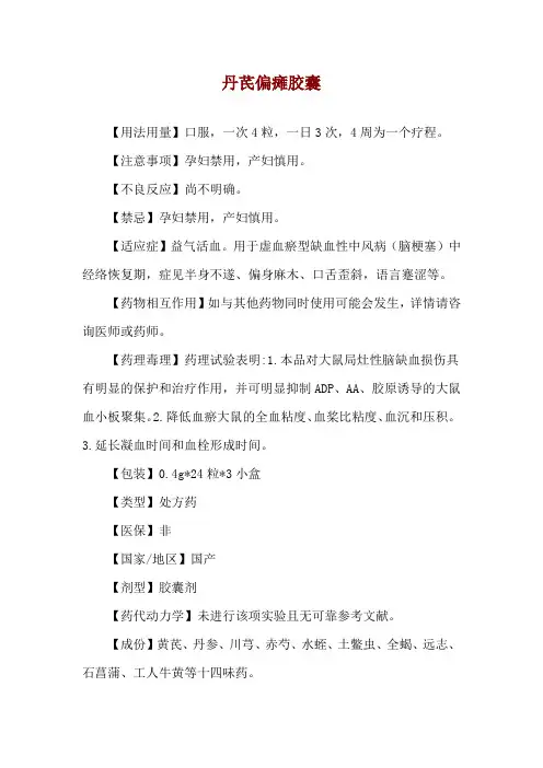
丹芪偏瘫胶囊
【用法用量】口服,一次4粒,一日3次,4周为一个疗程。
【注意事项】孕妇禁用,产妇慎用。
【不良反应】尚不明确。
【禁忌】孕妇禁用,产妇慎用。
【适应症】益气活血。
用于虚血瘀型缺血性中风病(脑梗塞)中经络恢复期,症见半身不遂、偏身麻木、口舌歪斜,语言蹇涩等。
【药物相互作用】如与其他药物同时使用可能会发生,详情请咨询医师或药师。
【药理毒理】药理试验表明:1.本品对大鼠局灶性脑缺血损伤具有明显的保护和治疗作用,并可明显抑制ADP、AA、胶原诱导的大鼠血小板聚集。
2.降低血瘀大鼠的全血粘度、血桨比粘度、血沉和压积。
3.延长凝血时间和血栓形成时间。
【包装】0.4g*24粒*3小盒
【类型】处方药
【医保】非
【国家/地区】国产
【剂型】胶囊剂
【药代动力学】未进行该项实验且无可靠参考文献。
【成份】黄芪、丹参、川芎、赤芍、水蛭、土鳖虫、全蝎、远志、石菖蒲、工人牛黄等十四味药。
丹芪偏瘫胶囊对神经系统的作用

Neuroprotective and neuroproliferative activities of NeuroAid (MLC601,MLC901),a Chinese medicine,in vitro and in vivoC.Heurteaux a ,*,C.Gandin a ,M.Borsotto a ,C.Widmann a ,F.Brau a ,M.Lhuillier b ,B.Onteniente b ,zdunski a ,*aInstitut de Pharmacologie Moléculaire et Cellulaire,Centre National de la Recherche Scienti fique (CNRS),Universitéde Nice Sophia Antipolis,660Route des Lucioles,06560Valbonne,France bINSERM UMR 861,Universitéd'Evry,5rue Henri Desbrueres,91030Evry cedex,Francea r t i c l e i n f oArticle history:Received 4September 2009Received in revised form 1December 2009Accepted 4January 2010Keywords:NeuroAid StrokeFocal ischemia Excitotoxicity Neurogenesis Proliferationa b s t r a c tAlthough stroke remains a leading cause of death and adult disability,numerous recent failures in clinical stroke trials have led to some pessimism in the field.Interestingly,NeuroAid (MLC601),a traditional medicine,particularly used in China,South East Asia and Middle East has been reported to have bene ficial effects in patients,particularly in post-stroke complications.Here,we demonstrate in a rodent model of focal ischemia that NeuroAid II (MLC901)pre-and post-treatments up to 3h after stroke improve survival,protect the brain from the ischemic injury and drastically decrease functional de ficits.MLC601and MLC901also prevent neuronal death in an in vitro model of excitotoxicity using primary cultures of cortical neurons exposed to glutamate.In addition,MLC601/MLC901treatments were shown to induce neuro-genesis in rodent and human cells,promote cell proliferation as well as neurite outgrowth and stimulate the development of a dense axonal and dendritic network.MLC601and MLC901clearly represent a very interesting strategy for stroke treatment at different stages of the disease.Ó2010Elsevier Ltd.All rights reserved.1.IntroductionStroke affects numerous people every year.When brain cells die,the function of the body parts they control is impaired or lost,causing paralysis,speech and sensory problems,memory and reasoning de ficits,coma,and possibly death.Treatment for stroke is almost reduced to fibrinolysis,a therapy that unfortunately can be only used in a relatively low percentage of patients.Dozens of clin-ical trials have failed to show ef ficacy in humans for a variety of neuroprotective drugs (Ginsberg,2008).In addition,there are no effective,clinically approved methods that promote restoration of CNS function,days,weeks or months after stroke.The need for new therapeutic strategies is high.A slow but consistent recovery can be observed in the clinical practice over a period of weeks and months.Whereas the recovery in the first few days likely results from edema resolution and/or from reperfusion of the ischemic penumbra,a large part of the recovery afterwards is mainly due to brain which spontaneously recovers by the reorganization of surviving central nervous system elements inthe damaged areas (Cramer,2008).Neurogenesis and angiogenesis are key mechanisms of recovery after stroke (Zhang et al.,2008).The research of therapeutic agents able to stimulate proliferation,migration and differentiation of new neural cells that can replace those lost during a stroke episode is important for future.Due to the complexity of stroke disease,there is increasing evidence that the search for a “magic drug ”which speci fically acts on a single target is exceeded and that combination therapies comprising more than one active ingredient can represent a better strategy against stroke.Interestingly,combination therapy has been advocated for >2500years by prescriptions of formulae in tradi-tional Chinese Medicine (TCM),that consist of several types of medicinal herbs,based on clinical experience.As recently shown for promyelocytic leukemia,Chinese herbal medicines can represent a new promising area in drug discovery (Wang et al.,2008).The aim of this work is to address the possible bene ficial effects of MLC601and MLC901against stroke disease.MLC601(NeuroAid,Moleac Pte.Ltd,Singapore)is a TCM which is used extensively in China to facilitate recovery after stroke (Chen et al.,2009).It combines 9herbal (including Radix astragali,Radix salviae miltiorrhizae,Radix paeoniae rubra,Rhizoma chuanxiong,Radix angelicae sinensis,Carthamus tinctorius,Prunus persica,Radix polygalae and Rhizoma acori tatarinowii)and 5animal components (including Hirudo,Eupolyphaga seu steleophaga,Calculus bovisartifactus,Buthus*Corresponding authors.Tel.:þ33493957784;fax:þ33493957704.E-mail addresses:heurteau@rs.fr (C.Heurteaux),lazdunski@rs.fr (M.Lazdunski).Contents lists available at ScienceDirectNeuropharmacologyjo urn al homepag e:/locate/neuropharm0028-3908/$e see front matter Ó2010Elsevier Ltd.All rights reserved.doi:10.1016/j.neuropharm.2010.01.001Neuropharmacology 58(2010)987e 1001martensii and Cornu saigae tataricae).A simplified formula of MLC601called MLC901(NeuroAid II)based on its9herbal components is now also available.A multicenter,randomized, double-blind placebo-controlled study to investigate CHInese Medicine MLC601Efficacy on Stroke recovery(CHIMES)is ongoing in Asia(Venketasubramanian et al.,2009).Additional studies assessing immediate and long-term effects,alone or in combination with aspirin showed the safety of MLC601in normal subjects and stroke patients(Gan et al.,2008;Siow,2008).However,before this work was started,there was no scientific background for the use of this TCM against stroke.The purpose of this work is to analyze whether MLC601and MLC901have interesting neuroprotective and/or neurogenerative properties in in vitro and in vivo assays that are normally used in Western medicine to develop new drugs,pre-clinically,before assaying them in humans.We report here the protective effects of MLC601and MLC901on neuronal and brain injuries as well as positive effects on functional recovery after ischemic stroke.We also demonstrate in vitro neuronal proliferation and neurite outgrowth as well as in vivo neurogenesis induced by MLC601/MLC901.2.Materials and methods2.1.Neuronal cultureTime-pregnant(E14)C57Bl/6J mice were anesthetized with isopentane followed by cervical dislocation.Fetuses were removed and placed in cold HBSSþsolution. Cerebral cortices were dissected in cold HBSSþsolution and the meninges were removed.Cortical samples were cut in small pieces and were gently triturated with afire-polished glass Pasteur pipette in8ml HBSSþsolution.The mix wasfiltered (40m mfilter)and centrifuged at800rpm for8min.The supernatant was removed and the pellet was dissolved in2ml culture medium.Cells were plated on poly-D-lysine(Sigma e Aldrich Chimie,St Quentin Fallavier,France)-coated12well(24mm diameter)plates with glass coverslips(12mm diameter)(CML,Nemours,France)at a density of1Â106cells/well.Cultures were maintained at37 C in a humidified5% CO2atmosphere incubator in Neurobasal supplemented with B27,Glutamax,anti-biotics and used for experiments after16days.Glial growth was suppressed by addition of5-Fluoro-2-deoxyuridine(2m M)and Uridine(2m M)during the second day of culture.The degree of damage observed in the current in vitro system was similar to that previously reported in aging cultures of mouse cortical neurons (Lesuisse and Martin,2002).2.2.In vitro model of excitotoxicityAs model of excitotoxicity,we used glutamate at the concentration of10m M in magnesium-free glycine-supplemented PBS during10min,which induced signifi-cant damage as previously reported(Hartley et al.,1993).MLC601or MLC901was added at the concentration of1m g/ml,4days before and after treatment with glutamate.Control cells were incubated with vehicle alone.Cell survival and lactate dehydrogenase(LDH)release were estimated5,8and24h after glutamate treat-ment(n¼3cultures,36wells per experimental group).2.3.Cell injury assay:cell survival and lactate dehydrogenase(LDH)measurementsCell viability was assessed at Day8,10,12and14of cell culture and at5,8and 24h after glutamate treatment,by using the Cell Titer96(r)Aqueous One Solution Cell Proliferation Assay(Promega,Charbonnières-les-Bains,France)(n¼3cultures, 36wells per experimental group).This assay is a colorimetric method,which is based on the use of the3-(4,5-dimethylthiazol-2-yl)-5-(3-carboxymethoxyphenyl)-2-(4-sulfophenyl)-2H-tetrazolium inner salt(MTS),a marker of mitochondrial activity and an electron-coupling reagent(phenazine ethosulfate,PES).The MTS tetrazolium compound is bioreduced by cells into a colored formazan product that is soluble in tissue culture medium.This conversion is presumably accomplished by NADPH or NADH produced by dehydrogenase enzymes in metabolically active cells.The quantity of formazan product as measured by the absorbance at490nm is directly proportional to the number of living cells in culture.According to the manufacturer's recommendations,the assay was performed as follows:the totality of cell culture medium was removed and replaced by500m l of Neurobasal mediumþCell Titer96 Aqueous One Solution.Cells were incubated for4h at37 C in the humidified5%CO2 atmosphere incubator.The reaction was stopped with2%SDS.Optical density was measured4h later at490nm utilizing a microplate reader(Labsystem Multiscan RC, VWR International,Fontenay sous Bois,France).Background absorbance at620nm was subtracted.Results were expressed in Optical Density(ODÂ10À3).To correlate the mitochondrial activity measured by OD in the wells to the cell viability,a calibration curve was performed giving the effect of cell number on absorbance at 490nm.The correlation coefficient was0.99,indicating a linear response between cell number and absorbance at490nm.Data are expressed as the percentage of cell viability,which is calculated by dividing the absorbance value of MLC601/MLC901-treated samples by that of the untreated controls within each group.Neuronal injury was quantitatively assessed by the measurement of LDH release from cultured neurons at Day8,10,12and14of cell culture and at5,8and24h after glutamate treatment(Day6of culture)(Koh and Choi,1987).LDH release assay provides a measure of cytoplasmic membrane integrity.100m l cell culture medium was transferred from culture wells to96-well plates and mixed with100m l reaction solution according to LDH assay kit(Roche Diagnostic:Cytotoxicity Detection). Quantification was done by measuring the Optical Density(OD)30min later at 492nm on a microplate reader(Labsystem Multiscan RC,VWR International,Fon-tenay sous Bois,France).Background absorbance at620nm was subtracted.As recommended by the manufacturer,neurons exposed to a lysis solution(PBS con-taining0.1%Triton X-100)were used as positive control and set as100%LDH release. Data are expressed as ratio of LDH efflux/cell viability.All in vitro experiments were monitored by one researcher blinded to the treatment status(n¼3cultures,36wells per experimental group).Results corre-sponded to the mean of three independent experiments with triplicate determi-nation.Statistical analyses of cell viability and LDH results were assessed using one factor ANOVA test following by post-hoc test(P<0.05).2.4.Focal ischemia2.4.1.AnimalsAll experiments were performed according to policies on the care and use of laboratory animals of European Community legislation.The local Ethics Committee approved the experiments(protocol numbers NCA/2006/10-1and NCA/2006/10-2). All efforts were made to minimize animal suffering and reduce the number of animals used.Adult male C57/Bl6mice,weighing22e26g(7e9weeks old)were used in this study.Animals housed under controlled laboratory conditions with a12-h dark e light cycle,a temperature of21Æ2 C,and a humidity of60e70%for at least one week prior to drug treatment or surgery.Mice had free access to standard rodent diet and tap water.The researchers,who carried out the ischemic surgery and measured infarct volumes were blinded in regard to the treatment code.2.4.2.Model of focal ischemiaIschemia was induced by occlusion of the left middle cerebral artery(MCA)using an intraluminalfilament technique(Heurteaux et al.,2006a;Huang et al.,1994).After a midline neck incision was made,the left common and external carotid arteries were isolated and ligated with a4e0silk suture thread(Ethicon).A yasargil aneurysm clip (BMH31,Aesculap,Tuttlingen,Germany)was temporarily placed on the internal carotid artery.A6e0coatedfilament(Doccol,Redlands,CA,USA)was introduced through a small incision into the common carotid artery and13mm distal to the carotid bifurcation for occlusion of MCA origin.Animals were kept at37 C for1h, after which time the thread was carefully withdrawn to allow reperfusion of MCA territory.To control MCAO severity regional cerebral bloodflow(rCBF)was deter-mined by laser-Dopplerflowmetry(Perimed)using aflexible0.5-mmfiber optic extension to the master probefixed on the intact skull over the ischemic cortex(2mm posterior and6mm lateral from the bregma).Sham-operation was performed inserting the thread into the common carotid artery without advancing it to occlude MCA.Animals were allowed to regain full consciousness on a heating pad before returning to the cage.2.4.3.Physiological parametersGeneral anesthesia was induced with3%isoflurane and maintained with1% isoflurane by means of an open facemask for each mouse.Mice were allowed to breathe spontaneously.A subset of animals(n¼5per group)were monitored for physiological parameters including mean arterial blood pressure(MABP),rectal temperature,arterial blood gases and pH before,during and after ischemia.The right femoral artery was catheterized with PE-10polyethylene tubing and connected to a blood pressure transducer(Harvard Apparatus)for continuous monitoring of MABP(mm Hg).A heparinized blood sample(75m l)was then obtained from the catheterized femoral artery.Blood P a O2,P a CO2and pH were measured using an Acid-Base Laboratory system(ABL555,Radiometer).Core temperature was continuously monitored with a thermometer(3-mm probe diameter;Harvard Apparatus), inserted into the rectum and maintained at physiological temperatures using a thermostatically controlled heating blanket(Harvard Apparatus).Core tempera-ture was maintained before,during and3h after ischemia at physiological values by using the homeothermic blanket control.2.4.4.Determination of infarct volumeMice were sacrificed at30h after reperfusion.To visualize the evolution and the extent of infarct volume by TTC(2.3.5-triphenyltetrazolium chloride)staining, brains were removed and sectioned into six1mm-thick coronal slices using a tissue chopper(Phymep,France).Coronal slices were immediately immersed into2%TTC (Sigma,France)for20min at room temperature in the dark followed byfixation in 4%paraformaldehyde solution overnight prior to analysis(Heurteaux et al.,2006a).C.Heurteaux et al./Neuropharmacology58(2010)987e1001 988Areas of infarction,outlined in light appeared in white on coronal TTC-stained slices. To confirm the extent of the cerebral lesion,cresyl violet staining on coronal frozen brain sections(10m m-thick)was performed using a solution of1%cresyl violet in 0.25%acetic acid and mounted with Entellan.The striatal and cortical areas of infarction,outlined in light were measured on each section using a computer image analysis system and corrected for brain edema according to Golanov and Reis(1995). Infarct volume,expressed in mm3was calculated by a linear integration of the corrected lesions areas as previously described(Heurteaux et al.,2006a).2.5.Drug treatmentsMLC601(NeuroAid)and MLC901(NeuroAid II)were provided by Moleac (Singapore).The composition of MLC601(0.4g per capsule)was the following:0.57g Radix astragali,0.114g Radix salvia miltiorrhizae,0.114g Radix paeoniae rubra,0.114g Rhizoma chuanxiong,0.114g Radix angelicae sinensis,0.114g Carthamus tinctorius, 0.114g Prunus persica,0.114g Radix polygalae,0.114g Rhizoma acori tatarinowii, 0.095g Buthus martensii,0.0665Hirudo,0.0665g Eupolyphaga seu steleophaga, 0.0285g Calculus bovisartifactus,0.0285g Cornu saigae tataricae.In MLC901,Buthus martensii,Hirudo,Eupolyphaga seu steleophaga,Calculus bovisartifactus and Cornu saigae tataricae have been removed.For in vitro experiments,the concentration used in each24mm well was1m g/ml.A capsule containing400mg MLC601or MLC901 was diluted in40ml Neurobasal medium corresponding to a concentration of10mg/ ml(Stock solution)at37 C during60min.Cell treatment with MLC601or MLC901 started at Day3of culture during14days(corresponding to17days of culture).For in vivo experiments,MLC901pre-treatment was given in drinking water at the concentration of6mg/ml.One capsule of MLC901was dissolved in66ml water under stirring with an agitator for1h at37 C.The solution was thenfiltered with0.22m m filter.For in vivo post-treatment,mice were intraperitoneally injected with a single dose of2m g/ml MLC901or MLC601solution diluted in saline(as vehicle)in a total volume of500m l/mouse weighing25g at the onset of ischemia and6h after reperfusion(Post-treatment Onset)or3and24h following the end of ischemia(Post-Treatment3H).The dose used for in vivo pre-treatment has been selected based on the concentrations used in humans(oral administration:4capsules three times a day)(Chen et al.,2009)and reported to the mouse weight and its daily water intake. The dose used in the post-treatment corresponded to the doses used on cortical neurons in culture(see Results Section3.1).Each treatment group had its own control.Theflowchart illustrating the experimental design is given in Fig.1.2.6.Motor performance testsTo explore the functional recovery after ischemia,behavioral testing was per-formed3days following ischemia with the rotarod and the actimeter tests,which were monitored by one researcher blinded to mouse treatment code.2.6.1.Accelerated rotarodThe rotarod test has been used to assess motor coordination and balance alterations after ischemic brain injury in the rodent(Rogers et al.,1997).The rotarod apparatus consists of a striated rod(diameter3cm)subdivided into5areas(width:5cm)by disks 25cm in diameter.Mice(n¼10per group)were conditioned to the accelerating rotarod (Ugo Basile,France)for three days before MCA occlusion.To this end,mice werefirstFig.1.Flowchart illustrating the different in vivo paradigms.In A,B and C,mice were subjected to a60min middle cerebral artery occlusion(MCAO).(A)MLC901pre-treatment administered during42days(6weeks)in drinking water(6mg/ml).(B)MLC901Post-treatment:ONSET.MLC01was intraperitoneally injected(1m g per mouse)at the onset and6h after ischemia.(C)MLC901Post-treatment:3H.MLC901or MLC601was intraperitoneally injected(1m g per mouse)3and24h after ischemia.(D)MLC901pre-treatment and neurogenesis.MLC901was administered during42days(6weeks)in drinking water(6mg/ml).24h later,mice received4BrdU intraperitoneal injections(75mg/kg)at2h interval.C.Heurteaux et al./Neuropharmacology58(2010)987e1001989placed on the apparatus during30s with no rotation and thereafter for2min with a constant low speed(4rpm).They were tested until they achieved a criterion of remaining on the rotating spindle for1min.This procedure was performed only thefirst day of training.After10min rest,each mouse then received a single baseline trial on the accelerating rotarod in which the spindle increased in speed from4to40rpm over a period of6min.The same protocol was applied at DayÀ3,DayÀ2and DayÀ1.The test trial was performed at Dayþ3and Dayþ7after MCA occlusion.The maximum duration the animals were able to walk on the rotarod before falling was measured(maximum value:6min).The trial was ended if the mice gripped the device and spun around for2 consecutive revolutions.Mice were tested over three daily trials in the accelerated condition(4e40rpm).The daily mean value was taken for each mouse and used for statistical analysis.2.6.2.Spontaneous locomotor activityMice were placed individually into an activity-monitoring system(Imetronic, France),consisting of clear plexiglas cages each with3banks of photoelectric emitters and detectors.Total locomotor activity i.e.quantification of the total number of activity counts(photocell beam breaks)was recorded for24h.A loco-motor activity test for24h was performed before ischemia surgery and three days after MCA occlusion.Total activity corresponded to different movements of animals: coming-and-going between the back and the front of the cage,climbing,and other movements in the back or the front of the cage(n¼10per group).2.7.Immunohistochemistry on cortical neurons in culture or brain sectionsCortical cells on coverslips or brain sections werefixed with4%para-formaldehyde/PBS,permeabilized in0.3%polyoxyethylensorbitan monolaurate (Tween20,Sigma)for10min and blocked with2.5%donkey serum/PBS for2h at room temperature.Cells or sections were incubated with an anti-doublecortin(DCX) antibody(1/200,Santa Cruz SC-8066),a mouse anti-synaptotagmin1(1:100, Stressgen,Euromedex),a rabbit anti-GAP43(1:300,Abcam Limited)or a rabbit anti-mature-BDNF(1/200,Chemicon International,Hampshire,UK)in2%donkey serum/ phosphate buffer saline overnight(Heurteaux et al.,2006b).After3washes in phos-phate buffer saline(PBS),cells or sections were incubated in anti-goat Alexa-488-coupled antibodies(FluoProbes)in2%donkey serum for2h,washed three times in PBS for5min each.Then,neurons were incubated in Hoechst solution(3m l in10ml, Sigma e Aldrich Chimie,Saint Quentin Fallavier,France)for10min to label cell nuclei. After2washes in PBS and1wash in water,coverslips or sections were dried and mounted on glass slides with Fluoroprep(Biomérieux:75521).Cells or sections were observed using confocal epifluorescence microscopy.Confocal microscopy observa-tions were performed using a Laser Scanning Confocal Microscope(TCS SP,Leica) equipped with a DMIRBE inverted microscope and an argon e krypton laser(laser excitation488nm,acquisition500e600nm every10nm).Signal specificity was assessed in negative control coverslips by omitting primary antibody.Images were acquired as single transcellular optical sections and averaged over at least four scans per frame.Epifluorescence microscopy images of protein labelling were captured with identical time of exposition after spectral correction of the autofluorescence back-ground.Analysis of thefluorescence intensity was performed by using the NIH Image J software(/ij),which allowed to extract thefluorescent intensity levels of cells of eachfluorescent image saved as a16-bit TIFFfile(n¼3cultures,12 wells per experimental group,total15fields per condition).Results are given as ratio of meanfluorescence intensity in AU(arbitrary unit)/number of labelled cellsÆSEM of three experiments.The differentiated neurites of cortical neurons in culture were observed by DCX immunostaining at Day14of treatment.Neurite outgrowth was determined on epifluorescence microscopy by measuring total length of neurites in culture dishes at different times of treatment using a cell photo image and Neurite Tracer Image J software(Pool et al.,2008).2.8.Analysis of in vivo neurogenesis on brain sectionsBrdU treatment consisted of4injections(75mg/kg,i.p.each,2h interval).Brains were removed at24h after the last injection.Serial sections of paraformaldehyde-perfused-brains were cut(40m m)throughout the entire hippocampus on a vibratome (Leica).Every sixth section throughout the hippocampus was processed for immu-nohistochemistry(Heurteaux et al.,2006b)using a monoclonal mouse anti-BrdU (1/200;BD Biosciences,Le Pont de Claix).For BrdU chromogenic immunodetection, sections were then incubated for1h in biotin-conjugated species-specific secondary antibodies(diluted1/100,Vector Laboratories),followed by a peroxidase e avidin complex solution according to the manufacturer's protocol.The peroxidase activity of immune complexes was visualized with DAB staining using the VectaStain ABC kit (Vector Laboratories).BrdU-labeled cells of granular and subgranular layers were counted in each section(n¼8mice per group,8sections per mouse,3independent experiments)at400Âunder a light microscope by a blind experimenter.The phenotype of BrdU-positive cells was determined usingfluorescent double-labelling with the following antibodies and dilutions:anti-sheep BrdU(1:200,Interchim, Montluçon,France),anti-goat DCX(1/200,Santa Cruz Laboratories,Heidelberg, Germany),anti-mouse NeuN(neuron specific nuclear protein,1/250,Millipore, St Quentin en Yvelines,France),GFAP(Glial Fibrillary Acidic Protein,1/250,Dako cytomation,Trappes,France)and secondary antibodies conjugated with Alexa Fluor 488or594(1/1000;Molecular Probes,Leiden,Netherlands).Confocal microscopy observations were performed with a Laser Scanning Confocal Microscope(TCS SP, Leica,Rueil Malmaison,France).Counting of BrdU/DCX and BrdU/NeuN-positive cells were performed on each section(n¼8per group and8sections per mouse,3 independent experiments).2.9.Human embryonic stem cells(hESC)cultureTo assess the effects of MLC901on human cells,neural progenitors were derived from the SA001(Cellartis AB,Sweden)embryonic stem cells(hESC)line.Neural rosettes were derived in DMEM-12enriched with N2/B27,FGF2(10ng/ml)and bFGF (10ng/ml).After10days of induction,rosettes were harvested,dissociated and cultured in non-adherent conditions to form largefloating rosette clusters.Rosette clusters were gently dissociated and plated on polyornithin(15m g/ml,Sigma)e laminin(15m g/ml,Sigma)-coated dishes for enrichment of an adherently growing monolayer of neural precursors(NSC).These culture conditions generated a synchronized and homogenously nestin-positive NSC population that can be frozen and thawed for further differentiation.Cell density was adjusted as required before seeding on96-well plates.MLC901was added6h after seeding at concen-trations varying from0to100m g/ml,and the experiment was ended after2days. Cells were either directly used for counting orfixed for30min with4%para-formaldehyde in phosphate-buffered saline(PBS).For immunohistochemistry, human specific anti-nestin polyclonal antibodies(Millipore,ab5922)were applied for1h and revealed with Alexa-488conjugated goat anti-rabbit IgG(Molecular Probes,A11008).Observations were performed on a Zeiss Axiovert200fluorescent microscope.2.10.Statistical analysesData were expressed as meanÆS.E.M.Statistical analysis of differences between groups was performed by using unpaired t test or ANOVA.Where F ratios were significant,statistical analyses were extended and post-hoc comparisons made by using Tukey's test multiple comparison tests.Correlation analyses used Pearson's linear regression.In all analyses,the level of significance was set at P<0.05.3.Results3.1.MLC601and MLC901protect cortical neuronsagainst death associated with aging in cultureCortical cells werefirst exposed to three concentrations of MLC601:0.1,0.5and1.0m g/ml from Day1until Day14of treatment. The doses used werefirst selected based on a previous study on the anti-inflammatory effects of Radix astragali which constitutes the major component of both MLC601and MLC901(Ryu et al.,2008). From these results we then conducted pilot studies using a wide range of MLC601/MLC901concentrations to search the best protec-tion(data not shown).Cell survival was studied at Day8,10and14. Fig.2A shows the dose-response effect of MLC601treatment.Until Day8there was no significant differences in neuronal protection on cells treated with MLC601concentrations of0.1e0.5e1.0m g/ml as compared to control(P>0.05)(n¼36wells per group).The protection induced by1m g/ml MLC601appeared at Day10of treat-ment(*P<0.05versus control group).At Day14the concentration of 1m g/ml MLC601induced a significant increase(50%)in neuronal survival as compared to control(**P<0.01versus control group).We then compared the protective effects of MLC601and MLC901treatments against neurodegeneration of cortical cells over time in culture by using cell viability and LDH measurements.At the concentration of1m g/ml,which corresponds to the best results obtained on cell viability with MLC601,both treatments induced,as soon as Day10of treatment,a significant increase in neuronal viability as compared to respective controls(*P<0.05,**P<0.01) (Fig.2B).The highest efficacy of both treatments was observed at Day14with w48%increase of cell survival(**P<0.01).There was no significant difference of efficacy between MLC601and MLC901 at the different stages of culture(n¼36wells per group)(Fig.2B).It is well known that increased cell suffering that leads to cell death is associated with increased LDH pared to respective controls both treatments significantly reduced the ratio LDH release/cell viability after12and14days of treatment(*P<0.05C.Heurteaux et al./Neuropharmacology58(2010)987e1001 990。
中风脑卒中痉挛性瘫痪PPT课件

xx
LOGO
Contents
1 2 3 4 5 6
研究背景 研究思路 研究方案 偏倚分析 数据管理 质量控制
xx
LOGO
一、 研究背景
发达国家中风病发病 率和死亡率出现明显 下降 中国中风发病率和男 性死亡率、女性死亡 率在全球处于较高水 平 2000年以后中国中 风发病率开始出现下 降趋势,但由于人口 的增长,中风病总人 数持续上升
MAS, SSS Fugl-Meyer mRK, mBI,PRO
•血常规、尿常规、便 常规、凝血功能、肝肾 功能等
人口学资料,危险因素、合并 症、并发症、治疗情况。 24h内完成相关资料评估
xx
LOGO
临床评估与检测项目---治疗后28天
治疗后28天.
.
现 用 药 情 况
血常规 尿常规 便常规 凝血四项 肝功能 肾功能 心电图
中枢性瘫痪的特点
1
2
3
严重影响患者 肢体运动功能 的康复和生存 质量,同时严 重影响了其社 会交往及精神 心理状态
神经科和康复 科医师所面临 的一个棘手的 问题,康复训 练,药物、手 术和电刺激的 治疗效果仍有 待于提高。
针灸疗法治 疗痉挛性瘫 痪有效,但 缺乏循证医 学依据。
xx
LOGO
二、研究思路:
MAS, SSS Fugl-Meyer mRK, mBI,PRO
相 关 费 用 收 集
.
xx
LOGO
临床评估与检测项目------随访
治 疗 后 三 个 个 月 现用药情况
改良Ashworth量表 改良Rankin量表 修订的Barthel指数 简化Fugl-Meyer运动功能评价 斯堪的纳维亚卒中量表 基于中风痉挛性偏瘫患者报告的临床结局 评价量表 复发率 死亡率 不良反应/事件 依从性评价 脱落原因分析
石学敏院士:开创石氏中风单元疗法,让中医所及之处,皆有针灸
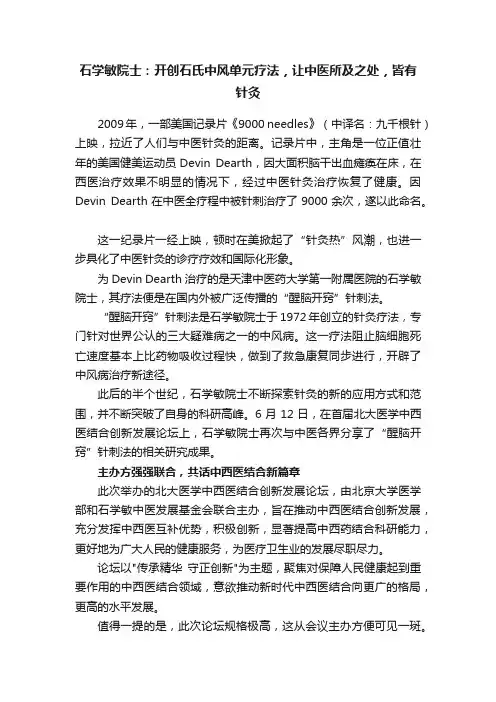
石学敏院士:开创石氏中风单元疗法,让中医所及之处,皆有针灸2009年,一部美国记录片《9000 needles》(中译名:九千根针)上映,拉近了人们与中医针灸的距离。
记录片中,主角是一位正值壮年的美国健美运动员Devin Dearth,因大面积脑干出血瘫痪在床,在西医治疗效果不明显的情况下,经过中医针灸治疗恢复了健康。
因Devin Dearth在中医全疗程中被针刺治疗了9000余次,遂以此命名。
这一纪录片一经上映,顿时在美掀起了“针灸热”风潮,也进一步具化了中医针灸的诊疗疗效和国际化形象。
为Devin Dearth治疗的是天津中医药大学第一附属医院的石学敏院士,其疗法便是在国内外被广泛传播的“醒脑开窍”针刺法。
“醒脑开窍”针刺法是石学敏院士于1972年创立的针灸疗法,专门针对世界公认的三大疑难病之一的中风病。
这一疗法阻止脑细胞死亡速度基本上比药物吸收过程快,做到了救急康复同步进行,开辟了中风病治疗新途径。
此后的半个世纪,石学敏院士不断探索针灸的新的应用方式和范围,并不断突破了自身的科研高峰。
6月12日,在首届北大医学中西医结合创新发展论坛上,石学敏院士再次与中医各界分享了“醒脑开窍”针刺法的相关研究成果。
主办方强强联合,共话中西医结合新篇章此次举办的北大医学中西医结合创新发展论坛,由北京大学医学部和石学敏中医发展基金会联合主办,旨在推动中西医结合创新发展,充分发挥中西医互补优势,积极创新,显著提高中西药结合科研能力,更好地为广大人民的健康服务,为医疗卫生业的发展尽职尽力。
论坛以"传承精华守正创新"为主题,聚焦对保障人民健康起到重要作用的中西医结合领域,意欲推动新时代中西医结合向更广的格局,更高的水平发展。
值得一提的是,此次论坛规格极高,这从会议主办方便可见一斑。
主办方北京大学医学部是我国创办的第一所西医院校,承担着培养高水平医学人才、引领医学原创性研究的国家重任,石学敏中医发展基金会也在推动我国中医药科研成果、人才培养和传承发扬上发挥着重要作用。
丹芪胶囊的功能主治
丹芪胶囊的功能主治简介丹芪胶囊是一种中药制剂,由多种中药材制成。
其主要成分为淡竹叶、香雪兰等。
丹芪胶囊在中医药中被广泛应用,并具有多种功能主治。
下面我们将介绍丹芪胶囊的功能主治及其相关信息。
功能主治丹芪胶囊具有多种功能主治,以下是其常见的功效:1.清热解毒:丹芪胶囊具有清热解毒的作用,可以用于治疗一些由于体内热量过高引起的症状,如咽喉肿痛、口腔溃疡等。
2.润肺止咳:丹芪胶囊可以增加肺部的润滑性,减少咳嗽症状。
它常用于治疗因肺热引起的咳嗽、胸闷等症状。
3.改善免疫力:丹芪胶囊具有一定的免疫调节作用,可以改善免疫系统的功能,增强人体的抵抗力。
4.抗氧化:丹芪胶囊富含多种抗氧化成分,可以中和体内自由基,减少氧化损伤,起到抗衰老的作用。
5.调节内分泌:丹芪胶囊可以调节内分泌系统的功能,对于一些因内分泌失调引起的症状,如月经不调、失眠等具有一定的疗效。
6.抗炎消肿:丹芪胶囊中的成分具有抗炎消肿的作用,可以用于治疗一些炎症性疾病,如湿疹、皮炎等。
用法用量丹芪胶囊的用法用量如下:•成人:每次口服2粒,每日3次。
•儿童:按体重适量酌减。
请在医生指导下正确使用。
注意事项在使用丹芪胶囊时,需要注意以下事项:1.孕妇禁用:孕妇应避免使用丹芪胶囊,以免对胎儿产生影响。
2.过敏体质慎用:对丹芪胶囊中的任何成分过敏的人应慎用,如出现过敏反应应立即停用。
3.不宜与某些药物同时使用:丹芪胶囊可能与某些药物发生相互作用,建议在使用时遵医嘱,并告知医生所使用的其他药物。
4.饮食调节:在使用丹芪胶囊期间,应避免辛辣刺激性食物的摄入,以利于药效的发挥。
请在医生指导下正确使用,避免不必要的不良反应。
总结丹芪胶囊是一种中药制剂,具有多种功能主治,包括清热解毒、润肺止咳、改善免疫力、抗氧化、调节内分泌、抗炎消肿等。
使用时需要注意用法用量和注意事项,遵医嘱使用,以免发生不必要的不良反应。
天津市食品药品监督管理局关于恢复药品丹芪偏瘫胶囊在津销售的通知
天津市食品药品监督管理局关于恢复药品丹芪偏瘫胶
囊在津销售的通知
文章属性
•【制定机关】天津市食品药品监督管理局
•【公布日期】2008.06.18
•【字号】津食药监市[2008]285号
•【施行日期】2008.06.18
•【效力等级】地方规范性文件
•【时效性】现行有效
•【主题分类】药政管理
正文
天津市食品药品监督管理局关于恢复药品丹芪偏瘫胶囊在津
销售的通知
(津食药监市[2008]285号)
各分局、稽查执法大队:
根据《药品广告审查办法》第二十一条规定,市局于2008年5月13日下发了《关于对千佛灵芝孢子粉胶囊等20种药品实施暂停销售行政强制措施的通知》(津食药监市[2008]219号),暂停药品丹芪偏瘫胶囊在我市的销售。
通知发出后,天津市石天药业有限责任公司已按规定在原刊登媒体发布了丹芪偏瘫胶囊“违法广告更正启事”。
经研究,,局决定从即日起恢复丹芪偏瘫胶囊(国药准字Z20010105)在我市的销售,请各单位做好有关通知工作。
特此通知
二○○八年六月十八日附件:违法广告更正启事的样件(略)。
丹芪胶囊的功能主治
丹芪胶囊的功能主治功能丹芪胶囊是一种中药,以丹参(Salvia miltiorrhiza)为主要成分,通过一系列药理作用发挥多种功能,主要包括:1.血管扩张作用:丹芪胶囊可以通过促进一氧化氮的释放,扩张血管,降低血压,提高血液流动性,改善微循环,从而起到保护心脑血管的作用。
2.抗炎作用:丹芪胶囊中的丹参酮等成分具有明显的抗炎作用,可以抑制炎症反应,减轻组织损伤和炎症引起的不适。
3.抗血栓作用:丹芪胶囊中的丹参酮可以抑制血小板聚集和血栓形成,减少心脑血管病发作的风险。
4.抗氧化作用:丹芪胶囊中的黄酮类和酚类成分具有较强的抗氧化作用,能够清除自由基,减少氧化损伤。
5.抗纤维化作用:丹芪胶囊能够抑制纤维化过程,减少疤痕组织形成,促进受损组织的修复与恢复。
主治丹芪胶囊在临床上被广泛应用,主要用于以下疾病和症状的治疗:1.心脑血管疾病:丹芪胶囊可以改善心脑血液循环,预防和治疗冠心病、心绞痛、心肌梗死、中风等心脑血管疾病。
2.高血压:丹芪胶囊可降低血压,提高血管弹性,改善高血压患者的微循环,减少相关并发症的发生。
3.糖尿病:丹芪胶囊可改善糖尿病患者的血液循环,减少糖尿病引起的心脑血管病变风险。
4.肾脏疾病:丹芪胶囊具有保护肾脏功能的作用,可以预防和治疗肾血管疾病、肾小球肾炎等肾脏疾病。
5.免疫调节:丹芪胶囊具有调节免疫功能的作用,可以增强机体的免疫力,减少疾病的发生和复发。
6.女性疾病:丹芪胶囊对于月经不调、痛经等女性疾病也有一定的疗效。
在使用丹芪胶囊时,应遵医嘱,在医生指导下选择合适的剂量和疗程。
不同人群的体质和疾病状态有所差异,应根据个体情况进行治疗。
注意事项使用丹芪胶囊时需要注意以下事项:1.丹芪胶囊属于中药制剂,副作用较少,但仍可能出现个别不良反应,如腹泻、恶心、头晕等。
如出现不适,应及时停药并告知医生。
2.孕妇、哺乳期妇女和儿童患者慎用丹芪胶囊,必要时需在医生指导下使用。
3.丹芪胶囊可能与其他药物发生相互作用,包括抗凝药和降压药等,使用时请告知医生所用的其他药物。
针灸中药治疗中风
B
足内翻:丘墟透照海。
C
血管性痴呆:上星、百会、四神聪、风池、四白、太冲。
合并上消化道出血:可用三七粉冲服、白及煎服。
C
合并肺感染:可用千金苇茎汤加味。
B
大便不通:大承气汤口服或灌肠。
D
应用活血化瘀中药制剂可贯穿于脑梗塞治疗的整个过程,在此基础上可辨证加药,如丹芪偏瘫胶囊、灯盏花素片等。
A
尿路感染:八正散。
自然发生的大脑皮质功能重组是有限的,要提高功能恢复程度并使患者能够适应环境与独立生活,功能训练极为重要。国外有人作了这样的实验,使5只猴子的手指运动皮支区梗塞,不进行任何训练,3-5个月后给予皮层内微电极刺激,2个月后,在梗死临近部位没有发现手指代表区扩大,而增加手指的技巧训练后则发现梗死区周围手的运动皮质区域有所扩大,证明了运动训练可促进脑功能的重建。
当CBF下降至10-20 ml/(100g·min)或灰质血流下降至正常的40%,白质降至正常的35%时,脑组织就会出现缺血反应。若完全阻断脑血液循环6秒钟,神经元的代谢即受影响;阻断2分钟,脑细胞的电活动停止;阻断5分钟后则开始发生脑组织损害。
1
脑的血供系统
颈内动脉系统:主要供应大脑半球 椎基底动脉:主要供应脑干、小脑。
过去认为其死亡后不可再生,目前的研究结果已证实了脑中的干细胞在一定的条件下可分化成脑细胞,但是这种条件是非常苛刻的,尚没有从该途径治疗脑损伤的临床应用。因此,最大程度的减少脑细胞的损伤数目,促进临近脑细胞的功能代偿仍然是目前的治疗方法。
01
02
一、了解脑细胞、脑循环的特点
脑循环的特点 脑是人体最娇嫩的器官,脑的重量只占人体重的1/50,但是安静状态下脑血流要占心输出量的1/5,并且与其它器官的血液供应无关。由于脑组织几乎无能量的贮备,因此,需要连续地供应血和葡萄糖才能保证脑的正常代谢。正常脑血流(CBF)平均为50-55ml/(100g·min),灰质血流比白质高3-4倍。
神经性偏瘫该如何治疗呢
神经性偏瘫该如何治疗呢很多的人患有神经性的疾病,人体神经系统是很复杂的,因此患者一旦患有了神经类的疾病就需要患者及时的采取方法进行治疗,这样才能够保证患者的身体的健康,那么神经性偏瘫该如何治疗呢?那么下面就请专家来介绍一下吧。
脑梗偏瘫不是不治之症,关键在于是否能够对症治疗用药,而且能够有信心配合坚持恢复锻炼,引发脑梗的因素就是脑动脉血管有梗塞的血脂斑块造成了堵塞,脑部缺血,脑神经细胞有坏死的情况,表现为肢体偏瘫,手指脚趾麻木,语言功能障碍等等,因人而异,因梗塞部位的不同而表现不同的症状,很多病人所在地区医院没有很好的专科用药进行治疗,一般都是开的常规性药物,比如阿司匹林类,大活络丹,华佗再造丸等等,这些用药对脑梗偏瘫的恢复治疗效果微乎其微,很多时候反而病情越拖越久越严重,认为不可治愈的心理,进而放弃治疗,实际上脑梗偏瘫后遗症的治疗要从根本致病因素入手,必须有效的乳化清除梗塞血栓斑块,让持续缺血缺氧的脑组织神经细胞得到有效的“养分”。
那我告诉你只有含有CH3成分的药物才能有效清除!比如国药准字药物类的玉金方胶囊,对脑梗效果就很好,治疗预防相结合。
而针对后遗症的治疗,那就是神经性功能恢复了,也就是我们说的通经络,那么在此基础上可建议诸如丹芪偏瘫胶囊等用药来进行恢复,另外日常的家庭护理必须到位,要让患者树立康复的信心,坚持每日的功能性恢复锻炼,不放弃治疗,这一类康复的病人在医院里很多,只是自己看到的都是住院的患病病人而已,康复的病人看到的都是正常人一样,没人认为是患病的,所以作为一个患病康复的过来人,我给你说这么多,希望患者能够坚强,要有信心,要对症治疗,要不懈练习恢复,一定可以恢复。
以上就是专家为我们介绍的这个问题的看法,患者要想治疗的顺利就需要按照医生的治疗的步骤严格的进行治疗,尤其是患者要避免出现其他的因素影响到治疗的顺利进行,尤其是保证患者的健康最为重要,那么最后祝患者能够早日康复。