Smad7在兔下颌骨牵张成骨过程中的表达
感觉神经缺失对兔下颌骨牵张成骨影响的生物力学观察

感觉神经缺失对兔下颌骨牵张成骨影响的生物力学观察(作者:___________单位: ___________邮编: ___________)作者:雷德林,王磊,曹健,杜兆杰,杨镟凝【摘要】目的研究离断下牙槽神经对兔下颌骨牵张成骨新生骨痂生物力学性能的影响。
方法对20只新西兰白兔进行双侧下颌骨牵张成骨,左侧(实验侧)在手术显微镜下暴露并切取约0.6cm下牙槽神经,保护下牙槽血管束,右侧(对照侧)仅暴露下牙槽神经而不切断。
术后动物随机分为2组,分别在固定期14、28天取材。
对双侧兔下颌骨新生骨痂进行外径测量、苏木精伊红(HE)染色及生物力学测试。
应用SPSS12.0软件包进行配对t检验,分析离断下牙槽神经侧与对照侧牵张区新生骨痂上述指标的差异。
结果双侧牵张区新生骨痂长度与外径未见明显差异(P0.05);离断下牙槽神经侧牵张间隙可见神经组织完全变性,对照侧牵张间隙内神经组织完好。
三点弯曲测试显示固定期14、28天时实验侧最大载荷及挠曲强度分别比对照侧低40.1%、26.8%(P0.05)。
结论离断下牙槽神经可引起牵张区新生骨痂生物力学强度不足,提示感觉神经系统在牵张成骨的成骨过程中起到一定调控作用。
【关键词】感觉神经;下颌骨;生物力学Abstract:Objective To investigate the effects of inferior alveolar nerve resection on mandibular distraction osteogenesis in rabbit.Methods Twenty new Zealand white rabbits underwent bilateral distraction osteogenesis with a rate of 1mm/d.The inferior alveolar nerve of one side was resected under the surgical microscope,with great care of protecting the inferior alveolar vessel bundle.The contralateral side received sham operations.The rabbits were sacrificed at consolidation time of 14 and 28 days.After three dimension measurements,neoformative callus underwent HE staining and three points bending test.A paired t test was performed using SPSS 12.0 software package.Results The three dimension measurement of both sides were of no significant difference(P0.05).The nerve tissues in the experimental side were severely damaged,whereas the nerve tissues in the control side were intact.Three point bending test showed that the bending strength on the denervated side was 40.1% and 26.8% lower than the contralateral side at 14 days and 28 days after consolidation repectively(P0.05).Conclusion The inferior alveolar nerve resection can result in mechanical weakness of new bone,thus suggesting that the peripheral nerves play a role during the new boneformation in the distraction osteogenesis.Key words:sensory nerve;mandible;biomechanics牵张成骨(distraction osteogenesis)是通过特殊的装置,使被切开的骨断端在受到缓慢的牵引力条件下,形成新骨,以延长或拓宽骨骼的外科新技术。
兔下颌骨牵张成骨实验动物模型的建立
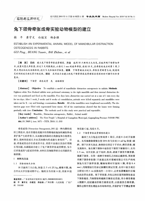
和 实 验 C组 , 组 6只 。 每
12 下颌骨 牵张成骨模型的建立 .
者扩张产生的牵张力 , 从而激发机体的 自我修复和 自我再 生
潜力 , 使两骨 段延 长并 在其 间隙 内形成 新生 骨组 织… 。 目
前 , 张成 骨 还 存 在 着 成 骨 不 良 、 段 不 连 接 以及 治 疗 周 期 牵 骨
tke u o — r ya itlg xa ia in . s t Al fte ma dil swe elngh ne u c sf ly. a n o tf rX a nd hsoo y e m n to s Re uls lo h n b e r e te d s c e su l Thedi— s
t ci n g p e e f ld wi e e ea e o e t s e l o h x mi ain h we h tt e b n s w r omi g r t a s w r l t rg n r t d b n i u .A l ft e e a n t s s o d t a h o e e e f r n a o i e h s o g a u l i i . n l s n T e meh d s d i h ssu y w r r ci a a d rp aa l . r d al w t t y h me Co cu i h t o s u e n ti td e e p a t l n e e t be o c
d i1 .9 9ji n 17 — 3X.00 1 . 1 o:0 36 /. s.6 1 3 i, O 是一种内源性 组 Ds at nO t gns D ) t i e s 织工程技术 , 指在骨缝处或截开的骨断端处施加机械拉伸或
兔下颌骨放射后牵引成骨术式选择的实验研究

双 侧 下领 骨 截 断 、 侧牵 引 的截 骨 办 式 可 作 为 兔 下 颌 骨放 射 后 牵 引成 骨 术 的饿 骨 模 式 。 单 【 键 词 】 },成 , 张 ; 颁 骨 ; 关 iI 牵 1 n ’ 下 兔
d i O 99 .s. 7 —8 0 0 010 4 o: . 6 /i n1 1 0 . 1 .l0 1 3 js 6 0 2 【 图 分 类 号】 R 1 中 38 【 献标志码】 A 文 【 章编 号 】 l 7—8 02 1)110 .4 文 6 1 0 (0o 1.2 70 0
,
sd it ce n o f a in wa d n t eoh r i e Co t l r u :h r d ae i e s s o s da dd — i ed s a td a dn x t sma eo t e d . n r o p t ei a i d s t mi n i r i o h s og r t d wa o e e src e , n o o ea in wa o eOl h t e i e Eih a slt r dsr c in wa c i ae t aeo 4 m m t td a d n p r t sd n it eo h rs . g t y ae, it to sa t t da r t f a o d d a v a 0
兔下颌骨牵张成骨实验动物模型的建立

兔下颌骨牵张成骨实验动物模型的建立中文摘要目的:建立兔下颌骨牵张成骨实验动物模型。
方法:选用30只新西兰大白兔,随机分为5组,每组6只。
在右侧下颌骨第一前磨牙前方和颏孔之间行骨切开术,用自制牵张器固定。
经过7天潜伏期,以每次0.25mm,每天2次的速度牵张,连续6天。
在术后每周进行X线观察,观察模型建立后牵张成骨(distraction osteogenesis.DO)的可行性。
分别在牵张结束,固定期第2、4、6、8周处死动物,切取标本行组织学观察,免疫组化观察和定量CT测定。
结果:大体标本示:实验动物手术后均成活、健康状况良好。
全部动物右侧下颌骨被成功的延长3mm,安置D0装置后伤口愈合良好,自制的DO装置固定牢靠。
X 线示:随固定时间的延长,牵张区均可见新骨逐渐形成,密度影逐渐增强。
组织学观察示:牵张结束牵张区中央为沿牵张力方向排列的胶原纤维束为主;固定期2周牵张间隙内可见沿牵张方向走行的较细的骨小梁结构;固定期4周:牵张区己被沿牵张方向走行的较粗的骨小梁结构所充满;固定期6周牵张区骨小梁沿牵拉方向呈编织状;固定期8周可见成熟的板层骨。
随着牵张时间的延长,新骨在牵张区内不断生成,成熟。
牵张区新骨的成骨方式为膜内成骨。
免疫组化染色示成骨细胞内Ⅰ型胶原呈阳性表达。
在固定期2、4周,成骨细胞内Ⅰ型胶原表达强于固定期6、8周。
结论:1. 根据兔下颌骨离体标本设计的内置式兔下颌骨牵张器,完全符合DO 动物实验的要求。
2. 兔下颌骨牵张成骨中新骨的成骨方式为膜内成骨3.兔作为牵张成骨动物模型具有很高的可行性和可重复性。
关键词:牵张成骨下颌骨动物模型兔The establishment of an animal model of mandibular distraction osteogenesis in rabbitsabstractObjective:To establish an animal model of mandibular distraction osteogenesis in rabbitsMethods:30 white New Zealand rabbits were divided into 5 groups randomly. Each group has 6 rabbits. Osteotomy was performed on right mandible between the first premolar and mental foramen .Bone stumps were repositioned and fixed with self-devised distraction equipment. After 6 days latency period,the distraction was carried out with the frequency of 2 times a day and with the rate of 0.25 mm once for 6 days. The new generation bone were studied by X- ray every week after the operation. When distraction completed , and the rabbits sacrificed after 0, 2 ,4 ,6 ,and 8 weeks. The specimens were take for Quantitative Computed Tomography(QCT),immunohistochemistry and histology examination.Results:specimens macrography All rabbits were in good condition with in the experiment period. The right mandible of every rabbits were lengthend 3mm successful.During the experiment period ,well healing was found between the distraction device and surrounding tissues.The instrument fixed well.X-ray With the time of fixation passing,the density of new bone was enhanced gradually.Histology showed that at the end ofdistraction period the distraction zone was filled with fibers arrayed from the distraction direction;on the second week of consolidation period the new bone trabecula extended from the distraction direction; on the fourth week of consolidation period the distraction zone was filled with mature bone trabecula arrayed from the distraction direction; on the sixth week of consolidation period ,woven bone was found in the distraction zone;on the eighth week of consolidation period, lamellar bone was found in the distraction zone.New bone gradually formed and matured within distraction gap along the time of study. The new bone regeneration was formed by intramembranous ossification.Immunohistochemistry show that the expression of CollagenⅠwas observed in the osteoblasts.The expression of CollagenⅠin the osteoblasts on the fourth week of consolidation period and on the second week of consolidation period was stronger than other time.Conclusion: 1.The internal fixed distracter that designed according to the structure of the mandible of rabbits maybe used for animal experiment.2.The new bone regeneration was formed by intramembranous ossification.3.Rabbit is a good animal for developing an experimentalmodel on study of distraction osteogenesis.Key words:Distraction Osteogenesis(DO) Animal model Mandible Rabbit前言牵张成骨(distraction osteogenesis.DO)是一种内源性组织工程技术,指在骨缝处或截开的骨断端处用特制的牵张装置以一定的速度和频率牵开,从而激发机体的自我修复和自我再生潜力,使两骨段延长并在其间隙内形成新生骨组织,以达到延长骨组织的目的。
Smad7转染人骨髓间充质干细胞的初步实验研究

Smad7转染人骨髓间充质干细胞的初步实验研究陈康;蔡晓峰;庹瑶;赖佳;李旭清【摘要】Objective Human bone marrow mesenchymal stem cells were transfected by Smad7 and labeled with green fluores-cent mark.BMMSCs were implanted into rabbit glaucoma operational model and observed surviving condition.Methods Through BP and LR reaction,Smad7 with green fluorescent mark was inserted into human bone marrow mesenchymal stem cells by bacterio-phage,filtered positive colony and picked out cell line.15 New Zealand white rabbits were enforced trabeculectomy with BMMSC, then following up cell survivalcondition.Results Smad7 expressed stable in human bone marrow mesenchymal stem cells with sat-isfactory green fluorescent mark.BMMSC survived in rabbit trabecula with stable green fluorescent and effective ocular press.Con-clusion Smad7 with green fluorescent mark could be inserted into human bone marrow mesenchymal stem cells stably,and has ef-fective results in rabbit model.%目的:将 Smad7转染到人骨髓间充质干细胞(BMMSC),并进行绿色荧光标记,将其植入兔青光眼手术模型,观察BMMSC 存活情况。
目标基因测序技术检测与下颌前突相关的BMP-Smad信号通路基因突变

目标基因测序技术检测与下颌前突相关的BMP-Smad信号通路基因突变李偲;陈凤山【摘要】目的:采用目标基因捕获联合二代测序技术探索BMP-Smad信号通路基因与下颌前突(mandibular prognathism)相关的变异.方法:从同济大学附属口腔医院收集176例下颌前突患者及155例正常对照,分别采集5 mL静脉血并提取基因组DNA.采用NimbleGen捕获富集系统对病例对照组的23个目标基因的编码区和侧翼区进行捕获富集,通过Illumina Hiseq 2000测序平台进行测序.通过比较变异位点基因型及等位基因分布频率的差异,探索与下颌前突相关的突变位点.结果:共发现7个与下颌前突相关的常见变异,分别位于5个基因上:chr4:81975103(BMP3)、rs7078571(BMPR1A)、chr2:148686946(ACVR2A)、rs1128919(ACVR2A)、rs2070489(ACVR2B)、chr18:48610378(SMAD4)、chr18:48610376(SMAD4).未发现罕见变异的分布差异有显著性.结论:通过目标基因测序的方法检测到了病例对照组中的常见变异和罕见变异,并发现了与下颌前突相关的可能致病基因BMP3、BMPR1A、ACVR2A、ACVR2B、Smad4.未检测到下颌前突的罕见致病位点.%Objective: To identify variants of the genes in BMP-Smad signaling pathway that predisposing to mandibular prognathism in Chinese population. Methods: 176 mandibular prognathism individuals and 155 controls were recruited and provided informed consent from affiliated stomatology hospital of Tongji University, 5 mL peripheral blood were obtained from each individual and genomic DNA was extracted. The coding and flanking regions of 23 candidate genes in the BMP-smad signaling pathway were captured by usingNimbleGen Targeted Enrichment System and then sequenced on the Illumina Hiseq2000 platform. Comparing the allelic and genotypic distribution in the cases and controls, the variants related to mandibular prognathism were identified. Results:7 common variants in 5 genes were identified, which associated with mandibular prognathism in the following loci:chr4:81975103 in BMP3, rs7078571 in BMPR1A, chr2:148686946 and rs1128919 in ACVR2A, rs2070489 in ACVR2B chr18:48610378,chr18:48610376 in SMAD4. No significant difference in rare variants between case and control were found. Conclusions: Via targeted sequencing analysis, new genes that associated with mandibular prognathism were identified, which were BMP3, BMPR1A, ACVR2A, ACVR2B, Smad4. We do not identify any rare variants related to this disease, which might affect this disease.【期刊名称】《口腔颌面外科杂志》【年(卷),期】2016(026)006【总页数】7页(P399-405)【关键词】下颌前突;目标基因捕获;新一代测序;BMP-Smad信号通路;突变【作者】李偲;陈凤山【作者单位】同济大学口腔医学院, 同济大学附属口腔医院口腔正畸科,上海牙组织修复与再生工程技术中心,上海 200072;同济大学口腔医学院, 同济大学附属口腔医院口腔正畸科,上海牙组织修复与再生工程技术中心,上海 200072【正文语种】中文【中图分类】R782下颌前突(mandibular prognathism,MP)是临床较为常见的一种严重的颌面部发育畸形,初诊患者多为青少年,常规根据其骨性错颌严重程度选择不同的治疗方案[1-4]。
转化生长因子在复合异种部分脱钙骨修复兔下颌骨缺损中表达的实验研究的开题报告

转化生长因子在复合异种部分脱钙骨修复兔下颌骨缺损中表达的实验研究的开题报告
一、研究背景
随着人口老龄化程度的加剧和骨缺损相关疾病的不断增多,神经生长因子在骨修复治疗中的临床应用愈加广泛。
为了进一步探究神经生长因子在骨修复过程中的作用和机制,本研究以转化生长因子(TGF)作为研究对象,从分子层面深入探究其在复合异种部分脱钙骨修复兔下颌骨缺损中的表达情况和作用机制,为临床治疗提供理论参考和实践指导。
二、研究目的
本研究旨在探究转化生长因子在复合异种部分脱钙骨修复兔下颌骨缺损中的表达情况,分析其作用机制,为临床治疗提供重要的理论依据和实践指导。
三、研究方法
1.实验对象及组分:
本研究选取30只健康有满口牙齿、体重相近的新西兰大白兔为实验对象,分为对照组和实验组。
2.实验方法:
对照组采用普通缝线进行硬膜下注射;实验组采用TGF进行硬膜下注射。
3.实验过程:
制作下颌骨缺损模型后,加入TGF进行硬膜下注射,注射进程术后第3天开始并持续14天。
4.实验指标:
主要观察指标包括下颌骨缺损面积、骨组织生长情况、血管新生情况等,并进行统计分析。
四、研究意义
本研究将深入探究转化生长因子在复合异种部分脱钙骨修复兔下颌骨缺损中的作用机制,为临床治疗提供更为全面、深入的指导和参考,为改善人类生命质量、推动医疗技术水平的提升贡献力量。
失神经支配在胫骨牵张成骨延长过程中的骨再生及Runx2表达

失神经支配在胫骨牵张成骨延长过程中的骨再生及Runx2表达郑科;宋冬惠;冯兴梅;祝颂松;胡静;叶斌【摘要】BACKGROUND:During the healing of fractures, removal of sciatic nerve can result in insufficient mechanical rigidity of newborn woven bone. However, there are less reports concerning the denervation effects during distraction osteogenesis. OBJECTIVE:To observe the effect of removal of the sciatic nerve on bone regeneration and the expression of Runt-related transcription factor 2 (Runx2) protein during distraction osteogenesis in a rabbit model. METHODS:Twenty-four adult male New Zealand rabbits were selected and underwent left tibial osteodistraction to construct animal models of distraction osteogenesis. Before distraction, the animals were randomly divided into group R (resecting the left sciatic nerve) and group I (intact left sciatic nerve). Six weeks after completion of distraction, the animals were kil ed and the lengthened tibias were harvested for radiography, three-dimensional CT reconstruction, histological evaluation, connectivity density (Conn.D) evaluation. RESULTS AND CONCLUSION:New regenerated bone was present and Runx2 protein was expressed in the distraction gaps of al animals at the end of the study, as revealed by radiography, three-dimensional CT reconstruction, and histological observation. However, less new bone formation and a lower degree of mineralization and expression of Runx2 protein were observed in group R compared with group I. The results suggest that thedenervation appears to have an inhibitory effect on bone formation and the expression of Runx2 protein during distraction osteogenesis.%背景:研究发现,去除坐骨神经会导致骨折愈合过程中新生编织骨机械硬度不足,而目前对有关失神经因素在牵张成骨过程中作用的相关报道较少。