化学发光分析仪使用说明书
罗氏 cobas e 801 电化学发光免疫分析仪使用说明书 - 检测抗 HBs 抗体的试剂盒
Elecsys Anti-HBs IIREFSYSTEM********************** 300cobas e 801EnglishSystem information Short name ACN (application code number) AHBS 2 10138 Immunoassay for the in vitro quantitative determination of human antibodies to the hepatitis B surface antigen (HBsAg) in human serum and plasma.Anti-HBs assays are used within the scope of hepatitis B vaccination to check the necessity and success of vaccination. In addition, anti-HBs assays are used to monitor the course of disease following acute hepatitis B infection. This test is not intended for diagnosis.The e lectro c hemi l uminescence i mmuno a ssay “ECLIA” is intended for use on the cobas e 801 immunoassay analyzer.Note: Please note that the catalogue number appearing on the package insert retains only the first 8 digits of the licensed 11-digit Catalogue Number: 07026854190 for the Elecsys Anti-HBs II assay. The last 3 digits -190 have been replaced by -119 for logistic purposes. SummaryAnti-HBs is a specific (generally IgG) antibody that is directed against the hepatitis B surface antigen (HBsAg).1,2 Anti ‑HBs can be detected several weeks after the disappearance of hepatitis B surface antigen.3,4 Anti ‑HBs can be formed following a hepatitis B infection or after hepatitis Bvaccination.3,4 Antibodies are formed against the HBsAg determinant a, which is common to all subtypes, and against subtype-specific determinants.1,5,6Anti ‑HBs assays are used within the scope of hepatitis B vaccination to check the necessity and success of vaccination.2,4,7 In addition, anti ‑HBs assays are used to monitor the course of disease following acute hepatitis B infection.3The Elecsys Anti ‑HBs II assay uses a mixture of purified antigens fromhuman serum (HBsAg subtype ad), and recombinant HBsAg subtype ay from CHO (Chinese Hamster Ovary) cells. Test principleSandwich principle. Total duration of assay: 18 minutes.▪ 1st incubation: Anti ‑HBs in the sample (24 μL), biotinylated HBsAg(ad/ay), and HBsAg (ad/ay) labeled with a ruthenium complex a) react to form a sandwich complex.▪ 2nd incubation: After addition of streptavidin-coated microparticles, thecomplex becomes bound to the solid phase via interaction of biotin and streptavidin.▪ The reaction mixture is aspirated into the measuring cell where themicroparticles are magnetically captured onto the surface of the electrode. Unbound substances are then removed with ProCell II M. Application of a voltage to the electrode then induces chemiluminescent emission which is measured by a photomultiplier.▪ Results are determined via a calibration curve which is instrument-specifically generated by 2‑point calibration and a master curve provided via the cobas link.a) Tris(2,2'-bipyridyl)ruthenium(II)-complex (Ru(bpy)32+)Reagents – working solutionsThe cobas e pack (M, R1, R2) is labeled as AHBS 2. M Streptavidin-coated microparticles, 1 bottle, 13.2 mL:Streptavidin-coated microparticles 0.72 mg/mL; preservative. R1 HBsAg~biotin, 1 bottle, 16.7 mL:Biotinylated HBsAg (ad/ay) human/recombinant, > 0.5 mg/L; MES b) buffer 85 mmol/L, pH 6.5; preservative. R2 HBsAg~Ru(bpy)32+, 1 bottle, 15.8 mL:HBsAg (ad/ay) human/recombinant, labeled with ruthenium complex > 0.3 mg/L; MES buffer 85 mmol/L, pH 6.5; preservative.b) MES = 2-morpholino-ethane sulfonic acidAHBS 2 Cal1 Calibrator 1, 1 bottle of 1.3 mL:Anti ‑HBs (human) in human serum; preservative.AHBS 2 Cal2 Calibrator 2, 1 bottle of 1.3 mL:Anti ‑HBs (human) in human serum; preservative.Precautions and warnings For in vitro diagnostic use.Exercise the normal precautions required for handling all laboratory reagents. Disposal of all waste material should be in accordance with local guidelines. Safety data sheet available for professional user on request.This kit contains components classified as follows in accordance with the Regulation (EC) No. 1272/2008:n ‑Octyl ‑N,N ‑dimethyl ‑3‑ammonio ‑1‑propanesulfonateEUH 208 May produce an allergic reaction.Product safety labeling primarily follows EU GHS guidance. All human material should be considered potentially infectious.The calibrators (AHBS 2 Cal1 and AHBS 2 Cal2) have been preparedexclusively from the blood of donors tested individually and shown to be free from HBsAg and antibodies to HCV and HIV. The testing methods applied were FDA ‑approved or cleared in compliance with the European Directive 98/79/EC, Annex II, List A.The HBsAg starting material used was inactivated prior to labeling with biotin or ruthenium by heating to 60 °C for 15 hours. In addition, any virus particles remaining were removed by ultracentrifugation.However, as no inactivation or testing method can rule out the potential risk of infection with absolute certainty, the material should be handled with the same level of care as a patient specimen. In the event of exposure, the directives of the responsible health authorities should be followed.8,9 Avoid foam formation in all reagents and sample types (specimens, calibrators and controls). Reagent handlingThe reagents (M, R1, R2) in the kit are ready-for-use and are supplied in cobas e packs. CalibratorsThe calibrators are supplied ready ‑for ‑use in bottles compatible with the system.Unless the entire volume is necessary for calibration on the analyzer, transfer aliquots of the ready ‑for ‑use calibrators into empty snap ‑cap bottles (CalSet Vials). Attach the supplied labels to these additional bottles. Store the aliquots at 2‑8 °C for later use.Perform only one calibration procedure per aliquot.All information required for correct operation is available via the cobas link. Storage and stability Store at 2‑8 °C. Do not freeze.Store the cobas e pack upright in order to ensure complete availability of the microparticles during automatic mixing prior to use. Stability of the cobas e pack: unopened at 2‑8 °Cup to the stated expiration date on the cobas e 801 analyzer 16 weeksStability of the calibrators: unopened at 2‑8 °C up to the stated expiration date after opening at 2‑8 °C 16 weeks on the cobas e 801 analyzer at 20‑25 °Cuse only onceadhering to the snap ‑cap.Specimen collection and preparationOnly the specimens listed below were tested and found acceptable.Serum collected using standard sampling tubes or tubes containing separating gel.K2‑EDTA and K3‑EDTA plasma.Criterion: Slope 1.00 ± 0.15 + intercept 0 ± 2 IU/L + bias at 10 IU/L: ≤ 30 %. Stable for 3 days at 20‑25 °C, 6 days at 2‑8 °C, 3 months at ‑20 °C(± 5 °C). The samples may be frozen 5 times.For plasma treated with lithium heparin, lithium heparin with gel or sodium heparin, the values found were on average up to 20 % lower than those obtained in serum. For plasma treated with sodium citrate, the values found were on average up to 30 % lower than those obtained with serum.The sample types listed were tested with a selection of sample collection tubes or systems that were commercially available at the time of testing, i.e. not all available tubes of all manufacturers were tested. Sample collection systems from various manufacturers may contain differing materials which could affect the test results in some cases. When processing samples in primary tubes (sample collection systems), follow the instructions of the tube manufacturer.Centrifuge samples containing precipitates and thawed samples before performing the assay.Do not use heat‑inactivated samples.Do not use samples and controls stabilized with azide.Ensure the samples and calibrators are at 20‑25 °C prior to measurement. Due to possible evaporation effects, samples and calibrators on the analyzers should be analyzed/measured within 2 hours.The performance of the Elecsys Anti‑HBs II assay has not been established with cadaveric samples or body fluids other than serum and plasma. Materials providedSee “Reagents –working solutions” section for reagents.▪ 2 x 6 bottle labelsMaterials required (but not provided)▪REF 11876317122, PreciControl Anti‑HBs, 16 x 1.3 mL▪REF 11776576322, CalSet Vials, 2 x 56 empty snap-cap bottles▪REF***********,DiluentUniversal,45.2mLsamplediluent▪▪cobas e 801 analyzerAccessories for the cobas e 801 analyzer:▪REF***********,ProCellIIM,2x2Lsystemsolution▪REF 04880293190, CleanCell M, 2 x 2 L measuring cell cleaning solution ▪REF***********,ReservoirCups,8cupstosupplyProCellIIMand CleanCell M▪REF***********,PreCleanIIM,2x2Lwashsolution▪REF***********,AssayTip/AssayCuptray,6magazinesx6magazine stacks x 105 assay tips and 105 assay cups, 3 wasteliners▪REF***********,LiquidFlowCleaningCup,2adaptorcupstosupply ISE Cleaning Solution/Elecsys SysClean for Liquid Flow CleaningDetection Unit▪REF***********,PreWashLiquidFlowCleaningCup,1adaptorcupto supply ISE Cleaning Solution/Elecsys SysClean for Liquid Flow Cleaning PreWash Unit▪REF 11298500316, ISE Cleaning Solution/Elecsys SysClean,5 x 100 mL system cleaning solutionAssayFor optimum performance of the assay follow the directions given in this document for the analyzer concerned. Refer to the appropriate operator’s manual for analyzer‑specific assay instructions.Resuspension of the microparticles takes place automatically prior to use. Place the cooled (stored at 2‑8 °C) cobas e pack on the reagent manager. Avoid foam formation. The system automatically regulates the temperature of the reagents and the opening/closing of the cobas e pack. Calibrators:Place the calibrators in the sample zone.Read in all the information necessary for calibrating the assay.CalibrationTraceability: This method has been standardized against the 1st WHO Reference Standard 1977.The predefined master curve is adapted to the analyzer using AHBS 2 Cal1 and AHBS 2 Cal2.Calibration frequency: Calibration must be performed once per reagent lot using AHBS 2 Cal1, AHBS 2 Cal2 and fresh reagent (i.e. not more than24 hours since the reagent kit was registered on the analyzer).Renewed calibration is recommended as follows:▪after 12 weeks when using the same reagent lot▪after 28 days when using the same cobas e pack on the analyzer▪as required: e.g. quality control findings with PreciControl Anti‑HBs outside the defined limitsQuality controlFor quality control, use PreciControl Anti‑HBs.Controls for the various concentration ranges should be run individually at least once every 24 hours when the test is in use, once per cobas e pack, and following each calibration.The control intervals and limits should be adapted to each laboratory’s individual requirements. Values obtained should fall within the defined limits. Each laboratory should establish corrective measures to be taken if values fall outside the defined limits.If necessary, repeat the measurement of the samples concerned.Follow the applicable government regulations and local guidelines for quality control.CalculationThe analyzer automatically calculates the analyte concentration of each sample in IU/L.Interpretation of the resultsNumeric result Result message Interpretation< 10 IU/L Non-reactive Negative for anti-HBs≥ 10 IU/L Reactive Positive for anti-HBsvary depending on the testing procedure used. Results obtained from a single sample using tests from different manufacturers can therefore differ by up to a factor of 4 (or even a factor of 10 in rare cases). If there is a change in the assay procedure used during the monitoring of vaccination protection, then the anti‑HBs values obtained upon changing over to the new method must be confirmed by parallel measurements by both methods. Vaccination strategies in certain risk groups are based on the measured anti‑HBs concentration. Respective recommendations are given by national or regional guidelines. Limitations - interferenceThe effect of the following endogenous substances and pharmaceutical compounds on assay performance was tested. Interferences were tested up to the listed concentrations and no impact on results was observed. Endogenous substancesCompound Concentration testedBilirubin ≤ 513 μmol/L or ≤ 30 mg/dL Hemoglobin ≤ 0.621 mmol/L or ≤ 1000 mg/dL Intralipid ≤ 1500 mg/dLBiotin ≤ 41 nmol/L or ≤ 10 ng/mL Rheumatoid factors ≤ 1200 IU/mLAlbumin ≤ 7.0 g/dLIgG ≤ 7.0 g/dLIgA ≤ 1.6 g/dLIgM ≤ 1.0 g/dL2 / 42017-09, V 1.0 Can EnglishCriterion: Recovery for samples from Limit of Detection to 10 IU/L:≤ ± 2 IU/L, and samples > 10 IU/L: ≤ ± 20 % of initial value.Samples should not be taken from patients receiving therapy with high biotin doses (i.e. > 5 mg/day) until at least 8 hours following the last biotin administration.Pharmaceutical substancesIn vitro tests were performed on 16 commonly used pharmaceuticals. No interference with the assay was found.In addition, the following special drugs used in hepatitis B therapy were tested. No interference with the assay was found.Special drugsDrug Concentration testedmg/LPeginterferon alfa‑2a ≤ 0.18Peginterferon alfa‑2b ≤ 1.6Lamivudine ≤ 300Adefovir ≤ 10Entecavir ≤ 10Tenofovir ≤ 600Telbivudine ≤ 245Due to high-dose hook effect c), results from anti‑HBs concentrations of> 200000 IU/L may be found below the upper limit of the measuring range of 1000 IU/L. In rare cases, a high-dose hook effect from anti HBs concentrations of < 20000 IU/L cannot be excluded. Therefore in case of any unexpected low result the sample should be diluted 1:100 (refer to chapter “Dilution”) and tested again.In rare cases, interference due to extremely high titers of antibodies to streptavidin and ruthenium can occur. The test contains additives which minimize these effects.c) High-dose hook effect: A sample with a true concentration clearly above the measuring range, but found within the measuring range.Limits and rangesMeasuring range2‑1000 IU/L (defined by the Limit of Detection and the maximum of the master curve). Values below the Limit of Detection are reported as< 2 IU/L.Values above the measuring range are reported as > 1000 IU/L (or up to 100000 IU/L for 100‑fold diluted samples).DilutionSamples with anti‑HBs concentrations above the measuring range can be diluted with Diluent Universal. The recommended dilution is 1:100 (either automatically by the analyzer or manually). The concentration of the diluted sample must be > 10 IU/L.After manual dilution, multiply the result by the dilution factor.After dilution by the analyzer, the software automatically takes the dilution into account when calculating the sample concentration.Manual dilution can also be made with negative human serum.Note: Antibodies to HBsAg are heterogeneous. In some isolated cases, this may lead to non-linear dilution behavior.Specific performance dataRepresentative performance data on the analyzer is given below. Results obtained in individual laboratories may differ.PrecisionPrecision was determined using Elecsys reagents, samples and controls in a protocol (EP05‑A3) of the CLSI (Clinical and Laboratory Standards Institute): 2 runs per day in duplicate each for 21 days (n = 84). The following results were obtained:cobas e 801 analyzerRepeatability d)Intermediateprecision e)Sample MeanIU/LSDIU/LCV%SDIU/LCV% Human serum 1 4.33 0.224 5.2 0.272 6.3 Human serum 2 12.0 0.237 2.0 0.277 2.3 Human serum 3 475 6.81 1.4 7.55 1.6 PC f) Anti-HBs 1 < 2.00 - - - -PC Anti-HBs 2 83.8 1.08 1.3 1.28 1.5d) Repeatability = within-run precisione) Intermediate precision = between-run precisionf) PC = PreciControlAnalytical specificityNo cross-reactions with HAV, HCV, HEV, CMV, EBV, HIV, Rubella, Toxoplasma gondii, Treponema pallidum, rheumatoid arthritis, autoimmune response or alcoholic liver disease were observed.Measurements were performed on each of the pathogens listed above using ≥ 8 serum or plasma samples which were positive for antibodies to the above-mentioned pathogens.Relative sensitivityPerformance of the Elecsys Anti‑HBs II assay has been assessed by testing a total of 669 samples at two different study sites. 296 samples from vaccinated persons and 373 samples from patients recovered from a hepatitis B infection have been measured with the Elecsys Anti‑HBs II assay and another commercially available fully automated anti‑HBs assay. Discrepant samples were tested with additional anti‑HBs assays to achieve a consensus.Characterization ofsamplesN ElecsysAnti‑HBs IIreactiveAnti‑HBscomparisontest reactiveSensitivity%Anti-HBs positive:vaccinees 296 296 296 100Anti-HBs positive:recovered from ahepatitis B infection373 373 373 100 Total 669 669 669 100 Relative specificityPerformance of the Elecsys Anti‑HBs II assay has been assessed by testing 2673 samples from blood donors negative for anti‑HBs at two different study sites and 1623 anti‑HBs negative samples from laboratory routine at three different study sites. Discrepant samples were tested with additional anti‑HBs assays to achieve a consensus.Characterization of samples N ElecsysAnti‑HBs IIfalsepositiveSpecificity%Anti-HBs negative: blood donors 2673 6 99.78 Anti-HBs negative: routinesamples1623 9 99.45 References1Seeger C, Zoulim F, Mason WS. Hepadnaviruses. In: Field’s Virology, Knipe DM, Howley RM (eds), 2007 5th edition, Lippincott Williams andWilkins, Philadelphia, USA. Chapter 76, pp2977-3029.2WHO. Hepatitis B vaccines. Wkly Epidemiol Rec 2009;84:405-420.3Liaw YF, Chu CM. Hepatitis B virus infection. Lancet2009;373:582-592.4Caspari G, Gerlich WH. The serologic markers of hepatitis B virus infection – proper selection and standardized interpretation. Clin Lab2007;53:335-343.5Kramvis A, Kew M, François G. Hepatitis B virus genotypes. Vaccine 2005;23:2409-2423.6Michel ML, Tiollais P. Hepatitis B vaccines: protective efficacy and therapeutic potential. Pathol Biol 2010;58:288-295.7Elgouhari HM, Abu-Rajab Tamimi TI, Carey WD. Hepatitis B virus infection: understanding its epidemiology, course, and diagnosis. Cleve Clin J Med 2008;75:881-889.8Occupational Safety and Health Standards: Bloodborne pathogens. (29 CFR Part 1910.1030). Fed. Register.9Directive 2000/54/EC of the European Parliament and Council of18 September 2000 on the protection of workers from risks related toexposure to biological agents at workFor further information, please refer to the appropriate operator’s manual for the analyzer concerned, the respective application sheets, the product information and the Method Sheets of all necessary components (if available in your country).A point (period/stop) is always used in this Method Sheet as the decimal separator to mark the border between the integral and the fractional parts of a decimal numeral. Separators for thousands are not used.SymbolsRoche Diagnostics uses the following symbols and signs in addition to those listed in the ISO 15223‑1 standard:CONTENT Contents of kitSYSTEM Analyzers/Instruments on which reagents can be used REAGENT ReagentCALIBRATOR CalibratorVolume after reconstitution or mixingGTIN Global Trade Item NumberCOBAS, COBAS E, ELECSYS and PRECICONTROL are trademarks of Roche. INTRALIPID is a trademark of Fresenius Kabi AB.All other product names and trademarks are the property of their respective owners. Additions, deletions or changes are indicated by a change bar in the margin.© 2016, Roche DiagnosticsRoche Diagnostics GmbH, Sandhofer Strasse 116, D-68305 Mannheim。
BIO-RAD ChemiDocXRS+化学发光凝胶成像仪中文简易说明书
ChemiDocXRS+化学发光凝胶成像仪的应用一、实验流程:1、实验前需提前半小时打开冷CCD镜头的开关,打开仪器的开关,最后打开软件。
2、实验时先将抽屉拉开放置核酸凝胶或转印膜(若是蛋白凝胶需放在暗箱中的白光板上,不用时白光板竖立卡好在暗箱中),关上抽屉,检查暗箱门是否关好。
确认好后,打开ImageLab软件,设置拍照程序,当选择不同凝胶拍摄方法时,会提示滤光片的位置和使用激发光的类型(例如拍摄化学发光会提示:无光源,无滤光片,此时滤光片的位置要调到“0档”),严格按照软件中的提示操作。
等拍照结束后保存拍照程序及最终生成的图片,便于后续数据的分析及程序的再次调用。
普通核酸凝胶和蛋白质凝胶的成像步骤与GelDocXR+的操作一样,也可参照《ImageLab中文操作指南》。
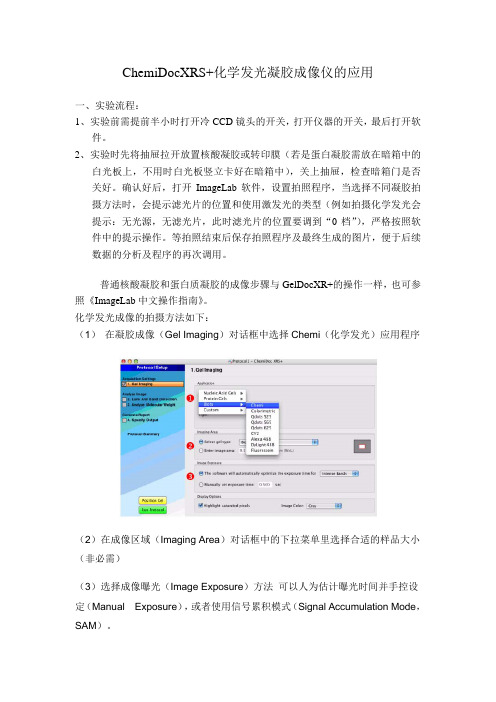
化学发光成像的拍摄方法如下:(1) 在凝胶成像(Gel Imaging)对话框中选择Chemi(化学发光)应用程序(2)在成像区域(Imaging Area)对话框中的下拉菜单里选择合适的样品大小(非必需)(3)选择成像曝光(Image Exposure)方法可以人为估计曝光时间并手控设定(Manual Exposure),或者使用信号累积模式(Signal Accumulation Mode,SAM)。
在建立一个化学发光的程序时最难的任务就是图像曝光的确定,因为您想获得一个充分利用了相机大的动态范围的图像。
过短的图像曝光时间可能使高于膜背景的微弱信号不能被识别;过长的图像曝光时间能够使得某些条带饱和(也就是说,超出了相机准确报告信号的能力)。
如果这是您第一次用ChemiDoc XRS+分析化学发光样品,您需要在使用手控或信号累积模式(SAM)之前决定一个合适的成像时间框架。
获得这个信息的最好的方式是取得一个相对短的手工曝光并利用Image Lab里的工具估计最适的曝光时间。
手控曝光1. 选择手控设定曝光时间(Manually setexposure time)并在读秒框中输入10秒。
万孚生物POCT仪器说明书
万孚生物POCT仪器说明书
【药品名称】
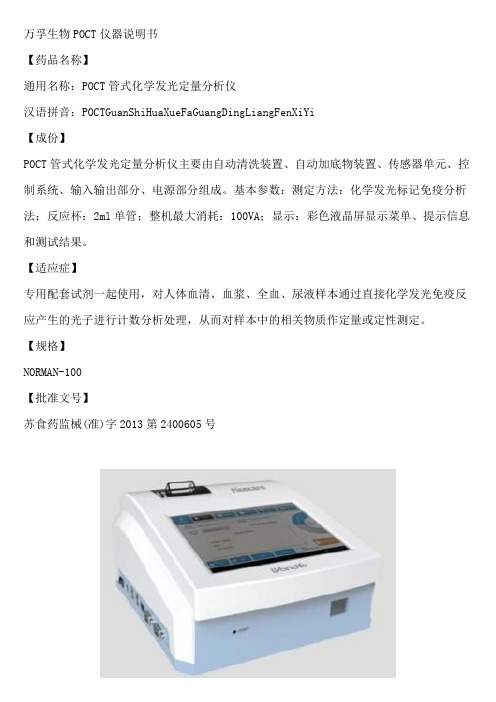
通用名称:POCT管式化学发光定量分析仪
汉语拼音:POCTGuanShiHuaXueFaGuangDingLiangFenXiYi
【成份】
POCT管式化学发光定量分析仪主要由自动清洗装置、自动加底物装置、传感器单元、控制系统、输入输出部分、电源部分组成。
基本参数:测定方法:化学发光标记免疫分析法;反应杯:2ml单管;整机最大消耗:100VA;显示:彩色液晶屏显示菜单、提示信息和测试结果。
【适应症】
专用配套试剂一起使用,对人体血清、血浆、全血、尿液样本通过直接化学发光免疫反应产生的光子进行计数分析处理,从而对样本中的相关物质作定量或定性测定。
【规格】
NORMAN-100
【批准文号】
苏食药监械(准)字2013第2400605号。
LiCA500自动光激化学发光检测仪标准化操作规程

LiCA500 自动光激化学发光检测仪标准化操作规程(SOP)一.检测原理LICA500系统仪器与光激化学发光试剂配套使用,用于检测体液内特异性的抗原和抗体。
LiCA500采用双针臂式结构使用一次性吸头,自动进行加样、稀释、振荡、温育、680纳米红光激发、PMT光电倍增管记录发光信号后拟合计算量值和传送结果。
二.运行环境为保证仪器的正常运行,仪器应当在满足下列条件的环境下使用:1.灰尘少、换气良好的环境。
2.避免阳光直接照射仪器。
3.室内温度保持在10℃~30℃。
4.室内相对湿度保持20%~80%。
5.远离电磁场干扰源。
6.电源220V±22V AC 50Hz±1Hz 具有良好的接地7.(台式机)操作台至少保留工作空间至少长 200 厘米,宽 80 厘米。
称重量280公斤以上8.需要准备一个台面放置操作电脑及显示器,供后期操作三.安全条款1.操作过程中请使用橡胶手套,不要直接接触待测试样本及相关试剂。
2.仪器上或周围不要使用可燃性的危险品,避免引起火灾等重大事故。
3.仪器运行过程中,不要将身体或者肢体伸入仪器,避免带来伤害。
4.在电源开关处理ON状态下,触摸线路板有触电的危险。
5.禁止非专业的人员打开仪器的面板,对仪器进行拆卸。
6.请勿用肉眼直视光源灯光,避免伤害视力。
四.开机前准备1.确认LiCA500电源线连接正常。
2.确认电脑显示器及主机电源线连接正常。
3.确认电脑主机与仪器CAN接口使用CAN线连接正常。
五.开机步骤1.打开仪器电源开关(仪器左侧面)。
2.按下黄色按钮2-3秒,仪器机械模块上电,并自动完成仪器初始化过程。
3.打开电脑及显示器电源。
待电脑启动后双击桌面LiA500图标启动软件。
六.实验前准备1.标本严格按照7-10分钟3500转/分的参数进行处理,避免有血凝块及纤维蛋白原。
2.检查实验资源,通过软件工作台资源显示功能完成:●检查清洗液瓶中的清洗液是否足够,若清洗液不足,加满清洗液;●检查通用液是否盛满,若通用液不足,加一瓶通用液;●检查枪头是否足够,若不够,则添加枪头;●检查板条是否足够,做不够,则添加板条;●废液抽屉是否处于清空状态,若废液抽屉内废液需要清理,在工作台面上小心地抽取废液抽屉,清理完废液抽屉中的废液,将废液抽屉推送至原来位置后继续之后的操作。
全自动化学发光免疫分析仪Immulite2000标准操作规程完整
3.抗体捕获法:抗体捕获形式是指被测量样本中的物质是抗体。试剂是使用一种抗人的特种抗体。抗体捕获形式通常使用两次孵育和洗涤,第一次孵育和洗涤是为了除去样品中过多的干扰物质。第二次孵育和洗涤是为了测量样品中的抗体。抗体捕获形式主要是检测人体内的IgG和IgM。下面以检测体内IgG为例来说明抗体捕获类型:
2. 竞争法:竞争法使用ALP标记的抗原在检测单位中进行反应。
2.1 标记抗原的液相试剂加到检测单位中,标记有ALP的抗原(Ag※ALP)与样品中的抗原共同竞争捕
获抗体。
2.2充分反应后形成抗原-抗体-抗原ALP的复合物。
2.3通过高速离心将游离物分离。
2.4底物加入后,在发光区发光,根据光量子数,对应曲线,系统马上计算出抗原的浓度。
DHS
铁蛋白
FER
性激素结合球蛋
SBG
叶酸
FOL
雄烯二酮
AND
白介素-6
I6
非结合雌三醇
FE3
全段甲状旁腺激素
iPT
总三碘甲状腺原氨酸
T3
降钙素
CAL
总甲状腺素
T4
地高辛
DGX
游离三碘甲状腺原氨酸
T3F
总IgE
TIE
游离甲状腺素
T4F
β-2 微球蛋白
BMG
第三代促甲状腺素
TSH
甲状腺结合球蛋白
TBG
甲状腺球蛋白
TG
抗甲状腺球蛋白抗体
ATG
抗甲状腺过氧化物酶抗体
ATA
甲胎蛋白
AF
前列腺酸性磷酸酶
PAP
肌酸激酶同工酶
CMB
肌红蛋白
化学发光仪(AlphaFluorChemQ)操作说明书

化学发光仪(Alpha FluorChem Q)操作说明书一、仪器使用注意事项1.请勿频繁开关CCD,一天开关一次;2.样品分析之前,请先打开仪器和软件,必须等CCD的温度降至-20℃才可以进行样品成像操作;3.分析样品前,请在托盘上铺一张干净的保鲜膜;4.仪器使用完毕后,请清洁仪器的内部和外部;请用自来水冲洗样品托盘,切勿使用抹布、刷子和纸巾擦拭托盘;5.此台仪器不能用于照EB胶;6.如实填写仪器使用记录,爱护仪器。
二、化学发光成像操作步骤1.开机:依次打开电脑、插线板开关、仪器开关(在仪器后下方),软件;关机:依次关闭软件、仪器开关、插线板开关、电脑。
图1-12.打开电脑桌面上的FluorChem Q软件,单击Acquire按钮,仪器进入降温状态,待温度降至-20℃才可以进行使用(仪器温度降至-20℃且稳定30min后使用,效果会更好)。
3.准备样品:铺一张干净保鲜膜在托盘上,把western blotting膜放在托盘白色框内,加入试剂。
图1-24.打开仪器门,把托盘放入仪器内第1层;关闭仪器门,打开仪器上方小门(左推即开),调节光圈数至0.95。
图1-35.预览结果方法设置(也可以不预览结果,直接进行下面第6步操作):5.1 左键单击Live按钮5.2 Protocal:Chemi High-Med5.3 Display:选中Chemi Display和Show Saturati5.4 Speed/Resolution: Medium/HighNoise reduction: Level 25.5 单击Preview预览结果5.6 想采集预览图片信息,点击Acquire按钮开始采集图片信息图1-46.根据预览结果设定采集信号的方法:6.1 左键单击MovieMode按钮6.2 设定采集照片总数Total fr.,例如设为106.3 选中Stack Frames6.4 不选中Auto exposure,设定手动曝光时间Exposure,例如设为20s(根据信号强弱设定)6.5 按照图1-4选择其它选项6.6 左键单击Copy to En按钮6.7 点击Go按钮即开始采集图片信息图1-57.保存图片7.1点击要保存的照片,点击图标保存单张图片,点击图标保存所有图片照片最好以Tiff格式保存,方便发表文章用;7.2 或者点击要保存的照片,单击File,单击Save保存原始图片;7.3 或者点击要保存的照片,单击File,单击Save Modified保存图片(此方式保存的图片所有图片软件都能打开)。
化学发光仪使用说明
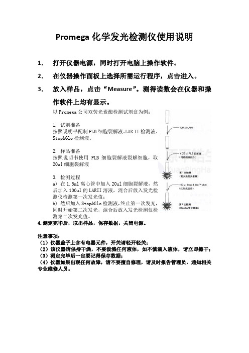
Promega化学发光检测仪使用说明
1.打开仪器电源,同时打开电脑上操作软件。
2.在仪器操作面板上选择所需运行程序,点击进入。
3.放入样品,点击“Measure”。
测得读数会在仪器和操作软件上均有显示。
以Promega公司双荧光素酶检测试剂盒为例:
1. 试剂准备
按照说明书配制PLB细胞裂解液、LAR II检测液、
Stop&Glo检测液。
2. 样品准备
按照说明书使用PLB细胞裂解液裂解细胞,取
20ul细胞裂解液
3. 检测过程
a) 在1.5ml离心管中加入20ul细胞裂解液,然
后加入100ul的LARII溶液,混合后放入发光检
测仪检测第一次发光值;
b) 然后加入Stop&Glo检测液,终止第一次发光,
同时开始第二次发光,混合后放入发光检测仪检
测第二次发光值。
4.测定完毕后,取出样品,保存数据,关闭电源。
注意事项:
(1)仪器盖子上含有电器元件,开关请轻开轻关;
(2)该仪器请保持干燥,不要泼溅任何液体,如不慎滴入液体,请立即擦干;(3)测定完毕后一定要记得保存数据;
(4)仪器如果出现任何故障,请不要擅自修理,请及时报告管理员,通知相关专业维修人员。
全自动化学发光免疫分析仪使用流程
全自动化学发光免疫分析仪使用流程下载提示:该文档是本店铺精心编制而成的,希望大家下载后,能够帮助大家解决实际问题。
文档下载后可定制修改,请根据实际需要进行调整和使用,谢谢!本店铺为大家提供各种类型的实用资料,如教育随笔、日记赏析、句子摘抄、古诗大全、经典美文、话题作文、工作总结、词语解析、文案摘录、其他资料等等,想了解不同资料格式和写法,敬请关注!Download tips: This document is carefully compiled by this editor. I hope that after you download it, it can help you solve practical problems. The document can be customized and modified after downloading, please adjust and use it according to actual needs, thank you! In addition, this shop provides you with various types of practical materials, such as educational essays, diary appreciation, sentence excerpts, ancient poems, classic articles, topic composition, work summary, word parsing, copy excerpts, other materials and so on, want to know different data formats and writing methods, please pay attention!I. 准备工作。
在开始使用全自动化学发光免疫分析仪之前,确保做好以下准备工作:1. 样本准备。
- 1、下载文档前请自行甄别文档内容的完整性,平台不提供额外的编辑、内容补充、找答案等附加服务。
- 2、"仅部分预览"的文档,不可在线预览部分如存在完整性等问题,可反馈申请退款(可完整预览的文档不适用该条件!)。
- 3、如文档侵犯您的权益,请联系客服反馈,我们会尽快为您处理(人工客服工作时间:9:00-18:30)。
化学发光分析仪使用说明书
一、产品概述
本产品是一种化学发光分析仪,用于定量测量样品中的特定分析物。
该分析仪基于化学发光原理,通过测量样品产生的光信号强度来确定
目标分析物的浓度。
本使用说明书将详细介绍该仪器的使用方法和操
作步骤,以帮助用户正确操作并获得准确的分析结果。
二、安全警告
使用本化学发光分析仪前,请务必阅读本章节内容并严格遵守以下
安全要求,以确保操作人员和仪器安全:
1. 仅限专业人士操作:本仪器仅适用于熟悉化学实验操作且具备相
关知识的专业人士。
2. 电源要求:请使用指定电源提供商提供的电源,并确保电源稳定
可靠。
3. 防护措施:在操作过程中,请佩戴防护手套、护目镜等必要的个
人防护装备。
4. 样品处理:遵循各种样品处理和废物处理的标准操作规程。
5. 化学品储存:储存和处理化学试剂时,请严格遵守安全操作规程。
三、仪器组成
本化学发光分析仪主要包括以下组成部分:
1. 主机:包含仪器的核心控制系统和显示屏,用于显示分析结果和操作界面。
2. 试剂仓:用于存放化学试剂和控制试剂加入的时间和量。
3. 样品槽:用于接收待测样品。
4. 光学系统:用于测量样品产生的发光信号。
5. 数据处理模块:用于对测量数据进行处理和存储。
四、使用步骤
1. 准备工作:
a. 将仪器放置在平稳的工作台面上,并接通电源。
b. 打开仪器主机,待系统自检完成后,进入操作界面。
2. 样品处理:
a. 打开样品槽盖,将待测样品倒入样品槽中。
b. 根据操作要求,在试剂仓加入必要的化学试剂。
c. 关闭样品槽盖,确保样品和试剂处于封闭状态。
3. 测量操作:
a. 在操作界面选择待测分析物和相应的测量方法。
b. 设置测量参数,如测量时间、波长等。
c. 点击“开始测量”按钮,仪器将自动开始测量过程。
d. 测量完成后,记录测量结果。
4. 数据处理与导出:
a. 在操作界面选择数据处理功能。
b. 根据需要选择数据处理方法,如拟合曲线、计算浓度等。
c. 完成数据处理后,选择导出数据的格式和路径。
五、故障排除
1. 仪器显示异常:如果仪器显示屏出现异常或无法正常操作,请检查电源连接和电源稳定性。
2. 测量结果异常:如果测量结果异常,请检查样品槽和试剂仓是否封闭良好,并确保样品和试剂的质量和配比正确。
3. 其他故障:如遇到其他无法解决的故障,请及时联系售后服务人员。
六、维护与保养
1. 定期校准:为保证测量结果的准确性,请定期进行仪器校准,并根据校准结果调整仪器参数。
2. 仪器清洁:及时清除样品槽和试剂仓内的残留物,保持仪器的清洁和正常运行。
3. 安全存放:长时间不使用仪器时,请将其存放在干燥、防尘的环境中,避免损坏和污染。
七、技术支持和售后服务
如需技术支持或售后服务,请联系售后服务热线:XXX-XXXX。
八、免责声明
本化学发光分析仪仅适用于科学研究和实验室使用,不得用于其他任何用途。
用户在使用本仪器时,应遵循相关法律法规并承担相应责任。
九、版权和知识产权
本化学发光分析仪使用说明书受版权保护,未经许可,不得以任何形式进行复制、传播和修改。
通过本使用说明书,您应对化学发光分析仪的使用和操作有了更清晰的了解。
请您务必按照本说明书的要求正确操作,以确保仪器的安全和测量结果的准确性。
如有任何疑问或需要进一步的帮助,请联系我们的技术支持和售后服务团队。
祝您使用愉快!。