不同神经移植体移植修复自体周围神经缺损时的再生效果比较
多种移植体修复周围神经的比较实验研究

多种移植体修复周围神经的比较实验研究
卫晓恩;韩西城
【期刊名称】《中国修复重建外科杂志》
【年(卷),期】1996(10)1
【摘要】修复神经缺损的材料甚多,但至今尚未见各种材料的比较研究。
为了比较自体神经、肌肉、静脉、肌腱及硅橡胶修复神经缺损的效果,选用SD大白鼠,切断腓总神经,制成0.6cm缺损,分别用不同移植物桥接缺损。
于术后6周、12周在电生理、胫前肌称重、远端轴突计数及组织学等方面进行综合分析评价。
结果表明:自体神经移植在各方面均优于其它移植体,而静脉组又优于另外3组。
对各种移植体中神经再生的特点及其成熟过程进行了讨论,并探讨了形成这种差别的有关神经再生微环境的影响。
【总页数】4页(P12-15)
【关键词】周围神经缺损;自体神经移植
【作者】卫晓恩;韩西城
【作者单位】山西省人民医院创伤显微外科;山西医学院第一附属医院骨科
【正文语种】中文
【中图分类】R651.305
【相关文献】
1.骨骼肌桥接与自体神经移植修复周围神经缺损的比较研究 [J], 金国华;胡迎青
2.一种新的修复周围神经损伤的移植体—动物实验研究 [J], 翁文杰;石桂秀
3.不同神经移植体移植修复自体周围神经缺损时的再生效果比较 [J], 黄启顺;甘萌;郑怀远;周学武;周攀;陈振兵
4.多种移植体修复周围神经缺损的研究进展 [J], 文小泉;王志明
5.4种自体神经移植方法修复周围神经缺损比较的实验研究 [J], 袁伟东;付银环;田跃;仝吉庆;潘进社;张煜
因版权原因,仅展示原文概要,查看原文内容请购买。
游离神经移植修复周围神经缺损50例疗效分析

【 关键词 】 游离神经移植修复 ; 周围神经损伤 ; 治疗效果
A n a l y z e t h e E f i c a c y o f 5 0 C a s e s o f F r e e Ne r v e Gr a f t R e p a i r o f P e r i p h e r a l N e r v e I n j u r y / WA N G J i e , WA NG
Ha o,HUANG F e i ,e t id a / Me d i c a l I n n o v a t i o n o f Ch i n a,2 0 1 5 ,1 2( 1 5): 0 3 8 - 0 4 0
【 A b s t r a c t 】O b j e c t i v e : T o e x p l o r e t h e f r e e n e r v e g r a f t r e p a i r c l i n i c a l o u t c o me s i n p a t i e n t s w i t h p e i r p h e r a la t me n t ,t h e p a t i e n t s we r e d i v i d e d i n t o t wo g r o u p s, 2 5 c a s e s i n e a c h ro g u p. Th e c o nt r o l g r o u p wa s t r e a t e d b y a s i mp l e n e ve r a f t ,t h e e x p e ime r n t a l g r o u p Wa s i mp l e me n t e d t h e f r e e v se a u l a r i z e d n e ve r t r a n s pl a n t t r e a t me n t ,t he e f f e c t o f t wo ro g up s we r e c o mpa r e d. Re s ul t :92 . O % o f pa t i e n t s i n t h e e x pe r i me n t a l ro g u p a f f i r me d r e h a b i l i t a t i o n p r o g r a m e v a l u a t i o n.
不同神经导管在周围神经损伤修复中的应用
基于以上原因,人们一直以来都在不断探索并寻找能够替代神经移植的桥接物。早在1882年,Gluck就尝试使用一个中空的骨管用于修复狗30毫米的神经缺损。1898年,Forssman提出的神经趋化生长学说,为神经导管的应用提供了理论基础。20世纪80年代,Lundborg和他的同事们用著名的Y形硅胶管套接大鼠周围神经缺损的实验证实了神经再生具有趋化特性,并提出了神经再生室模型,神经导管的研究开始进入了系统化和深入化的阶段。如今,使用神经导管来替代神经移植修复神经缺损已经被临床认可;其优点包括减少成纤维细胞的浸润、减轻神经瘤与瘢痕形成、有利于神经营养因子的积聚,并没有相关的供区并发症等。理想的神经导管材料应满足神经细胞生长的基本要求,要有特定的三维结构支架,可接纳再生轴突的长入,对轴突起机械引导作用;应有适当的孔径和孔隙率,利于导管内外物质交换;具有可降解性及降解速率可调控性,使其降解速度与神经再生同步;具有良好的组织相容性和无毒性[1],和合适的细胞相容性等特征,有利于雪旺细胞的粘附、增殖、分化;导管要有一定的机械强度和柔韧性,以抵抗周围组织的压迫及适应一定的活动范围,同时易于加工及消毒等。
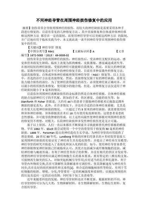
人去细胞异体神经一期移植重建手指神经缺损
人去细胞异体神经一期移植重建手指神经缺损尽管周围神经损伤伴缺损的治疗金标准仍然是自体神经移植修复,但是近些年来各种各样的人工生物套管以及同种异体神经在神经缺损修复当中扮演着越来越重要的角色,其中最具潜力替代自体神经的移植物是人脱细胞神经。
与传统方法相比,人去细胞神经不存在任何供体受区的损伤,且来源广泛。
化学去细胞处理后的异体神经既保留了原有的结构又降低了免疫源性,且术后无需服药。
尽管同种异体神经临床的使用还处于初期阶段,但修复指神经缺损的临床效果都较为确定。
在既往的研究当中,神经移植重建都在损伤后1周或数周以后进行,主要考虑到伤口稳定以及减少术后炎症的发生比率。
缺点就是患者需要进行2次手术,增加了住院和康复时间。
来自中国宁波市第六医院的李学渊所在团队首次将人体去细胞异体神经应用于手外伤急诊手术修复。
针对急诊15例患者的18指神经损伤伴缺损,彻底清创后,以人体去细胞神经移植重建指神经。
重建后随访6-24个月。
静态两点辨别觉结果优良率为89%;轻触觉明显改善率为78%。
提示人体去细胞神经移植一期重建手外伤指神经缺损临床操作可行,为缺损周围神经的重建提供了新趋向。
相关文献发表于《中国神经再生研究(英文版)》杂志2015年1月第1期。
以人体去细胞神经移植重建指神经损伤伴缺损Article: " One-stage human acellular nerve allograft reconstruction for digital nerve defects," by Xue-yuan Li1, Hao-liang Hu1, Jian-rong Fei1, Xin Wang1, Tian-bing Wang2, Pei-xun Zhang2, Hong Chen1 (1 Department of Hand Surgery, Ningbo No.6 Hospital, Ningbo, Zhejiang Province, China; 2 Department of Trauma and Orthopedics, Peking University People’s Hospital, Beijing, China)Li XY, Hu HL, Fei JR, Wang X, Wang TB, Zhang PX, Chen H (2015) One-stage human acellular nerve allograft reconstruction for digital nerve defects. Neural Regen Res 10(1):95-98.欲获更多资讯:Neural Regen ResOne-stage human acellular nerve allograft reconstruction for digital nerve defectsAutologous nerve graft repair has become a gold standard for the treatment of peripheral nerve injury combined with defects. However, a variety of biological conduits and nerve allografts have played increasingly important roles in the repair of nerve defects. Human acellular nerves have become the most promising substitute for autologous nerve grafts.Compared with traditional methods, human acellular nerves do not induce damage to affected areas of the donor, and there is a wide variety of sources. With developments in medical technology, nerve allografts after acellular treatment have been shown to retain the original structure and reduce immunogenicity. Moreover, patients do not have to take medicine following surgery. The clinical use of nerve allografts is still in the early stages, but studies have shown positive clincial repair outcomes of digital nerve defects.In previous studies, nerve graft reconstruction was conducted at 1 week or several weeks after injury, resulting in wound stability and reduced incidence rate of postoperative inflammation. The disadvantage of this method is that patients require two surgeries, which increased hospitalization and recovery time.Xue-yuan Li, Ningbo No.6 Hospital, China and his colleagues first used human acellular nerve allograft to reconstruct < 5-cm digital nerve defects in 18 digits of 15 patients with nerve injury from the emergency department. The patients were followed up for 6–24 months after reconstruction. Mackinnon-Dellon static two-point discrimination results showed excellent and good rates of 89%. Semmes-Weinstein monofilament test demonstrated that light touch was normal, with an obvious improvement rate of 78%. These findings confirmed that human acellular nerve allograft for one-stage reconstruction of digital nerve defect after hand injury is feasible, which provides a novel trend for peripheral nerve reconstruction. The relevant article was published in the Neural Regeneration Research (Vol. 10, No. 1, 2015).Reconstruction of nerve injury accompanied by defects using human accellular nerve allograft.Article: " One-stage human acellular nerve allograft reconstruction for digital nerve defects," by Xue-yuan Li1, Hao-liang Hu1, Jian-rong Fei1, Xin Wang1, Tian-bing Wang2, Pei-xun Zhang2, Hong Chen1 (1 Department of Hand Surgery, Ningbo No.6 Hospital, Ningbo, Zhejiang Province, China; 2 Department of Trauma and Orthopedics, Peking University People’s Hospital, Beijing, China)Li XY, Hu HL, Fei JR, Wang X, Wang TB, Zhang PX, Chen H (2015) One-stage human acellular nerve allograft reconstruction for digital nerve defects. Neural Regen Res 10(1):95-98.。
原位神经移植与自体静脉小间隙套接修复周围神经损伤的疗效比较

原位神经移植与自体静脉小间隙套接修复周围神经损伤的疗效比较张文龙;董乐乐;崔成立【期刊名称】《内蒙古医学杂志》【年(卷),期】2012(044)011【摘要】目的:观测周围神经损伤后应用原位神经移植与自体静脉小间隙套接修复方法的疗效比较,为临床治疗周围神经损伤选择合适的修复方法提供实验基础.方法:将45只Wistar大鼠的左右后肢共90侧随机分为3组,每组30侧.A组:将股神经切断3 mm作为移植段,将移植段与神经远近两断端行原位外膜吻合.V组:将股神经切断3 mm,同时切取长约5~7 mm的同侧股静脉,将神经两断端送入游离的静脉两端,断端之间留一3 mm的间隙,将神经外膜与静脉壁缝合.N组:对照组.术后16周,对股神经进行电生理及组织学检测.结果:再生神经纤维均可通过A组吻合口和V 组的套接间隙,A、V两组神经传导速度及股四头肌张力与N组比较差异均无显著性(P>0.05),组间差异也无显著性(P>0.05).结论:应用神经原位移植、自体静脉小间隙套接均可以修复周围神经损伤,且静脉小间隙套接修复可获得与神经原位移植相似的修复效果.【总页数】4页(P1281-1283,封4)【作者】张文龙;董乐乐;崔成立【作者单位】包头医学院第一附属医院骨科,内蒙古包头 014010;包头医学院第一附属医院骨科,内蒙古包头 014010;包头医学院解剖教研室,内蒙古包头014010【正文语种】中文【中图分类】R322.85【相关文献】1.神经碎片联合NGF在自体神经外膜小间隙修复周围神经损伤 [J], 马华;2.神经碎片联合NGF在自体神经外膜小间隙修复周围神经损伤 [J], 马华3.可降解生物套管小间隙套接法修复周围神经损伤的临床观察 [J], 张培训;卢浩;殷晓峰;白露;王艳华;安帅;张殿英;付中国;姜保国;寇玉辉;韩娜;党育;薛峰;王天兵;徐海林;陈建海;杨明4.替代神经外膜缝合─小间隙套接法修复周围神经损伤 [J], 姜保国;李剑5.小间隙吻合静脉套接加生物蛋白胶修复周围神经损伤58例 [J], 邓忠虎;王玉禄;周凤莲因版权原因,仅展示原文概要,查看原文内容请购买。
组织工程神经修复周围神经缺损过程中髓鞘再生的研究

BRG1;术后3w,Auto组CAMK2B与施万细胞有较好定位。
8. 相关基因的实时定量PCR检测结果显示,基因随时间变化的表达趋势与基因芯片检测结果基本一致。
结论:1. 周围神经缺损经不同类型的移植物桥接修复后,髓鞘再生过程比较相似,均需经历施万细胞去分化,增殖、迁移引导轴突再生,施万细胞分化、包绕轴突重新形成髓鞘,但人工移植物修复时,髓鞘再生还需经历“板层压缩”的过程。
2. 自体神经修复时,髓鞘再生进程要明显快于人工移植物修复;丝素支架可引导施万细胞迁移,支持细胞SKP-SCs在髓鞘再生过程中,可以促进施万细胞迁移和引导轴突再生。
3. 髓鞘再生过程中参与基因网络构成的分子,总体来说各组差别不大,但由于髓鞘再生进程的差异,不同组之间具体分子的作用时相不一样。
关键词:周围神经损伤,组织工程神经,施万细胞,髓鞘再生,基因网络分析作者:王亚先指导教师:顾晓松教授顾芸副教授Remyelination during the Repair of Peripheral Nerve Defects Using Tissue Engineered NervesAbstractObjective:The present study was aimed to investigate the basic biological process of remyelination and the potential gene network underlying the complex biological process after the repair of rat sciatic nerve defect with different types of nerve grafts. We sought to provide new content for the remyelination study during peripheral nerve regeneration.Methods:1. Construction of tissue engineered nerve graft (TENG) in vitro. The artificial tissue engineered nerve was constructed in a perfused rotatory cell culture system (RCCS), and finally, to form a trinity structure composed of chitosan conduit, silk fibroin fibers and rat skin-derived precursor Schwann cells (SKP-SCs).2. Preparation of rat sciatic nerve defect/repair model. The rats sciatic nerve defect, measuring 10mm in length, were bridged by chitosan/silk fibroin-based scaffold (Sca group ), TENG (TENG group) and autologous nerve graft (Auto group), respectively. The experiment was divided into 8 time points, including 1d, 4d, 1w, 2w, 3w, 4w, 8w, 12w, in addition, a sham operation group was set as control at each time point (Sham group).3. Morphological observation of remyelination. The bridge segments in 3 groups (Sca, TENG and Auto) were harvested at 1d, 4d, 1w, 2w and 3w after operation. The biological process of remyelination was respectively observed by optical microscope and transmission electron microscope.4. Microarry detection and bioinformatics analysis. The graft segments together with both nerve stumps (2mm) were collected at 1d, 4d, 7d, 1w, 2w, 3w, 4w, 8w, 12w post-surgery and nerve segment of sham group in the corresponding position, respectively. Microarray data analysis was performed after RNA extraction and microarray detection, with the aid of several bioinformatics analysis software, i.e. MeV (MultiExperiment Viewer), PCA (Principal Components Analysis), IPA (Ingenuity Pathway Analysis) and Venn diagram. The expression of the target genes were verified by immunohistochemistry and quantitative real-time PCR.Results:1. One day post-surgery, there were few Schwann cells showing a proliferating state. Nerve degeneration was limited at the broken edge of the proximal and distal nerve stumps in all 3 groups. In the Auto group, the morphology of myelin sheath of graft segment was close to that of the normal, but the structure of myelin sheath was slightly loose, and the intra-axonal granules disappeared.2. Four days post-surgery, a large number of Schwann cells in the proximal and distal nerve stumps showed active proliferation condition in all 3 groups, and a few of SKP-SCs in the TENG group as well. A small amount of Schwann cells migrated from the proximal nerve stumps in the TENG and Auto groups. Furthermore, nerve degeneration of graft segment in the Auto group was remarkable at this stage.3. One week post-surgery, more Schwann cells migrated from the proximal and distal nerve stumps, besides, the regenerating axons extended from the proximal nerve stump along Schwann cells toward the distal stump in all 3 groups. Schwann cells did not yet wrap around the regenerating axons in the Sca and TENG groups. Degeneration peaked at this time in the Auto group, while a few axons were wrapped by Schwann cells. Supplementary experiment of the TENG group showed that 10 days post-surgery, there was well co-located between the SKP-SCs and regenerating axons in the proximal stump.4. Two weeks post-surgery, a poorly characterized tissue bridge forms, connecting the proximal and distal stumps in the Sca and TENG groups. The number of migrating Schwann cells increased significantly in both groups, and axons from proximal stump continued to regenerate along the cell bridge. The distal Schwann cells in TENG group contained suspected cell debris of SKP-SCs. Compared with the Sca and TENG groups, regenerating axons in Auto group had already grown to a long distance through its original structure. A small amount of axons were wrapped by Schwann cells, without lamellar structure formation in the Sca and TENG groups. Degeneration came to a close in the Auto group, and a large number of axons were wrapped by Schwann cells arranging in fascicles, in which some had formed a thin myelin sheath.5. Three weeks post-surgery, Schwann cells migrated from the proximal and distal nerve stumps gathered together in the middle of the lumen. A small amount of regenerating axons passed through the bridge into the distal nerve stump in the TENG group while a large number of regenerating axons crossed the bridge into the distal stump in the Autogroup. In the Sca and TENG groups, there was a large number of loosely structured myelin sheath, by contrast, myelin sheath possessed dense structure in the Auto group.6. Microarray data clustering analysis fitted perfectly the biological process of remyelination. Gene network analysis combined with the Venn diagram showed that there was little difference about molecular involved in the network among the three groups.7. The immunohistochemical staining showed that JNK translocated into the Schwann cell nucleus 1 day post-surgery in the proximal nerve stumps in all 3 groups, RAC1 and BRG1 showed well co-located with Schwann cells 2 weeks post-surgery in all 3 groups, BRG1 also showed well co-located with SKP-SCs 10 days post-surgery in the TENG group, CAMK2B displayed well co-located with Schwann cells 3 weeks post-surgery in the Auto group.8. The quantitative real-time PCR detection of the related genes involved in remyelination showed that the expression pattern of genes was similar to the microarray results.Conclusions:1. The biological process of remyelination during peripheral nerve regeneration was similar among different groups, i.e. dedifferentiation of Schwann cells, proliferation and migration of Schwann cells, differentiation and myelination of Schwann cells and so on, while the regenerating myelin sheath needed to undergo the process of lamellar compaction when repaired by the artificial grafts.2. The result of remyelination in Auto group was much better than that in Sca or TENG group within 3weeks post-surgery. SKP-SCs as the supporting cells could promote Schwann cells migration and guide axon regeneration.3. Genes involved in the network displayed little difference among the three groups, but the action phase of the specific gene i.e. initiation and termination was significantly different due to the different regeneration rate of remyelination.Keywords:peripheral nerve injury, tissue engineered nerve graft, Schwann cells, remyelination, gene network analysisWritten by: Yaxian WangSupervised by: Prof. Xiaosong GuA/Prof. Yun Gu目录前言 (1)参考文献 (4)材料与方法 (7)结果 (14)讨论 (67)结论 (73)参考文献 (73)综述 (78)参考文献 (87)英文缩略词表 (96)博士生期间发表科研论文及学术会议情况 (98)博士生期间参加科研项目 (100)致谢 (101)前言髓鞘是神经系统的重要组成部分,为高等脊椎动物所特有,中枢与周围神经系统中,髓鞘分别是由少突胶质细胞(Oligodendrocytes)及施万细胞(Schwann cells)的质膜反复缠绕神经元轴突所形成的一层脂类膜结构,其主要成分为髓磷脂,故又称髓磷脂鞘[1]。
不同自体皮神经移植修复神经缺损后S-100蛋白的表达

・
12 l 6
医药 杂志 2 1 00年 l 2月 第 2 7卷 第 1 2期来自Pa rcJMe d&
不 同 自体皮神 经移植修复神经缺损后 S 10 白的表达 一0 蛋
魏 国兴 , 牛 军 , 文生 , 付 郭广 惠 , 永庆 常
【 摘 要】 目的 探讨 不 同结 构 皮 神 经移 植 修 复 神 经 缺损 的再 生 效 果 。 法 方 新西兰兔 2 7只 , 随机 分 为 A、 C B、
,
te h PL A, Xixin , He n 453 00 Chi n ag na 0 , na
【 bta t Obet e T net a h f c o asl tt n wt d f e tctnos nre o nua A s c】 r jci o ivsgt te e et ft npa a o i ie n uaeu evsf erl v i e f r n i h fr r
不同神经移植体移植修复自体周围神经缺损时的再生效果比较
2 0 1 3年 6月
华 中科 技 大 学 学 报 ( 医学版)
Ac t a Me d Un i v S c i Te c hn o l Hu a z h o n g
V0 1 . 4 2 No . 3 P . 29 0
肠肌湿重 , 术后 2 、 4 周 时 3组 间 差 异 无 统 计 学 意 义 ; 术后 8 周 时 运 动 神 经 移 植 组 和 混 合 神 经 移 植 组 重 于 感 觉 神 经 移 植 组
( 均 P <O . 0 5 ) 。术 后 8周 时 , 在运 动神 经移 植 组 和混 合 神 经 移 植 组 中可 见 大 量 神 经 纤 维 生 长 , 神 经纤维 的数量 、 轴 突 的 密度均大于感觉神经移植组 ( 均 P <O . 0 5 ) ; 再 生 神 经 轴 突 在 运 动 神 经 移 植 组 更 加 成 熟 。结 论 不 同 的 神 经 移 植 材 料 对 自体 神 经 移 植 再 生 效 果 有 着 显 著 影 响 运 动 神 经 及 混 合 神 经 移 植 修 复 自体周 围神 经缺 损 时 的再 生效 果 优 于感 觉 神 经 移
J un . 2 O1 3
不 同神经移植体移植修 复 自体周 围神 经缺损 时的 再 生 效果 比较 *
黄启 顺 , 甘 萌 , 郑怀远 , 周 学武 , 周 攀, 陈振 兵
华 中科 技 大学 同济 医学 院 附属 协 和 医 院手 外 科 , 武汉 4 3 0 0 2 2
Sc i e n c e a nd Te c h no l o gy , Wu han 43 00 2 2, Chi na
Ab s t r a c t 0b j e c t i v e To c o mp a r e e f f e c t s o f d i f f e r e n t a u t o l o g o u s n e r v e g r a f t s o n t h e r e g e n e r a t i o n o f t h e p e r i p h e r a l mi x e d
- 1、下载文档前请自行甄别文档内容的完整性,平台不提供额外的编辑、内容补充、找答案等附加服务。
- 2、"仅部分预览"的文档,不可在线预览部分如存在完整性等问题,可反馈申请退款(可完整预览的文档不适用该条件!)。
- 3、如文档侵犯您的权益,请联系客服反馈,我们会尽快为您处理(人工客服工作时间:9:00-18:30)。
不同神经移植体移植修复自体周围神经缺损时的再生效果比较黄启顺;甘萌;郑怀远;周学武;周攀;陈振兵【摘要】目的比较不同自体神经移植材料在移植修复周围混合神经缺损时的再生效果.方法取45只SD大鼠,随机分成3组,分别切除0.5 cm右侧坐骨神经,建立神经缺损模型.运动神经移植组:取左侧后肢的股神经运动支移植修复缺损的坐骨神经;感觉神经移植组:取左侧后肢的腓肠神经移植修复缺损的坐骨神经;混合神经移植组:取左侧后肢的坐骨神经移植修复.分别于术后2、4和8周,采用神经夹捏实验测量神经再生长度,处死动物后取下术侧腓肠肌称取肌肉湿重.术后8周取胫神经远侧吻合口远端3 mm神经组织行甲苯胺蓝染色,同时电镜下观察神经纤维的直径及髓鞘的厚度,采用图像分析软件进行图片分析,计数神经纤维总数并计算神经纤维的密度.结果神经再生长度,术后2周时3组间差异无统计学意义;术后4、8周时运动神经移植组及混合神经移植组大于感觉神经移植组(均P<0.05).腓肠肌湿重,术后2、4周时3组间差异无统计学意义;术后8周时运动神经移植组和混合神经移植组重于感觉神经移植组(均P<0.05).术后8周时,在运动神经移植组和混合神经移植组中可见大量神经纤维生长,神经纤维的数量、轴突的密度均大于感觉神经移植组(均P<0.05);再生神经轴突在运动神经移植组更加成熟.结论不同的神经移植材料对自体神经移植再生效果有着显著影响.运动神经及混合神经移植修复自体周围神经缺损时的再生效果优于感觉神经移植.%Objective To compare effects of different autologous nerve grafts on the regeneration of the peripheral mixed nerve. Methods Nerve defect models were established by removing 0. 5 cm right sciatic nerve in SD rats (n= 45 ). Then ,the animals received motor ,sensory or mixed nerve grafts ,which originated from the femoral nerve ,sural nerve and sciatic nerve of the contralateral hind limb. At 2 ,4 and 8 weeks postoperation ,the nerve pinch test was performed to measure the length of the regenerated nerve ,and the wet weight of the gastrocnemius muscle of the operated limb was measured. The tissues (3 mm in length) of the tibial nerve distal to the anastomosis were stained by toluidine bluestaining ,and the diameter of the nerve fibers and the thickness of the myelin were determined by electron microscopy on week 8 after operation. Image analysis software was used to analyze the pictures ,and the total number of nerve fibers was counted and the density of nerve fibers calculated. Results Histomorphometry of the regenerated nerves at 8 weeks demonstrated robust nerve regeneration in both motor and mixed nerve graft groups. In contrast ,poor nerve regeneration was observed in sensory nerve graft group ,as evidenced by significantly decreased nerve fiber counts and nerve density when compared with mixed and motor groups (P<0. 05 ). Moreover ,the length of the regenerated nerve in the motor and the mixed nerve graft groups were greater than that in the sensory nerve graft group at 4 and 8 weeks post operation (P<0. 05 ). The wet weight of the gastrocnemius muscle in the motor and the mixed nerve graft groups were greater than that in the sensory nerve graft group at 8 weeks post operation (P<0. 05 ). Conclusion Motor or mixed nervegrafts ,rather than sensory nerve grafts ,contribute to the regeneration of the peripheral mixed nerve.【期刊名称】《华中科技大学学报(医学版)》【年(卷),期】2013(042)003【总页数】4页(P290-293)【关键词】周围神经;自体神经移植;运动神经;感觉神经【作者】黄启顺;甘萌;郑怀远;周学武;周攀;陈振兵【作者单位】华中科技大学同济医学院附属协和医院手外科,武汉,430022;华中科技大学同济医学院附属协和医院手外科,武汉,430022;华中科技大学同济医学院附属协和医院手外科,武汉,430022;华中科技大学同济医学院附属协和医院手外科,武汉,430022;华中科技大学同济医学院附属协和医院手外科,武汉,430022;华中科技大学同济医学院附属协和医院手外科,武汉,430022【正文语种】中文【中图分类】R651.3目前,对于周围神经损伤后缺损的修复,除了改变神经走行方向、屈曲其所跨过的关节以弥补较短的神经缺损外,大部分还需要用移植的办法进行解决。
神经移植方法甚多,有自体、异体、异种神经移植,非神经组织移植和人工神经移植等[1-2]。
在这些方法之中,无张力的自体神经移植仍然是神经缺损修复最主要的方法,其余的都存在着或多或少的、这样或那样的问题。
周围神经缺损是一种混合神经缺损,采用自体神经移植修复时常用腓肠神经或桡神经浅支,而这二者均为感觉神经,在结构及超微结构上与运动神经纤维有所区别。
临床中也发现,用废弃的混合神经移植修复神经缺损,术后神经恢复效果比用单纯感觉神经移植修复的效果好,其中神经移植体结构可能是影响再生效果的一个重要方面。
为此,我们建立了大鼠坐骨神经缺损模型,并采用自体不同类型神经移植体修复,以研究不同类型神经移植体的结构差异对混合神经缺损修复后再生效果的影响。
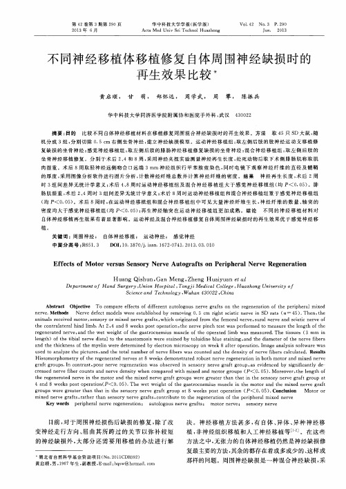
1 材料与方法1.1 动物模型及分组清洁级3周龄SD大鼠45只(华中科技大学同济医学院实验动物学部提供,编号:13314),随机分为3组,每组15只。
将右侧坐骨神经自梨状肌下缘0.5cm处切断,并将远端切除0.5cm,制成神经缺损模型。
运动神经移植组:取左侧后肢的股神经运动支约1.5cm长,重叠3股,移植修复;感觉神经移植组:取左侧后肢的腓肠神经桥接1.5cm,重叠3股,移植修复;混合神经移植组:取左侧后肢同部位坐骨神经0.5cm移植修复。
见图1。
术后动物分笼饲养。
1.2 神经再生长度检测于术后2、4、8周分别从各组中随机抽取5只动物,1%戊巴比妥(50mg/kg)腹腔内注射麻醉,暴露出右侧坐骨神经移植修复段,观察神经大体生长情况。
用显微外科镊以1mm的间隔由远而近夹捏神经,标记出最早出现腓肠肌收缩反应的部位,测量此点与近端吻合口之间的距离,此即运动神经最大的再生长度,以此作为神经再生速度的观察指标[3]。
图1 坐骨神经缺损的神经移植修复手术Fig.1 Nerve transplantation for the repair of sciatic nerve defect1.3 肌肉湿重检测于术后2、4、8周将各组进行神经再生长度检测后的动物处死,将腓肠肌自股骨内外髁起点至跟骨结节止点完整取下,用测量仪准确称取肌肉湿重。
1.4 组织形态学测定术后第8周,神经再生长度测定后将动物处死,取胫神经远侧吻合口远端3mm神经组织,将取得的神经样本用2.5% 戊二醛溶液前固定,饿酸固定液后固定,梯度浓度乙醇脱水,环氧树脂618包埋,切片厚度为1μm,甲苯胺蓝染色,在光学显微镜下观察再生神经纤维总数和华勒氏变性的程度。
制备超薄切片,枸橼酸铅及醋酸双氧铀染色,电镜下测量神经纤维的直径及髓鞘的厚度。
所有镜下组织图像采用形态测定软件(Leco Inst-ruments,St.Joseph,MI)进行分析。
在放大1 000倍镜下,每根神经随机选择4个区域进行分析,镜下计数神经纤维的数目并测出神经束的横截面积,神经纤维的总数/神经束面积即为神经纤维密度(根/单位面积)。
1.5 统计学分析实验所得数据用均数±标准差(±s)表示,采用SPSS 12.0统计软件对数据进行分析,组间计量资料比较采用t检验,以P<0.05为差异有统计学意义。
2 结果2.1 神经再生长度术后2周,各实验组神经再生长度差异无统计学意义。
术后4、8周,运动神经移植组及混合神经移植组神经再生长度差异无统计学意义,但均大于感觉神经移植组,差异均有统计学意义(均P<0.05),见表1。
2.2 肌肉湿重术后2、4周时3组间腓肠肌湿重差异无统计学意义。
术后8周时,运动神经移植组及混合神经移植组间腓肠肌湿重差异无统计学意义,但均大于感觉神经移植组,差异有统计学意义(均P<0.05),见表2。