GFP绿色荧光蛋白在水稻中的表达
GFP及其在植物分子生物学研究中的应用
[%] 多管水母中 "#$ 生色团的化学结构和附近序列
通过调整光圈减少进光量, 或选择适合的滤光片就 可以消除背景, 从而检测出 "#$ 荧光。所以检测 "#$ 时一般不会出现假阳性结果。如要观察组织 内部 "#$ 荧光, 又不损伤材料, 可以使用激光扫描 共聚焦显微镜 ( 4E+FE4*7 7*@6/ @4*++9+, 094/E@4EGH) , 它 可以对组织内部进行分层扫描, 也可进行三维显 示, 是目前理想的荧光检测仪器。另外, 荧光激活 的细胞分选仪 (也叫流式细胞仪) 也可检出转化细
光的 "#$, 之后, "#$ 越来越受到人们的广泛关注。 由于野生型 "#$ 具有一定的缺点, 如用蓝光激发 时, 其荧光强度较低, "#$ 合成及折叠产生荧光的 过程慢, 蛋白质折叠受温度影响大, 表达量较低等,
[!!] 限制了 "#$ 的应用。!’’- 年, 采用定点 L690 等
突变方式, 把第 (- 位丝氨酸换为苏氨酸 ( 1(-2) 或 胱氨酸 ( 1(-?) , 生色团形成速度比野生型 "#$ 快 & 倍, 荧光强度提高了 & M ( 倍, 而且激发光谱和发射 光谱红移, 与 N6+977* "#$ 相似。此后, 人们根据不 同的研究需要, 不断地对 "#$ 进行改造, 以改善其 性能。表 ! 是在植物基因工程中最常用的 "#$ 变 种。 # "! &’( 的毒性 人们曾一度认为 "#$ 对植物 [!%] 细胞有毒。L*@67EFF 和 C0E@ 报道, 转 "#$ 基因拟 南芥的愈伤组织不能分化成再生植株, 推测绿色荧 光的光子干扰会产生自由基, 对细胞核造成氧化损 伤。但许多研究人员并没有观测到 "#$ 对植物细 胞明显的毒性, 认为 "#$ 对植物细胞有毒的证据不 足。动物和植物的适应性不同, 植物的形态、 生理 特征使其能适应光线, 而动物细胞可能会受到灼
绿色荧光蛋白作为报告基因在分子生物学中的应用
绿色荧光蛋白作为报告基因在分子生物学中的应用绿色荧光蛋白作为报告基因在分子生物学中的应用摘要:随着科学技术的不断更新和发展,绿色荧光蛋白在动物学、植物学、微生物学等领域的应用研究越来越广泛。
绿色荧光蛋白(green fluorescent protein,GFP)可作为报告基因,且具有分子量较小、荧光性质稳定、对生物体无毒性作用、检测时不需要底物等的特点。
本文就对荧光蛋白在分子生物学中的应用做一综述。
关键词:绿色荧光蛋白;报告基因;应用The Application of GFP As Reporter Gene In the Molecular Biology Abstract: With the upgrade and development of science and technology, the application of green fluorescent protein used in Zoology, Botany and microbiology is more extensive. As a reporter gene, GFP have some characteristics, such as low molecular weight, good fluorescent stability, non- toxicity to organisms. This paper reviews the application of GFP in the molecular biology. Key words: green fluorescent protein, reporter gene, application of GFP绿色荧光蛋白(green fluorescent protein,GFP)是一类来自于海洋生物如水母、水螅和珊瑚等腔肠动物内的一种生物发光蛋白,当受到紫外或蓝光激发时,能发射出绿色荧光。
GFP绿色荧光蛋白在水稻中的表达

绿色荧光蛋白基因在水稻基因转化中研究(续)
1.3 Quantitative assay of GFP values in transgenic rice plants(定量检测绿 ( 色荧光蛋白在转基因水稻植株中的表达值 蛋白在转基因水稻植株中的表达值) 色荧光蛋白在转基因水稻植株中的表达值) To detect GFP value in R0 transgenic rice plants, a quantitative assay was carried out. The second leaf from the top of each two-month-old transgenic rice plant was collected and immediately frozen in liquid nitrogen. The leaves were ground in liquid nitrogen and homogenized in ice - cold extraction buffer (100 mmol HEPES, pH 8.0, 10 mmol EDTA, 5 mmol DTT, 10% glycerol, 1% PVP, 25 fig mL-1 PMSF, 15 fig mL-1 leupeptin, 1% activated carbon). This ex-traction buffer was found by preliminary experiments to give minimum autofluorescence of rice leaf extractsr data not shown). After the first centrifugation (12 000 rpm for 15 min at 4B ) , the crude extract was centrifuged again (12 000 rpm for 20 min at 4B ) to further reduce insoluble particles which affect the quantification of fluorescence. Then the GFP values were quantified based on fluorescence units f g - 1 protein using a fluorometerr model SLM8000) after excitation at 385 nm and measure the emission maximum at 510 nm. Nontransformed tissues were used to estimate the autofluores-cence of the tissue. Protein concentration was determined according to Bradford (Bradford , 1976). Statistical analysis was carried out as described by Sokal and Rohlf (Sokal and Rohlf , 1969).
利用gfp报告基因优化农杆菌介导的水稻遗传转化
利用绿色荧光蛋白GFP研究稻瘟病菌与水稻的互作
利用绿色荧光蛋白GFP研究稻瘟病菌与水稻的互作作者:许有嫔廖海澄陈金华罗櫞宿加邹成东李伟滔王静马炳田贺闽陈学伟来源:《植物保护》2017年第06期摘要稻瘟病菌Magnaporthe oryzae严重威胁水稻的产量与质量,明确稻瘟病菌与水稻互作过程及机理,对防治稻瘟病具有重要意义。
本研究利用稻瘟病菌常用致病菌株GUY11和ZB25,构建了绿色荧光蛋白GFP的过量表达菌株,并通过荧光显微观察菌株侵染寄主水稻过程中侵染结构的形成与发育,包括孢子萌发、附着胞形成、侵染钉形成、侵染菌丝增殖、坏死斑形成及产孢。
另外,通过比较过量表达菌株对稻瘟病高抗水稻和易感水稻的侵染过程,发现侵染过程的差异主要集中于侵染钉的穿透和侵染菌丝的定殖。
本研究为分析稻瘟病菌对寄主水稻的定殖规律提供了一种有效工具。
关键词稻瘟病菌;绿色荧光蛋白;侵染过程;水稻中图分类号:S 435.111.41文献标识码:A DOI:10.3969/j.issn.05291542.2017.06.008Fluorescent microscopic analysis of the plant infection process ofMagnaporthe oryzae using green fluorescent proteinXu Youpin,Liao Haicheng,Chen Jinhua,Luo Yuan,Su Jia,Zou Chengdong,Li Weitao,Wang Jing,Ma Bingtian,He Min,Chen Xuewei(Collaborative Innovation Center for Hybrid Rice in Yangtze River Basin, State Key Laboratory ofHybrid Rice, Key Laboratory of Major Crop Diseases of Sichuan Department of Education,Rice Research Institute, Sichuan Agricultural University, Chengdu 611130, China)AbstractBlast disease caused by the fungus Magnaporthe oryzae leads to tremendous loss in production and quality of rice worldwide. To effectively control rice blast disease, we need to understand the infection process and pathogenic mechanism of M.oryzae. Green fluorescent protein (GFP) has been widely used as a labelling tool in life science research. Here we report the construction of avector overexpressing GFP and the successful introduction of the vector into two commonly used M.oryzae strains Guy11 and ZB25. By using the resulting fungal transformants overexpressing GFP,we observed the successional and dynamic plant infection processes by M.oryzae including conidial germination, germ tube elongation, formation of an appressorium and penetration peg,proliferation of infection hyphae within the rice tissue, appearance of blast lesion, development of conidiophore and production of conidia at the blast lesion. We also compared the infection processes of M.oryzae between interactions with compatible rice host and incompatible host, and found that M.oryzae underwent normal appressorium on both rice hosts. After the formation of appressorium,M.oryzae was able to develop penetration peg and invasive hyphae within rice tissue in a compatible interaction, but lost these abilities in an incompatible interaction. Our study proves that the M.oryzae expressing GFP facilitates our observation of the processes of M.oryzae infection on rice plant, thus providing an effective tool for examination of pathogenplant interaction.Key wordsMagnaporthe oryzae;green fluorescent protein (GFP);infection process;rice水稻是我国主要粮食作物之一,保障水稻生产安全对我国国民经济发展至关重要[1]。
绿色荧光蛋白及其在农作物研究中的应用
在研 究植物遗传转 化 、 因表达调 控及 蛋 白质定 位 与时 基
高峰 , 4 5n 在 7 l 还有 一 个肩 峰 。 田涛 等 证实 了 G P的活 n处 F 性 发色基 团是 一 个 三肽 St5Tr6Gy7 但 野生 型 G P发 e6一y6一 l , 6 F 光较 弱 , 至在某些植 物细胞 中并 不表 达_ 。G P荧光 特性 甚 7 F
c ce t crsac 1 e c aatr tc fGF a d i plc t n i ersac fco sweebif x u d d. mp sini eerh.1 h rcei iso P t a piai nt e rh o rp r r l e p n e i f h s n s o h e ey o
测都 需要 底物 和辅 助 因子 , 研究 材 料具有 破坏 性 , 对 在活 体
中的应用 受 到 限 制 , 能进 行实 时 动 态追 踪 _ 。源 于 水母 不 l (eur io a 等海 洋无 脊椎 动 物 的绿色 荧光 蛋 白 ( F ) A qo av r ) e ai G P 可吸收蓝光后 发出绿光 , 是生物发 光现 象 中能量 传递 的受体
YU L n h i t l ( it h ooy R sac e t f hn he og sU i ri , c a g H b i 4 0 2 i- u ea B o c n l e erh C ne o iaT r G re nv s y Yi n . u e 4 3 0 ) e g r C e e t h
绿色荧光蛋白(GFP)的基因克隆及表达
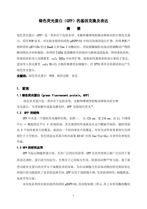
绿色荧光蛋白(GFP)的基因克隆及表达摘要绿色荧光蛋白(GFP)是一类存在于包括水母、水螅和珊瑚等腔肠动物体内的生物发光蛋白。
采用PCR技术,对实验室提供的质粒pEGFP-N1中的目的基因进行扩增。
所得PCR产物和质粒pET-28b经过BamH I和Nde I双酶切后,用琼脂糖凝胶电泳法检测酶切产物的酶切情况并回收凝胶,再利用T4DNA连接酶将目的基因与载体连接起来,得到重组质粒。
将重组质粒导入克隆菌E. coli DH5a中培养扩增,提取阳性菌落质粒进行重组子鉴定,进而导入表达菌E. coLi BL-21大肠杆菌感受态细胞中,经IPTG诱导目的基因表达产生绿色荧光蛋白。
关键词:绿色荧光蛋白 PCR 基因克隆表达1.前言1.1绿色荧光蛋白(green fluorescent protein,GFP)绿色荧光蛋白是一类存在于包括水母、水螅和珊瑚等腔肠动物体内的生物发光蛋白。
当受到紫外或蓝光激发时,GFP 发射绿色荧光[1]。
1.2 GFP 的结构GFP中央是一个圆柱形水桶样结构,如图二。
长420 nm,宽240 nm,由11 个围绕中心α螺旋的反平行β折叠组成,荧光基团的形成就是从这个螺旋开始的,桶的顶部由3个短的垂直片段覆盖,底部由一个短的垂直片段覆盖,对荧光活性很重要的生色团则位于大空腔内。
发色团是由其蛋白质内部第65-67位的Ser-Tyr-GLy自身环化和氧化形成。
1.3 GFP的研究应用GFP可标记细胞和蛋白质,具有广泛的应用前景。
GFP及其突变体已被广泛应用于基因表达调控、蛋白质空间定位、生物分子之间相互作用、转基因动物]2[等方面。
基于新型功能荧光蛋白的光学分子成像技术的发展,为在活细胞乃至活体动物内研究基因表达和蛋白质功能提供了更多的选择空间。
GFP还用于观察微生物、发育机理研究、细胞筛选、免疫学等方面。
本实验是利用实验室提供的质粒pEGFP-N1,其结构如图三所示。
其上有所用酶的酶切位点。
表达gfp水平
表达GFP水平
表达GFP水平通常指的是在细胞或组织中绿色荧光蛋白(Green Fluorescent Protein,GFP)的表达量或荧光强度。
GFP是一种来自水母(Aequorea victoria)的天然荧光蛋白,它能够在激发光的作用下发出绿色荧光。
在生物学研究中,GFP常被用作报告基因,通过基因工程技术将其与其他基因融合,用于研究基因表达、蛋白质定位等。
因此,表达GFP水平的高低可以反映目标基因或蛋白质的表达情况。
为了量化表达GFP水平,可以使用荧光显微镜或荧光成像系统对细胞或组织进行观察和测量。
通过比较不同样品之间GFP荧光的强度和分布,可以评估目标基因或蛋白质的表达水平和空间分布。
同时,还可以使用荧光定量PCR(qPCR)等技术对GFP mRNA的表达量进行定量分析。
- 1、下载文档前请自行甄别文档内容的完整性,平台不提供额外的编辑、内容补充、找答案等附加服务。
- 2、"仅部分预览"的文档,不可在线预览部分如存在完整性等问题,可反馈申请退款(可完整预览的文档不适用该条件!)。
- 3、如文档侵犯您的权益,请联系客服反馈,我们会尽快为您处理(人工客服工作时间:9:00-18:30)。
GFP简介
The green fluorescent protein (GFP) is a protein composed of 238 amino acid residues (26.9kDa) that exhibits bright green fluorescence when exposed to blue light.[1][2] Although many other marine organisms have similar green fluorescent proteins, GFP traditionally refers to the protein first isolated from the jellyfish Aequorea victoria. The GFP from A. victoria has a major excitation peak at a wavelength of 395 nm and a minor one at 475 nm. Its emission peak is at 509 nm, which is in the lower green portion of the visible spectrum. The GFP from the sea pansy (Renilla reniformis) has a single major excitation peak at 498 nm. 绿色萤光蛋白(green fluorescent protein),简称GFP,这种蛋白质最早是 由下村脩等人在1962年在维多利亚多管发光水母中发现。其基因所产生的蛋 白质,在蓝色波长范围的光线激发下,会发出绿色萤光。这个发光的过程中 还需要冷光蛋白质水母素的帮助,且这个冷光蛋白质与钙离子(Ca2+)可产生 交互作用。 由维多利亚多管发光水母中发现的野生型绿色萤光蛋白,395nm和475nm分 别是最大和次大的激发波长,它的发射波长的峰点是在509nm,在可见光绿 光的范围下是较弱的位置。由海肾(sea pansy)所得的绿色萤光蛋白,仅有在 498nm有一个较高的激发峰点。
绿色荧光蛋白基因在水稻基因转化中研究
1 Materials and Methods.(材料与方法) Methods. pJPM5 1.1 Construction of plasmids containing GFP gene in pJPM (构建含有GFP基因的pJPM 质粒) pJPM5 pJPM We used the pGHNC5 plasmid, reported by Allen et al. (1993, 1996), for the construction of pJPM5 ( Figure 1). Plasmid pJPMO was constructed by inserting a Klenow enzyme-filled EcoRI fragment, isolated from pBY505 (Wang and Wu, 1995; Zhao et al, 1989), which contains the Pin2 terminator, 35S promoter, bar gene and Nos terminator, into the Sma1 site of pGHNC5. Plasmid pJPM5 was constructed by inserting the rice actin1 promoter (McElroy et al, 1991) and a GFP gene isolated from pSBGXOO, into pJPMO. Plasmids pSBGXOO was constructed by Jukon Kim and Ray Wu ( unpublished results ).
绿色荧光蛋白基因在水稻基因转化中研究(续)
1.3 Quantitative assay of GFP values in transgenic rice plants(定量检测绿 ( 色荧光蛋白在转基因水稻植株中的表达值 蛋白在转基因水稻植株中的表达值) 色荧光蛋白在转基因水稻植株中的表达值) To detect GFP value in R0 transgenic rice plants, a quantitative assay was carried out. The second leaf from the top of each two-month-old transgenic rice plant was collected and immediately frozen in liquid nitrogen. The leaves were ground in liquid nitrogen and homogenized in ice - cold extraction buffer (100 mmol HEPES, pH 8.0, 10 mmol EDTA, 5 mmol DTT, 10% glycerol, 1% PVP, 25 fig mL-1 PMSF, 15 fig mL-1 leupeptin, 1% activated carbon). This ex-traction buffer was found by preliminary experiments to give minimum autofluorescence of rice leaf extractsr data not shown). After the first centrifugation (12 000 rpm for 15 min at 4B ) , the crude extract was centrifuged again (12 000 rpm for 20 min at 4B ) to further reduce insoluble particles which affect the quantification of fluorescence. Then the GFP values were quantified based on fluorescence units f g - 1 protein using a fluorometerr model SLM8000) after excitation at 385 nm and measure the emission maximum at 510 nm. Nontransformed tissues were used to estimate the autofluores-cence of the tissue. Protein concentration was determined according to Bradford (Bradford , 1976). Statistical analysis was carried out as described by Sokal and Rohlf (Sokal and Rohlf , 1969).
GFP的性质(续)
GFP的用途
GFP作为报告分子和细胞标记最明显的优势是无需底 物或辅因子参与;无论在活细胞还是在完整的转基因 胚胎和动物中,都能有效地监测基因转移的效率。 但在这方面的应用中,GFP最大的缺点就是没有放大 作用,它不能像酶一样能通过加工无数的底物分子而 将信号放大。所以一般都需强启动子以驱动GFP基因 在细胞内足量的表达。也可用亚细胞分辨率的显微成 像系统检测基因产物,靶入的基因被限制于一个细胞 器内,GFP的浓度种现成的荧光蛋白质,因此它特别容易使用。大多数 可以处理光的蛋白质都利用外来的分子吸收和释放光子。例如, 我们眼睛里的视紫红质利用维生素来感光。这些“发光团”必 须是专门为了发光而生成的,并且被仔细地插入到该蛋白质分 子内,不同的是,GFP控制光的部位是其自身的一部分,仅由 氨基酸构建而成,该部位含有一段三个氨基酸组成的特殊序列: 丝氨酸-酪氨酸-甘氨酸(有时丝氨酸会被相似的苏氨酸取 代)。当蛋白质链折叠时,这段短片段就被深埋在蛋白质内部, 然后,发生一系列化学反应:甘氨酸与丝氨酸之间形成化学键, 生成一个新的闭合环,随后这个环会自动脱水。最终,经过大 约一个小时的反应,周围环境中的的氧气攻击酪氨酸的一个化 学键,形成一个新的双键并合成荧光发色团。由于GFP可以形 成自己的发色团,它非常适合于基因工程。你根本不必担心操 作任何奇怪的发色团,你只需要利用遗传学的方法操纵细胞合 成GFP蛋白质,GFP就会自动折叠并开始发光。
绿色荧光蛋白基因在水稻基因转化中研究(续)
1.2 Production of transgenic rice plants(生产转基因大米植株) (生产转基因大米植株) Calli(愈伤组织) were induced in MS medium ( Murashige and Skong, 1962) from mature embryos of japonica rice ( Oryza saliva L. ) cv. TNG67. Fifteen-day-old embryogenic calli were bombarded with gold beads coated with either pJPM5, or with plasmid pSBG7OO according to the procedure of Cao et al. ( 1992) . Resistant calli were selected in plates contain-ing MS medium (Wang and Wu, 1995; Zhao et al, 1989), supplemented with 6 mg L-1 Bialaphos as the selective agent for 4 weeks (su bcultured every 2 weeks) . Resistant calli were transferred to plates containing MS regeneration media with 1 mg L-1 kinetin, 2mgL-1BAP, 0.25 mg L-1 NAA and 3 mgL-1 Bialaphos to regenerate into plants. Re-generated plants were transplanted into sterilized soil and grown in the greenhouse (30B day and 24 B night with a sup-plemented photoperiod of 10 h) . The presence of the transgenes in regenerated plants was first inferred by the herbicide resistance of the plants. To test for herbicide resistance, one leaf from each of the one-month-old transgenic rice plants was painted on both sides with 0. 25 % ( v/ v ) of the herbicide Basta ( containing 162 g L -1 glufosinate ammonium: Hoechst- Roussel Agri- Vet Co., Somerville, NJ) and 0.05% (v/v) Tween 20. One week later, the resistant or sensitive phenotypes were scored.