The subcoracoid space an anatomic study
华中科技大学学术期刊分类目录(T-D)_最新权威版
24.755 23.917 23.654 23.565 23.333 23.194 22.929 22.864 22.864 22.864 22.490 22.345 22.333 21.757 21.543 21.543 20.833 20.761 20.614 19.966 19.795 19.547 19.352 18.571 18.571 18.038 17.983 17.983 17.949 17.689 17.689 17.689 17.436 17.313 17.215 16.417 16.238 16.179 16.008 16.008 15.766 15.575 15.518 15.389 15.389 15.389 15.333 15.333 15.280 15.280 15.265 15.253 15.251 15.202
ONCOLOGY CLINICAL NEUROLOGY PLANT SCIENCES BIOCHEMICAL RESEARCH METHODS ASTRONOMY & ASTROPHYSICS MATERIALS SCIENCE, MULTIDISCIPLINARY PHYSICS, MULTIDISCIPLINARY BIOCHEMISTRY & MOLECULAR BIOLOGY CELL BIOLOGY MEDICINE, RESEARCH & EXPERIMENTAL MICROBIOLOGY PHARMACOLOGY & PHARMACY PHYSICS, PARTICLES & FIELDS CHEMISTRY, MULTIDISCIPLINARY PHARMACOLOGY & PHARMACY TOXICOLOGY CHEMISTRY, MULTIDISCIPLINARY CELL BIOLOGY NEUROSCIENCES INFECTIOUS DISEASES IMMUNOLOGY PHYSIOLOGY PHYSICS, MULTIDISCIPLINARY BEHAVIORAL SCIENCES NEUROSCIENCES ONCOLOGY CELL BIOLOGY DEVELOPMENTAL BIOLOGY ECOLOGY CHEMISTRY, MULTIDISCIPLINARY MAபைடு நூலகம்ERIALS SCIENCE, MULTIDISCIPLINARY NANOSCIENCE & NANOTECHNOLOGY GENETICS & HEREDITY MICROBIOLOGY MEDICINE, GENERAL & INTERNAL MICROBIOLOGY ASTRONOMY & ASTROPHYSICS MATERIALS SCIENCE, MULTIDISCIPLINARY BEHAVIORAL SCIENCES NEUROSCIENCES NEUROSCIENCES PSYCHOLOGY CLINICAL NEUROLOGY ECOLOGY EVOLUTIONARY BIOLOGY GENETICS & HEREDITY CHEMISTRY, PHYSICAL PHYSICS, CONDENSED MATTER BIOCHEMISTRY & MOLECULAR BIOLOGY CELL BIOLOGY PSYCHOLOGY MEDICINE, GENERAL & INTERNAL NEUROSCIENCES CARDIAC & CARDIOVASCULAR SYSTEMS
桡骨茎突的解剖学观察及其在针刀治疗腱鞘炎中的临床意义
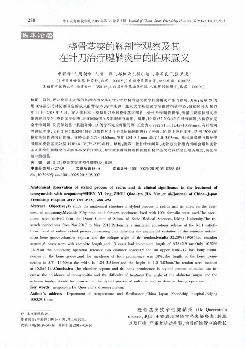
橈骨茎突的解剖学观察及其在针刀治疗腱鞘炎中的临床意义申毅锋=周俏吟巴贾雁I,邱祖云I,孙小洁1,李石良二张卫光4(1•中日友好医院针灸科,北京100029;2.成都中医药大学,四川成都610072;3.福建中医药大学,福建福州350108;4.北京大学基础医学院人体解剖教研室,北京100191)摘要目的:研究橈骨茎突部的解剖结构及其对针刀治疗挠骨茎突狭窄性腱鞘炎产生的影响。
方法:选取59例用10%福尔马林防腐固定的成人前臂标本,标本来源于北京大学基础医学院遗体捐献中心,研究时间为2017年11月-2018年5月。
在人体标本上模拟针刀松解梯骨茎突部第一骨性纤维鞘管操作,测量并观察伸肌支持带的解剖变异、挠骨茎突骨槽,纤维间隔情况及肌腱斜行角度。
结果:19例(32.20%)存在纤维间隔,6例存在完全纤维间隔、长度伴随整个肌腱延伸,13例为不完全纤维间隔、长度为6.78±2.91mm(2.45-10.88mm)o有纤维间隔的标本中,仅有2例(10.52%)的针刀操作对2个纤维间隔同时进行了松解。
40例上肢标本中,12例(30%)在饶骨茎突骨沟内有骨蜡。
骨11#长度5.71-14.60mm,宽度1.84-3.51mm,高度1.0〜3.03mm。
拇长展肌腱与拇短伸肌腱在挠骨茎突处呈15.8。
±4.13。
(7。
~23。
)斜行。
结论:腕第一腔室纤维间隔、挠骨茎突骨槽内骨U#会增加挠骨茎突狭窄性腱鞘炎的发病几率及治疗难度,拇长展肌腱与拇短伸肌腱在橈骨茎突处斜行应注意其角度,防止推割中的损伤。
关键词:针刀;挠骨茎突狭窄性腱鞘炎;解剖中图分类号:R274.9文献标识码:A 文章编号:1001-0025(2019)05-0288-05doi:10.3969/j.issn.1001-0025.2019.05.007Anatomical observation of styloid process of radius and its clinical significance in the treatment of tenosynovitis with acupotomy//SHEN Yi-feng,ZHOU Qiao-yin,JIA Yan,et al//Journal of China-Japan Friendship Hospital,2019Oct,33(5):288-292Abstract Objective:To study the anatomical structure of styloid process of radius and its effect on the treat-ment of acupotomy.Methods:Fifty-nine adult forearm specimens fixed with10%formalin were used.The spec- imens were derived from the Donor Center of School of Basic Medical Sciences,Peking University.The research period was from Nov.2017to May2018.Performing a simulated acupotomy release of the No.l osteofi-brous canal of radius styloid process,measuring and observing the anatomical variation of the extensor retinaculum,bone groove,chamber septum and the oblique angle of the tendon.Results:32.20%(19/59)had chamber septum,6cases were with complete length,and13cases had incomplete length of 6.78±2.91mm.Only10.52% (2/19)of the acupotomy operation released two chamber spaces.Of the40upper limbs,12had bony prominences in the bone groove,and the incidence of bony prominence was30%.The length of the bony prominences is 5.71-14.60mm,the width is 1.84-3.51mm,and the height is 1.0-3.03mm.The tendon were inclined at15.8±4.13°.Conclusion:The chamber septum and the bony prominence in styloid process of radius can increase the incidence of tenosynovitis and the difficulty of treatment.The angle of the abductor longus and the extensor tendon should be observed at the styloid process of radius to reduce damage during operation.Key words acupotomy;De Quervain*s disease;anatomyAuthor's address Department of Acupuncture and Moxibustion,China-Japan Friendship Hospital,Beijing 100029,China1*本文通讯作者。
胸廓出口综合征

胸廓出口综合征中国医学科学院北京协和医学院整形外科医院麻醉科薛富善首都医科大学宣武医院疼痛诊疗中心倪家骧在颈胸接合处可发现许多解剖变异,其中的一些可压迫穿经颈部进入臂部的血管和/或神经结构。
4种主要的胸廓出口综合征(thoracic outlet syndromes)是前斜角肌综合征(scalenus anticus syndrome)、肋锁综合征(costoclavicular syndrome)、颈肋综合征(cervical rib syndrome)和外展过度综合征(hyperabduction syndrome)(图56-6)。
一、病因学目前已将胸廓出口综合征归因于如下病变:颈肋、异常的第1胸肋、前斜角肌肥厚、中斜角肌止点异常、Sibson筋膜束、肋锁关节异常和臂外展过度等。
虽然已知一些患者存在有这些病变,但其却没有任何症状。
因而,对各种病变的具体情况仍有疑问。
一些结构异常可影响臂丛的不同部位,并可产生非典型性臂丛下干神经功能障碍。
详细的体格检查和诊断试验可表明存在这种非典型性神经功能障碍的可能。
然而,亦有一些确切证实的变异型胸廓出口综合征,并且患者的主诉情况和检查结果与解剖变异相一致。
1个典型的例子就是侵害臂丛下干的颈肋,其可导致手和前臂肌肉萎缩和肌力减退、前臂尺侧疼痛、臂和前臂尺侧感觉异常。
手术治疗这种先天性畸形常常可使患者的症状获得缓解。
一些病例是与正常结构的位置异常有关,而另一些病例则可能是由异常的肋骨、软骨或异常的肌肉附着点所致。
Sunderland已描述了这种病变的解剖学变异及其是如何导致相关结构压迫的。
遗憾的是,许多研究人员对用来缓解非特异性肩部、颈部、臂部和手部痛的各种手术的治疗效果做出了不确定的断言。
有人怀疑许多所谓的综合征是否真正存在。
与前几代的临床医师相比较,目前的临床医师似乎极少发现各种胸廓出口综合征患者。
二、症状和体征常见的胸廓出口综合征累及锁骨下血管和臂丛下干(C8和T1),可发现不同程度的血管和/或神经功能障碍,并伴有局部锁骨上疼痛。
小脑扁桃体下疝(Chiari畸形)
Wil正lia常ms—发产育伤是的病后源脑因素结构因后颅窝过度 Eme挤ry、压Ma而cke疝nzi入e—椎颅管内及内椎。管内压力梯度
家族遗传因素
外伤
2021/6/16
16
Chiari畸形的合并症:
脊髓空洞症 脑积水 枕骨大孔区畸形(颅底凹陷、扁平颅
底、寰枢椎畸形)
2021/6/16
27
Chiari畸形的治疗:
手术原则:去除脑干及下疝的小脑扁桃 体的骨性束缚,解除因疝出导致的硬 膜压迫,重建枕骨大孔正常的脑脊液 动力学,并保持硬膜的密闭性。
2021/6/16
28
单纯后颅窝减压:
2021/6/16
29
手术方式的改进:
2021/6/16
30
Chiari畸形的护理要点:
2021/6/16
20
Chiari畸形的分型(Ⅰ型):
小脑扁桃体下疝至枕骨大孔平面下,呈 锥状向椎管内疝入;最多见
2021/6/16
21
Chiari畸形的分型(Ⅱ型):
小脑扁桃体及后颅窝内容物包括脑干,第四脑室,小 脑蚓部均下疝至枕骨大孔下,常合并有脑积水
2021/6/16
22
Chiari畸形的分型:
12
神经结构(三):
2021/6/16
13
神经结构(四):
2021/6/16
14
血管结构:
2021/6/16
15
Chiari畸形的发病机制:
Chi公ari认—脑的积发水压病迫机学制说 :1995年,由Badie
Pen提fie出ld—,脊可髓栓能系发学生说 于胎儿的第3个月, Gar胚dne胎r—时液体期动,力由学说于中胚层体节枕骨部 POa'tR发taehni育l—l胚y不—胎后良时颅期,窝与导容正积致常与脑枕其干内骨屈容曲发物发大育育小异迟之常缓间有不滞关相后称的,假
系统解剖学 英文版.Myology(1)

Major Muscles of Back
★Latissimus dorsi 背阔肌
Origin: Spinacic and all lumbar vertebrae Median sacral crest Posterior part of iliac crest Insertion: crest of lesser tubercle Action: when trunk fixed, extends, adducts and medially rotates arm; when arm fixed, elevates trunk.
Muscles of the neck
Anterior group Suprahyoid muscles Digastric 二腹肌 Stylohyoid 茎突舌骨肌 Mylohyoid 下颌舌骨肌 Geniohyoid 颏舌骨肌 Action: elevate (raise) hyoid bone and depress mandible
Facial muscles 面肌
Epicranius 颅顶肌 Frontal belly 额腹 Occipital belly 枕腹 Galea aponeurotica 帽状腱膜 Orbicularis oculi 眼轮匝肌
Oral group 口周围肌
Orbicularis oris 口轮匝肌 Buccinator 颊肌
Masticatory muscles 咀嚼肌
Lateral pterygoid 翼外肌
superiorly to the medial
pterygoid
Pulls the mandible forward
前锯肌的肌构筑学研究
f 摘 要】 目的 研究前锯肌的构筑学特征, 探讨该肌 的生理功能, 为临床 肌移植提供形态学资料。方法 取成人尸 前锯 肌上部肌重 4 . 88g 肌长 1 .1 1 5 m、 47 . 、 + 7 08 + . c 肌纤维长 6 0 0 3m、 _7 . + . c 生理 6- 6
S u y o r h t cu e o e r t sa t ro t d n a c ie t r fs r a u n e ir
L h u t n XU in I o —i 。 E O a S a
(eat n f n t y,u y Mei l o ee uzo u y 5 30 ,hn) D pr t ao Z ni dc l g, i uZ ni 6 0 3C ia me o A m aC l G h
n e i r 09 + 4 3 g 1 . 8 1 1 e 1 .7 . m,.3 07 c a tr rwee 8 .5 2 . 1 , 88 + .6 m , 33 +1 c 5 7 + .8 m r s e t eyCo cu i n e s p r ra d mi d e o _ _ _ 41 _ e p c v l . n l so sTh u e o d i i n l p r o er t s a t r r ma ny e e tman an h s b l y o s a u a i mb mo e n d t e i ei r p r o e r ts a t fs r u ei i l x r a n o it it e t i t f c p l n l v me t a n r at f s rau a i i n h f o
t形吻合应用于全胸腹腔镜ivorlewis食管癌切除术的临床研究_董祎楠
T 形吻合应用于全胸腹腔镜Ivor Lewis 食管癌切除术的临床研究收稿日期:2015-11-19;修回日期:2016-01-06通讯作者:刘永煜,E -mail :profyongyuliu@董祎楠,张亮,孙楠,刘德贵,李继佳,佟状,刘永煜(中国医科大学肿瘤医院,辽宁省肿瘤医院,辽宁沈阳110042)摘要:[目的]评价全胸腹腔镜下Ivor Lewis 食管癌切除术中直线吻合器吻合应用的价值。
[方法]整个吻合过程借助腔镜下直线切割吻合器(强生Ethicon flex 60直线吻合器)完成。
首先,以直线切割吻合器斜向上方击发建立胃食管的侧—侧吻合形成吻合口的上、下两壁,再次使用直线切割吻合器沿管状胃的延长线切割击发,完成吻合口的制作,同时切除胃小弯及食管肿瘤。
当吻合完成后吻合口的平面呈矢状位,由上缘、下缘和前缘构成。
[结果]2014年2月至2015年12月,共有28例患者接受这种吻合方式的Ivor Lewis 食管癌切除术,最长随诊时间为22个月,最短1个月,无吻合口出血,无吻合口瘘及吻合口狭窄。
[结论]T 形吻合应用于全胸腹腔镜Ivor Lewis 食管癌切除术受空间制约小、操作过程简单、吻合可靠,是一种安全有效的全胸腔镜下胸腔内胃食管吻合方式。
关键词:全胸腹腔镜;T 形吻合;胸内食管胃吻合中图分类号:R735.1文献标识码:A 文章编号:1004-0242(2016)03-0233-04doi :10.11735/j.issn.1004-0242.2016.03.A015A Clinical Study of T Shaped Gastroesophageal Anasto -mosis for Totally Laparoscopic Thoracoscopic Ivor Lewis Esophagectomy for Esophageal CancerDONG Yi -nan ,ZHANG Liang ,SUN Nan ,et al.(Cancer Hospital of China Medical University ,Liaoning Cancer Hospital and Institution ,Shenyang 110042,China )Abstract :[Purpose ]To investigate the role of T shaped gastroesophageal anastomosis for totally laparoscopic thoracoscopic Ivor Lewis esophagectomy for esophageal cancer.[Methods ]A new technique for the endoscopic gastroesophageal anastomosis was completed just with a linear stapler (Ethicon Flex 60).In this technique ,a linear stapler was first fired upward to establish the side to side anastomosis of the esophagus and stomach.This created the anterior and posterior wall of the anasmotic site.The linear stapler was then fired along the extension line of the gastric conduit ,to complete the anastomosis and at the same time resected the lesser curvature of the stomach and the esophageal cancer.Upon completion ,the anastomotic plane was axial ,and contained a superior edge ,inferior edge ,and anterior edge.[Results ]From February 2014to December 2015,28esophageal cancer patients were performed the minimally invasive Ivor Lewis esophagectomy with this anastomosis.These patients were all followed up within 1~22months.None of these patients had any anastomotic bleeding ,leak ,or stenosis.[Conclusions ]This new technique is less restrict -ed by the limited space during minimally invasive Ivor Lewis procedure.The anastomotic tech -nique is easy to perform and appears to be reliable ,safe and effective judging from our limited clinical experience up to this date.Key words :totally iaparoscopic thoracoscopic ;T shaped gastroesophageal anastomosis ;intratho -racic esophagogastral anastomotic随着胸腔镜手术器械和手术技术的不断进步,微创Ivor Lewis 食管切除手术越来越多地运用于胸中、下段食管癌的手术治疗,并且在良好的术野、胸壁的低创伤、完整的肿瘤切除都优于传统的食管癌切除手术[1]。
人体解剖学HumanAnatomy
人體解剖學(Human Anatomy)-解剖位置(Anatomical Position)-解剖面(Anatomical Planes)橫切面(Transverse plane):上、下額切面(Frontal plane):前、後側切面(Sagittal plane):左、右-解剖方位(Anatomical Directions)上方(Superior)、下方(Inferior)前方(Anterior)、後方(Posterior)內側(Medial)、外側(Lateral)近端(Proximal)、遠端(Distal)關節的運動(Movements in Joints)1. 屈∕彎曲(flexion)2. 伸(Extension)3. 外展(Abduction)4. 內收(Adduction)5. 旋轉(Rotation)6. 迴旋(Circumduction)7.足背屈(Dorsiflexion)8.蹠屈(Plantarflexion)9. 內翻(Inversion)10. 外翻(Eversion)11. 旋外∕旋上/旋後(Supination)12. 旋內∕旋下/旋前(Pronation)13. 上舉(Elevation)14. 下壓(Depression)身體部位-頭顱(The Skull)腦(The Brain):大腦、中腦、小腦等-胸腔(The Thorax)呼吸及循環器官:心臟、肺臟等-腹腔(The Abdominal Cavity)消化、新陳代謝器官:胃、腸、肝、腎、膀胱-四肢(The Extremities)特點: 1. 對比2. 由少而多:1-2-8-191-2-7-19骨髂(The Bones)軀幹骨:-脊椎:26塊真頸椎7 ( Cervical Vertebrae)胸椎12 (Thoracic Vertebrae)椎腰椎 5 (Lumbar Vertebrae)偽骶骨 1 (5合1)椎尾骨 1 (4合1)-頭骨:22 塊-舌骨: 1 塊-胸骨: 1 塊-肋骨:24 塊-聽小骨: 6 塊四肢骨:-上肢骨:64 塊-下肢骨:62 塊共計:206 塊鎖骨、肩胛骨肱骨尺骨、橈骨1. 豆狀骨(Pisiform)2. 三角骨(Triquetrum)3. 月狀骨(Lunate)4. 舟狀骨(Scaphoid)1. 勾狀骨(Hamate)2. 頭狀骨(Capitate)3. 小菱形骨(Trapezoid)4. 大菱形骨(Trapezium)掌骨(Metacarpals):共5根指骨(Phalanges):除拇指為二根外,其他均各為三根共14根共32根x 2 = 64根髖骨股骨(Femur)、脛骨(Tibia)、腓骨(Fibula)髕骨∕膝蓋骨1.距骨2. 跟骨3. 舟狀骨4. 骰骨5. 楔狀骨:內、中、外,共三塊蹠骨:共五根(類比手之掌骨)趾骨:除大趾為二根外,其他均各為三根,共14根共31根x 2 = 62根骨髂種類:1. 長骨:尺骨、橈骨、股骨等2. 短骨:腕骨、跗骨3. 扁平骨:頭骨4. 不規則骨:脊椎骨5. 種子狀骨:膝蓋骨(近關節處,增進槓桿作用)骨髂功能:1. 身體支架2. 保護作用3. 運動4. 儲存:礦物質(鈣、磷)5. 造血骨髂(長骨)之構造1.關節軟骨(Articular Cartilage)2.生長區(骨端)(Epiphysis)3.骨幹(Diaphysis)4.生長板(Epiphysis plate)5.鬆質骨(Spongy bone)6.密質骨(Compact bone)7.哈氏系統(Haversian)8.骨髓腔(Medullary Cavity)9.動脈孔(Arterial opening)10.骨膜(Periosteum)骨髂表面之位置名稱1.粗隆、結節、突(Tuberosity、tubercle、process)2.切跡(Notch)3.棘(Spine)4.窩(Fossa)5.孔(Foramen)6.髁(Condyle)關節(Joints)A.纖維性關節(Fibrous joints)頭骨、牙齒B.軟骨性關節(Cartilaginous joints)骨盤、椎骨間盤C.滑液關節(Synovial joints)1.單軸關節:膝、肘、手指2.雙軸關節:橈骨、腕骨間,尺骨、腕骨間3.三軸關節:(Ball Socket joints)肩、髖關節肌肉(Muscles)-平滑肌(Smooth muscles):內臟-心肌(Cardiac muscles)-骨髂肌(Skeletal muscles)又稱:橫紋肌(Striated muscles)隨意肌(Voluntary muscles)骨髂肌的構造肌肉(Muscles)→肌束(Muscle Fasciculus)→肌纖維(Muscle Fiber)(肌細胞) →Myofibril →SarcomereActin (肌動蛋白)(Z帶-Z帶)Myosin (肌凝蛋白)-骨髂肌的排列與形狀:1. 紡錘狀(Fusiform)2. 羽毛狀(Pennate)-韌帶(Ligament)-肌腱(Tendon)-纖維軟骨(Fibrocartilage):半月軟骨-滑液膜(Synovial membrane)-關節囊(Joint Capsule)-滑液囊(Bursae)-鞬鞘(Tendon Sheath)上肢(Upper Extremity)-肩部(The Shoulder)骨髂:肩胛骨(Scapula)鎖骨(Clavicle)肱骨(Humerus)-鎖骨、肩胛骨組成肩帶(Shoulder Girdle)-肱骨、肩胛骨組成肩關節(Shoulder Joint)-肩胛骨的部位名稱:1.尖峰突(Acromion Process)2.喙狀突(Coracoid Process)3.肩胛棘(Spine)4.棘上窩(Supraspinous Fossa)5.棘下窩(Infraspinous Fossa)6.肩胛下窩(Subscapular Fossa)7.肩窩∕關節盂(Glenoid Fossa)-肩部的關節:共有四個關節1.胸鎖關節(Sternoclavicular Joint)胸骨與鎖骨2.肩峰鎖關節(Acromioclavicular Joint)簡稱A-C Joint肩峰突(Acromion Process)與鎖骨3.肩喙鎖關節(Coracoclavicular Joint)喙狀突(Coracoid Process)與鎖骨4.肩關節(Glenohumeral Joint)關節盂(Glenoid Fossa)與肱骨肩部的運動(The Movement of the Shoulder)-肩帶(Shoulder Girdle)的運動1.內收(Adduction):挺胸2.外展(Abduction):駝背3.上舉(Elevation)4.下壓(Depression)5.向上旋轉(Upward Rotation)6.向下旋轉(Downward Rotation)-肩關節(Shoulder Joint)之運動1.外展(Abduction)2.內收(Adduction)3.屈∕彎曲(Flexion)4.伸(Extension)5.水平屈(Horizontal Flexion)6.水平伸(Horizontal Extension)7.向外旋轉(Outward Rotation)(Lateral Rotation)8.向內旋轉(Inward Rotation)(Medial Rotation)-與肩帶動作有關之肌肉-1.斜方肌(Trapezius)起點:a. 枕骨(Occipital)b. C7 ~ T12 (有部份在項韌帶上,但不在頸椎上,除C7外)終點:a. 鎖骨b. 肩峰∕肩峰突c. 肩胛棘主要作用:當頸部固定時 a. 上舉b. 向上旋轉c. 內收d. 下壓當肩帶固定時 a. 頸部左右彎曲b. 頸部左右旋轉2.菱形肌(Rhomboideus)起點:a. 項韌帶(Ligamentum Nuchae)b. C7 ~ T5終點:肩胛骨主要作用: a. 內收b. 上舉3.提肩胛肌(Levator Scapulae)起點:C1 ~ C4之橫突(Transverse processes)終點:肩胛骨內緣之上角(superior angle)主要作用:上舉4.前鋸肌(Serratus Anterior)起點:第一至第九肋骨終點:肩胛骨內面之全部內緣主要作用: a. 外展b. 向上旋轉5.胸小肌(Pectoralis minor)起點:第三~五肋骨終點:肩胛骨之喙狀突主要作用: a. 外展b. 下壓c. 向下旋轉-與肩關節動作有關之肌肉-1.三角肌(Deltoid)起點: a. 前:外側1∕3之鎖骨b. 中:肩峰(Acromion)c. 後:肩胛棘(Spine)終點:肱骨(Humerus)之前1∕3處主要作用:前 a. 水平彎曲b. 彎曲中 c. 外展中、後 d. 水平伸展2.棘上肌(Supraspinatus)起點:肩胛骨之棘上窩終點:肱骨之大粗隆前上方(Great tubercle)主要作用:外展3.棘下肌(Infraspinatus)起點:肩胛骨之棘下窩終點:肱骨大粗隆後上方主要作用: a. 向外旋轉b. 水平伸展4.小圓肌(Teres Minor)起點:肩胛骨外緣終點:肱骨之大粗隆後下方主要作用: a. 水平伸展b. 向外旋轉5.肩胛下(內)肌(Subscapularis)起點:肩胛下窩終點:肱骨之小粗隆(Lesser tubercle)主要作用: a. 向內旋轉b. 伸展6.闊背肌(Latissimus Dorsi)起點: a. 髂脊(Crest of the ilium)b. T7~S5c. 最下面的3或4根肋骨(背部)終點: a. 肱骨b. 肩胛骨主要作用: a. 伸展b. 內收c. 向內旋轉9.胸大肌(Pectoralis Major)起點: a. 鎖骨(內側2∕3)b. 胸骨c. 第一~六(或七)肋骨終點:肱骨之節結間溝(Bicipital Groove)主要作用: a. 水平彎曲b. 向內旋轉c. 內收(胸骨)d. 伸展(胸骨)e. 彎曲(鎖骨)7.喙肱肌(Coracobrachialis)起點:肩胛骨之喙狀突終點:肱骨內側之下半部主要作用: a. 內收b. 水平彎曲8.圓大肌(Teres major)起點:肩胛骨下角之外緣終點:肱骨結節間溝之內緣主要作用: a. 向內旋轉b. 伸展c. 內收肘部(The Elbow)骨髂:1.肱骨(Humerus)2.尺骨(Ulna):內側-靠近小指邊3.橈骨(Radius):外側-靠近拇指邊骨髂部位之名稱:-肱骨(Humerus):1.內上髁(Medial Epicondyle)2.外上髁(Lateral Epicondyle)3.內上髁脊(Medial Supra condylar ridge)4.外上髁脊(Lateral Supra condylar ridge)-尺骨(Ulna):1.鷹嘴突(Olecranon Process):近2.尺骨粗隆(Ulnar Tuberosity)3.尺側莖狀突(Ulnar Styloid Process):遠4.冠狀突(Coronoid Process):近-橈骨(Radius):1.橈骨頭(Radial Head)2.橈骨頸(Radial Neck)3.橈骨粗隆(Radial Tuberosity)4.橈側莖狀突(Radial Styloid Process)肘部之關節:1.肘關節(The Elbow Joint)-肱骨滑車(Trochlea)與尺骨之滑車切跡(Trochlear Notch)-肱骨小頭(Capitulum)與橈骨頭2.橈尺關節(Radioulnar Joint)-橈骨之橈骨頭及尺骨之橈骨切跡-近腕關節處(即遠端)之橈尺關節剛好相反(與近端):尺骨頭與尺骨切跡肘部之動作:1.肘關節:彎曲與伸展2.橈尺關節:-旋內∕旋下(Pronation)-旋外∕旋上(Supination)肘部之肌肉:1.肱二頭肌(Biceps Brachii)起點: a. 短頭:喙狀突b. 長頭:肩上窩結節(Supraglenoid Tubercle)終點:橈骨粗隆主要作用: a. 彎曲b. 旋外∕上2.肱肌(Brachialis):於肱二頭肌之下起點:肱骨之下半前方終點:尺骨之粗隆與冠狀突主要作用:彎曲3.肱橈肌(Brachioradialis)起點:肱骨之外上髁脊終點:橈側莖狀突主要作用:彎曲4.肱三頭肌(Triceps Brachii)起點:長頭:肩下窩結節(Infraglenoid Tubercle)外側頭:肱骨後上方(~1∕3)內側頭:肱骨後下方(約2∕3)終點:尺骨之鷹嘴突主要作用:伸展5.肘肌∕肘後肌(Anconeus)起點:肱骨外上髁之後方終點:a. 鷹嘴突外側面b. 尺骨外側之近端主要作用:伸展並拉開關節滑膜6.旋內(下)圓肌(Pronator Teres)起點: a. 肱骨頭:肱骨內上髁b. 尺骨頭:尺骨冠狀突內側終點:橈骨外側之中段主要作用:旋內∕下7.旋內(下)方肌(Pronator Quadratus)起點:尺骨前下方(約1∕4處)終點:橈骨前下方(約1∕4處)主要作用:旋內∕下8.旋外(上)肌(Supinator)起點: a. 肱骨外上髁b. 尺骨外側之近端部位終點:橈骨外側之近端1∕3部位主要作用:旋外(上)手腕與手(The Wrist and Hand)骨髂:-尺骨(Ulna)、橈骨(Radius)-腕骨(Carpals):共有二排:近端、遠端由內側(Medial)到外側(Lateral)的順序是:-近端排:1.豆狀骨(Pisiform)2.三角骨(Triquetrum)3.月狀骨(Lunate)4.舟狀骨(Scaphoid)-遠端排:1.勾狀骨(Hamate)2.頭狀骨(Capitate)3.小菱形骨(Trapezoid)4.大菱形骨(Trapezium)-掌骨(Metacarpals):共5根-指骨(Phalanges):單數為Phalanx除拇指為二根外,其他均各為三根共14根手腕與手關節:1.腕關節(Wrist joint)2.腕骨間關節(Intercarpal joints)3.腕掌關節(Carpometcarpal joints)4.掌指關節(Metacarpophalangeal joints)5.指間關節(Interphalangeal joints)腕與各手指之動作-腕關節1.內收(Ulnar Flexion,Adduction):向尺骨2.外展(Radial Flexion,Abduction):向橈骨3.彎曲4.伸展-大拇指1.內收(Abbuction)2.外展(Abduction)3.彎曲(Flexion)4.伸展(Extension)-手指1.內收2.外展3.彎曲4.伸展腕與手指之肌肉1.橈側屈腕肌(Flexor Carpi Radialis)起點:肱骨內上髁終點:第二掌骨近端之前方主要作用: a. 腕彎曲b. 腕外展2.掌長肌(Palmaris Longus)起點:肱骨內上髁終點:掌腱膜(Palmar Aponeurosis)主要作用:腕彎曲3.尺側屈腕肌(Flexor Carpi Ulnaris)起點:二個頭a. 內上髁b. 鷹嘴突內緣及尺骨近端之後方前端的2∕3處終點:豆狀骨(Pisiform)主要作用: a. 腕之彎曲b. 腕之內收4.屈指淺肌(Flexer Digitorum Superficialis)起點: a. 肱骨之內上髁b. 尺骨之冠狀突c. 橈骨中段之前方終點:4根指骨(中間指骨,及第二節)的內、外側主要作用: a. 手指彎曲b. (手)腕彎曲5.屈指深肌(Flexor Digitorum Profundus)起點:尺骨前方近端3∕4處終點:手掌四根手指的最後(末)一節指骨主要作用: a. 手指彎曲b. (手)腕彎曲-以上五條肌肉為屈肌群(腕、手指)6.橈側伸腕長肌(Extensor Carpi Radialis Longus)起點:肱骨之外上髁脊終點:第二掌骨近端(掌背)主要作用: a. 腕伸展b. 腕外展7.橈側伸腕短肌(Extensor Carpi Radialis Brevis)起點:肱骨外上髁終點:第三掌骨近端(掌背)主要作用: a. 腕伸展b. 腕外展8.尺側伸腕肌(Extensor Carpi Ulnaris)起點:肱骨之外上髁終點:第五掌骨近端之內側主要作用: a. 腕之伸展b. 腕之內收9.伸指肌(Extensor Digitorum)起點:肱骨之外上髁終點:四根手指(掌背)的第二及第三節指骨之近端主要作用: a. 手指伸展b. 腕之伸展10.伸小指肌(Extensor Digiti Minimi)起點:伸指肌之共同肌腱上終點:伸指肌之小指肌腱上主要作用: a. 小指伸展b. 腕之伸展11.伸食指肌(Extensor Indicis)起點:尺骨後方遠端1∕3處終點:伸指肌之食指肌腱上主要作用: a. 食指伸展b. 腕之伸展-以上六條肌肉為伸肌群(腕、手指)-以下四塊肌肉是拇指之主要肌肉:12.伸拇指長肌(Extensor Pollicis Longus)起點:尺骨後方之中間1∕3處終點:拇指末節後方(掌背)之近端處主要作用:拇指伸展13.伸拇指短肌(Extensor Pollicis Brevis)起點:橈骨後方遠端1∕2處(即橈骨後半段)終點:拇指前節後方之近端處主要作用:拇指伸展14.拇指外展長肌(Abductor Pollicis Longus)起點: a. 尺骨外側之近端1∕3處b. 橈骨後方之中間1∕3處終點:第一蹠骨外側之近端主要作用: a. 拇指外展b. 腕之外展15.屈拇指長肌(Flexor Pollicis Longus)起點:橈骨前方之中段終點:拇指末節前方之近端主要作用: a. 拇指彎曲b. 拇指內收軀幹(Torso,or Trunk)-由脊椎(Vertebrae)、肋骨(Ribs)及胸骨(Sternum)所組成-26脊椎組成脊柱(Spinal Column或Vertebral Column)-脊椎的基本結構雖同,但在不同部位其外形及大小會改變。
neck
Chapter 2 The Neck
Department of Anatomy, Harbin Medical University
September 2001
Section 1
1、Division of neck
Summary
Suprahyoid region:Submental triangle Anterior region Submandibular triangle Infrahyoid region:Carotid triangle Sternocleidom astoid region Occipital triangle Lateral region Greater supraclavicular fossa Muscular triangle
Cervical part of trachea Position:上起环状软骨下缘(C6下缘),下至胸骨颈静脉切迹。
前面:皮肤、浅筋膜、颈深筋膜浅层、胸骨上间隙及颈静脉弓、舌骨 下肌、气管前筋膜、气管前间隙。第2~4气管软骨前方有甲状腺峡 后者下方有甲状腺最下动脉、甲状腺下动脉、甲状腺奇静脉丛。 在幼儿,胸腺、左头臂静脉、主动脉弓常高出胸骨颈静脉切迹。 后面:食管,两者之间的气管食管旁沟内有喉返神经。 两侧:甲状腺侧叶及甲状旁腺。
内有颈前静脉、颈外侧静
脉末端。
2)middle layer (pretracheal layer) Obuccopharyngeal fascia :
位于后上部,覆盖颊肌 和咽缩肌。
Pretracheal fascia:
位于前下部,向下入胸 腔与心包上部融合
Sheath Thyroid sheath:后部增厚,形成甲状腺悬韧带,连于喉软骨及上位气管软骨 Carotid sheath:气管前层分两层包绕颈总动脉、颈内动脉、颈内静脉和迷走神经 Space Pretracheal space:
系统解剖学实验重点结构(英文班和MBBS
Chapter1 Osteology1.bone matrix2.periosteum3.bone marrow4.diploё5.vertebral foramen6.intervertebral foramen7.transverse foramen8.transverse process9.spinous process10.vertebra prominens11.superior articular process12.inferior articular process13.costal fovea14.sacral promontory15.sacral cornu16.anterior sacral foramen17.posterior sacral foramen18.sacral hiatus19.sternal angle20.jugular notch21.xiphoid process22.costal groove23.pterion24.temporal fossa25.infratemporal fossa26.pterygopalatine fossa27.angle of mandible28.head of mandible29.mental foramen 30.zygomatic arch31.articular tubercle32.infraorbital foramen33.supraorbital foramen34.frontal sinus35.sphenoid sinus36.Trigeminal impression37.clivus38.dorsum sellae39.superior nasal concha40.middle nasal concha41.inferior nasal concha42.superior nasal meatus43.middle nasal meatus44.inferior nasal meatus45.sphenoethmoidal recess46.anterior nasal aperture(piriform aperture)47.posterior nasal aperture48.cribriform plate49.optic canal50.supraorbital fissure51.foramen rotundum52.foramen ovale53.foramen spinosum54.external opening of carotidcanal55.foramen lacerum56.internal acoustic pore57.external acoustic pore58.jugular foramen59.foramen magnum60.internal opening ofhypoglossal canal61.external opening ofhypoglossal canal62.stylomastoid foramen63.anterior fontanelle64.external occipitalprotuberance65.acromion66.superior angle of scapula67.inferior angle of scapula68.coracoid process69.glenoid cavity70.surgical neck71.groove for radial nerve72.sulcus for ulnar nerve73.olecranon74.olecranon fossa75.iliac crest76.anterior superior iliac spine77.anterior inferior iliac spine78.arcuate line79.pecten pubis80.pubic tubercle81.ischial tuberosity82.greater sciatic notch83.greater trochanter84.lesser trochanter85.neck of femurteral condyle of femur87.medial condyle of femurteral condyle of tibia89.medial condyle of tibia90.neck of fibulaChapter2 Arthrology91.articular cavity92.articular disc93.intervertebral disc94.anterior longitudinal ligament95.posterior longitudinalligament96.supraspinal ligament97.interspinal ligament98.ligamenta flava99.costal arch100.infrasternal angle101.articular capsule of shoulder joint102.glenoid labrum103.anular ligament of radius 104.radial collateral ligament 105.ulnar collateral ligament 106.iliofemoral ligament107.ligament of head of femur 108.acetabular labrum109.anterior cruciate ligament 110.posterior cruciate ligament 111.tibial collateral ligament 112.fibular collateral ligament113.patellar ligament114.medial meniscusteral meniscus116.greater sciatic foramen117.lesser sciatic foramen118.linea terminalis (terminal line)119.pubic symphysis120.sacrospinous ligament121.sacrotuberous ligamentChapter3 Myology122.masseter123.temporalis124.scalene fissure125.sternocleidomastoid126.trapeziustissimus dorsi128.erector spinae129.pectoralis major130.pectoralis minor131.serratus anterior132.intercostales externi133.intercostales interni134.central tendon of diaphragm135.aortic hiatus136.esophageal hiatus137.vena caval foramen138.conjoined tendon (inguinalfalx)139.obliquus externus abdominis(external oblique muscle)140.obliquus internus abdominis(internal oblique muscle)141.inguinal ligament142.tendinous intersections143.rectus abdominis144.deltoid145.supraspinatus146.infraspinatus147.teres major148.teres minor149.biceps brachii150.long head of biceps brachii151.triceps brachii152.pronator teres153.brachioradialis154.thenar155.gluteus maximus156.gluteus medius157.piriformis158.iliotibial tract159.sartorius160.quadriceps femoris161.rectus femoris162.vastus medialis163.vastus lateralis164.vastus intermedius165.biceps femoris166.semitendinosus167.semimembranosus168.gracilis169.tibialis anterior170.tibialis posterior171.triceps surae172.extensor digitorum longus 173.flexor digitorum longus Chapter5 Alimentary system 174.oral vestibule175.isthmus of fauces176.palatopharyngeal arch 177.palatoglossal arch178.crown of tooth179.root of tooth180.enamel181.filiform papilla182.fungiform papilla183.vallate papilla184.foliate papilla185.sublingual caruncle 186.sublingual fold187.parotid gland188.parotid duct189.submandibular gland 190.sublingual gland191.tubal torus192.pharyngeal opening ofauditory tube193.pharyngeal recess194.piriform recess195.superior esophagealconstriction196.middle esophagealconstriction197.inferior esophagealconstriction198.cardia (of stomach)199.fundus of stomach200.body of stomach201.pylorus (of stomach)202.lesser curvature203.greater curvature204.pyloric canal205.pyloric valve206.suspensory ligament ofduodenum(ligament of Treitz)207.major duodenal papilla208.colic band209.haustra of colon210.ileocecal valve211.left colic flexure(splenicflexure)212.right colic flexure213.vermiform appendix214.McBurney’s point215.anal column216.anal valve217.anal pecten218.dentate line219.white line220.porta hepatis221.fossa for gallbladder222.falciform ligament223.right lobe of liver224.left lobe of liver225.caudate lobe of livermon hepatic duct227.cystic ductmon bile duct229.pancreas230.pancreatic duct231.Calot’s triangleChapter6 Respiratory system232.arytenoid cartilage233.epiglottic cartilage234.arch of cricoid cartilagemina of thyroid cartilageryngeal prominence237.conus elasticus238.median cricothyroid ligament239.vocal ligament240.vestibular fold241.vocal fold242.fissure of glottis243.rima vestibuli244.aperture of larynx245.ventricle of larynxryngeal vestibule247.cricothyroid muscle248.carina of trachea249.right and left principalbronchi250.hilum of lung251.root of lung252.oblique fissure253.horizontal fissure254.costal pleura255.diaphragmatic pleura256.mediastinal pleura257.cupula pleura258.pulmonary pleura (visceralpleura)259.pleural cavity260.costodiaphragmatic recess261.pulmonary ligament262.mediastinumChapter7 Urinary system263.renal column264.renal pyramid265.renal papillae266.minor renal calyx267.major renal calyx268.renal pelvis269.renal pedicle270.renal sinus271.fibrous capsule272.fatty renal capsule273.renal fascia274.superior stricture of ureter275.middle stricture of ureter276.inferior stricture of ureter277.fundus of bladder278.body of bladder279.neck of bladder280.apex of bladder281.trigone of bladder282.ureteric orifice283.interureteric foldChapter8 Male reproductive system284.spermatic cord285.contorted seminiferoustubules286.epididymis287.seminal vesicle288.prostate289.ampulla ductus deferentis290.ejaculatory duct291.internal urethral orifice292.external urethral orifice293.membranous part of urethra294.cavernous body of urethra295.cavernous body of penis296.subpubic curvature297.prepubic curvature298.three strictures of maleurethra299.three dilations of male urethraChapter8 Female reproductive system300.suspensory ligament of ovary301.proper ligament of ovary302.uterine part of uterine tub303.isthmus (medial constrictedpart) of uterine tube304.ampulla of uterine tub305.infundibulum of uterine tube306.fundus of uterus307.body of uterus308.cervix (neck) of uterus309.cavity of uterus310.canal of cervix of uterus311.round ligament of uterus312.broad ligament of uterus313.fornix of vagina314.vaginal vestibule315.suspensory ligament of breast(Cooper’s ligament)316.the narrow sense of perineumChapter10 Peritoneum317.lesser omentum318.greater omentum319.omental foramen (foramen ofWinslow)320.omental bursa321.hepatoduodenal ligament322.ligamentum teres hepatis323.coronary ligament324.mesoappendix325.mesentery326.transverse mesocolon327.sigmoid mesocolon328.hepatorenal recess329.rectovesical pouch330.Douglas' pouch (rectouterine pouch)331.vesicouterine pouch Chapter11 cardiovascular systemHeart332.cardiac apex333.cardiac base334.anterior interventriculargroove335.posterior interventriculargroove336.coronary sulcus337.interatrial groove338.left atrioventricular orifice339.right atrioventricular orifice340.aortic orifice341.pulmonary orifice342.mitral valve343.tricuspid valve344.aortic valve345.pulmonary valve346.orifice of coronary sinus347.orifice of inferior vena cava348.orifice of superior vena cava349.orifices of pulmonary veins350.fossa ovalis351.pectinate muscles352.chordae tendineae 353.trabeculae carnae354.septomarginal trabecula355.papillary muscle356.right auricle357.left auricle358.right fibrous trigone (centralfibrous body)359.left fibrous trigone360.left coronary artery361.right coronary artery362.anterior interventricular artery363.posterior interventricularartery364.fibrous pericardium365.serous pericardium366.transverse sinus ofpericardium367.oblique sinus of pericardiumArteries368.pulmonary trunk369.left pulmonary artery370.right pulmonary artery371.arterial ligament372.ascending aorta373.aortic arch374.brachiocephalic trunk375.subclavian artery376.vertebral artery377.thyrocervical trunk378.internal thoracic artery379.axillary artery380.brachial artery381.radial artery382.ulnar artery383.superficial palmar arch384.deep palmar archmon carotid artery386.external carotid artery387.superior thyroid artery388.facial artery389.superficial temporal artery390.maxillary artery391.middle meningeal artery392.internal carotid artery393.carotid sinus394.thoracic aorta395.posterior intercostal artery396.abdominal aorta397.celiac trunk398.left gastric artery399.splenic artery400.left gastroepiploic arterymon hepatic artery402.proper hepatic artery403.cystic artery404.right gastric artery405.gastroepiploic artery406.gastroduodenal artery407.superior mesenteric artery408.ileocolic artery409.appendicular artery410.jejunal arteries411.ileal arteries412.right colic artery413.inferior mesenteric artery 414.left colic artery415.sigmoid artery416.renal arteries417.testicular (ovarian) arteries mon iliac artery419.uterine artery420.external iliac artery421.posterior humeral circumflex artery422.femoral artery423.deep femoral artery424.anterior tibial artery425.posterior tibial artery426.internal iliac artery427.internal pudendal artery 428.dorsal artery of footVeins429.left and right superiorpulmonary veins430.left and right inferiorpulmonary veins431.internal jugular vein432.external jugular vein433.venous angle434.subclavian vein435.brachiocephalic vein436.superior vena cava437.cephalic vein438.basilic vein439.median cubital vein440.axillary vein441.azygos vein442.inferior vena cava443.rectal venous plexus444.hepatic portal vein445.internal iliac vein446.external iliac vein447.superior mesenteric vein448.inferior mesenteric vein449.splenic vein450.left and right gastric veins451.paraumbilical vein452.the great saphenous vein453.small saphenous vein454.femoral veinChapter12 Lymphatic system455.thoracic duct456.right lymphatic duct457.cisterna chyli458.right and left lumbar trunks459.axillary lymph nodes460.bronchopulmonary lumphnodes461.supraclavicular lymph nodes462.mesenteric lymph nodes463.superficial inguinal lymphnodes464.splenic notchChapter14 Visual organ465.cornea466.sclera467.sinus venosus sclerae(Sclemm’s canal)468.anterior chamber469.posterior chamber470.iris471.pupil472.iridocorneal angle473.ciliary body474.lens475.vitreous body476.retina477.optic disc478.macula lutea479.palpebral conjunctiva480.conjunctival fornix481.medial rectusteral rectus483.superior rectus484.inferior rectus485.superior obliquus486.inferior obliquusChapter15 Vestibulocochlear organ487.auricle (pinna)488.external acoustic meatus489.tympanic membrane490.tegmen tympani491.carotid wall (anterior wall)492.jugular wall (floor)byrinthine wall (medialwall)494.auditory ossicles495.fenestra vestibuli496.fenestra cochleae497.bony semicircular canals498.membranous semicircularducts499.scala vestibuli500.scala tympani501.cochlear duct502.utricle503.macula utriculi504.saccule505.macula sacculi506.membranous ampulla507.crista ampullaris (ampullary crest)an of CortiChapter17 Central nervous systemSpinal cord509.anterior median fissure510.posterior median sulcus511.cervical enlargement512.lumbosacral enlargement513.conus medullaris 514.cauda equine515.filum terminale516.anterior horn(column) ofspinal cord517.posterior horn(column) ofspinal cord518.nucleus proprius of theposterior horn519.intermediate zone520.anterior funiculus521.posterior funiculusteral funiculus523.anterior white commissure524.fasciculus gracilis525.fasciculus cuneatus526.anterior spinothalamic tractteral spinothalamic tract528.anterior spinocerebellar tract529.posterior spinocerebellar tractteral corticospinal tract531.Anterior corticospinal tractBrainstem532.bulbopontine sulcus533. pyramid534. decussation of pyramid535. hypoglossal nerve536. glossopharyngeal nerve537. vagus nerve538. accessory nerve539. basilar sulcus540. trigeminal nerve541. cerebral peduncle542. interpeduncular fossa543. oculomotor nerve544. gracile tubercle545. cuniate tubercle546. striae medullares547. hypoglossal triangle548. vagal triangle549. facial colliculus550. vestibular area551. acoustic tubercle552. middle cerebelar peduncle553. inferior cerebral peduncle554. superior cerebral peduncle555. fouth ventricle556. rhomboid fossa557. superior colliculus558. inferior colliculus559. cerebral aqueduct560. nucleus of oculomotor nerve561. accessory nucleus ofoculomotor neve562. nucleus of trochlear nerve563. motor nucleus of trigeminalnerve564. sensory nucleus oftrigeminal nerve(mesencephalic nucleus oftrigeminal nerve, pontinenucleus of trigeminal nerve,spinal nucleus of trigeminalnerve)565. nucleus of abducent nerve 566. nucleus of facial nerve 567. nucleus ambiguus568. accessory nucleus569. nucleus of hypoglossal nerve 570.vestibular nuclei571.cochlear nuclei572.dorsal nucleus of vagus nerve 573.superior salivatory nucleus 574.inferior salivatory nucleus 575.solitary nucleus576.red nucleus577.substantia nigra578.gracilis nucleus579.cuneate nucleus580.medial lemniscus581.decussation of mediallemniscus582.spinothalamic lemniscus 583.trigeminal lemniscusteral lemniscus585.pyramidal tract586.corticospinal tract587.corticonuclearCerebellum588.primary fissure589.vermis 590.cerebellar hemispheres591.tonsil of cerebellum592.flocculonodular lobe593.superior cerebellar peduncle594.middle cerebelar peduncle595.inferior cerebellar peduncle596.dentate nucleus597.fastigial nucleus598.globose nucleus599.emboliform nucleusDiencephalon600.dorsal thalamus601.epithalamus602.hypothalamus603.metathalamus604.subthalamus605.internal medullary lamina606.ventral posteromedial nucleus607.ventral posterolateral nucleus608.medial geniculate bodyteral geniculate body610.pineal body611.habenular triangle612.optic chiasma613.tuber cinereum614.mammillary body615.third ventricleTelencephalon616.longitudinal cerebral fissure617.transverse cerebral fissure618.central sulcusteral sulcus620.parietooccipital sulcus621.calcarine sulcus622.insula623.precentral gyrus624.postcentral gyrus625.paracentral lobule626. superior, middle, inferiorfrontal gyri627.transverse temporal gyri628.supramarginal gyrus629. angular gyrus630.cingulate gyrus631.parahippocampal gyrus632.dentate gyrus633.Hippocampus634.Cuneus635.Lingual gyrus636.corpus callosum637.Internal Capsule638.corpus striatum639.lenticular nucleus640.caudate nucleus641.claustrum642.amygdaloid bodyteral ventriclesChapter20 Meninges and bloodvessels of brain and spinal cord, andcerebrospinal fluid644.dura matter645.epidural space646.arachnoid647.subarachnoid space648.spinal pia mater649.cerebral falx650.tentorium of cerebellum 651.superior sagittal sinus652.inferior sagittal sinus653.transverse sinus654.straight sinus655.sigmoid sinus656.cavernous sinus657.anterior cerebral artery 658.middle cerebral artery659.posterior cerebral artery 660.anterior communicating artery 661.posterior communicatingartery662.basilar artery663.cerebral arterial circle(circle of Willis)Chapter18 Peripheral nervoussystemSpinal nerves664.spinal ganglion665.anterior (ventral) root666.posterior (dorsal) root667.cervical plexus668.brachial plexus 669.lumbar plexus670.sacral plexus671.lesser occipital nerve672.greater auricular nerve673.transverse cervical nerve674.supraclavicular nerves675.phrenic nerve676.long thoracic nerve677.thoracodorsal nerve678.axillary nerve679.musculocutaneous nerve680.radial nerve681.median nerve682.recurrent branch of mediannerve683.ulnar nerve684.intercostal nerve685.femoral nerve686.saphenous nerve687.obturator nerve688.iliohypogastric nerve689.ilioinguinal nerve690.superior gluteal nerve691.inferior gluteal nerve692.pudendal nerve693.sciatic nerve694.tibial nervemon peroneal(fibular)nerve696.superficial peroneal(fibular)nerve697.deep peroneal (fibular) nerveCranial nerves698. olfactory nerve699.optic nerve700.oculomotor nerve701.trochlear nerve702.trigeminal nerve703.trigeminal ganglion704.ophthalmic nerve705.maxillary nerve706.mandibular nerve707.lingual nerve708.supraorbital nerve709.infraorbital nerve710.auriculotemporal nerve711.inferior alveolar nerve712.abducent nerve713.chorda tympani714.the muscular branches offacial nerve (temporalbranches, zygomatic branches,buccal branches, marginalmandibular branch, cervicalbranch)715.vestibulocochlear nerve716.glossopharyngeal nerve717.vagus nerve718.superior laryngeal nerve719.recurrent laryngeal nerve720.accessory nerve721.hypoglossal nerve Autonomic nervous system722. paravertebral ganglia723. prevertebral ganglia724. sympathetic trunk725. communicating branches (communicating rami) 726. ciliary ganglion727. pterygopalatine ganglion728. submandibular ganglion729. superior cervical ganglion 730. greater splanchnic nerve731. lesser splanchnic nerve Chapter19 Nervous pathways 732. central thalamic radiation 733. optic tract734. optic radiation735. auditory radiation736. trapezoid body737. the upper motor neuron738. the lower motor neuron739. corticonuclear tract(corticobulbar tract)740. corticospinal tract741. pretectal area。
- 1、下载文档前请自行甄别文档内容的完整性,平台不提供额外的编辑、内容补充、找答案等附加服务。
- 2、"仅部分预览"的文档,不可在线预览部分如存在完整性等问题,可反馈申请退款(可完整预览的文档不适用该条件!)。
- 3、如文档侵犯您的权益,请联系客服反馈,我们会尽快为您处理(人工客服工作时间:9:00-18:30)。
The Subcoracoid Space An Anatomic Study
CHRISTIAN GERBER, M.D.,* FRANCOIS TERRIER, M.D.,** ROLF ZEHNDER PH.D.,~ AND REINHOLD GANZ, M.D.$
Soft tissue impingement under the coracoid process has recently been identified as a cause of painful
shoulder disability. In this study the normal space between the humeral head and the coracoid process in two functional positions of the arm was measured in an attempt to delineate anatomic risk factors pre- disposing to subcoracoid impingement. Forty-seven normal shoulders were studied by computerized to- mography (CT) in adduction, 20 additionally in for-
ward flexion/internal rotation, which is the position most frequently causing subcoracoid impingement. The distance between the humeral head and the coracoid tip averaged 8.7 mm for the adducted arm
and 6.8 mm for the flexed arm. Modifications of the coracohumeral relationship were found to affect the subcoracoid clearance roughly 1.5 times more in flexion than with the arm at the side. Subcoracoid impingement appeared particularly likely during forward flexion of a shoulder with a coracoid tip close to the scapular neck and projecting far lat- erally.
Impingement of the tendinous cuff beneath the coracoacromial arch is an established cause
acromioclavicular joint are recognized sites of impingement.6-9," Not only congenital ana- tomic variation^,',^,^ but also traumatic skeletal
change^,^ and surgical can lead
to symptomatic impingement on the coracoid process. The skeletal configurations that pre- dispose to impingement of the cuff between the tip of the coracoid process and the humeral head are unknown. Furthermore, the effects of advertent or inadvertent surgical modifi- cations of the subcoracoid space4 have not been studied. This computed tomography (CT) study was done to define the normal space between the tip of the coracoid process and .the humeral head (termed the subcoracoid space) in vivo and to assess the significance of individual skeletal variations as predisposing factors for subcoracoid impingement.
MATERIALS AND METHODS of chronic shoulder disability.8 The anteroin- SCAPULAR AND
CoRACoHUMERAL
romial ligament and the undersurface of the ferior surface of the acromion, the coracoac- RELATIONSHIP IN INTERNAL, NEUTRAL, AND
EXTERNAL ROTATION
From the University of Bern and the AO/ASIF Docu- * Department of Orthopaedic Surgery, University of
** Department of Diagnostic Radiology, University of
t AO/ASIF Documentation Center, Bern, Switzerland.
$ Professor and Chairman, Department of Orthopaedic Surgery, University of Bern, Switzerland. Reprint requests to Christian Gerber, M.D., Department of Orthopaedic Surgery, University of Bern, Inselspital, 3010 Bern, Switzerland.
mentation Center, Bern, Switzerland. Bern, Switzerland. Bern, Switzerland.
Received: August 26,
1985.
Nineteen male (28 shoulders) and 15 female (19 shoulders) volunteers participated in the study (age range, 24-59 years; average, 32.9 years). None of the individuals had any history of previous pain, dysfunction, or injury to the shouldefls). Clinical
examination revealed normal range of motion, no atrophy or overt weakness, a negative impingement
sign," a negative modified impingement test: and
the absence of (atraumatic) uni- or multidirectional
instability.' True anteroposterior (AP) and axillary roentgenograms were normal. The individuals were studied in the supine po- sition in a Siemens SOTATOM CT Scanner (Sie-
132 Number 215
February, 1987 The Subcoracoid Space 133
FIG. 1. Computer tomogram through the tip of the coracoids of two shoulders, as used for measurements. The right shoulder is normal; an anatomic variant of the coracoid accounts for the idiopathic subcoracoid impingement syndrome of the left shoulder. Note the left/right difference of the distances between the
humeral head and the coracoid tip (c = coracoid tip; s = subscapularis).
mens, Erlangen, Germany). Both shoulders were flat on the examining table. With the humerus at the side, 4-mm CT cuts of both shoulders were taken from the top of the humeral head to the inferior glenoid rim. The cuts were initially taken in neutral rotation (elbows flexed to go”, forearms perpendic- ular to the examining table), and full external and internal rotation (elbows at the side, forearms under lumbar spine). In the first 18 shoulders studied, the distance between the humeral head and the tip of the coracoid was consistently smallest in internal rotation. The coracohumeral relationship was con- sequently evaluated in additional patients but only in internal rotation. Computerized tomography cuts through the tip of the coracoid process were used for all measurements (Fig. 1). All metric and angular measurements were repeated three times and the mean value was recorded. Unilateral recording was due to absence of an internal rotation film depicting exactly the tip of the coracoid in eight patients and to an abnormal opposite shoulder in 13 others. The following variables were defined and measured on adequate computertomograms (Fig. 2): The scapuloglenoid angle (a). A reference line “a” was drawn through the anterior and posterior glenoid rim representing the plane of the glenoid fossa. A second reference line “b” ran from the middle of the glenoid through the medial rim of the scapular blade. This line was approximately parallel to the dorsal contour of the scapula and represented the plane of the scapular body. The an- gle formed between “a” and a perpendicular line to “b” represented the anterior or posterior glenoid tilt and was called the scapuloglenoid angle “a ”.