LAMBDA软硬组织激光
激光辅助知识

Anderson于2004年首先发表, 立即得到世界各地专家认同并迅速
应用于临床治疗。它既有侵袭性治疗的快速和显著效果,又具有 非侵袭性治疗副作用小,恢复时间短的优势,集二者的优点为一 体。
什么是3D点阵激光技术?
• 3D点阵激光技术即三维点阵激光技术:可以调
控点阵的密度(即小光斑间隔距离),又可以调 控点阵的穿透深度、脉宽和能量,以达到无损 点阵模式、微损点阵模式及有损点阵模式。有 效降低激光创伤面积,同时利用小光斑周围的
Er-Glass激光比Co2激光的优势
• 铒玻璃激光能比Co2激光穿透更深,色素沉着更小。能使靶组 织均匀吸收同样宽度和深度的激光,能达到最高能量和最有 效的治疗结果;而二氧化碳激光只是作用在皮肤表面。
• 另外由于二氧化碳激光被水吸收很厉害,能有效作用在靶组
织上的有效能量比较少,要达到同样的治疗结果,必然要加
激光美容知识
什么是激光美容技术?
• 激光是一种高能量的光,穿透力非常强,方向性非常好,只向一 个方向发射,在传播中始终像一条笔直的线,不易发散,颜色单 一。即具有高能量,相干性及单色性的特点。 • 激光自十九世纪被发现后,已开始运用于工业、医疗、美容等各 个领域。医疗中常用到的如激光诊断,激光手术,激光治疗近视
大能量,这样造成色素沉着、皮肤灼伤的概率要大很多。铒
激光更趋向于采用特定焦距的透镜作用在深层的靶组织,从
长远的影响来看铒玻璃点阵激光是很好的一种治疗方式。
正常皮肤组织加速治疗部位愈合的速度,取得
最好的临床疗效。
点阵激光作用原理
• 激光在医疗的发展治疗上,是以高功率和小光点来穿透皮肤。以往这样的治疗方式, 会导致健康皮层随着需要治疗的皮肤而遭受破坏,术后需要较长的修复及照护期。而3D变频 飞梭激光是根据分段光热裂解作用而发展出来的科技。局部的以极细微的激光光束治疗皮肤, 每个激光光点直径可控制在60-100微米,是极细致的激光光束,达到独特的伤疤疗愈成效。 • 每一个激光光束所产生的微热区MTZ (Micro-Thermal Zone)都被健康的皮层环绕。许多 乳突真皮层的胚胎干细胞和黑色素细胞因此消失。3D变频飞梭激光除疤不但能让表皮组织快 速再生,同时也能促进胶原蛋白重组,深度可达0.2~ 1.5mm。 • 3D变频飞梭激光采分段换肤(Fractional)的方式,一次治疗20%~30%而非全面性破坏, 激光光束能够精准的穿透组织层及角质层,就如同光线穿透一扇窗一般,并且使组织层毫无 损伤。这种治疗比起一次治疗整个皮肤区域,更能让皮肤快速治疗,运用皮肤的自愈机制, 创造新生健康且能取代不完美皮肤的全新皮层。 3D变频飞梭激光治疗不仅具有非侵入式的 无伤害性特色,同时更能达成如同侵入式治疗般,对于皮肤表面的再生,产生极佳效果。再 深的痘疤疤痕也能获得最好的改善。
二氧化碳点阵激光对瘢痕的防治作用
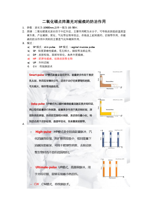
二氧化碳点阵激光对瘢痕的防治作用1.参数:波长为10600nm,功率一般为10-50W.2.原理:二氧化碳激光波长位于中红外区,主要作用靶为水分子,可导致皮肤组织温度显著升高,产生凝固,炭化,气化等生物学效应,在临床上起到烧灼,切割等作用。
在瘢痕的防治作用中用到的主要是气化和凝固作用。
3.模式:a)SP模式:skin pulse DP模式:vaginal mucosa pulseb)SP:轻度萎缩性瘢痕,毛孔粗大,细纹等浅部应用。
c)DP:皮肤松弛,面部年轻化,各类中度瘢痕。
d)HP:肥厚性瘢痕,祛除皮肤赘生物e)UP:外科切割f)CW:传统换肤术4.5.禁忌症i.治疗前(至少1个月)、治疗时、治疗后应避免阳光和紫外(UV)灯照射。
ii.治疗前(至少1周)停用下列药物:iii.抗凝剂(如阿司匹林、肝素等)iv.类维生素A—这些药物在愈合过程中会带来伤疤等问题(如异维A酸等)v.光敏剂(如四环素[抗生素]、萘普生[非甾体类抗炎药]、金诺芬[抗风湿药]、雌激素和孕激素[口服避孕药]、氯喹[抗疟药]等)vi.近期有表皮脱落性治疗(脱皮、磨皮、维生素A酸、之前的激光换肤或皮肤磨削术)或外科治疗(如除皱术等)。
vii.有皮肤病或瘢痕瘤。
viii.有孢疹病毒感染。
6.注意事项:i.激光治疗前应当注意是否有怀孕,是否糖尿病,是否局部有金属物。
ii.在只使用二氧化碳激光辐射治疗时,不能皮肤和电极是不能接触水的。
iii.在使用射频时,应当使电极接触部分充分湿润,放置局部电流不稳,灼伤皮肤。
iv.在射频使用过程中,在不能保证电极充分接触皮肤的情况下,不能应用。
v.激光治疗后应用防晒霜SPF50左右。
vi.为了减轻炎症反应,激光治疗后可以冰敷,每天生理盐水清洗脸部(3次/天,共三天)清洗完成后可以用红霉素软膏涂抹,3天后恢复正常保湿及防晒。
vii.激光之后后3天避免接触热水。
知情同意书点阵激光需多次治疗,影响治疗次数和疗效的因素有:病种、部位、时间、病变深浅、年龄、性别、对点阵激光治疗的反应、个体差异、生活习惯等,局部治疗每次间隔3-4周,全面部治疗每次间隔4-6周。
腺泡状软组织肉瘤的免疫治疗进展

腺泡状软组织肉瘤的免疫治疗进展
周铁;李茹恬
【期刊名称】《中国肿瘤外科杂志》
【年(卷),期】2024(16)1
【摘要】腺泡状软组织肉瘤(ASPS)是一种极罕见的、预后差的恶性肿瘤,发病率占所有软组织肉瘤<1%。
其标准治疗手段主要是手术切除,化疗敏感性差。
近年来免疫治疗为局部晚期或转移性ASPS的治疗提供了新的治疗方案,尤其是免疫检查点抑制剂为治疗ASPS最有效的免疫治疗药物,也是非常有前景的新领域。
该文回顾性分析了免疫检查点抑制剂及与靶向、放疗等联合模式免疫治疗ASPS的临床疗效,就ASPS免疫治疗的现状与进展进行综述。
其中伴有远处转移的ASPS患者接受免疫治疗可获得较好的临床疗效,尤其是阿昔替尼联合帕博利珠单抗,在免疫治疗过程中,需选择合适的生物标志物,关注免疫微环境对疗效的影响及根据程序性死亡受体-1/配体-1(PD-1/PD-L1)的表达模式来指导个体化治疗。
【总页数】6页(P100-105)
【作者】周铁;李茹恬
【作者单位】徐州市医科大学附属医院内科;南京大学医院附属鼓楼医院/南京鼓楼医院肿瘤中心
【正文语种】中文
【中图分类】R73
【相关文献】
1.腺泡状软组织肉瘤组织起源和分子遗传学研究进展
2.腺泡状软组织肉瘤药物治疗现状与进展
3.腺泡状软组织肉瘤生物学特征及靶向治疗的研究进展
4.舌根腺泡状软组织肉瘤误诊异位甲状腺1例
5.软组织肉瘤免疫治疗进展
因版权原因,仅展示原文概要,查看原文内容请购买。
激光医学

激光医学1.世界上第一台激光器是在何时何地由谁研制成功的?1960年美国科学家梅曼(Maiman)在加利福尼亚州休斯研究所,研制成功了世界上第一台“红宝石激光器”。
2.激光的全称?激光的全称为“受激辐射光放大”,英文light amplification by stimulated emission of radiation,简称laser。
3.激光的本质是什么?激光和普通光并无本质上的差别,它们都是电磁波,都具有波粒二象性。
4.电磁波和辐射能。
根据麦克斯韦的电磁场理论,变化的电场产生磁场,变化的磁场产生电场。
若在空间某一区域存在周期性变化的电场,在它邻近的区域则会产生周期性变化的磁场。
这种变化的磁场又在较远的区域引起变化的电场。
如此下去,变化的电场和磁场不断地交替产生,使变化的电磁场由近至远传播出去,这种变化的电磁场在空间的传播称为电磁波。
辐射能是指以电磁波的形式传播的能量。
5.哪些现象证明了光具有波动性?光在传播过程中的干涉、衍射和偏振现象,有力地证明了光具有波动性。
干涉是波动的基本特征,产生干涉现象的波叫相干波。
所谓干涉是指两列相干波相遇,在叠加的区域出现波的强度重新稳定的分布,在某些区域合振动始终加强,而在另一些区域合振动始终减弱的现象。
衍射现象是指光通过夹缝后,传播方向发生了改变,这种现象在激光的发射中也存在。
光的干涉现象和衍射现象说明光具有波动的性质,但还没有说明光是横波还是纵波。
光的偏振现象则可以说明光是横波,即振动方向与传播方向相垂直。
6.原子的能级、基态、激发态。
根据玻尔理论可得,原子的能量是按绕核旋转的电子的不同轨道半径一级一级分开的。
我们把代表不同原子能量值的档级称为能级。
其中,原子的最低能级称为基态,除此以外的高能级称为激发态。
7.什么是自发辐射?原子从高能级跃迁到低能级有哪些释放能量的方法?原子不受外界影响时,处于高能级的原子有一定的几率自发地向低能级跃迁而发光,这种发光过程称为自发辐射。
外文翻译---激光的组织接合:激光束光斑尺寸和研究概况
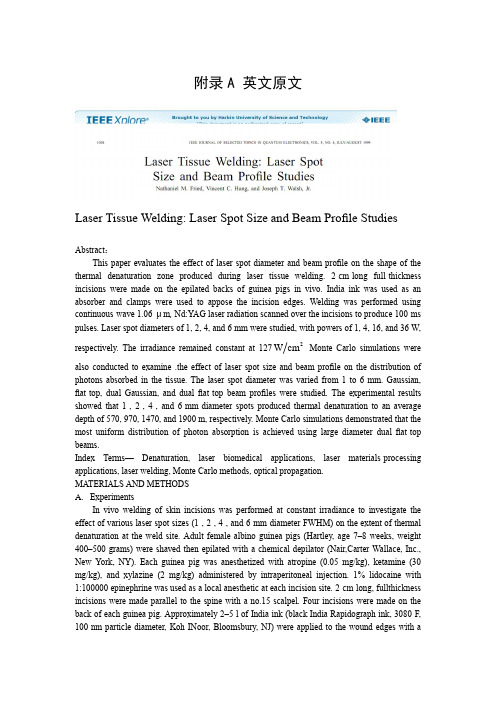
附录A 英文原文Laser Tissue Welding: Laser Spot Size and Beam Profile Studies Abstract :This paper evaluates the effect of laser spot diameter and beam profile on the shape of the thermal denaturation zone produced during laser tissue welding. 2-cm-long full-thickness incisions were made on the epilated backs of guinea pigs in vivo. India ink was used as an absorber and clamps were used to appose the incision edges. Welding was performed using continuous-wave 1.06-μm , Nd:YAG laser radiation scanned over the incisions to produce 100-ms pulses. Laser spot diameters of 1, 2, 4, and 6 mm were studied, with powers of 1, 4, 16, and 36 W, respectively. The irradiance remained constant at 1272cm W Monte Carlo simulations were also conducted to examine .the effect of laser spot size and beam profile on the distribution of photons absorbed in the tissue. The laser spot diameter was varied from 1 to 6 mm. Gaussian, flat-top, dual Gaussian, and dual flat -top beam profiles were studied. The experimental results showed that 1-, 2-, 4-, and 6-mm-diameter spots produced thermal denaturation to an average depth of 570, 970, 1470, and 1900 m, respectively. Monte Carlo simulations demonstrated that the most uniform distribution of photon absor ption is achieved using large diameter dual flat -top beams.Index Terms — Denaturation, laser biomedical applications, laser materials-processing applications, laser welding, Monte Carlo methods, optical propagation.MATERIALS AND METHODSA. ExperimentsIn vivo welding of skin incisions was performed at constant irradiance to investigate the effect of various laser spot sizes (1-, 2-, 4-, and 6-mm-diameter FWHM) on the extent of thermal denaturation at the weld site. Adult female albino guinea pigs (Hartley, age 7–8 weeks, weight 400–500 grams) were shaved then epilated with a chemical depilator (Nair,Carter-Wallace, Inc., New York, NY). Each guinea pig was anesthetized with atropine (0.05 mg/kg), ketamine (30 mg/kg), and xylazine (2 mg/kg) administered by intraperitoneal injection. 1% lidocaine with 1:100000 epinephrine was used as a local anesthetic at each incision site. 2-cm-long, fullthickness incisions were made parallel to the spine with a no.15 scalpel. Four incisions were made on the back of each guinea pig. Approximately 2–5 l of India ink (black India Rapidograph ink, 3080-F, 100-nm particle diameter, Koh-INoor, Bloomsbury, NJ) were applied to the wound edges with amicropipette. The animal was then placed prone on a translation stage, in preparation for surgery. Clamps were used to temporarily appose the incision edges during welding.Welding was performed with a continuous-wave (CW), Nd:YAG laser (Lee Laser, Model 703T) emitting 1.06m μ radiation that was coupled into a 600m μ -core diameter optical fiber (Thor Labs, Newton, NJ). A stepper-motor-driven translation stage (Newport, Irvine, CA) scanned the laser beam along the axis of the weld site at speeds that effectively produced 100-ms-long pulses. Seventy scans were made along each weld; the beam stopped at the end of the weld site for 10 s after each scan. To minimize thermal damage to the skin beyond the weld area, high-reflecting metal plates placed on each end of the incision blocked the beam. Experiments were performed at constant irradiance (1272cm w ) comparing laser spot diameters of 1, 2, 4, and 6 mm [full-width at full-maximum(FWHM)], with laser output powers of 1, 4, 16, and 36 W, respectively. The beam profile, as measured by scanning a 200- m-diameter pin hole across the beam, was approximately Gaussian for all spot diameters. The power delivered to the tissue was measured before each weld with a power meter (Molectron PowerMax 5100, Portland, OR). It shows the experimental configuration use d for dye-assisted laser skin welding and summarizes the laser parameters for this study.After welding, the anesthetized guinea pig was euthanized with an intracardiac overdose of sodium pentobarbitol (Nembutal, Abbott Laboratories, North Chicago, IL). The dorsal skin, including epidermis and dermis, was excised with a scalpel and then sectioned. Samples were processed using standard histological techniques, including storage in 10% formalin, processing with graded alcohols and xylenes, parafin embedding, s ectioning, and hemotoxylin and eosin staining. A minimum of seven samples was processed for each laser spot diameter and beam profile. The 6-mm-diameter spot study was discontinued after grossly obvious burns developed at the wound site.Thermal denaturation measurements were made using a transmission light microscope (Nikon, Japan) fit with crossed linear polarizers (Prinz, Japan). Thermal denaturation was measured laterally from the center of the weld site at three different depths: the papillary dermis, mid-dermis, and base of the dermis. The depth to which one observed denaturation was recorded and divided by the skin thickness to obtain the fraction of a full-thickness weld that was achieved. Measurements were made consistently to the point at which complete thermal denaturation of the tissue was observed.Statistical analyzes were conducted on the histological data. ANOV A was used to determine statistical significance of thermal denaturation measurements between laser spot size groups.B. Monte Carlo SimulationMonte Carlo simulations were run to investigate the effect of various spot sizes (1–6-mm diameters) and beam profiles (Gaussian versus flat -top and single versus dual beam) on the distribution of absorbed radiation. All simulations were run using code available over the public domain . Several changes were made in the Monte Carlo code to adapt it for use with the geometry of this application. First, because the vertical ink layer in the tissue disrupted the cylindrical symmetry assumed in the Original program, the data were stored in Cartesian rather than cylindrical coordinates and a convolution program was not used to generate the laser beam profile. The beam profile was, instead, created using a random number generator ; a large number of photons was used to create the desired beam profile. Second, the vertical ink layer was modeledas an infinite absorber extending from the skin surface to the base of the dermis with a uniform thickness of 100 m. The experimentally measured absorption coefficient for the ink, was 3500 cm. Even though histologic analysis of the welds showed variable staining of the tissue with a lateral thickness varying from 40 to 100 m, since the ink layer thickness was much greater than the probability that a photon could cross the ink layer was negligible, and the assumption that was infinite is reasonable.Third, the skin was modeled as a single dermal tissue layer with the assumption that the epidermis and subcutaneous tissue have optical properties similar to that of the dermis. Finally, even though the optical properties of tissue are known to be temperature-dependent, with the dermal scattering coefficient initially increasing with temperature for temperatures less than 60 C then decreasing sharply at higher temperatures and the dermal ab sorption coefficient decreasing with increasing temperature , the optical properties in this model were assumed to be static. This assumption, which avoided a complete optical-thermal model, will result in a slight underestimation of the penetration depth of the photons in the dermis. The optical properties of guinea pig skin at a wavelength of 1.06m μ have not been well characterized. The optical properties for human, pig, and rat dermis were therefore. compiled from several sources. The optical properties used in the Monte Carlo simulations are listed in Table II. Note that in the experimental irradiations, the irradiance was held constant at 127 2cm W . For the simulated irradiations, the mean irradiance over the full-width, halfmaximum of each beam was constant (10 photons per 1-mm-diameter area). The grid element size in the tissue was fixed at 100 m, and the dimensions of the tissue (length width depth) were 1.0 cm1.0 cm0.5 cm, respectively. The tissue thickness was, in part, chosen based on the knowledge that human skin may be thicker than guinea pig skin, ranging in thickness from 1 to 4 mm. Simulations were run on a Pentium 133 MHz PC computer (Micron, Nampa, ID)running Microsoft Windows 95 (Microsoft, Redmond, WA) III. RESULTSA. ExperimentsHistologic analysis showed that only shallow welds were achieved using a 1-mm-diameter laser irradiation area. Thermal denaturation was observed only to a depth of 570±100m μ (mean ±S.D.,n=7) or 30% of the average dermal thickness of 1900±200m μ , see Table III. Thermal denaturation lateral to the incision was limited to m μ30100± near the tissue surface. An image of a weld created with a 1-mm-diameter spot is shown in.When the laser spot diameter was increased to 2 mm, thermal denaturation was observed down to the middle layers of the dermis, as show. The thermal denaturation extended to an average depth of m μ210970±(n=7)(p<0.001) or 50% of the dermal thickness. This depth was significantly greater than achieved with a 1-mm-diameter spot Significantly more lateral thermal denaturation was also measured at the surface of the skin, m, than for the 1-mm-diameter spot.Increasing the spot diameter to 4 mm resulted in welds with an average depth of m μ1901470±(n=7) , or 80% of the dermal thickness 。
第五章-肿瘤激光治疗学
第一代光敏剂
第一代光敏剂 HpD 是由 8 种组分组成的混 合制剂,其有效成分主要是双血卟啉醚或酯 (DHE) ,约占药物总量的 20-30% 左右。
光敏素 Ⅱ ( Photofrin Ⅱ) 是 HpD 二期 精制、提纯以后的产物, DHE 等有效成分的 含量在 80% 以上。
光源和传导系统
靠性也是决定治疗效果重要的可控因素。
光敏剂
定义:在光化学反应中,只吸收光子并将能 量传递给那些不能吸收光子的分子,促其发 生化学反应,而自己则不参与化学反应,这 类分子就称为光敏剂。
有氧分子参与的伴随生物效应的光敏反应称 为光动力反应,把可引发光动力反应破坏细 胞结构的药物称为光动力药物,即光敏剂。
早期的光源: 利用灯泡来做体表照射,特别是 皮肤,通过过滤取得所需波长的光,去掉其它 能引起发热的光。这种光源的不足之处是在光 的传递、光的控制、精确性方面都受到限制。
激光以其单色性好、方向性好、功率大、亮
度高、相干性好的优点,可以更有效地激发光 动力反应。
光动力治疗对激发光源的要求
激光波长在450-1000nm 之间,治疗表浅病变一般 选用绿光和黄光,治疗深部病变或瘤体较大的肿 瘤多选择红光和近红外光;
目前,在欧美日等许多发达国家,光动力 治疗作为一种肿瘤治疗的新技术,已经获 得政府主管机构的审查批准,在越来越多 的医院成为一种新的常规治疗手段,基础 研究不断深入,临床应用日益广泛。
产业界也在加快新型光敏药物和配套设备 的研究制步伐,以满足医疗市场不断增长 的需要。
我国对光动力治疗的研究起步并不晚,完 成的临床病例数更堪称世界第一,在上个 世纪八十年代曾经出现过一个研究热潮。
1984年, Roswell Park 癌症研究所从 HpD 中分离 出高效组分 , 命名为 photofrinII( 即后来商品化 的 PHOTOFRIN II) 。自此,世界上大多使用 photofrinII 作为基本的光敏剂。
两种不同波长激光小梁成形术后急性期组织学形态比较
两种不同波长激光小梁成形术后急性期组织学形态比较陈蕾;李昂;刘冬娟;刘哲丽;柳力敏【期刊名称】《中国激光医学杂志》【年(卷),期】2002(11)3【摘要】目的探讨选择性激光小梁成形术 (SLT)的优越性及可能的作用机制。
方法选取因恶性肿瘤而摘除的人眼球 5只 ,经检查房角结构正常 ,摘除后立即行SLT和氪绿激光小梁成形术 ,每只眼球的颞侧做180°范围的SLT ,Q 开关Nd∶YAG激光波长 5 32nm ,光斑直径4 0 0 μm ,每脉冲持续 3ns,能量为 1 5~ 2 1mJ,总能量 5 2 4 6mJ,共 5 0个非重叠光斑。
每只眼的鼻侧180°范围做氪绿激光(5 2 1nm)小梁成形术 ,光斑直径5 0 μm ,脉冲时间 0 1s ,总能量6 0 0~ 80 0mJ,共 5 0个光斑。
术后取小梁组织制成切片 ,于光镜和电镜下进行观察比较。
结果两种激光均能有效地进行小梁成形术 ,但SLT对小梁损伤小 ,且仅对色素性小梁内皮细胞起作用,对非色素细胞无损伤作用。
氪绿激光小梁成形术则破坏小梁网细胞。
结论SLT治疗开角型青光眼安全有效 ,损伤小 ,其房水流出量的增加可能与激光刺激小梁干细胞 ,使其参与小梁结构重建 ,恢复小梁正常功能 ,增强滤过有关。
【总页数】5页(P161-165)【关键词】激光小梁成形术;组织学;SLT;开角型青光眼【作者】陈蕾;李昂;刘冬娟;刘哲丽;柳力敏【作者单位】中国医科大学附属第一医院眼科【正文语种】中文【中图分类】R775.2;R779.63【相关文献】1.三种不同波长激光猴眼小梁成形术的组织学观察和比较 [J], 孙静芬;叶纹;钟一声;王康孙2.两种改良悬雍垂腭咽成形术后悬雍垂形态的比较 [J], 王巍;孙沛勇;张文华;王娜3.三种波长的激光小梁成形术临床疗效比较 [J], 孙静芬;叶纹;余梓逵;王康孙4.两种不同固定策略的重睑成形术对先天性单睑就医者术后形态美观度的影响 [J], 柴剑;张艳;王莹5.氩激光小梁成形术——单色和双色波长激光的比较 [J], 王夷传因版权原因,仅展示原文概要,查看原文内容请购买。
激光退火对Inconel718微观组织和硬度的影响_刘六法
3期
刘六法等:激光退火对 Inconel 718 微观组织和硬度的影响
·503·
图可以看出,退火层厚度随激光扫描速度增大而 减小。这是由于激光移动快时,沿扫描方向单位 长度的表面接受激光照射时间短,因此所吸收的 激光能少。
退火层内的炭化物粒子和δ粒子的变化利用光 学显微镜和扫描电镜(SEM)进行观测,微细的 γ"和 γ′ 粒子的变化则利用透射电镜(TEM)进行观察。
3 结果与讨论
3.1 退火层的形成 试样经激光照射后,表面层的状态随激光工艺参
数的变化而不同,一般可以分为 3 种:表面被熔化; 退火层形成而没有熔化;退火层没有形成。所有试样 的中心(母材)没有变化。上述 3 种表面层的光学显微 镜照片如图 1 所示。
定厚度表面层内的硬度降低到标准退火合金的水平,而不影响试样内部母材的硬度。显微组织观察显示表面层的基体
强化相(γ"和 γ′ ) 在激光照射过程中被固溶,而其它二次相没有变化。γ"和 γ′的固溶被确定是表面层硬度下降的原因。在
其它试验条件不变时,确立了退火层生成时由激光散焦距离和扫描速度描述的工艺参数范围。
Inconel 718 有很高的氢脆敏感性,特别是在一般 工业应用的热处理状态,即时效状态下尤其严重 [4-12]。 由于氢脆能够引起延迟失效,该合金在氢环境中应用 时有发生延迟失效的潜在危险性。确立能有效降低氢 脆敏感性的方法是一个极其重要的课题。虽然退火态 的 Inconel 718 具有远低于时效态合金的氢脆敏感性, 但其强度水平(比时效态低 40%)一般难以符合应用要 求。因此,特别需要有一种既能保持时效合金的强度 水平,又能降低氢脆敏感性的科学方法。由于氢脆裂
1Cr18Ni9Ti激光相变硬化层组织及性能
1Cr18Ni9Ti激光相变硬化层组织及性能邱星武【摘要】为了探讨1Cr18Ni9Ti不锈钢组织及性能的变化,采用激光相变硬化处理的方法,利用扫描电子显微镜、X射线衍射仪、显微硬度计、磨损试验机、恒电位仪等研究了激光相变硬化层的组织及性能.进行了理论分析和实验验证,取得了激光相变硬化层的硬度、耐磨性、耐蚀性数据.结果表明,激光相变硬化层主要由奥氏体、马氏体、Fe-(Cr,Ni)以及Fe等组成.随着激光功率的增大,平均显微硬度先增加后减小,在功率为750W时,平均显微硬度达最大值,为223.5HK;在功率为550W时,耐磨性最好,磨损率为基体的56%.激光相变硬化处理后耐蚀性增强;最小的维钝电流密度是基体的33%,最大的钝化稳定区长度是基体的7倍.这一结果对研究1Cr18Ni9Ti不锈钢组织及性能的转变是有帮助的.%To investigate the changeof microstructure and properties, laser transformation hardening was carried out on 1Crl8Ni9Ti stainless steel, and the microstructure and properties of laser transformation hardened layers were investigated by means of scanning electron microscope, X-ray diffractometer, microhardnessmeter, abrasive wear testing machine, potentiostat etc. The hardness, wear resistance, corrosion resistance data of laser transformation hardening layer were obtained by theoretical analysis and experimental verification. The result indicated that the laser transformation hardening layer was mainly consisted of austenite, martensite, Fe-( Ct, Ni), Fe etc.With the increase of the laser power, the average microhardness of laser hardening layers increased first and then decreased. The average microhardness reached the highest (223.5HK) when laser power was 750W.Wear resistance reached the iest ( whose wear rate is 56% of the substrate ) when the power was 550W. Corrosion resistance was improved after laser transformation hardening, passivation current density of the smallest dimension was 33% of the substrate, the maximum length of the passive zone of stability was seven times of the substrate. The results are helpful to study the change of microstructure and properties of 1Crl8Ni9Ti stainless steel.【期刊名称】《激光技术》【年(卷),期】2011(035)003【总页数】4页(P425-427,432)【关键词】激光技术;激光相变硬化;1Cr18Ni9Ti;显微组织;硬度;耐磨性;耐蚀性【作者】邱星武【作者单位】四川建筑职业技术学院材料工程系,德阳,618000【正文语种】中文【中图分类】TG156.99引言激光加工技术实现了光、机、电技术相结合,是一种先进制造技术,目前正处于向传统制造技术中许多工艺过程积极渗透的阶段[1-4]。