Identification of Differently Expressed Genes in Chemical Carcinogen-induced Rat Bladder Cancers
ICHM7(step4)基因毒性杂质评估和控制◆中英

ASSESSMENT AND CONTROL OF DNA REACTIVE(MUTAGENIC) IMPURITIES IN PHARMACEUTICALS TOLIMIT POTENTIAL CARCINOGENIC RISK为限制潜在致癌风险而对药物中DNA活性(诱变性)杂质进行的评估和控制M7Current Step 4 versiondated 23 June 2014This Guideline has been developed by the appropriate ICH Expert Working Group and has been subject to consultation by the regulatory parties, in accordance with the ICH Process. At Step 4 of the Process the final draft is recommended for adoption to the regulatory bodies of the European Union, Japan and USA.M7Document History 文件历史The document is provided "as is" without warranty of any kind. In no event shall the ICH or the authors of the original document be liable for any claim, damages or other liability arising from the use of the document.The above-mentioned permissions do not apply to content supplied by third parties. Therefore, for documents where the copyright vests in a third party, permission for reproduction must be obtained from this copyright holder.ASSESSMENT AND CONTROL OF DNA REACTIVE (MUTAGENIC) IMPURITIES IN PHARMACEUTICALS TO LIMIT POTENTIALCARCINOGENIC RISK为限制潜在致癌风险而对药物中DNA活性(诱变性)杂质进行的评估和控制ICH Harmonised Tripartite GuidelineICH三方协调指南Having reached Step 4 of the ICH Process at the ICH Steering Committee meeting on 5 June 2014, this Guideline is recommended for adoption to the three regulatory parties to ICHASSESSMENT AND CONTROL OF DNA REACTIVE (MUTAGENIC) IMPURITIES IN PHARMACEUTICALS TO LIMIT POTENTIALCARCINOGENIC RISK为限制潜在致癌风险而对药物中DNA活性(诱变性)杂质进行的评估和控制1. INTRODUCTION概述The synthesis of drug substances involves the use of reactive chemicals, reagents, solvents, catalysts, and other processing aids. As a result of chemical synthesis or subsequent degradation, impurities reside in all drug substances and associated drug products. While ICH Q3A(R2): Impurities in New Drug Substances and Q3B(R2): Impurities in New Drug Products (Ref. 1, 2) provides guidance for qualification and control for the majority of the impurities, limited guidance is provided for those impurities that are DNA reactive. The purpose of this guideline is to provide a practical framework that is applicable to the identification, categorization, qualification, and control of these mutagenic impurities to limit potential carcinogenic risk. This guideline is intended to complement ICH Q3A(R2), Q3B(R2) (Note 1), and ICH M3(R2): Nonclinical Safety Studies for the Conduct of Human Clinical Trials and Marketing Authorizations for Pharmaceuticals (Ref. 3).原料药合成牵涉到使用活性化学物质、试剂、溶剂、催化剂和其它工艺助剂,导致在所有原料药及其制剂中会残留有化学合成或其降解产物、杂质。
拟南芥alpha-Dioxygenase 2原核表达,纯化及亚细胞定位预测
,2002 a,b)。van der Biezen .(1996)首次利用转
座子标签法从在番茄中分离到一个新基因,该基因
突变导致生长缓慢、植株矮小柔弱、结实率低等,因
而命名为
。Sanz (1998)通过差异显示
技术从烟草中分离到参与细菌诱导的超敏反应过程
的新基因,并命名为 (pathogeninduced oxyge
spectively. A fusion protein about 98 kD was detected by SDSPAGE and Western blot in the induced recombinant BL21 (DE3)RIPL
codon+ strain, and it accounts for 38% of the total bacterial proteins. The target protein was purified by the GST chromatography col
第5 期
张美祥等: 拟南芥 Alphadioxygenase 2 原核表达、纯化及亚细胞定位预测
817
们蛋白序列的相似性为 71.4%。拟南芥 DOX1 在蛋
白序列、表达模式与最初的从烟草中分离到的
相似,其表达受病原及抗逆过程诱导(Koeduka
,2005),拟南芥 DOX2 与最初从番茄中分离到
nase),到目前为止研究者已先后从番茄、烟草、拟南
芥、水稻、小麦等植 物中分离 到 18 个 同源基 因
(Hamberg
,2005), 并 将 其 统 一 归 类 为 al
phadioxygenase。
拟南芥中脂肪酸 alphadioxygenase 存在两个拷
采用体外染色体畸变试验检测锐钛型纳米二氧化钛的遗传毒性
收稿日期:2021-08-12;修订日期:2021-11-16作者信息:杜秀明,E-mail:**********。
*周庆云,E-mail:**********采用体外染色体畸变试验检测锐钛型纳米二氧化钛的遗传毒性杜秀明1,2,洪丽玲1,2,徐灵芝1,2,周庆云1,2,* (1.上海化工研究院有限公司,上海200062;2.上海化工院检测有限公司,上海200062)In vitro chromosome aberrationevaluation of anatase titaniumdioxide nanoparticlesDU Xiuming1,2,HONG Liling1,2,XU Lingzhi1,2,ZHOU Qingyun1,2,*(1.Testing Center,Shanghai Research Institute of Chemical IndustryCo.,Ltd.,Shanghai200062;2.Shanghai Institute of ChemicalIndustry Testing Co.,Ltd.,Shanghai200062,China)【摘要】目的:按《OECD化学品测试准则473:体外哺乳动物染色体畸变测试》的要求进行试验,检测锐钛型纳米二氧化钛诱发体外培养的哺乳动物细胞染色体畸变的能力,以评价其是否属于致突变物。
方法:试验组受试物终浓度分别设置为0.0078、0.0312、0.125、0.5和2mg/mL,同时设立溶剂对照组和阳性对照组。
试验分为有代谢活化系统短时处理组(+S9,4h)、无代谢活化系统短时处理组(-S9,4h)和无代谢活化系统连续处理组(-S9,24h)3种处理方式,将培养的中国仓鼠肺细胞(CHL)暴露于锐钛型纳米二氧化钛混悬液中,染毒相应时间后收获细胞,经低渗固定后,制片并染色。
每个浓度组选取300个分散良好的中期分裂相细胞进行染色体畸变分析。
1.Beclin1

J OURNAL OF V IROLOGY, 0022-538X/98/$04.00ϩ0Nov.1998,p.8586–8596Vol.72,No.11Copyright©1998,American Society for Microbiology.All Rights Reserved.Protection against Fatal Sindbis Virus Encephalitis by Beclin,a Novel Bcl-2-Interacting ProteinXIAO HUAN LIANG,1LINDA K.KLEEMAN,1HUI HUI JIANG,GERALD GORDON,2JAMES E.GOLDMAN,3GAIL BERRY,2BRIAN HERMAN,2†AND BETH LEVINE1*Departments of Medicine1and Pathology,3Columbia University College of Physicians andSurgeons,New York,New York10032,and Department of Cell Biology andAnatomy,University of North Carolina at Chapel Hill,Chapel Hill,North Carolina275992Received5May1998/Accepted6July1998bcl-2,the prototypic cellular antiapoptotic gene,decreases Sindbis virus replication and Sindbis virus-induced apoptosis in mouse brains,resulting in protection against lethal encephalitis.To investigate potential mechanisms by which Bcl-2protects against central nervous system Sindbis virus infection,we performed a yeast two-hybrid screen to identify Bcl-2-interacting gene products in an adult mouse brain library.We iden-tified a novel60-kDa coiled-coil protein,Beclin,which we confirmed interacts with Bcl-2in mammalian cells, usingfluorescence resonance energy transfer microscopy.To examine the role of Beclin in Sindbis virus pathogenesis,we constructed recombinant Sindbis virus chimeras that express full-length human Beclin(SIN/ beclin),Beclin lacking the putative Bcl-2-binding domain(SIN/beclin⌬Bcl-2BD),or Beclin containing a pre-mature stop codon near the5terminus(SIN/beclin stop).The survival of mice infected with SIN/beclin was significantly higher(71%)than the survival of mice infected with SIN/beclin⌬Bcl-2BD(9%)or SIN/beclin stop (7%)(P<0.001).The brains of mice infected with SIN/beclin had fewer Sindbis virus RNA-positive cells,fewer apoptotic cells,and lower viral titers than the brains of mice infected with SIN/beclin⌬Bcl-2BD or SIN/ beclin stop.Thesefindings demonstrate that Beclin is a novel Bcl-2-interacting cellular protein that may playa role in antiviral host defense.The cellular antiapoptotic gene bcl-2represents a novel class of antiviral host defense molecules which function both by restricting viral replication and by preventing virus-induced cell death.Bcl-2blocks apoptosis in vitro induced by several different RNA viruses,including Sindbis virus(18,41),influ-enza virus(13,26),reovirus(31),Semliki Forest virus(34), LaCrosse virus(29),and Japanese B encephalitis virus(21). Previously,we have shown that Bcl-2overexpression in virally infected neurons in vivo also protects mice against fatal en-cephalitis caused by the prototypic alphavirus,Sindbis virus (17).The protective effects of Bcl-2against fatal Sindbis virus encephalitis were associated with a reduction both in neuronal apoptotic death and in central nervous system(CNS)viral replication.A similar antiviral effect of Bcl-2overexpression has been observed during Sindbis virus infection in cultured AT3cells(41)as well as during influenza virus infection of MDCK cells(26),Japanese B encephalitis virus infection of N18cells(21),and Semliki Forest virus infection of AT3cells (34).Although the role of endogenous Bcl-2in antiviral de-fense has yet to be evaluated,these studies support the hy-pothesis that Bcl-2may be important in protecting cells against viral infections.Most previous studies examining the effects of Bcl-2on viral infections have been with neurotropic RNA viruses(e.g.,Sind-bis virus,reovirus,Semliki Forest virus,LaCrosse virus,and Japanese B virus)(18,21,29,31,34,41).Although Bcl-2may affect viral replication and virus-induced apoptosis with non-neurotropic viruses(e.g.,influenza virus)(13,26),host mech-anisms to inhibit apoptosis may be of particular importance during viral infections of vital,nonrenewable cell populations such as neurons.In such instances,virus-induced apoptotic death of neurons may result in irreversible CNS pathology and death of the host organism(1,17,19,25).Therefore,while apoptosis in other cell types may be an adaptive host defense strategy that reduces total viral burden for the organism(re-viewed in references11,16,36,and39),unique strategies may have evolved to permit control of CNS viral replication without inducing apoptotic death of infected neurons.It is thus possi-ble that cellular genes that play a role in preventing apoptosis during normal neuronal development(e.g.,bcl-2and bcl-x L) (reviewed in reference23)may also be important in regulating CNS viral replication and in defending against apoptosis in-duced by neurotropic viruses.To further understand how the cellular gene bcl-2exerts antiapoptotic and antiviral effects during CNS viral infection, we performed a yeast two-hybrid screen to identify Bcl-2-in-teracting gene products in adult mouse brain.In this study,we describe the identification of a novel Bcl-2-interacting gene product,which we named Beclin because of its predicted coiled-coil structure(hence,the“-in”suffix)and its interaction with Bcl-2(Becl).Like Bcl-2overexpression,Beclin overex-pression in neurons in vivo can inhibit Sindbis virus replication, reduce CNS apoptosis,and provide protection against fatal Sindbis virus infection.A Beclin construct lacking the putative Bcl-2-binding domain provides no protection and has no anti-viral activity.Thus,ourfindings identify a novel protein,Bec-lin,which may play a role in host defense against Sindbis virus infection.In addition,they suggest that interactions with Bcl-*Corresponding author.Mailing address:Department of Medicine, Columbia University College of Physicians and Surgeons,630W.168th St.P&S8-444,New York,NY10032.Phone:(212)305-7312.Fax: (212)305-7290.E-mail:Levine@.†Present address:Department of Cellular and Structural Biology, University of Texas Health Science Center at San Antonio,San An-tonio,TX78284.85862-like proteins may be important for the protective activity of Beclin.MATERIALS AND METHODSPlasmids.To construct vectors for transient expression in mammalian cells, the open reading frame of human bcl-2,flag epitope-tagged human beclin,and flag epitope-tagged beclin deleted of nucleotides238to453were cloned into pSG5(Stratagene).To construct viral cDNA clones,the previously described neurovirulent double subgenomic Sindbis virus vector dsTE12was used(17). Full-lengthflag epitope-tagged human beclin,human beclin containing a stop codon inserted at nucleotide position270,human beclin containing an in-frame deletion of nucleotides238to453,human bcl-2,and human bcl-2containing a stop codon at nucleotide position118were cloned into the Bst EII site of dsTE12 to generate plasmids SIN/beclin,SIN/beclin stop,SIN/beclin⌬bcl-2BD,SIN/bcl-2, and SIN/bcl-2stop,respectively.To construct vectors for yeast two-hybrid studies, the sequences encoding amino acids(aa)1to218of human bcl-2,1to212of bcl-x L,1to149of bcl-x S,and1to171of bax were cloned into pGBT9in frame with the GAL4DNA-binding domain.To avoid difficulties with targeting of proteins to the nucleus,the sequences encoding C-terminal transmembrane domains of bcl-2family members were omitted.Control pGBT9plasmids con-taining lamin(pLAM5Ј)and p53(pVA3)inserts were obtained from Clontech. Yeast two-hybrid screen.Saccharomyces cerevisiae SFY526cells were cotrans-formed with pGBT9/bcl-2and106cDNA molecules from an adult mouse brain library fused to a GAL4activation domain vector(pGAD10;Clontech),plated onto SD medium lacking tryptophan and leucine,incubated at30°C for4days, and then screened for LacZ activity by a colony liftfilter assay.Putative inter-acting clones were isolated by manipulation in leuB Escherichia coli,and further tested against pGBT9and control plasmids.A positive-galactosidase reaction between pGBT9/bcl-2and clone F1was obtained within10to15min.For analysis of interactions between Beclin and Bcl-2family members,pGBT9plas-mids containing bcl-2family members were cotransformed with fragments of human beclin(1to450,262to450,and1to708)fused to the GAL4activation domain in pGAD424,and transformants were screened for LacZ activity. Sequencing and analysis of human beclin.The partial nucleotide sequence of mouse beclin obtained from sequencing clone F1was aligned with an overlapping clone GT197isolated from human breast(32).Primers immediately upstream and downstream of the predicted open reading frame were used to amplify the coding sequence of human beclin from a normalized human infant brain cDNA library(37).The resulting PCR products from several independent reactions were cloned into pCRII and sequenced in both directions,using Sequenase(U.S. Biochemical)as well as automated sequencing.The resulting nucleotide se-quence and deduced amino acid sequences were used to scan various data banks (GenBank,EMBL,SwissProt,and PIR)for homologous sequences,using the BLAST algorithms(2).The amino acid sequence was also analyzed by the PROSITE program to identify functional motifs and by the COILS program to identify coiled-coil regions(22).Northern blot analysis.Human and mouse multiple tissue blots(Clontech) were hybridized with32P randomly labeled human or mouse beclin probes(nu-cleotides1to485)as instructed by the manufacturer(Clontech).Equal loading [2g of poly(A)RNA]was confirmed by hybridization to a-actin probe. Production of recombinant viruses.The viruses SIN/beclin,SIN/beclin stop, SIN/beclin⌬Bcl-2BD,SIN/bcl-2,and SIN/bcl-2stop were generated from viral cDNA clones as previously described(17).Briefly,5Ј-capped transcripts were synthesized from cDNA clones linearized with Pvu I(for beclin-containing vi-ruses)or Xho I(for bcl-2-containing viruses),transcribed in vitro with SP6DNA-dependent RNA polymerase,and transfected into BHK cells by using Lipofectin according to the manufacturer’s instructions.Twenty-four hours after transfec-tion,virus particle-containing supernatants of transfected cell monolayers were collected,frozen in aliquots at70°C,and used for all subsequent experiments. Titers of stock viruses were determined by plaque assay titration on BHK-21 cells.Animal experiments.Ten-day-old litters of CD1mice were inoculated intra-cerebrally into the right cerebral hemisphere with1,000PFU of each recombi-nant virus in0.03ml of Hanks’balanced salt solution.For mortality experiments, three to six separate litters were inoculated with each virus,and mortality was determined by daily observation of the mice for3weeks after infection.For virus titration and histopathology experiments,three to six mice per experimental group were sacrificed at days1,2,and4after inoculation.The right cerebral hemisphere was dissected and stored atϪ70°C,and freeze-thawed tissues were used to prepare10%homogenates in Hanks’balanced salt solution for plaque assay titration.The left cerebral hemisphere wasfixed by immersion in3% paraformaldehyde.Histopathology.Paraformaldehyde-fixed mouse brains were embedded in par-affin,and a series of4-m parasagittal sections were cut at the level of the olfactory bulb,extending caudally from the bulb to the cerebellum and medulla. For each brain,sequential sections were stained by hematoxylin and eosin to detect histopathology,in situ end labeling(ISEL)to detect apoptotic nuclei,in situ hybridization to detect Sindbis virus RNA,and immunoperoxidase to detect flag-Beclin protein expression.ISEL and in situ hybridization were performed by methods identical to those described previously for SIN/bcl-2-infected mouse brains(17).Immunoperoxidase staining to detectflag-Beclin protein expression in SIN/beclin-,SIN/beclin⌬Bcl-2BD-,and SIN/beclin stop-infected mouse brains was performed by using the monoclonal anti-flag antibody M2(5g/ml;VWR) and the avidin-biotin peroxidase method(Vectastain ABC kit;Vector Labora-tories)according to the manufacturer’s instructions.The number of virus RNA-positive and ISEL-positive cells in each brain section was quantitated with Image-ProPlus software.To calculate the number of positive cells per brain section,nonoverlapping0.25-mm2microscopicfields spanning the entire brain section were scanned with a10ϫobjective and constant settings for brightness,contrast,and threshold values for positive events.The number of positive events(i.e.,RNA-positive cells or ISEL-positive cells)for each brain section was determined by adding the sum of all individualfields analyzed.The total number of positive events per brain section was divided by the total area of the brain section to yield the average number of RNA-positive or ISEL-positive cells per square millimeter of brain.Sections of hippocampus and anterior cortex from an adult human were stained with843,a polyclonal antibody against a human Beclin peptide corre-sponding to aa1to15(1:200dilution;Eurogenetics,Seraing,Belgium),and human Beclin was detected by the avidin-biotin peroxidase method.Plasmid transfections.Plasmids pSG5/bcl-2and pSG5/flag-beclin or pSG5/ bcl-2and pSG5/flag-beclin⌬Bcl-2BD(1g of each)were transiently transfected into COS7cells by using Superfect(Qiagen)according to the manufacturer’s instructions.Protein detection.For immunofluorescence studies,COS7cells werefixed24h after transfection with100%ethanol.Expression offlag-Beclin andflag-mutant Beclin⌬Bcl-2BD mutant constructs was detected with an anti-flag epitope anti-body(M2;1:20;VWR)andfluorescein isothiocyanate(FITC)-conjugated horse anti-mouse immunoglobulin G.Bcl-2expression was detected with a polyclonal rabbit anti-Bcl-2antibody(1:100;Pharmingen)and rhodamine-conjugated goat anti-rabbit antibody.SERCA(endoplasmic reticulum Ca2ϩATPase)was de-tected with an anti-SERCA antibody(1:500;Research Design,Inc.).Western blot analysis to detectflag-Beclin expression in BHK cells infected with SIN/ beclin,SIN/beclin stop,and SIN/beclin⌬Bcl-2BD was performed with either an-tibody M2(20g/ml)or anti-human Beclin peptide antibody843(1:200)and enhanced chemiluminescence detection as instructed by the manufacturer(Am-ersham).FRET.Fluorescence resonance energy transfer(FRET)microscopy was per-formed as previously described on COS7cells cotransfected with bcl-2and beclin expression vectors(9).The donor(FITC)filter set had the following parameters: excitation(ex)ϭ450to490nm;dichroic mirror(dm)ϭ510nm;emission(em)ϭ590long pass.The acceptor(rhodamine)filter set had the following param-eters:exϭ546nm;dmϭ580nm,emϭ590long pass.Images obtained with these twofilter sets were used to directly quantify the intensities of eachfluoro-phore.The signal recorded from the FRETfilter set(exϭ450to490nm;dmϭ580nm;emϭ590nm long pass)is from energy that has transferred from FITC to rhodamine molecules.A mapping program described previously(9)was used to mapfluorescent cells and to quantify the intensity within each cell.Quanti-tative analysis of these mapped images required solving three equations,one for eachfilter set,which accounted for the excitation and detection of both labels in all threefilter sets as well as the concentrations of the donor and acceptor molecules and the probability of transfer.The measured quantities are expressed as follows,in which thefirst letter(uppercase)indicates thefilter set(A,accep-tor;F,FRET;D,donor)and the second letter(lowercase)indicates the labels present(a,acceptor alone;f,acceptor and donor;d,donor alone).A solution of the equation is Eϭ1/[aconc(RKϩ1)],where aconcϭ(AdFfϪFdAf)/[(AdFa/ Aa)ϪFd];Rϭ(DaFf/FaϪDf)/[aconc((Fa/Aa)ϪFdDa/DdAa)ϪFϭFdDf/ Dd];and K is proportional to the product of the ratio of the quantum yield of the two labels and the ratio of the absolute detection efficiencies of the two labels. Nucleotide sequence accession numbers.The GenBank accession numbers for human and mouse beclin are AF077301and AF077302,respectively.RESULTSIdentification of beclin,a novel gene on chromosome17q21. Previously,we demonstrated that Bcl-2inhibits Sindbis virus replication and prevents Sindbis virus-induced apoptosis in mouse neurons(17).To further understand the mechanisms by which Bcl-2protects against Sindbis virus infection in neurons, we performed a yeast two-hybrid screen of an adult mouse brain library for complementary DNAs encoding proteins that bind to the cell death inhibitor Bcl-2.We constructed a bait plasmid(pGBT9/bcl-2)by fusing human bcl-2(lacking the C-terminal signal-anchor sequence to ensure translocation to the nucleus)to the GAL4DNA-binding domain,which was co-transformed with an oligo(dT)and random hexamer-primed adult mouse brain cDNA fusion library in a GAL4activation domain vector,pGAD10.Of1million transformants,one positive colony was identified by the5-bromo-4-chloro-3-in-dolyl--D-galactopyranoside(X-Gal)filter assay.SequencingV OL.72,1998PROTECTION AGAINST SINDBIS VIRUS INFECTION BY BECLIN8587analysis of the cDNA plasmid rescued from this colony(F1) revealed a termination codon42bp downstream from the GAL4activation domain,several predicted short open reading frames between nucleotides124and1843and a longer pre-dicted open reading frame(with a good Kozak consensus se-quence and multiple stop codons upstream)spanning from nucleotide1855to the3Јend of the insert.Thus,either the 14-aa fusion protein was interacting with Bcl-2or one of the downstream open reading frames encoded a protein that con-tains its own activation domain and interacts with Bcl-2.To identify the Bcl-2-interacting region of F1,we fused nucleo-tides1to1854and1855to2500to the GAL4activation do-main in pGAD424and tested for interactions with Bcl-2.Nu-cleotides1855to2500,but not1to1854,encoded a protein that interacts with Bcl-2fused to the GAL4DNA-binding do-main(Table1)but not with control GAL4DNA-binding do-main plasmids containing p53,lamin(Table1),or Sindbis virus glycoproteins(data not shown).A database search revealed that the nucleotide sequence of F1(1855to2500)overlapped with sequences of several clones isolated from a normalized infant human brain cDNA library in the Merck EST(epitope tag sequence)database as well as clones from human breast(GT197)(32)and humanfibroblast (B32)cells(8).These clones contain only partial open reading frames of a novel gene that encodes a protein with coiled coils. As explained above,we assigned the name beclin to this gene because of the interaction of its encoded protein with Bcl-2 and the predicted coiled-coil structure of its encoded protein. Clones GT197and B32were both isolated in the generation of transcription maps of the breast cancer susceptibility locus on chromosome17q21and are mapped to a region located ap-proximately100kb centromeric to the gene BRCA1.They lie within a400-kb minimal deletion unit mapped by Tangir et al. in sporadic epithelial ovarian cancers(38).We aligned the overlapping partial clones in GenBank with our mouse beclin sequence to obtain a predicted sequence of the full-length open reading frame of human beclin and iso-lated human beclin from a normalized human infant brain cDNA library(37).Human beclin is predicted to encode a novel450-aa,60-kDa protein containing a coiled-coil region with25to28%homology with myosin-like proteins(Fig.1).It shares93%identity at the nucleotide level and98%identity at the amino acid level with the mouse beclin sequence identified in the yeast two-hybrid screen.Human Beclin is also homolo-gous with the Caenorhabditis elegans T19E7.3gene product (GenBank accession no.U42843)and the S.cerevisiae gene product Lph7p(GenBank accession no.U43503)(38and37% identical over145and137residues,respectively),indicating a high degree of evolutionary conservation.PROSITE analysis of human Beclin identified several potential glycosylation, phosphorylation,and myristoylation sites but no other func-tional sequence motifs.Ubiquitous expression of beclin mRNA in mouse and human tissues.To examine the tissue-specific pattern of beclin expres-sion,we hybridized mouse and human multiple-tissue North-ern blots with a beclin-specific probe.RNA blot analysis re-vealed that expression of beclin mRNA is widespread in both mouse and human adult tissue(Fig.2A).A beclin-specific probe hybridized to a2.2-kb transcript present at highest levels in human skeletal muscle but at detectable levels in all tissues examined.The size of this transcript is approximately the same as that observed previously for clones GT197and B32(8,32). In some tissues,additional1.7-and1.4-kb transcripts were observed,suggesting the presence of alternatively spliced tran-scripts.Beclin is expressed in human neurons.To examine whether Beclin protein is expressed in neurons(the primary CNS target cell type for Sindbis virus infection),we performed immuno-peroxidase staining of adult human brain sections.We used rabbit immune serum843,generated against a human Beclin peptide corresponding to aa1to15.843reacts with a61-kDa protein in lysates prepared from BHK cells after infection with a recombinant Sindbis virus containing aflag epitope-tagged human beclin insert(SIN/beclin)(Fig.3B).This protein mi-grates identically to the major band detected with an anti-flag epitope antibody in SIN/beclin-infected BHK cell lysates(Fig. 3A)and to in vitro translatedflag-Beclin(data not shown). Immunoperoxidase staining of human brain sections from the hippocampus and frontal cortex revealed Beclin immunoreac-tivity in many neurons throughout these regions as well as in some glial cells.In neurons,Beclin demonstrated a granular, punctate pattern of staining that was found almost exclusively in the region of the perikaryon(Fig.2B).Beclinimmunoreac-FIG.1.Deduced amino acid sequence of human Beclin.The boxed area represents the Bcl-2-binding domain of human Beclin(Table1),and the under-lined area corresponds to the region that is predicted to have a coiled-coil conformation.TABLE1.Summary of yeast two-hybrid assay resultsGAL4activation domain plasmid b-Galactosidase reaction with GAL4binding domain construct aEmpty Bcl-2Bcl-x L Bcl-x S Bax Lamin p53EmptyϪϪϪϩϪϪϪF11–2563ϪϩϩNDϪϪϪ1–1855ϪϪϪNDϪϪϪ1856–2563(MusBeclin1–708)ϪϩϩNDϪϪϪHuman Beclin1–708ϪϩϩNDϪϪϪBeclin1–450ϪϩϩNDϪϪϪBeclin1–258ϪϪϪNDϪϪϪBeclin262–450ϪϩϩNDϪϪϪBeclin451–708ϪϪϪNDϪϪϪBeclin1–1353ϪϪϪNDϪϪϪaϩ,positive reaction within15min;Ϫ,lack of positive reaction at24h;ND,not determined.b Nucleotide positions of genes fused to the plasmid.8588LIANG ET AL.J.V IROL.tivity was also observed in the media of blood vessels,in the ependymal cells,and in the choroid plexus.No staining was observed in human brains stained with rabbit preimmune se-rum 843(data not shown).Interaction of Beclin and Bcl-2.We performed additional yeast two-hybrid studies to confirm that human beclin ,like mouse beclin ,encodes a protein that interacts with human Bcl-2and to further define the Bcl-2-interacting region of human Beclin (Table 1).We found that the region of human Beclin that corresponds to the mouse gene product isolated in the yeast two-hybrid screen (aa 1to 236)also interacts with Bcl-2.Further deletion mutation analysis revealed that aa 88to 150of Beclin were sufficient to mediate an interaction with Bcl-2.Interestingly,the coding sequence for this region of Beclin is deleted in some human infant brain cDNA clones in the Merck EST database,suggesting that Beclin exists in at least two forms,including one that contains a Bcl-2-binding domain and one that lacks this domain.Full length-Beclin does not interact with Bcl-2in the yeast two-hybrid system.As noted below,when expressed in transient transfection assays,flag-tagged full-length human Beclin displays a punctate immuno-reactivity pattern suggestive of association with intracellular organelles and is associated with the insoluble fraction after cell lysis.In contrast,a flag-tagged truncated Beclin (aa 1to 236)(corresponding to the region isolated in the yeast two-hybrid screen)displays a diffuse cytoplasmic staining pattern and is soluble after cell lysis.These differences between full-length and truncated Beclin are thought to account for differ-ences in ability to translocate to yeast nuclei and interact with Bcl-2in the yeast two-hybrid assay.To directly examine whether full-length human Beclin and Bcl-2interact in mammalian cells,we performed FRET studies of COS7cells cotransfected with Bcl-2and flag epitope-tagged Beclin.Beclin is predicted to be a coiled-coil protein that may be associated with the cytoskeleton,and it partitions with the insoluble fraction following cell lysis.For this technical reason,biochemical analyses of in vivo interactions between Bcl-2and Beclin cannot be performed.FRET is a fluorescence technique that can be used as a spectroscopic ruler to study and quantify the interactions of cellular components with each other (re-viewed in references 6,12,and 35).In FRET,a fluorophore (donor)in an excited state may transfer its excitation energy to a neighboring chromophore (acceptor)nonradiatively through dipole-dipole interactions.The efficiency of this process varies as the inverse of the sixth power of the distance separating the donor and acceptor chromophores and,in practice,requires the distance between the donor and acceptor fluorophores to be short (usually less than 50Å).The dependence of the energy transfer efficiency on the donor-acceptor separation provides the basis for the utility of this phenomenon in the study of cell component interactions.FRET has been used by a number of investigators to examine interactions ofcellularconstituents (reviewed in 6,12,and 35)such as endosomal fusion events,ligand-dependent growth factor receptor aggre-gations,interactions of viral and cellular proteins with regula-tors of apoptosis (20),and interactions of cellular cytoskeletal components (33).Prior to measuring FRET,we first confirmed the colocaliza-tion of full-length Bcl-2and Beclin in transfected COS7cells,using confocal laser microscopy (Fig.4).Bcl-2is known to associate with the outer mitochondrial membrane,endoplas-mic reticulum,and perinuclear membranes,and it displays a punctate pattern of cytoplasmic immunoreactivity (reviewed in reference 28).We found that flag-Beclin invariably displayed a pattern of immunoreactivity identical to that of Bcl-2in all cotransfected COS7cells examined by confocal laser micro-scopic analysis (Fig.4A to C).Furthermore,we found that deletion of the putative Bcl-2-binding domain from Beclin did not alter its pattern of immunoreactivity;flag-Beclin ⌬Bcl-2BD appeared to have a pattern of staining similar to that of full-length Beclin,and like full-length Beclin,it colocalized with Bcl-2in cotransfected cells (Fig.4D to F).This granular pat-tern of Beclin immunoreactivity in the perinuclear region is similar to that observed for endogenous Beclin in human neu-rons (compare Fig.2B,4A,and 4D).After confirming the colocalization of Bcl-2and Beclin and of Bcl-2and Beclin ⌬Bcl-2BD,we used FRET analysis to de-termine whether Bcl-2and Beclin physically interact.We com-pared in transfected COS7cells the amounts of FRET between Bcl-2and full-length Beclin,Bcl-2and Beclin ⌬Bcl-2BD,and Bcl-2and a control protein,SERCA.Quantitative analysis of microscopic images (following corrections for cross-talk be-tween filter sets and donor and acceptor concentrations)showed significantly more FRET in cells with labeled full-length Beclin and Bcl-2(E Beclin-Bcl-2ϭ0.000578Ϯ0.000262;mean Ϯstan-dard error of the mean [SEM],n ϭ410)than in cells with labeled Beclin ⌬Bcl-2BD and Bcl-2(E Beclin ⌬Bcl-2BD-Bcl-2ϭ0.000189Ϯ0.000151;n ϭ2,946)(P ϭ0.0068,t test)or in cells with labeled SERCA and Bcl-2(E SERCA-Bcl-2ϭ0.0000639Ϯ0.0000390;n ϭ775)(P ϭ0.0021,t test).These quantitative analyses indicate that Beclin and Bcl-2exhibit FRET and pro-vide evidence of an interaction between these two proteins in mammalian cells.Furthermore,deletion of the Bcl-2-binding domain of Beclin mapped in yeast two-hybrid studies does not alter the spatial orientation of transfected Beclin,but it does significantly decrease FRET.This observation suggests that the FRET observed between full-length Beclin and Bcl-2re-flects a specific association of these proteins in vivo,rather than an artifact secondary to overexpression.Selective interaction of Beclin with death repressor mem-bers of the Bcl-2family.To investigate whether Beclin inter-acts with other Bcl-2family members that positively or nega-tively regulate apoptosis,we fused bax ,bcl-x S ,and bcl-x L into the GAL4binding domain vector and tested for interactions with Beclin in the yeast two-hybrid system (Table 1).The Bcl-x S GAL4binding domain construct activated transcription by itself and therefore could not be tested for interactions with Beclin.The same region of Beclin (aa 88to 150)that inter-acted with Bcl-2also interacted with Bcl-x L ,a related Bcl-2family member that inhibits apoptosis (4).In contrast,Beclin did not react with Bax,a family member that promotes apo-ptosis (27).This pattern of interaction,i.e.,with Bcl-2and Bcl-x L ,but not Bax,is identical to that observed for all previ-ously identified Bcl-2-interacting proteins outside the Bcl-2family (reviewed in reference 30).Mutations in Bcl-2and Bcl-x L that block death repressor activity also block binding to Beclin.Cheng et al.have shown that Bcl-2and Bcl-x L overexpression can delay Sindbis virus-induced death of BHK cells (5).To evaluate whether Bcl-2–Beclin and Bcl-x L –Beclin interactions may be related to this ability of Bcl-2and Bcl-x L to inhibit Sindbis virus-induced apoptosis,we constructed pGBT9vectors containing bcl-2and bcl-x L constructs with mutations in the conserved BH1domain that are known to block death repressor activity.A Gly-Ala mutation at amino acid position 145of Bcl-2completely abro-gates Bcl-2death repressor activity in interleukin-3depriva-tion-,␥-irradiation-and glucocorticoid-induced apoptosis (42)and also blocks Bcl-2binding to Beclin in the yeast two-hybrid system (Table 2).Similarly,substitution of aa 136to 138of Bcl-x L (VNX 3AIL)completely abolishes death repressor ac-tivity in Sindbis virus-induced apoptosis (5)and also blocks Bcl-x L binding to Beclin (Table 2).These mutations inBcl-2FIG.3.Expression of flag-Beclin protein constructs by the virus vectors SIN/beclin ,SIN/beclin stop,and SIN/beclin ⌬Bcl-2BD.(A and B)Western blot analyses of virus-infected BHK cell lysates with either anti-flag epitope antibody M2(A)or polyclonal rabbit anti-Beclin antiserum (B).Lane 1,SIN/beclin ;lane 2,SIN/beclin stop;lane 3,SIN/beclin ⌬Bcl-2BD;lane 4,empty Sindbis virus vector.(C to E)Immunoperoxidase staining using anti-flag epitope antibody M2of mouse brains 2days after infection with SIN/beclin (C),SIN/beclin stop (D),or SIN/beclin ⌬Bcl-2BD (E).Magnification,ϫ111.8590LIANG ET AL.J.V IROL .。
PRRS
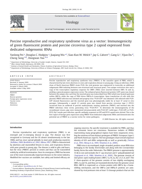
Porcine reproductive and respiratory syndrome virus as a vector:Immunogenicity of green fluorescent protein and porcine circovirus type 2capsid expressed from dedicated subgenomic RNAsYanlong Pei a ,Douglas C.Hodgins a ,Jiaqiang Wu a ,1,Siao-Kun W.Welch b ,Jay G.Calvert b ,Gang Li c ,Yijun Du d ,Cheng Song a ,d ,Dongwan Yoo d ,⁎aDepartment of Pathobiology,University of Guelph,Guelph,Ontario,Canada N1G 2W1bP fizer Animal Health,Kalamazoo,MI 49001,USA cInstitute of Animal Health and Husbandry,Chinese Academy of Agricultural Sciences,Beijing,China dDepartment of Pathobiology,University of Illinois at Urbana-Champaign,2001South Lincoln Ave,Urbana,IL 61802,USAa b s t r a c ta r t i c l e i n f o Article history:Received 31January 2009Returned to author for revision 3March 2009Accepted 31March 2009Available online 6May 2009Keywords:PRRSVReverse geneticsForeign gene expression vector Vaccine vector Nidovirus ArterivirusPorcine reproductive and respiratory syndrome virus (PRRSV)is the causative agent of PRRS,which is characterized by late-term abortions in sows and respiratory disease in young ing an infectious cDNA clone of North American PRRSV strain P129,the viral genome was engineered to transcribe an additional subgenomic RNA initiating between non-structural and structural genes.Two unique restriction sites and a copy of the transcription regulatory sequence for ORF6(TRS6)were inserted between ORFs 1b and 2a,yielding a general purpose expression vector.The enhanced green fluorescent protein (GFP)gene was cloned between the unique sites such that the inserted gene was transcribed from TRS2which was located upstream within ORF1b,while the copy of TRS6drives ORF2a/b transcription.Upon transfection of cells with this plasmid,PRRSV infection was initiated and progeny virus “P129-GFP ”was obtained.Cells infected with P129-GFP showed fluorescence and the inserted gene was phenotypically stable for at least 37serial in vitro passages.Subsequently,a capsid (C)protein gene was cloned from porcine circovirus type 2(PCV2)recovered from an outbreak of porcine multisystemic wasting syndrome (PMWS)and inserted into the PRRSV infectious clone vector,generating virus “P129-PCV ”.To determine the immunogenicity of the recombinant viruses,pigs were immunized intramuscularly with P129-WT (wild-type),P129-GFP,or P129-PCV2.By 5weeks post-infection,speci fic antibody responses to GFP and PCV2capsid were elicited.This is the first report of foreign gene expression using PRRSV from dedicated subgenomic RNAs and demonstrates the potential use of PRRSV as a vaccine vector for swine pathogens.©2009Elsevier Inc.All rights reserved.IntroductionPorcine reproductive and respiratory syndrome (PRRS)is an emerged and re-emerging disease in pigs.The disease was first recognized in Germany and the US almost simultaneously in the late 1980s and has since spread globally to most pork producing countries (Keffaber,1989;Ben field et al.,1992;Albina,1997).PRRS is characterized by abortions and mummi fied fetuses in sows,and respiratory distress with poor growth in young pigs.The disease is mild in gilts and boars,but the virus (PRRSV)persists in semen and thus can be transmitted widely by arti ficial insemination.Since its emergence,PRRS has become one of the most economically important diseases in the swine industry.Modi fied-live vaccines are available,but safety and limited ef ficacy areongoing concerns.No speci fic treatment is available for PRRS,and thus the economic losses are enormous.Numerous isolates of PRRSV representing many geographical regions have been sequenced,revea-ling the existence of two distinct genotypes of PRRSV:European (type I)and North American (type II).The two genotypes share overall sequence identity of 63%and differ antigenically as well as genetically (Nelson et al.,1993;Meng et al.,1995;Wootton et al.,2000).PRRS virus is an enveloped,single-stranded,positive-sense RNA virus belonging to the family Arteriviridae and along with the Coronaviridae family,forms the order Nidovirales .The PRRSV genome is approximately 15kb in size and includes the 5′cap structure and 3′polyadenylated tail (Sagripanti et al.,1986;Wootton et al.,2000).The genome consists of nine genes:open reading frames (ORFs)1a,1b,2a,2b,3,4,5,6,and 7.The 5′three-quarters of the genome consists of two slightly overlapping ORFs,1a and 1b,and they are translated directly from the genome-sense RNA.ORF1b is expressed as a fusion protein with ORF1a by a frame-shifting mechanism,and the ORF1a and ORFla/b proteins are auto-cleaved by viral proteases into at least 13cleavage products that are theVirology 389(2009)91–99⁎Corresponding author.E-mail address:dyoo@ (D.Yoo).1Current address:Shandong Key Laboratory of Animal Disease Control and Breeding,Shandong Academy of Agricultural Sciences,Jinan,Shandong,China.0042-6822/$–see front matter ©2009Elsevier Inc.All rights reserved.doi:10.1016/j.virol.2009.03.036Contents lists available at ScienceDirectVirologyj o u r n a l ho m e p a g e :w w w.e l s ev i e r.c o m /l o c a t e /y v i r onon-structural proteins Nsp1α,Nsp1β,and Nsp2through Nsp12.The Nsps are thought to be involved in genome replication and subgenome transcription(van Dinten et al.,1999;van Marle et al.,1999).The viral structural proteins are encoded by ORFs2a,2b,and3through7which are located downstream of ORFs1a and1b.These genes are expressed as a3′-coterminal nested set of subgenomic(sg)RNAs(de Vries et al., 1990).The5′untranslated leader sequences of the sgRNAs are derived from the5′end of the viral genome and fused to the body segments of the sgRNAs at conserved hexanucleotide motifs[5′UCAAC(U/C)3′] located immediate upstream of every transcription unit(de Vries et al., 1990;den Boon et al.,1996).The conserved hexanucleotide motif and poorly conservedflanking sequences form secondary structures in the sgRNAs that make up the transcriptional regulatory sequences(TRS)that are necessary for sgRNA formation(Pasternak et al.,2000).With the exception of sgRNA7,sgRNAs are structurally polycistronic but,with the exception of sgRNA2a/b,functionally monocistronic as only the5′most proximal gene of each sgRNA is translated.ORFs2a,2b,and3through7 code for GP2(glycoprotein2),E(envelope),GP3,GP4,GP5,M (membrane)and N(nucleocapsid)proteins,respectively,and they make up virion particles(Meulenberg et al.,1995).The recent development of infectious clones for PRRSV has allowed specific alterations of viral genomes and generation of mutant viruses(Yoo et al.,2004).However,manipulation of arterivirus genomes is complicated by the condensed organization of the viral genome.In the case of the European genotype of PRRSV,each gene overlaps slightly except for ORFs 1b and2a(Meulenberg et al.,1993),although the overlap of genes seems less important for virus replication and growth for equine arteritis virus (de Vries et al.,2000).For the North American genotype,structuralgenes Fig.1.Genomic organization of P129-WT,P129-GFP,and P129-PCV2.(A)Genomic organization and sequence of the region surrounding the ORF1b/ORF2junction of the unmodified “wild-type”P129strain of PRRSV(P129-WT).The boxed sequence indicates the core hexanucleotide of TRS2.Amino acids indicate translated sequences of polyprotein1a/b(ORF1b),the GP2protein(ORF2a),and the E protein(ORF2b).The initiation codons are indicated in bold,and the E protein sequence is italicized.Numbers in parenthesis indicate genomic sequence positions.Stars indicate translation stops.(B)P129-GFP.Unique AflII and Mlu I sites and a copy of TRS6were introduced into the non-coding region between ORF1b and ORF2a,creating expression vector pCMV-S-P129-1bMCS2.The GFP gene was amplified by PCR and inserted between the AflII and Mlu I restriction sites.(C)P129-PCV2.The PCV2 capsid gene was amplified by PCR and cloned between the AflII and Mlu I restriction sites.92Y.Pei et al./Virology389(2009)91–99also overlap with the exceptions of ORFs 1b and 2a,and ORFs 4and 5(Nelsen et al.,1999;den Boon et al.,1991;Snijder and Meulenberg,1998).Thus,the entire genome of the North American type PRRSV possesses only 4short non-coding regions:191nucleotides of 5′untranslated region (UTR),1nucleotide between ORF1b and ORF2a,10nucleotides between ORF4and ORF5,and 151nucleotides of 3′UTR upstream of the polyadenylation tail.The 5′and 3′UTRs contain genome replication and transcription signals,and therefore offer limited sites for gene insertion and manipulation.The presence of overlapping genes hampers mutational analysis of the N-and C-termini of the structural proteins and also makes it dif ficult to insert heterologous genes into the viral genome.In the present study,we used a genomic cDNA clone of PRRSV (Lee et al.,2005)to generate a vector for foreign gene expression from a dedicated subgenomic transcription unit inserted in the region between the structural and non-structural genes.The vector was used to express GFP in vitro and in vivo .The inserted gene was tolerated by the virus,stable for at least 37passages in cell culture,and induced antibodies to GFP in young pigs.Using this approach,we also generated a recombinant PRRSV expressing the capsid protein gene of porcine circovirus type 2(PCV2).PCV2is a small DNA virus in the Circoviridae family,with a genome ofonly 1.76kb.PCV2ORF1is essential for viral DNA replication (Mankertz et al.,1998;Fenaux et al.,2000),while ORF2encodes the capsid protein containing type-speci fic epitopes that are believed to be important for virus neutralization (Nawagitgul et al.,2000;Fenaux et al.,2004).Accumulating evidence suggests a major role for PCV2in postweaning multisystemic wasting syndrome (PMWS)and porcine dermatitis and nephropathy syndrome (PDNS)(Hasslung et al.,2005).These syndromes cause serious economic impacts in the swine industry today (Chae 2005).We showed the recombinant PRRSV P129-PCV2induced an anti-PCV2antibody response in immunized pigs in the presence of maternal antibodies to PCV2.Our vector construction may be applicable not only for PRRSV,but also for other members of the families Arteriviridae and Coronaviridae .ResultsDevelopment of PRRSV as an expression vector for GFPTo explore the possibility of developing PRRSV as a gene expression vector,the GFP gene was inserted between the stop codon ofORF1bFig.2.(A)Restriction patterns of P129-WT (lane 1),P129-GFP (lane 2),and P129-PCV2(lane 3)genomic clones generated by Sma I digestion.Fragments of 590bp,4736bp and 10,779bp are expected from all three clones.The fourth fragment varies in size depending on the gene inserted at the non-structural and structural gene junction.Relative to the P129-WT fragment (2787bp,lane 1),the GFP-containing fragment from P129-GFP is 766bp larger (3553bp,lane 2)and the PCV2capsid-containing fragment from P129-PCV2is 754bp larger (3541bp,lane 3).(B –E)Recovery of recombinant PRRSV foci from full-length genomic clones.(F)Integration of the GFP gene in P129-GFP viral genome.Genomic RNA was extracted from P129-GFP virus and digested with DNase I prior to reverse transcription.Without reverse transcription,no products were ampli fied from P129-GFP using ORF7-speci fic PCR primers P129-7F and P129-7R (lane 1)or primers P129-1bF and P129-2aR that span the insertion site (lane 2).With reverse transcription,primers P129-7F and P129-7R ampli fied the ORF 7fragment (534bp)from both P129-WT (lane 3)and P129-GFP (lane 4).Using P129-1bF and P129-2aR,a product of 766bp larger (lane 6)than the product from P129-WT (lane 5)was ampli fied from P129-GFP.(G)Integration of the PCV2C gene in PRRSV.Genomic RNA from P129-WT (lanes 9,10,12)and P129-PCV2(lanes 7,8,and 11)was ampli fied using primers P129-1bF and pared to P129-WT (lane 9)a product that is 754bp larger was ampli fied from P129-PCV2(lane 7).Using PCV2C gene speci fic primers PCV2-F and PCV2-R,the expected 497bp product was generated from P129-PCV2(lane 8)but not from P129-WT (lane 10).Using PRRSV ORF4-speci fic primers P129-4F and P129-4R,the expected 567bp product was ampli fied from both P129-PCV2and P129-WT (lanes 11and 12,respectively).93Y.Pei et al./Virology 389(2009)91–99and the start codon of ORF2a (Fig.1A).This non-coding region is extremely short,comprising only one adenosine nucleotide.The TRS associated with ORFs 2a and 2b (TRS2;TGAACC)is positioned 26nucleotides upstream from the start of ORF2a and is embedded in ORF1b.Upon insertion of GFP to the region,TRS2will drive transcription of the GFP gene instead of ORFs 2a and 2b.Thus,a synthetic TRS (TTAACC)with flanking sequences derived from TRS6was introduced 22nucleotides downstream of GFP and 17nucleotides upstream from the ORF2a start (Fig.1B).TRS6was chosen because RNA secondary structure suggested that it was shorter than other PRRSV TRS elements and because the distance between the copy of TRS6driving ORF2a/b and the authentic TRS6ensured that potential intramolecular homologous RNA recombination would result in a non-viable (ORF2-5deleted)virus.The PRRSV genomic clone containing GFP was designated P129-GFP,and the insertion was con firmed by restriction patterns (Fig.2A,lane 2)and sequencing.MARC-145cells were transfected with P129-GFP and the production of virus was monitored daily for development of cytopathic effect (CPE)(Figs.2B,C,D).CPE was observed 4days post-transfection and the development of CPE was one day slower than for P129-WT.After three consecutive passages,P129-GFP virus was re-examined for GFP sequence integration in the viral genome (Fig.2F).While ORF7ampli fication products were identical in size for P129-WT (lane 3)and P129-GFP (lane 4),1bF and 2aR primers produced a larger size product from P129-GFP (lane 6)than from P129-WT (lane 5).The larger product was the expected size for correct insertion of GFP gene intothe viral genome.Replication of P129-GFP was slightly slower than that of P129-WT at passages 1to 3.However,plaques were comparable in size and morphology for P129-WT and P129-GFP,and the titers at passage 3were 5×10⁵plaque forming units (PFU)/ml and 1×10⁵PFU/ml,respectively.Fluorescence was evident in P129-GFP-infected cells (Fig.3A),demonstrating the expression of GFP during infection.To examine the genetic stability of recombinant PRRSV,P129-GFP was passaged 37times in MARC-145cells and GFP expression was monitored by fluorescence microscopy.Individual plaques of 20formed by P129-GFP were randomly chosen and examined for fluorescence.The selected plaques were all positive for fluorescence (Table 1),and sequencing of the viral RNA con firmed stability of the insert (data not shown).This data showed the genetic stability of P129-GFP after serial passages in cell culture and the stable expression of GFP.It also demonstrates that the region between ORF1b and ORF2a is a suitable site for foreign gene insertion for PRRSV.This was the first demonstration of the use of a nidovirus as an expression vector wherein the foreign gene is inserted in the region between the non-structural and structural protein codingsequences.Fig.3.Expression of GFP or PCV2capsid protein by P129-GFP and P129-PCV2in MARC-145cells.(A)Live cells infected with P129-GFP passage 3;(B)live cells infected with P129-GFP passage 37;(C)uninfected cells;(D)P129-PCV2passage 3fixed and stained with PCV2-speci fic antibody conjugated with FITC at 48h post-infection;(E and F)P129-PCV2infected cells fixed and co-stained with PCV2-speci fic antibody conjugated with FITC (E)or GP4protein-speci fic monoclonal antibody 169(F).Table 1Stability of GFP expression in P129-GFP virus.Virus Titer (PFU/ml)GFP expressing plaques Passage 38.3×10e620positive/20plaques Passage 372.4×10e720positive/20plaques94Y.Pei et al./Virology 389(2009)91–99Construction of PRRSV expressing PCV2capsidPCV2is associated with porcine multisystemic wasting disease (PMWS,now termed PCVAD [PCV-associated disease])and porcine dermatitis and nephropathy syndrome (PDNS).PCV2is transmitted by the oro-nasal route and shed in the bronchial secretions and feces,thus the transmission route is similar to that of PRRSV.The capsid (C)protein is the major antigen able to elicit protective immunity against PCV2.Thus,using the PRRSV vector system described above,an additional recombinant PRRSV was constructed to carry the PCV2C gene.A 702bp C gene was cloned by PCR from a lung tissue positive for PCV2.Sequencing of the C gene showed 99–100%amino acid identity to published sequences available in the GenBank database.The P129-PCV2clone was constructed by inserting the C gene into PRRSV in the same way as the GFP gene insertion for P129-GFP (Fig.1C).The insertion of the C gene was con firmed by restriction digestion pattern (Fig.2A,lane 3)and sequencing.The P129-PCV2recombinant virus was recovered from MARC-145cells by transfection (Fig.2E),and the insertion of the C gene in the viral genome was con firmed by RT-PCR of the viral RNA (Fig.2G).The titer of passage 3virus was 2×10⁵PFU/ml and the plaque morphology was indistinguishable from P129-WT.Infection of cells with P129-PCV2and staining with PCV2-speci fic antibody produced distinct fluorescence (Fig.3E),which shows the expression of the C protein by P129-PCV2.The capsid protein of PCV2is arginine-rich and normally shuttles into the nucleus during PCV2replication (Liu et al.,2001),and similarly,the PRRSV N has also been shown to localize in the nucleus and nucleolus (Lee et al.,2006;Pei et al.,2008).Thus,in cells infected with P129-PCV2,the synthesis of legitimate PCV2capsid should be evident by the translocation of capsid into the nucleus.Thus,the C protein expression by P129-PCV2was con firmed by co-staining of virus-infected cells with PRRSV N protein-speci fic antibody and PCV2-speci fic antiserum (Fig.4).While the PRRSV N protein was found in the both cytoplasm and the nucleolus as usual (panel A),the PCV2capsid protein was speci fically localized to the nucleus and nucleolus (panel C)in the same cell,clearly demonstrating the expression of PCV2capsid protein by the recombinant PRRSVP129-PCV2.Fig.4.Co-expression of the PRRSV N protein (green)and the PCV2capsid protein (red)during infection of P129-PCV2.MARC-145cells were infected with P129-PCV2and stained 24h post-infection with PRRSV N-speci fic MAb SDOW-17(A and B)or PCV2-speci fic pig serum (C and D).Arrows indicate the PRRSV N protein in the nucleolus (panel A)in addition to the cytoplasm and the PCV2capsid protein in the nucleus and nucleolus (panel C).Panel E shows the merge of A and C.Panel F shows the merge of B and D.Table 2PCR primers and their genomic Sequence (5′–3′)aGenomic position b PurposePCV2-F cacggatattgtagtcctggt 1093–1114PCV2PCR test PCV2-R ccgcaccttcggatatactgtc1565–1586PCV2PCR testPCV2-orf2F gatgcttaagatgacgtatccaaggtggcg 1715–1734PCV2ORF2ampli fication PCV2-orf2R gtacacgcgtcattaagggttaagtcccccc 1031–1050PCV2ORF2ampli ficationP129-F1F aacagaagagttgtcgggtccac11,699–11,721P12911,783–12,055,ampli fication P129-F1R gctttcacgcgtccccacttaagttcaattcaggcctaaagttggttca 12,031–12,055Introduction of A flII and Mlu IP129-F2F gcgacgcgt gttccgtggcaacccctttaaccagagtttcagcggaaga atgaaatggggtctatacaaagcctcttcgaca 12,056–12,089P12912,056–12,697,ampli fication and introduction of Mlu I and TRS6P129-F2R aacagaacggcacgatacaccacaaa 13,819–13,844P12912,056–12,679,ampli fication P129-7F tcatccgattgcggcaaatg 14,724–14,743P129ORF7ampli fication P129-7R agaatgccagcccatca 15,242–15,258P129ORF7ampli fication P129-4F gtttcacctagaatggctg 13,213–13,231ORF4ampli fication P129-4R ccccaacatacttgaacattc 13,750–13,770ORF4ampli ficationP129-1bF ggtgaggactgggaggattac 11,921–11,941ORF1b –ORF2a,region ampli fication P129-2aRcagtacgtagcattggaacc12,758–12,777ORF1b –ORF2a,region ampli ficationa Restriction sites are underlined.TRS6and surrounding sequences are indicated in bold.bGenomic positions for PCV2primers were based on GenBank accession AF027217.Genomic position for P129primers were based on GenBank accession AF494042.95Y.Pei et al./Virology 389(2009)91–99Infection of pigs and antibody responses to GFP and PCV2capsid protein To determine antibody responses to GFP and the PCV2capsid protein,pigs were immunized with the recombinant PRRS viruses.Fifteen PRRSV-free pigs at 4weeks of age were randomly allotted to 3groups of 5pigs each.Animals were immunized twice on days 0and 21by intramuscular injection of 5×10⁵PFU per animal with either P129-WT,P129-GFP,or P129-PCV2.Following inoculation,the animalswere maintained for 5weeks for clinical observation and serum collection.Clinical signs of PRRS were minimal and comparable in all 3groups (data not shown).Mild clinical signs were not unexpected,since the infectious cDNA clone used in these studies was not attenuated.Tonsil samples were collected at necropsy on day 35and assessed by RT-PCR for the presence of PRRSV ORF7using primers P129-7F and P129-7R (Table 2)as well as the GFP and PCV2capsid inserts using primers P129-1bF and P129-2aR.All 15pigs were positive for ORF7,indicating infection and persistence of PRRSV in the tonsils.PCR products from the ORF1b/ORF2a junction were not detected in these tonsil samples,possibly due to the much lower molar ratio of ORF1b-containing RNA template relative to ORF7-containing RNA in infected cells.Alternatively,it is possible that the GFP and PCV2capsid genes might have been unstable in vivo and lost in the inoculated pigs over time.To examine antibody responses in these pigs,ELISAs were conducted for PRRSV,GFP,and PCV2C protein.All pigs produced good levels of antibodies to PRRSV (Fig.5A),and the antibody titers were comparable among groups.P129-GFP elicited speci fic antibodies to GFP,whereas pigs immunized with P129-PCV2or P129-WT were negative for GFP (Fig.5B).The GFP antibodies increased following first immunization,and the second immunization at day 21boosted the antibody response somewhat (Fig.5B).Similarly,antibodies for PCV2C protein were detected in pigs immunized with P129-PCV2(Fig.5C).In these pigs,however,PCV2antibodies were detected at day 0in all 3treatment groups and tended to wane over time.These antibodies likely represent maternal antibodies taken up in colostrum shortly after farrowing at the farm of origin and prior to experimental infection.However,an increase in anti-PCV2antibodies was observed at 28days in the P129-PCV2group only (Fig.5C)due to boosting effects from the second immunization at day 21.In contrast,anti-PCV2antibodies in the other two treatment groups waned gradually from day 0until at least until day 35.To further determine the speci fic antibody responses to GFP and the PCV2C protein,Western blots were conducted using sera from these pigs.Serum from the P129-GFP group was reactive with GFP (Fig.6A),consistent with the ELISA data (Fig.5B).Similarly,serum from the P129-PCV2group showed a strong reaction with C pro-tein (Fig.6B).Weaker reactions were observed using sera from the P129-WT and P129-GFP groups,consistent with the presence of maternalantibodies.Fig.5.ELISA showing induction of speci fic antibodies in sera from pigs inoculated with P129-GFP,P129-PCV2,or P129-WT viruses.(A)antibodies to PRRSV N protein;(B)antibodies to GFP;(C)antibodies to PCV2Cprotein.Fig. 6.Western blots showing induction of speci fic antibodies in sera from pigs inoculated with P129-GFP,P129-PCV2,or P129-WT at day 0or day 35post-inoculation.Blots contain GFP protein (A)or PCV2virions (B).Arrows indicate positions of GFP and PCV2capsid protein.C denotes positive control (anti-GFP monoclonal antibody).M indicates molecular weight markers.The virus used to infect pigs that contributed to the serum pools is indicated above each lane.96Y.Pei et al./Virology 389(2009)91–99DiscussionThe primary target cell of PRRSV is the alveolar macrophage,and pigs are the only animal species known to be susceptible to PRRSV infection.Therefore,development of PRRSV as a vaccine vector would be useful for the delivery of porcine pathogen genes to the respiratory tract of the pig.Arterivirus genomes are organized in a complex way. Most genes overlap one another in different reading frames,making it difficult to engineer the genome for foreign gene insertion.An early approach involved engineering the3′terminal region of the N gene (Groot Bramel-Verheije et al.,2000).Modification of the3′terminal sequence of N gene was possible and caused only minimal effects on virus replication and growth.However,no more than7amino acids were inserted.More recently,Nsp2,a large product of proteolytic cleavage of the ORF1a and ORF1a/b polyproteins,was found to be remarkably heterogeneous in sequence,with several hypervariable regions.Therefore,the GFP gene was inserted in-frame into or between the hypervariable regions to create nsp2-GFP fusion proteins (Fang et al.,2006;Kim et al.,2007).This approach was successful and allowed for the production of recombinant viruses.However,in all cases the inserted GFP gene was not phenotypically stable and lost greenfluorescence after several passages in cell culture(Fang et al., 2006;Kim et al.,2007;Han et al.,2007).In the present study,we inserted GFP and the PCV2capsid genes into the short region separating the non-structural protein genes from the structural protein genes.In contrast to the nsp2-GFP fusion proteins described above,we expressed GFP as a separate transcription unit resulting in an additional sgRNA.This approach has the advantage of eliminating the need to alter the coding sequence of a viral gene,and also minimizes effects on expression and post-translational modification of viral gene products.As a result,our recombinant virus was stable for at least37cell culture passages without loss of the gene or the greenfluorescent phenotype.PRRSV is known to induce immune suppressive effects in pigs (Charerntantanakul et al.,2006)and can persist up to6months in infected pigs(Wills et al.,1997).For these reasons,co-infection of PRRSV with other pathogen such as PCV2can result in much more severe clinical outcome than either agent alone(Harms et al.,2001; Kim et al.,2003).Therefore,a dual-purpose vaccine capable of protecting pigs against both PRRS and PCV2would be advantageous. Our study demonstrates the potential of PRRSV as a viral vaccine vector.Prior to the purchase of animals for infection studies using P129-PCV2,all pigs were screened for PCV2by PCR.PCR is the gold standard for detection of PCV2,whereas antibody screening is not routinely conducted in diagnostic laboratories due to cross-reactivity between PCV2and PCV1which is ubiquitous and widely distributed in thefield (Magar et al.,2000).Although all pigs entering the present study tested negative for the presence of PCV2by PCR,antibodies were detected in sera collected on day0.These antibodies were most likely of maternal origin,since they decreased in concentration over the duration of the experiment in pigs receiving P129-WT or P129-GFP. Serological studies show that maternal antibodies for PCV2decay during thefirst2months of life(Rodríguez et al.,2002;Larochelle et al.,2003).Western blots and ELISA gave comparable results in this regard.In the current study,antibodies to PCV2only increased after day21of the study and did so only in pigs receiving P129-PCV2, suggesting that the increases were most likely specific responses to P129-PCV2vaccination.At the termination of the study(day35post-infection),tonsil samples in all3groups were positive for the PRRSV N gene by RT-PCR.The presence of the inserted GFP and PCV2genes in these same samples could not be confirmed using primersflanking the region of the gene insertion.No products were amplified,even from pigs infected with the P129-WT virus.Failure to amplify the ORF1b/ORF2a junction is likely the result of template RNA concentra-tions below the level of detection of the PCR assay,and is consistent with the observation that the copy number of ORF1b(present only on genomic RNA)is much lower than the copy number of ORF7(present on all sgRNAs as well as genomic RNA).In conclusion,a PRRSV gene expression vector was generated, capable of expressing a foreign gene from an additional transcription unit located in the region between the non-structural and structural genes of the virus.The recombinant PRRSVs expressing the GFP or PCV2capsid genes were generated and shown to replicate well in cell culture.The addition of766nt(GFP)or754nt(PCV2capsid)of foreign genetic material,representing approximately5%of the PRRSV genome,was tolerated with no evidence of compensatory deletions or rearrangements elsewhere in the genome.Pigs inoculated with these recombinant PRRSVs produced foreign gene specific antibodies.Our study demonstrates the potential of PRRSV to function as a vector for development of multivalent vaccines against swine diseases.Materials and methodsCells and virusesMARC-145African green monkey kidney cells(Kim et al.,1993) were maintained as previously described(Lee et al.,2003).Dulac porcine kidney cells,kindly provided by L.Babiuk(Vaccine and Infectious Disease Organization,SK,Canada),were grown in Modified Eagle's Medium(MEM)supplemented with5%fetal bovine serum (FBS;Gibco BRL),penicillin(100U/ml),and streptomycin(50μg/ml). Cells were maintained at37°C with5%CO2.Stocks of recombinant viruses derived from infectious clones were prepared by passaging three times on MARC-145cells.Titers of PRRSV were determined by standard plaque assays on MARC-145cells using6-well plates(35mm diameter)in duplicate.Plaques were stained with0.01%neutral red. For isolation of PCV2,lung tissues were obtained from a PCR-positive pig(Ontario18099)submitted to Animal Health Laboratory of the University of Guelph(Guelph,ON,Canada).The tissues were homogenized in PBS and thefiltrate was used to infect Dulac cells. At3days post-infection,cells were stained using a porcine circovirus hyperimmune serum(VMRD,Pullman,WA,USA)to confirm infection. On day4post-infection,cells were harvested and freeze–thawed three times.Cell debris was removed by centrifugation at5000×g, and the supernatant was stored at−80°C until use.Construction of a PRRSV expression vector and recombinant PRRSVsFor construction of the PRRSV expression vector,the regions flanking the ORF1b/ORF2a junction were amplified by PCR using the shuttle plasmid p2-7D4(containing genomic positions11,504to 15,395)as template.Two DNA products corresponding to positions 11,783to12,055and12,056to12,697were amplified.The primer set P129F1-F(containing an Eco47III site)and P129F1-R(containing AflII and Mlu I sites)was used for the upstream product(Table2).The primers set P129F2-F(containing Mlu I site and TRS6)and P129F2-R (containing a Bsr GI site)was used to amplify the downstream product(Table2).The twoflanking products were digested with Eco 47III–Mlu I and Mlu I–Bsr GI,respectively,and included in a three-way ligation with Eco47III–Bsr GI-digested full-length genomic cDNA clone pCMV-S-P129(Lee et al.,2003).The resulting construct pCMV-S-P129-1bMCS2contained a complete PRRSV genome with unique AflII and Mlu I sites and a copy of TRS6inserted between ORF1b and ORF2a.Transfection of MARC-145cells with this construct produced viable virus that replicated normally(data not shown).For insertion of foreign genes,pCMV-S-P129-1bMCS2was digested with AflII–Mlu I and ligated to either the GFP gene or the PCV2capsid gene into which AflII and Mlu I sites were introduced during PCR (Fig.1).Recombinant genomic clones were screened by Sma I digestion(Fig.2),and selected clones were sequenced to confirm the presence of insertions.The PCV2capsid protein gene was cloned97Y.Pei et al./Virology389(2009)91–99。
Clin Cancer Res-2009(乳腺癌)

DNA Methylation Markers Predict Outcome in Node-Positive,Estrogen Receptor-Positive Breast Cancer with Adjuvant Anthracycline-Based ChemotherapyOliver Hartmann,1Frede rique Spyratos,4Nadia Harbeck,2Dimo Dietrich,1Anne Fassbender,1Manfred Schmitt,2Serenella Eppenberger-Castori,3VincentVuaroqueaux,3Florence Lerebours,4KatrinWelzel,1Sabine Maier,1Achim Plum,1Stephan Niemann,1John A.Foekens,5Ralf Lesche,1and JohnW.M.Martens 5AbstractPurpose:We have shown that DNAmethylation of the PI TX2gene predicts risk of distant recurrence in steroid hormone receptor-positive,node-negative breast cancer.Here,we present results from a multicenter study investigating whether PI TX2and other candidate DNAmeth-ylation markers predict outcome in node-positive,estrogen receptor-positive,HER-2-negative breast cancer patients who received adjuvant anthracycline-based chemotherapy.Experimental Design:Using a microarray platform,we analyzed DNAmethylation in regu-latory regions of PITX2and 60additional candidate genes in 241breast cancer ing Cox regression analysis,we assessed the predictive power of the individual marker/marker panel candidates.Clinical endpoints were time to distant metastasis,disease-free sur-vival,and overall survival.Anested bootstrap/cross-validation strategy was applied to identify and validate marker panels.Results:DNAmethylation of PI TX2and 14other genes was correlated with clinical outcome.In multivariate models,each methylation marker added significant information to established clinical factors.Afour-marker panel including PI TX2,BMP4,FGF4,and C20orf55was identified that resulted in improvement of outcome prediction compared with PITX2alone.Conclusions:This study provides further evidence for the PITX2biomarker,which has now been successfully confirmed to predict outcome among different breast cancer patient populations.We further identify new DNAmethylation biomarkers,three of which can be combined into a panel with PITX2to increase the outcome prediction performance in our anthracycline-treated primary breast cancer population.Our results show that a well-defined panel of DNAmethylation markers enables outcome prediction in lymph node-positive,HER-2-negative breast cancer patients treated with anthracycline-based chemotherapy.I n breast cancer,currently available methods are inadequate todetermine precisely the aggressiveness of the disease and the likelihood of response to a certain treatment in individual patients.Therefore,biomarkers predicting the tumor’s meta-static potential and its response to a specific treatment are urgently needed.This is particularly true for patients for whom current guidelines (1)recommend anthracycline-based chemo-therapy.These patients comprise a clinically distinct interme-diate-to high-risk subgroup,for which in view of the short and long-term toxicity as well as the availability of new effective anthracycline-free chemotherapy options (2),the widespread use of adjuvant anthracyclines is currently under debate (3).Unfortunately,reliable biomarkers for benefit from anthracy-cline therapy are still lacking.DNA methylation of CpG dinucleotides within gene regulatory regions,associated with suppression of gene expression,is a common and early event in cancer (4–6).Specific DNA methylation patterns for tumor subtypes including breast cancer have been reported and associated with clinical outcome (7–16).Cancer Therapy:ClinicalAuthors’Affiliations:1Epigenomics AG,Berlin,Germany;2Department of Obstetrics and Gynecology,T echnical University Munich,Munich,Germany;3Stiftung T umorbank,Basel,Switzerland;4Centre ReneHuguenin,St.Cloud,France;and 5Erasmus Medical Center,Rotterdam,The Netherlands Received 1/22/08;revised 8/25/08;accepted 9/2/08.Grant support:Epigenomics and European Union Sixth Framework Program grant LSHC-CT-2003-504586(J.W.M.Martens,J.A.Foekens,S.Eppenberger-Castori,F.Spyratos,M.Schmitt,and N.Harbeck).The costs of publication of this article were defrayed in part by the payment of page charges.This article must therefore be hereby marked advertisement in accordance with 18U.S.C.Section 1734solely to indicate this fact.Note:Supplementary data for this article are available at Clinical Cancer Research Online (/).Preliminary results of this study were presented as a poster presentation at 29th San Antonio Breast Cancer Symposium,December 14-17,2006[Lesche et al.,Breast Cancer ResT reat 2006;100:A6009].Requests for reprints:John W.M.Martens,Department of Medical Oncology,Erasmus Medical Center,P.O.Box 2040,3000CARotterdam,The Netherlands.Phone:31-10-7044372;Fax:31-10-7044377;E-mail:j.martens @erasmusmc.nl.F 2009American Association for Cancer Research.doi:10.1158/R-08-0166www Clin Cancer Res 2009;15(1)January 1,2009315We have recently found and validated that DNA hyper-methylation of the PITX2promoter is associated with a high risk of recurrence in node-negative,steroid hormone receptor-positive breast cancer who received tamoxifen as their only systemic adjuvant therapy(13,17).These studies further showed that DNA methylation can be successfully measured in formalin-fixed,paraffin-embedded breast cancer specimens, which is one of the strengths of DNA methylation technologies and considered a prerequisite for routine clinical application (17).A follow-up study revealed that PITX2methylation is a strong and pure prognostic factor in patients with node-negative,steroid hormone receptor-positive breast cancer who did not receive any adjuvant systemic treatment(15).In the present study,we have used a microarray technology to study whether methylation of PITX2and60other candidate genes could be predictors for outcome in breast cancer patients with node-positive,steroid hormone receptor-positive,and HER-2-negative tumors receiving adjuvant anthracycline-based chemotherapy.In our study,we focused on patients with lymph node-positive,estrogen receptor(ER)-positive,and HER-2-negative breast cancer,as these patients comprise a clinically distinct subgroup for which anthracycline-based adjuvant chemotherapy is currently recommended(1).Our study suggests that reliable prediction of clinical outcome for these breast cancer patients may be possible using a well-defined panel of DNA methylation markers.Materials and MethodsPatients and samplesThe study cohort comprised384node-positive breast cancer patients whose tumor samples were recruited from four clinical centers:Erasmus Medical Center;Centre Rene´Huguenin;Stiftung Tumorbank;and Department of Obstetrics and Gynecology,Technical University of Munich.Appropriate consent,according to institutional requirements, has been obtained for all patients.The study protocol was approved by the local ethics committees.To be eligible for this study,patients had to fulfill all of the following criteria:(a)histologically confirmed invasive breast cancer,(b)primary tumor stage pT1to pT3,(c)histologically confirmed lymph node involvement(pN>1),(d)surgery before2002(potential follow-up of at least5years),(e)standard adjuvant anthracycline-based chemother-apy[no dose-dense therapy,no other primary systemic chemotherapy (except hormonal therapy),and no additional taxane],and(f) availability of clinical follow-up data.Among the384patients recruited for the study,284were reported as ER-positive.HER-2status was determined in all samples using the Light Cycler HER-2/neu DNA quantification Kit(Roche Applied Science). HER-2-positive patients(n=43)who nowadays would receive trastuzumab treatment were then excluded,leaving a total of241 node-positive,ER-positive,and HER-2-negative breast cancer patients available for analysis.For the whole cohort,the percentages of patients with distant metastasis at5and10years were estimated as70.1% (64.3-76.3%)and56.1%(48.5-65.0%),respectively;5-and10-year disease-free survival(DFS)were64.5%(58.6-71.0%)and47.4%(40.0-64.2%),respectively;and5-and10-year overall survival(OS)were 85.8%(81.4-90.5%)and67.7%(59.8-79.1%),respectively.The median follow-up81.5months.DNA extraction and bisulfite treatmentGenomic DNA was extracted from snap-frozen tumor tissue or tumor cell nuclei pelleted at100,000Âg as described(14).Bisulfite conversion of the DNA was done using the EpiTect Kit(Qiagen).Candidate gene selectionThe61candidate genes(listed in the Supplementary Material)to be analyzed on the microarray were selected based on(a)genome-wide screens for prognostic markers in ER-positive and ER-negative breast cancer,6(b)P TX2pathway analyses,and(c)literature.PCR amplification and microarray hybridizationPCR amplification and microarray hybridization were done as described previously(7,14).In total,64PCR amplificates representing 61genes were pooled and hybridized to the microarray on which detection oligonucleotides for methylated(CG)and nonmethylated (TG)gene copies were spotted.This allowed for simultaneous quantitative measurement of unmethylated and methylated copies of the genes.Microarrays included4oligonucleotide pairs for each of the 64PCR amplificates(total of256pairs).Each probe pair covered between one and three CpG dinucleotides in the regulatory regions of the respective candidate gene.The methylation score for each CpG site was calculated from the fluorescence intensity values of the methylated (FI m)and unmethylated(F I u)oligonucleotides.To stabilize the variance,the score was transformed using the generalized log transformation(g LOG):methylation score=g LOG(FI m/FI u)(18). For statistical analysis,methylation scores for each amplificate were determined by averaging measurements from all probe pairs belonging to one amplificate using the median.Multiple amplificates from the same candidate gene entered data analysis independently.Valid microarray results were obtained for all samples.Clinical endpointsThe primary clinical endpoint in this study was time to distant metastasis(TDM).Secondary endpoints were DFS and OS.For TDM, only distant recurrences were considered.Ipsilateral and locoregional recurrences were not considered as events or censoring events. Contralateral breast cancer,development of other primary tumors, death(from any cause)without observed recurrence,and loss for follow-up were considered censoring events.For DFS,all recurrences were considered as events,whereas death(from any cause)before recurrence,contralateral breast cancer,and development of other 6Unpublisheddata.Cancer Therapy:Clinical Clin Cancer Res2009;15(1)January1,2009316primary tumors or loss for follow-up were considered censoring events. For OS,only death(from any cause)was considered as event and patients were censored only when lost for follow-up.Statistical analysesThe relation between clinical endpoints and DNA methylation score was analyzed for each amplificate by linear univariate Cox proportional hazards models(19,20).Here,likelihood ratio tests(LRT)were done to test for a significant relationship of methylation scores of each amplificate with clinical endpoints.Hazard ratios(HR)for continuous variables were calculated relative to an increment of the interquartile range of that variable(for the increment from the25%quantile to the 75%quantile of the measurements;ref.20).Multivariate regression analysis,testing the association between clinical endpoint and multiple methylation scores and/or clinical variables,was done by linear Cox proportional hazards models.In that case,LRT was done to test for a significant association of the outcome variables(TDM,DFS,and OS) with the derived model consisting of clinical variables and/or methylation scores.In addition,Wald tests(testing the hypothesis that the variable in question provides significant information to the multivariate model)were calculated.Survival curves were plotted according to Kaplan-Meier(21).Log-rank tests were used to test for differences between survival curves.To describe and compare the predictive performance of each variable and each multivariate model, the concordance index(C index;refs.20,22)was calculated.To penalize overfitting by entering multiple factors,the bootstrap-corrected version of the C index is given(20,22).To correct for multiple testing in univariate testing(64amplificates), significance levels were adjusted using false discovery rate correction using the linear step-up procedure proposed in ref.23.Marker panel feature selection.A nested cross-validation/bootstrap procedure was applied to perform feature selection and panel validation (24,25).A schematic illustration of the methodology is shown in the Supplementary Material.To identify a marker panel for outcome prediction,the63amplificates[all but the amplificate designed in the promoter of transcripts A and B for PITX2(PITX2P2)]were first ranked by univariate performance(Cox proportional hazards model,LRT). Then,marker panels containing PITX2P2and increasing numbers of the best19single markers were evaluated with respect to their prognostic predictability,which was estimated using the bootstrap-corrected C index with B=200bootstrap runs(20).A gene was selected for the final predictor model if the C index of the model including this marker was larger than that of the model excluding the marker.A C index was considered‘‘larger’’if at least an increase of1%over the model containing PITX2P2was observed.The extensive search was limited to a maximum of20amplificates altogether,balancing the critical sample size to build a reasonable regression model(number of distant metastases)on the one side and the request to find markers complementing previously included ones(and therefore not necessarily good univariate markers)on the other.Marker panel validation.The cross-validated prediction score is an unbiased estimate of the performance of the marker panel in a future data set.It corrects for the optimism introduced due to feature selection within the same data set(24,25).A schematic illustration of the methodology is presented in the Supplementary Material.In brief,the generalization error for the marker panel selection was estimated by repeating the feature selection procedure using a20-fold cross-validation,starting with all64amplificates in each cross-validation run.We trained on95%of the samples and computed the cross-validated predictive index for the samples in the remaining5%of samples using the Cox model developed for the training set.To combine results from all runs,the predictive indices for all samples were transformed into risk percentiles within each run.The nested cross-validation procedure itself was replicated100times using random permutations of the data set to determine the estimation error of the cross-validated C index.As cross-validated risk percentiles are correlated among cases,the usual null distributions of the test statistics are not valid.To derive P values of both the likelihood ratio and Wald tests (Cox model)as well as the log-rank test(Kaplan-Meier analysis)for the cross-validated prediction score,the null distribution was estimated using100repetitions of the entire procedure for permutations of survival times and censoring indicators.To further illustrate the performance of the cross-validated prediction score,the score was analyzed in a similar way as described above for our individual amplificates.All results using a single cross-validated prediction score were generated using results of the first replicate of the nested cross-validation procedure.Statistical analysis was done using R version2.4.17and the R package‘‘Design’’version2.1. ResultsPatient and tumor characteristics.Patient and tumor char-acteristics(n=241)are given in Supplementary Material,and these are comparable with previously published cohorts with similar inclusion criteria(26).These clinical standards may differ somewhat from current guidelines as shown by the fact that only104(43%)patients received additional adjuvant endocrine treatment,although their tumors had positive steroid hormone receptor status.All patients had received adjuvant anthracycline-based chemotherapy according to clinical stand-ards at the time of their primary therapy.Tumor size(HR, 1.77;P=0.0225),grade(HR, 2.07; P=0.004),and adjuvant endocrine therapy(HR,0.49; P=0.0025)were significantly associated with TDM in this cohort(Table1).Adjuvant endocrine therapy,grade,and progesterone receptor(PgR)were significantly associated with DFS(data not shown).Tumor size,grade,endocrine therapy, and PgR status were significantly associated with OS(data not shown).For all clinical endpoints,risk of recurrence was lower for patients who received adjuvant endocrine therapy. Univariate analysis of methylation markers.Because we have observed DNA methylation of PITX2as a strong marker in previous studies,we first analyzed the performance of this marker.The methylation score measured with the amplificate designed for the promoter of transcripts A and B of PITX2 (PITX2P2),located upstream of the first transcriptional start, showed that PITX2P2hypermethylation was associated with a high risk of distant recurrence in this patient cohort[HR,1.66; 95%confidence interval(95%CI), 1.21-2.28;P=0.002;C index=0.624;see Table1and Fig.1].PITX2P2hyper-methylation was also associated with poor DFS(HR,1.47;95% CI,1.11-1.96;P=0.0084)and OS(HR,2.07;95%C,1.40-3.06;P=0.0003).However,methylation of the amplificate designed for the promoter regulating transcript C of PITX2 (P I TX2P1)was not associated with distant recurrence in this patient cohort(HR,1.23;95%CI,0.89-1.68;P=0.21). Among the other62tested amplificates,15amplificates, derived from14genes,were associated with TDM(false discovery rate<5%;Table2).Methylation of BMP4,FOXL2, LMX1A,C20orf55,BARX1,PLAU(both amplificates),FGF4, NR5A1,BRCA1,TLX3,NR2E1,LHX4,ZNF1A1,and CCND2 and BMP4,FOXL2,LMX1A,FGF4,and BRCA1showed C indices between0.58and0.63comparable with that of PITX2P2.For DFS,BMP4,FOXL2,and C20orf55and for OS all15amplificates described above were significantly associated with the respective clinical endpoint(false discovery rate<5%; 7DNA Methylation Markers and Anthracyclines Clin Cancer Res2009;15(1)January1,2009317data not shown).For all markers,with the exception of C20orf55,lower methylation scores were associated with better clinical outcome.Multivariate analysis of methylation markers and clinical variables.PITX2P2methylation was a significant marker in the multivariate analysis including age at surgery,pathologic T stage and grade,PgR status,and adjuvant endocrine therapy (P=0.05;Table1).To show clinical usefulness of a biomarker, it is necessary that the marker increases the predictive performance of established clinical variables.For the multivar-iate model including PITX2P2,the bootstrap-corrected C index, which is an unbiased estimate of the effect size for regression models,is larger than that for the model including the clinical variables only(0.647compared with0.61,respectively;Table 3).Therefore,PITX2P2provides additional information for prediction of TDM independent of established clinical variables.Most of the15amplificates identified in univariate analysis were also a significant marker in multivariate analysis when combined with age,pathologic T stage,PgR status,and adjuvant endocrine therapy.The bootstrap-corrected C indices of the multivariate models(range,0.620-0.665)were larger than those of the multivariate model using only clinical variables(0.610;Table2).Marker panel selection and performance prediction.Having established the role of several DNA methylation markers(PITX2 and15other markers)for outcome prediction,we assessed if combining these markers into a panel resulted in improved performance compared with that of the individual markers alone.Overfitting becomes a critical issue if feature selection, model building,and performance estimation are done within one data set(27).Therefore,the bootstrapped-corrected C index is used in the feature selection step.To select relevant markers that could improve the performance of our already well-established marker PITX2,we evaluated the gain in estimated effect size(C index)by stepwise adding the best markers from univariate analysis to PITX2P2.Figure2A illustrates the results of the feature selection step.With PITX2P2as‘‘anchor,’’BMP4, C20orf55,and FGF4were selected to define a four-marker panel for prediction of TDM in the currentcohort.Fig.1.Prediction of TDM survivalprobabilities based on PITX2P2DNAmethylation.Kaplan-MeierTDM curves ofpatients categorized in subgroups basedon PI TX2P2DNAmethylation scores(P=0.00014).The methylation score wasgrouped into quartiles.Log-rank test wasused to test for significant separationbetween the groups.C index was0.624.Patients at risk for the different quartiles areindicated in12-mo intervals.Cancer Therapy:Clinical Clin Cancer Res2009;15(1)January1,2009318To estimate the performance regarding outcome prediction of the four-marker panel,feature selection was nested within a cross-validation procedure.This corrects for overfitting due to feature selection,and the resulting cross-validated C index is an unbiased estimate of the performance of the marker panel in a future data set.The C index of the cross-validated prediction score is0.668compared with0.624for PITX2P2alone (Table3).We replicated the estimation procedure100times, thereby generating a range of C indices for our four-marker panel.As a result,a median C index of0.654(range,0.622-0.676)was obtained for the prediction score,and in98of100 replications,the estimated performance of our four-marker panel was better than that of PITX2P2alone(Fig.2B).Based on these results,we conclude that a four-marker panel can indeed improve outcome prediction over a single marker.To further illustrate the performance of our outcome predictor,we report the results of the first cross-validation replicate in more detail.The four markers selected for our marker panel represent a stable selection:C20orf55was selected in all cross-validation runs,FGF4in16of20runs, and BMP4in14of20runs.Only two other markers were selected in any run:APC and BRCA1(Fig.2C).The C index of the cross-validated prediction score for a multivariate model including the cross-validated prediction score as well as age at surgery,pathologic T stage,PgR,and adjuvant endocrine therapy was0.695(P<0.01,LRT;Table3).The Wald P value for the cross-validated prediction score in the multivariate model was<0.01.Kaplan-Meier estimates illustrate the difference(P<0.01,log-rank test;Fig.2D)between the performance of the identified marker panel(Fig.3,left)and PITX2P2alone(Fig.1).For all patients,the5-year survival prediction of the quartile with the highest cross-validated prediction scores for the four-marker panel is<50%,whereas that of the quartile with the lowest cross-validated prediction scores is>85%.Similar to TDM,significant prediction scores were derived for DFS(P=0.01)and OS(P<0.01).Since today all patients with steroid hormone receptor-positive breast cancer will receive adjuvant endocrine therapy,we performed a subgroup analysis for those patients in our cohort who had received adjuvant endocrine treatment. Although the size of cohort was limited to104patients,a highly statistically significant separation of survival groups was observed when stratifying the patients according to the prediction score(Fig.3,right).DiscussionAccording to current guidelines,adjuvant chemotherapy needs to be considered for the majority of patients with primary breast cancer,with the exception of a small subgroup with very favorable outcome prediction based on clinical factors(1).In node-positive breast cancer,anthracycline-based adjuvant chemotherapy has become the standard of care during the1990s(28).Moreover,several studies showed that adding a taxane(29),or,more recently,administering dose-dense chemotherapy regimes(including anthracyclines;ref.30), further improves outcome in node-positive breast cancer. However,not all patients will benefit from such more aggressive therapies and therefore only face their more severe side effects,because these patients either have an excellent prognosis or have tumors that do not respond to certain chemotherapies.Thus,prognostic and predictive markers are urgently needed to tailor the best possible therapy regimen for the individualpatients.DNA Methylation Markers and Anthracyclines Clin Cancer Res2009;15(1)January1,2009319In previous studies,we found that DNA methylation of PITX2reliably predicts the risk of distant recurrence in node-negative breast cancer (13,15,17).These results prompted us to analyze PITX2methylation in node-positive breast cancer and to measure other DNA methylation markers,with the goal to develop a marker panel for improved outcome prediction.For this purpose,we have analyzed a cohort of 241patients with lymph node-positive,ER-positive,and HER-2-negative breast cancer,who had all received adjuvant anthracycline-based chemotherapy.We focused on patients with these tumor characteristics,because they constitute a clinically relevant subgroup for which standardized treatment guidelines are available (1,26).Among the node-positive breast cancer patients,our subpopulation is considered to have a relatively favorable outcome because of their positive steroid hormone receptor status and the absence of HER-2amplification.Therefore,we hypothesized that it might be possible to further stratify this group of patients so that a good prognosis group could be defined that would derive sufficient benefit from conventional anthracycline-based therapy and would not require more aggressive therapy such as,for example,dose-dense regimens or addition of taxanes.In the current study,we were able to show that PITX2DNA methylation also predicts outcome in node-positive breast cancer.Because clinical factors such as grade,tumor stage,or age have been described previously as outcome predictors in primary breast cancer,we first calculated a base model.The predictive accuracy of the multivariate Cox model with established clinical variables,quantified using the bootstrap-corrected C index as an unbiased measure of the predictive strength of a model,was 0.604.Adding PITX2to the base model resulted in a significantly improved outcome prediction (C index =0.651).This confirms the utility of PITX2as a marker for outcome prediction in primary breast cancerandFig.2.Marker panel feature selection and marker panel validation.A,marker panel selection.Result of the feature selection step using bootstrap-corrected C index to select markers for the panel to predictTDM.Marker models built using PITX2P2and the first n amplificates (X axis ,markers ranked according to univariate performance)areevaluated using the bootstrap-corrected C index (Yaxis ).Closed circles,selected markers.These are,from left to right ,amplificates for PITX2P2,BMP4,C20orf55,and FGF4.B ,histogram of cross-validated C index from 100repetitions.C index for model with PITX2P2alone was 0.624(thick vertical black line );98of 100repetitions for the four-marker panel are larger.C,stability of marker selection.Frequency of the indicated markers in cross-validation runs.In each of the 20cross-validation runs,featureselection is repeated on 95%of the samples.C20orf55was selected in all cross-validation runs,FGF4in 16of 20runs,and BMP4in 14of 20runs.Only two other markers were selected by this procedure:APC and BRCA1,which were both selected in only 3of 20runs.D,histogram of the null distribution of the log-rank test statistic of our data set.Histogram shows the distribution of log-rank test statistic of 100data sets,of which survival time and censoring indicator were randomly permutated.This permutated data set was used to derive P values of the LRT (Cox model)for the cross-validated prediction score.Thick vertical black line,log-rank statistic for the cross-validated prediction score of the Kaplan-MeierTDM curves shown in Fig.3(left panel ).Cancer Therapy:ClinicalClin Cancer Res 2009;15(1)January 1,2009320。
诺禾致源有参转录组分析流程
Phred分值
10 20 30 40
不正确的碱基识别
1/10 1/100 1/1000 1/10000
碱基正确识别率
90% 99% 99.9% 99.99%
Q-sorce
Q10 Q20 Q30 Q40
测序错误率与碱基质量有关,受测序仪本身、测序试剂、样品等多个因素共同影响。对于RNA-seq技术,测序错误率分布具有两个特点: (1)测序错误率会随着测序序列(Sequenced Reads)长度的增加而升高,这是由于测序过程中化学试剂的消耗导致的,并且为illumina高通量测序平台都具有的特征。 (2)前6个碱基的位置也会发生较高的测序错误率,而这个长度也正好等于在RNA-seq建库过程中反转录所需要的随机引物的长度。所以前6个碱基测序错误率较高的原 因为随机引物和RNA模版的不完全结合(Jiang et al.)。
图1 测序错误率分布图 横坐标为reads的碱基位置,纵坐标为单碱基错误率
6 / 49
北京诺禾致源生物信息科技有限公司 2.2 A/T/G/C 含量分布检查
GC含量分布检查用于检测有无AT、GC 分离现象,而这种现象可能是测序或者建库所带来的,并且会影响后续的定量分析。 在illumina测序平台的转录组测序中,反转录成cDNA时所用的6bp 的随机引物会引起前几个位置的核苷酸组成存在一定的偏好性。而这种偏好性与测序的物种和 实验室环境无关,但会影响转录组测序的均一性程度(Hanse上应分别相等,且整个测 序过程稳定不变,呈水平线,而对于链特异性建库会出现GC分离的现象。对于DGE测序来说,由于随机引物扩增偏差等原因,常常会导致在测序得到的每个read前67个碱基有较大的波动,这种波动属于正常情况。
Ⅰ型胶原蛋白基因在胃癌、食管癌中的表达
Ⅰ型胶原蛋白基因在胃癌、食管癌中的表达【摘要】[目的]应用荧光差异显示技术筛选胃癌中特异表达的基因,并验证其在胃癌、食管癌中的表达,为进一步了解胃癌、食管癌发生、发展的分子机制提供帮助。
[方法]应用荧光差异显示技术(DD-PCR)分析胃癌及其相对应的正常胃黏膜组织,获得差异表达的基因片段,这些片段经过二次PCR再扩增后进行克隆和测序,通过在GenBank中同源性检索,查找与差异片段相对应的同源基因。
应用半定量PCR,荧光定量PCR等方法进一步验证该基因在胃癌及食管癌中的表达差异。
[结果]通过DD-PCR得到的差异片段中的一个片段对应于人Ⅰ型胶原蛋白基因。
该基因的读码框长4 395bp,编码1 464个氨基酸,编码蛋白分子量约139.01kD。
该基因在胃癌及食管癌组织中的表达量高于其对应的癌旁组织,并且在食管癌组织中的表达差异较胃癌中更明显。
[结论]人Ⅰ型胶原蛋白基因在胃癌及食管癌中均呈高表达,推测其可能在胃癌及食管癌的发生发展中起作用。
【关键词】基因Ⅰ型胶原蛋白胃肿瘤食管肿瘤荧光差异显示聚合酶链反应分子生物学研究表明癌基因、抑癌基因和细胞周期控制基因的异常表达参与了胃癌及其他恶性肿瘤的演进[1]。
目前许多胃癌特异表达基因已被鉴定及研究,如p53基因、谷胱甘肽S2转移酶基因等,但胃癌发生、发展的确切机制仍未阐明,因此寻找胃癌中新的特异表达的基因对胃癌的基础研究与临床研究有重要意义。
本试验应用荧光差异显示方法(DD-PCR)筛选到胃癌中差异表达的Ⅰ型胶原蛋白基因,并利用mRNA定量分析方法检测该基因在胃癌及食管癌组织中与其对应癌旁组织之间表达的差异。
1 材料与方法1.1 临床材料实验中标本来自2006年3月至2006年6月间于江苏大学附属医院接受手术治疗的胃癌患者12例及食管癌患者14例。
每例患者均取手术切除的癌组织及癌旁组织(距离癌组织约2~3c m),并经病理检查证实。
组织离体后立即在液氮中速冻,并置于-70℃冰箱中保存。
LncRNASNHG8对乳腺癌细胞MDA-MB-231的细胞生物学功能的影响
LncRNA SNHG8对乳腺癌细胞MDA-MB-231的细胞生物学功能的影响通信作者:李杰华,1978年5月,男,博士学位,主任医师,普通外科*******************基金项目:Rab25通过转录因子Snail调控三阴性乳腺癌上皮间充质转化的研究(2018GXNSFAA281255)摘要目的:探讨长链非编码RNA(LncRNA)SNHG8对乳腺癌细胞MDA-MB-231的细胞生物学功能的影响。
方法:收集乳腺癌患者的癌及癌旁标本,提取组织中的总RNA,实时荧光定量聚合酶链反应(qRT-PCR)技术检测乳腺癌组织和癌旁组织中SNHG8的相对表达量;体外培养乳腺癌细胞株MDA-MB-231,设置三组对照组,分别是不转入任何载体的正常对照组,转入空载慢病毒的阴性对照组,以及转入过表达慢病毒的SNHG8过表达组;CCK-8检测细胞增殖情况;划痕实验检测细胞迁移情况,transwell实验检测细胞侵袭情况,使用IBM SPSSStatitics25进行统计学分析。
结果:SNHG8在乳腺癌组织中表达量低于癌旁组织,差异具有统计学意义(P<0.05);正常对照组和阴性对照组的细胞在OD 值,细胞迁移率和穿膜细胞数无明显差异(P>0.05),正常组的凋亡率要低于阴性对照组(P<0.05);SNHG8过表达组的细胞OD值,细胞迁移率和穿膜细胞数明显低于空白对照组和阴性对照组(P<0.05)。
结论: LncRNA SNHG8抑制乳腺癌细胞MDA-MB-231的增值,迁移和侵袭。
关键词乳腺癌;长链非编码RNA;SNHG8背景据2021年GLOBOCAN全球癌症统计数据显示,全球乳腺癌每年新发病例高达226万例,乳腺癌已取代肺癌成为全球第一大癌症[1]。
在我国,乳腺癌是女性最常见的恶性肿瘤,发病率位居中国女性恶性肿瘤首位[2]。
乳腺在各种内外因素的作用和影响下,比如:激素[3]、遗传因素[4]、机体免疫功能下降[5];病毒[6]、放射线[7]等发生发展为乳腺癌。
新型肝癌免疫毒素构建表达与活性鉴定
华东师范大学2006届硕士学位论文特异性结合作用。
图2表示的是FAM标记的A54多肽与BEL7402的结合。
图1噬菌体肽A54与BEL7402特异性结合的免疫荧光鉴定F嘻lBindjngofPhagcA54toBEL7402ceUs锄alyzedbynuorescencemicmscopyA:BEL.7402incubatedwithphageA54;B:HL7702incubatedwithphagcA54C:BEL7402Ⅱegativecontrol:D:Hb7702negatiVecontrol图2荧光显微镜观察荧光多肽A54与BEL7402的特异性结合毕东师范人学2006届硕Ij学位论文。
F“}RI衢”m】L^PtP3}\●·—····—·——-氏c)l{l,S赶k1、心灏铆/扩N裂嘲s。
图3LAP(P3)-PE40KDEL—pIⅣ5表达载体的构建Fig.3ConstructionofexpressionVectorLAP(P3)一PE40KDEL—pLY5卜{pl2345620。
Oi0007i粥耳C哟2瓢》l(}【)兰墨堕丝盔兰星堡圭兰垡造兰图4LAPfP3)基因的PcR扩增结果F1擘JIdennficationofthegeneLAP(P3)byPCRl:DGL2000分子量标准j:A22IP3)基因:j;C1(P3)基因;I。
ane1:Lane3:Lane5:marKer:A22(P3)Cl(P3);A54(P3)基因B3(P3)基因:空白对照Lane2:A54{P3);Lane4:B3(P3);Lane6:negativecontr01图5重组质粒的限制性内切酶分析图Flg、【dentl矗catlonoftllerecombinantplasmidLAP(P3)一PE40KDEL-pL。
Y5byen巧med培estionEcoRI/SmaIj:^DNA/Ecom+HindIll分子量标准;2:EcoRI,SmaI双酶切A54(P31-PE40KDEL—pLY53:EcoRI/SmaI双酶切A22(P3)一PE40KDEL-pLY54:EcoRI/SmaI双酉每切B3(P3)-PE40KDEL—pLY5;5:EcoRI/SmaI双酶切Cl(P3)一PE40KDEL—pI。
- 1、下载文档前请自行甄别文档内容的完整性,平台不提供额外的编辑、内容补充、找答案等附加服务。
- 2、"仅部分预览"的文档,不可在线预览部分如存在完整性等问题,可反馈申请退款(可完整预览的文档不适用该条件!)。
- 3、如文档侵犯您的权益,请联系客服反馈,我们会尽快为您处理(人工客服工作时间:9:00-18:30)。
220 J Huazhong Univ Sci Technol[Med Sci] DOI 10 1007/sl 1596—009-0217-y 29(2):220—226,2009
Identification of Differently Expressed Genes in Chemical Carcino ̄en..induced Rat Bladder Cancers
Guangfu CHEN(陈光富) ,Franky L.CHAN(陈良) ,Xu ZHANG(张旭) ,Peter S.F CHAN(陈兆欢) ’ DepartmentofUrology,theGeneralHospital oftheChinesePeople'sLiberationArmy,Beijing100853,China 2FacultyofMedicine,theChinese University ofHongKong,HongKong,China
Summary:Possible altered gene expression paRems in bladder tumour carcinogenesis in rat bladder cancers induced bv BBN fN-butyl—N.(4.hydroxybuty1)nitrosamine1 was examined by eDNA mi— croarray analysis of gene expression profiles.Thirty Sprague—Dawley rats were given drinking water containing 0.05%BBN ad libitum for 24 to 28 weeks.Equal numbers of centrel rats were given tap water without BBN.Atier treatment,the rat bladders were excised for RNA extraction and histopa— thological examinations.Total RNAs were extracted from rat transitional cell carcinoma(TCC)tis— sues and micro—dissected normaI rat bladder epithelia.The atlas glass rat microarray was used.which included oligonucleotides of 1 08 1 rat genes.Some of the up.regulated genes in rat bladder TCCs were further confirmed by Northem bloRing.Our results showed that the transcriptions of 30 genes were significantly elevated in the rat bladder TCCs.and these included fly proto.oncogene. Lipocortin 2,COX IV。COX V a,and cathepsin D.Also,l 5 genes were significantly down.regulated in the rat bladder TCCs and they included B7.1.TNFr1.AP0A1 and VHL.The re— suits of cDNA microarray analysis demonstrated that normal rat bladder epithelia and bladder TCC exhibited difierent and specific gene statement profiles.The increased expressions of the identified genes may play an important role in the chemically induced bladder carcino£enesis. Key words:bladder cancer:anima1 model;cDNA microarray
Bladder cancer is ranked the ninth most common cancer and accounted for 3.3%of all malignancies on a worldwide basist :while in China,it is the commonest clinical urinary cancer and its incidence ranks the sixth commonest cancer in men and tenth in women.In 1 999. about 70 000 new cases of bladder cancer were diag. nosed in China【 .The maiority of studies on molecular biology and genetic changes of bladder cancer fclcus on the nuclear genome(such as oncogenes and…tum our sup‘ pressor genes,c-H.ras。c-erb—B2.p53,Rb) and the subsequent cellular events(such as DNA repair,genomic instability,disturbance of gene regulation,alterations of signa1 transduction and apoptosis pathways) .On the other hand,research on the role of mitochondria and mi— tochondrial fmt)DNA in bladder cancer has received much less attention.This study examined the gene ex. pression pattems in bladder tumor in rat bladder cancers induced by BBN『N—butyl—N.(4.hydroxybuty1)nitrosa. mine1 by cDNA microarray analysis. 1 MATERIALS AND METHoDS 1.1 Induction of Urinary Bladder Carcinomas in Rats byBBN A total of 60 male adult Sprague—Dawley(SD)rats Guangfu CHEN,E—mail:chengf@yahoo.corn Corresponding author This study was supposed by a Direct Grant for Research from the Research Grants Council of Hong Kong. (supplied by the Laboratory Animal Services Center, Chinese University of Hong Kong,China)were used. The rats were divided into carcinogen.treated groups and contrel group.The treated groups were given drinking water containing 0.05%(v/v)N_butV1.N (4.hydroxybuty1) nitrosamine(BBN:Tokyo Kasei Kogyo Co.Ltd..Japan) and the animals were treated for 24_28 weeks.according to protocols described by Fukushima el alt .The
drinking bottles with BBN.containing water were refilled at intervals of 1-2 days.Rats from the control group were given tap water without BBN.Rats from beth groups were sacrificed between 24 to 28 weeks.Urinary bladders with or without tumors were excised for histo1. ogy and snap—frozen in liquid nitrogen for subsequent DNA extraction. 1.2 Micro.disseetion of Bladder Mucosa and Tumors In order to collect relatively homogenous tissue of normal bladder epithelia.hyperplastic epithelia and small lesions of tumors for molecular analysis, mi- cro—dissection of tissues was performed by two means. Firstly,the fresh rat bladder was put on ice.and the mu— cosa or smal1 tumors from the freshly excised rat bladder were dissected quickly over an ice-bath under a dissect— ing microscope.Secondly.the tissues(normal bladder epithelia,hyperplastic epithelia and early tumour tissues) were also dissected using a laser capture mi- cro—dissection(LCM)system LM200 f0lympus。Japan). Sections before and after LCM were compared. 1.3 Histologieal Examination of the Normal Bladder Mucosa and Bladder Tumors The excised normal bladders and bladder tulTlor