隐匿性脑梗死和多血管床病变相关性研究
《2024年MMP-9、ADMA及sCD40L与缺血性脑卒中及颈动脉粥样硬化斑块的相关性研究》范文

《MMP-9、ADMA及sCD40L与缺血性脑卒中及颈动脉粥样硬化斑块的相关性研究》篇一摘要:本文探讨了基质金属蛋白酶-9(MMP-9)、对称性二甲基精氨酸(ADMA)和可溶性CD40配体(sCD40L)与缺血性脑卒中及颈动脉粥样硬化斑块之间的相关性。
通过对相关数据的统计分析,本文揭示了这些生物标志物在疾病发生、发展中的作用,为临床诊断和治疗提供了新的思路。
一、引言缺血性脑卒中是一种常见的脑血管疾病,其发病机制与颈动脉粥样硬化斑块密切相关。
近年来,越来越多的研究表明,基质金属蛋白酶-9(MMP-9)、对称性二甲基精氨酸(ADMA)和可溶性CD40配体(sCD40L)等生物标志物在缺血性脑卒中的发病过程中发挥重要作用。
因此,本文旨在探讨这些生物标志物与缺血性脑卒中及颈动脉粥样硬化斑块的相关性。
二、材料与方法1. 研究对象本研究共纳入缺血性脑卒中患者XX例,健康对照组XX例。
所有患者均进行颈动脉超声检查,以评估颈动脉粥样硬化斑块的情况。
2. 生物标志物检测采集所有研究对象的血液样本,检测MMP-9、ADMA和sCD40L的含量。
3. 统计分析采用SPSS软件进行数据分析,比较各组间生物标志物的差异,分析生物标志物与颈动脉粥样硬化斑块及缺血性脑卒中的相关性。
三、结果1. MMP-9、ADMA和sCD40L在缺血性脑卒中患者和健康对照组中的含量差异显著。
与健康对照组相比,缺血性脑卒中患者组MMP-9、ADMA和sCD40L的含量均有所升高。
2. 颈动脉超声检查结果显示,缺血性脑卒中患者组颈动脉粥样硬化斑块的检出率明显高于健康对照组。
同时,斑块患者的MMP-9、ADMA和sCD40L含量也高于无斑块患者。
3. 统计分析结果显示,MMP-9、ADMA和sCD40L的含量与颈动脉粥样硬化斑块的存在及缺血性脑卒中的发病具有显著相关性。
其中,MMP-9和sCD40L的含量与斑块的大小和数量呈正相关,ADMA的含量与斑块的稳定性相关。
纤维蛋白原及相关基因多态性与脑梗死类型的相关性研究

纤维蛋白原及相关基因多态性与脑梗死类型的相关性研究赵春阳【期刊名称】《中国社区医师》【年(卷),期】2015(000)009【摘要】目的:研究纤维蛋白原(Fg)及相关基因多态性与脑梗死类型的相关性。
方法:2013年8月-2014年8月收治脑梗死患者80例作为研究对象,选取同期健康体检者80例作为对照组,采用酶联合反应-限制性酶切法对其基因多态性进行分析,对其血浆 Fg 浓度以及分子聚合功能参数进行测定。
结果:PCI 组和 MCI 组的 FMPV 明显高于对照组, MCI组的Fg浓度以及FMPV/Amax显著高于对照组,且MCI组的FMPV/Amax明显比PCI组高,差异具有统计学意义(P<0.05)。
结论:纤维蛋白原及相关基因多态性与脑梗死类型具有密切关联性,临床诊断和治疗过程中应重点关注Fg浓度变化。
【总页数】2页(P89-90)【作者】赵春阳【作者单位】117000辽宁省本溪市第九人民医院【正文语种】中文【相关文献】1.β纤维蛋白原基因Bcl-Ⅰ多态性与血浆纤维蛋白原水平及脑梗死相关性研究 [J], 马学玲;王晓明;张桂茹;刘亢丁;赵臣堂;周岩2.纤维蛋白原β-148C/T、448G/A 基因多态性与脑梗死发病类型的相关性研究[J], 元小冬;王淑娟;高捷;裴焕珍;李洪芬3.β纤维蛋白原基因启动子区HaeⅢ多态性和血浆纤维蛋白原浓度与脑梗死的相关性研究 [J], 周盛年;潘姝;马玉燕;迟兆富;郭斌;李大年4.脑梗死中医证型与纤维蛋白原相关基因多态性的研究 [J], 霍绮雯;谭峰5.急性脑梗死患者CYP2C19基因多态性与纤维蛋白原及D-二聚体相关性研究 [J], 杨小蓉;蔡祥胜;卢汉威;秦建川因版权原因,仅展示原文概要,查看原文内容请购买。
老年脑梗死隐性误吸病人应用FOCUS-PDCA指导下护理的临床观察

饮水试验。 (4) U。 经过调查分析,观察到相应问题原因如
下,医护工作者对于吞咽功能障碍和相应程度的辨别水平相
对较差,对于患者病情的了解不足。 ( 5) S。 首先,针对神经
内科的相关医护工作者实施培训,加强他们对于存在吞咽功
能障碍患者出现隐性误吸情况的辨识水平。 其次,针对胃管
但观察组的改善情况更加明显,P<0.05具有统计学意义。 结论:FOCUS-PDCA 指导下,辅之以 GUSS 床旁评估及 VFSS 评估,更
加有利于患者的康复和护理,提升患者隐性误吸的检出率,有效避免了卒中相关性肺炎的产生。
关键词:脑梗死;隐性误吸;护理;FOCUS-PDCA
一、 资料和方法
( 一) 一般资料
相关性肺炎的产生。
参考文献:
[1] 纪蓉,宋爱霞,戈蕾,等.老年脑梗死隐性误吸病人应
用 FOCUS PDCA 指导下护理的临床观察[ J] .护理研究,2016
(7) :850-852,891.
作者简介:
李银银,贵州医科大学附属医院。
的检出率进行认真记录。 完成两个疗程的介入康复训练以
后,对患者的吞咽功能恢复情况加以评估,从而计算出隐性
·48·
误吸治疗的总有效率。
( 三) 统计学方法
实验研究中的相关数据借助于 SPSS 21.0 统计学软件进
行分析,计量资料用( x±s) 表示,进行 t 检验;计数资料用% 表
示,进行 x2 检验。 P < 0.05 表示存在显著差异,具有统计学
内科常规药物进行治疗,并辅之以康复训练;观察组在对照组基础上通过 FOCUS-PDCA 进行治疗,辅之以康复训练及护理干
金钠多联合丁苯酞软胶囊治疗老年脑梗死的效果观察

金钠多联合丁苯酞软胶囊治疗老年脑梗死的效果观察摘要:目的采用金钠多+丁苯酞软胶囊联合治疗脑梗死,分析其临床疗效及对患者神经功能的影响。
方法筛选2021年1月-2022年11月期间在本院治疗的脑梗死患者,筛选其中70例进行前瞻性分析。
随机数字表法将患者分为对照组、治疗组,每组35例。
对照组采用金钠多单一治疗,治疗组在此基础上联合丁苯酞软胶囊治疗,分级评定两组治疗效果。
借助量表评价两组神经功能、日常生活能力,比较两组患者不良反应发生率评估安全性。
结果相较于对照组,治疗组治疗总有效率与其对比差异有统计学意义(P<0.05),即更高;相较于对照组,治疗组干预后神经功能缺损量表评分更低,日常生活能力评分更高(P<0.05);相较于对照组,治疗组不良反应发生率与其差异无统计学意义(P>0.05)。
结论金钠多+丁苯酞软胶囊联合治疗脑梗死疗效确切,有利于改善患者神经功能,提升自理能力,具备较高的安全性。
关键词:金钠多;丁苯酞软胶囊;脑梗死;临床疗效;神经功能Abstract: Objective To use gold sodium polyphthalide +butylphthalide soft capsules in the treatment of cerebral infarction,to analyze the clinical efficacy and the effect on the neurological function of patients. Methods The patients with cerebral infarction treated in our hospital from January 2021 to November 2022 were screened and 70 of them were prospectively analyzed. The patients were randomly pided into control group and treatment group with 35 cases in each group. The control group was treated with gold sodium multiple single treatment, and the treatment group was treated withbutylphthalein soft capsules on this basis. The therapeutic effect of the two groups was graded. The neurological function and daily livingability of the two groups were evaluated by the scale, and theincidence of adverse reactions was compared between the two groups to evaluate the safety. Results Compared with the control group, thetotal effective rate of the treatment group was statisticallysignificant (P<0.05), that is, higher; Compared with the control group, the neurological deficit scale score was lower and the daily living ability score was higher in the treatment group after intervention(P<0.05). There was no significant difference in the incidence of adverse reactions in the treatment group compared with the control group (P>0.05). Conclusion The combination of gold sodiumpolyphthalide and butylphthalide soft capsules in the treatment of cerebral infarction is effective, which is conducive to improving the neurological function and self-care ability of patients, with high safety.Key words: auriculata; Butylphthalein soft capsules; Cerebral infarction; Clinical effect; Neural function脑梗死是一种以中老年人群为主要发病群体的脑血管疾病,可归结为坏死性损伤、脑组织润滑,高血脂、高血压等均是该病致病因素[1]。
急性脑梗死患者头颈CTA与NIHSS评分相关性分析

急性脑梗死患者头颈CTA与NIHSS评分相关性分析叶德刚;张惠英【期刊名称】《华北理工大学学报:医学版》【年(卷),期】2018(020)001【摘要】(1)目的探讨急性脑梗死患者的神经功能NIHSS评分与头颈动脉狭窄及部位的关系。
(2)方法选取198例急性脑梗死患者作为研究对象,其中男性患者为112例,女性患者为86例,均行头颈CTA检查,并记录血管有无狭窄及部位,并进行NIHSS评分,对所得数据行统计学分析。
(3)结果头颈血管有狭窄组的NIHSS评分为(12.15±0.87),头颈动脉无狭窄组的为(6.03±0.87),有狭窄组的NIHSS评分明显高与无狭窄组(P〈0.05);单纯颅内动脉狭窄NIHSS评分为(11.2±8.4),单纯颅外动脉狭窄的为(14.6±7.5),颅内外动脉均有狭窄的为(23.1±9.8),比较显示三组间差异有统计学意义(P〈0.05)。
(4)结论有血管狭窄的急性脑梗死患者较无血管狭窄的神经功能缺损更严重,NIHSS评分对急性脑梗死患者的血管狭窄部位有一定的预见性。
【总页数】4页(P40-43)【作者】叶德刚;张惠英【作者单位】[1]华北理工大学,河北唐山063000;[2]华北理工大学附属医院,河北唐山063000【正文语种】中文【中图分类】R81【相关文献】1.急性脑梗死患者头颈CTA与NIHSS评分相关性分析 [J], 叶德刚;张惠英2.急性脑梗死患者血清25羟维生素D水平与NIHSS评分、IMT的相关性分析[J], 陈燕;丁德谦;谢明辉3.急性脑梗死患者IMT、NIHSS评分与血压变异的相关性分析 [J], 伍能生;陈丹;许玉兰;邓士钦4.阿加曲班对急性脑梗死患者部分凝血指标及NIHSS评分、NDS评分、ADL评分的影响 [J], 刘素梅;王建伟;;;5.血清同型半胱氨酸、纤维蛋白原、胱抑素C水平与急性脑梗死患者NIHSS评分的相关性分析 [J], 高素颖; 冀瑞俊; 颜应琳; 于凯; 王拥军; 李芳; 朱东磊因版权原因,仅展示原文概要,查看原文内容请购买。
慢性脑供血不足研究进展

慢性脑供血不足研究进展【摘要】慢性脑供血不足(ccci)指各种原因导致脑动脉循环障碍,脑组织出现慢性广泛性的供血不足,大脑整体血液供应量减少而引起的一系列脑功能障碍疾病。
常见于中老年人,又称作中老年人健康的“隐形杀手”,因起病隐匿,早期症状不典型,无特异性,往往被人们忽视。
长期的慢性脑供血不足状态的脑组织产生慢性缺血缺氧,进一步发展可引起认知功能障碍、脑白质脱髓鞘、tia、脑梗死、血管性痴呆、甚至是出血性脑血管病等。
【关键词】脑供血;研究doi:103969/jissn1004-7484(x)201309806文章编号:1004-7484(2013)-09-5527-011实验研究11病理学研究慢性脑供血不足的组织病理学改变包括皮质萎缩、皮质和海马神经元变性、白质疏松、胶质细胞增生和毛细血管床的改变等。
一些基础和临床研究提示,慢性脑供血不足是皮质下动脉硬化性脑病、血管性痴呆、老年性痴呆和脑梗死的危险因素。
12生化研究慢性脑供血不足会使低灌注区域内的脑细胞长期处于缺氧、缺血状态,最终引起脑细胞过度氧化应激、自由基增加及线粒体功能障碍等,从而促使局部脑细胞死亡或激活细胞凋亡程序,脑供血不足在严重损伤脑细胞的同时,也破坏血脑屏障,从而使本来仅存在于中枢神经组织的一些蛋白或酶类,从坏死或凋亡的脑细胞内释放并通过破坏的血脑屏障进入血液。
脑维持其生命活动的主要物质基础是葡萄糖,且对缺血缺氧非常敏感,缺血可导致大脑组织耗氧量增加,葡萄糖代谢速率降低,不能满足脑组织活动所需能量。
2临床研究21病因一是血管因素,大脑由颈内动脉系统和椎基底动脉系统供血,任何可造成颈内动脉及椎基底动脉血管变硬或管腔变小的因素均可使脑组织出现供血不足,如动脉狭窄、动脉粥样硬化、动脉炎等。
二是血流动力学因素,通常血压的升高会使脑血流量相应增加,增加的脑血流量会加快脑动脉血管硬化的进展,造成血管狭窄,并且动脉硬化还会造成微小动脉玻璃样变,形成微循环障碍,长期如此即可出现ccci。
ABCA1基因多态性与老年脑梗死的关系的开题报告

ABCA1基因多态性与老年脑梗死的关系的开题报告一、研究背景及意义老年脑梗死是指脑部血管发生阻塞所导致的脑缺血性损伤,常见于65岁以上的老年人群。
老年脑梗死的发生率越来越高,给老年人的健康和生活带来了严重的威胁。
研究表明,遗传因素在老年脑梗死的发病机制中具有重要作用,而ABCA1基因则是老年脑梗死的一个重要候选基因。
ABCA1基因是编码ATP结合盒转运膜蛋白A1的基因,该基因位于9q31.1区域,可以促进胆固醇和磷脂的外流,具有重要的调节胆固醇代谢的作用。
近年来的研究表明,ABCA1基因的多态性与动脉粥样硬化、冠心病等心血管疾病的发病机制密切相关。
然而,目前尚未有关于ABCA1基因多态性与老年脑梗死的关系的研究报道。
因此,本研究旨在探讨ABCA1基因多态性与老年脑梗死的发生和发展之间的关系,为老年脑梗死的早期预防和有效治疗提供参考依据。
二、研究内容和方法本研究将招募两组研究对象,一组为老年脑梗死患者组,另一组为健康老年人对照组。
在两组对象中,将收集其口腔黏膜细胞样本,并对ABCA1基因 rs2066714、rs2230806 和 rs2230808位点的基因型进行PCR扩增和测序分析。
同时,通过问卷调查等方式获取两组对象的一些基本信息,包括性别、年龄、吸烟史、饮酒史、高血压、糖尿病等疾病史以及家族病史等,以便后期分析ABCA1基因多态性与老年脑梗死的关系。
三、研究预期成果本研究是第一次探讨ABCA1基因多态性与老年脑梗死的关系,在基础研究和临床实践方面都具有重要的参考意义。
研究结果将为老年脑梗死的遗传治疗、个性化治疗以及疾病早期干预等方面提供重要的理论和实践基础,从而更好地保障老年人群的身体健康和生活质量。
《2024年颈动脉斑块的HRMR-VWI特征与缺血性脑卒中发生的相关性研究》范文
《颈动脉斑块的HRMR-VWI特征与缺血性脑卒中发生的相关性研究》篇一摘要本研究旨在探讨颈动脉斑块的HRMR-VWI(高分辨率磁共振血管壁成像)特征与缺血性脑卒中发生的相关性。
通过对大量病例的回顾性分析,我们发现在颈动脉斑块患者中,HRMR-VWI 特征与缺血性脑卒中的风险密切相关。
本文将详细介绍研究方法、数据收集与分析、结果及讨论,以期为预防和治疗缺血性脑卒中提供理论依据。
一、引言缺血性脑卒中是一种常见的脑血管疾病,其发病原因之一是颈动脉斑块的形成。
颈动脉斑块的性质和分布对缺血性脑卒中的发生具有重要影响。
HRMR-VWI作为一种高分辨率的磁共振成像技术,能够清晰地显示颈动脉斑块的形态、结构和组成成分,为研究颈动脉斑块与缺血性脑卒中的关系提供了新的途径。
二、研究方法本研究采用回顾性分析方法,收集了近五年内我院收治的颈动脉斑块患者的临床资料。
所有患者均接受了HRMR-VWI检查,并对其检查结果进行了详细记录。
同时,我们收集了患者的年龄、性别、高血压、糖尿病、吸烟等基本情况,以及缺血性脑卒中的发生情况。
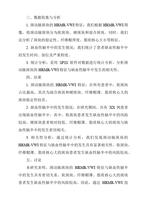
三、数据收集与分析1. 颈动脉斑块的HRMR-VWI特征:我们根据HRMR-VWI图像,将颈动脉斑块分为软斑块、硬斑块和混合斑块。
同时,我们还分析了斑块的稳定性、纤维帽厚度、脂质核心大小等特征。
2. 缺血性脑卒中的发生情况:我们统计了患者缺血性脑卒中的发生时间、部位及严重程度。
3. 统计分析:采用SPSS软件对数据进行统计分析,分析颈动脉斑块的HRMR-VWI特征与缺血性脑卒中发生的相关性。
四、结果1. 颈动脉斑块的HRMR-VWI特征:在所有患者中,软斑块占比最高,其次为混合斑块和硬斑块。
纤维帽薄、脂质核心大的斑块稳定性较差。
2. 缺血性脑卒中的发生情况:在研究期间,共有XX例患者出现缺血性脑卒中。
其中,软斑块患者发生缺血性脑卒中的风险较高,硬斑块患者相对较低。
纤维帽薄、脂质核心大的斑块与缺血性脑卒中的发生密切相关。
- 1、下载文档前请自行甄别文档内容的完整性,平台不提供额外的编辑、内容补充、找答案等附加服务。
- 2、"仅部分预览"的文档,不可在线预览部分如存在完整性等问题,可反馈申请退款(可完整预览的文档不适用该条件!)。
- 3、如文档侵犯您的权益,请联系客服反馈,我们会尽快为您处理(人工客服工作时间:9:00-18:30)。
隐匿性脑梗死和多血管床病变相关性研究 [摘要]目的研究隐匿性脑梗死(SCO和多血管床病变
(PolyVD)的相关性,为识别脑梗死高危人群提供依据。方
法纳入2015年2月〜2016年6月入住台州市中心医院神经 内科的患者, 95 例被确诊为首次隐匿性脑梗死的患者作为脑 梗死组, 95 例同期头晕、 头痛而经头颅 MRI 证实无梗死灶者 作为对照组。检测外周动脉区(颈动脉、锁骨下动脉、下肢 动脉)及冠状动脉、颅内动脉,比较各个区域血管床病变情 况。结果两组患者在性别、年龄、烟酒史及高血压病史、糖 尿病史、冠心病史、血脂基线水平等方面差异无统计学意义
(P>0.05)。检测外周血管区(颈动脉、锁骨下动脉、下肢动
脉)及冠状动脉、颅内动脉,相应动脉病变检出率,脑梗死 组分别为 65.3%、 24.2%、 33.7%, 21.1%、 32.6%,对照组分
别为 49.5%、 17.9%、 25.3%、 15.8%、 24.2%,其中脑梗死组 颈动脉斑块检出率高于对照组,差异有统计学意义( 2=4.842, P=0.028) 两组病变血管检出条数率分别为 35.4%、
27.4%,脑梗死组咼于对照组,差异有统计学差异(X 2=8.689,
P=0.003)。外周动脉、冠状动脉、颅内动脉的三个区域,两
组 1 个区域发生动脉粥样硬化病变分别是 37.9%、 72.6%,存 在 2个区域血管病变分别为 47.4%、 22.1%,存在 3个区域血 管病变分别为 14.7%、 5.3%,两组病变区域比较,差异有统 计学意义( Z=4.761, P=0.000 [关键词 ] 脑梗死;多血管床;外周动脉;冠状动脉
[中图分类号 ] R743.3;R766 [文献标识码 ] A [文章编号 ] 1673-9701(2016)33-0008-04 [Abstract] Objective To study the correlation between
silent cerebral infarction ( SCI) and polyvascular disease
PolyVD), and to provide evidence for identifying high risk population of cerebral infarction. Methods From February 2015 to June 2016, patients admitted to Department of Neurology ,
the Central Hospital of Taizhou City were enrolled in this study.
Among them , 95 patients who were diagnosed as first SCI were enrolled in the cerebral infarction group , 95 patients with dizziness and headache while had no cerebral infarction confirmed by head MRI were enrolled in the control group.
Peripheral arterial areas ( carotid artery , subclavian artery ,
lower extremity artery ), coronary artery and intracranial artery were examined , the vascular bed lesions were compared in each region. Results There were no significant differences in sex , age, history of alcohol and tobacco ,
hypertension , diabetes mellitus , coronary heart disease and blood lipid baseline level between the two groups (P>0.05) . The detection rate of cerebral arteries was 65.3% ,
24.2%,
33.7%, 21.1% and 32.6% respectively in the peripheral arterial areas (carotid artery , subclavian artery,
lower extremity artery ), coronary artery and intracranial artery , while the
control group was 49.5% , 17.9%, 25.3%,
15.8%, 24.2% respectively ,
in which, the carotid plaque detected was significantly higher in the cerebral infarction group than that in the control group , the difference was statistically significant
x 2=4.842, P=0.028) . The ratio of the number of blood vessels detected in the two groups were 35.4% and 27.4% ,
respectively , which was higher in the cerebral infarction group than the control group , the difference between the two groups was statistically significant ( x 2=8.689, P=0.003) . The incidence of one atherosclerotic lesion occured in the three regions of peripheral artery , coronary artery and intracranial artery between the two groups was 37.9% and 72.6% , while the incidence of two lesions occurred was 47.4% and 22.1% ,
respectively , the incidence of three lesions occurred was 14.7%
and 5.3%, respectively , showed statistically significant
differences between the two groups ( Z=4.761, P=0.000 [Key words] Cerebral infarction ; Polyvascular; Peripheral artery ; Coronary artery 脑血管病已居我国疾病死亡率首位,脑梗死具有高发病 率、高死亡率和高致残率 [1] 。其中,隐匿性脑梗死( silent
cerebral infarction,SC)或称无症状性脑梗死,是指经头颅
磁共振成像(MRI)等检查发现脑梗死,而在临床上无明显 相应的神经系统缺损症状和体征,或表现轻微容易被忽视,
disease,PolyVD),成为新的研究课题。 动脉粥样硬化是全身
性疾病,是脑梗死发生、发展的重要病因,被认为是预测脑 卒中发生的主要危险因素[2]。PolyVD指临床确认的2〜3处 动脉区域,主要表现为脑血管病( cerebrovascular disease, CVD)、冠心病(coronary artery disease,CAD)、外周动脉
疾 病(peripheral arterial disease, PAD)[3]。本研究探?SCI和 PolyVD 是否相关,为预测脑梗死高危人群提供依据。
1 资料与方法
1.1 一般资料
脑梗死组:纳入 2015 年 2 月〜 2016 年 6 月入住台州市
颅 MRI 确诊。纳入标准:依据全国第 4 届脑血管会议提出的 脑梗死的诊断标准[4]。排除标准:①出血性脑梗死;②动脉 炎致脑梗死; ③有明确栓子来源如心房颤动、 风湿性心脏病、
脂肪栓塞等所致脑梗死;④血液成分改变导致的脑梗死;⑤
列在 2015 年脑血管病分类中。 SCI是非致残性卒中 ,如何针 对病因精准有效干预,尤其是多血管床病变( polyvascular
中心医院神经内科患者, 95例为首次确诊SCI患者,通过头
肿瘤、全身免疫性疾病;⑥有脑卒中史。对照组:纳入 95