Current progress in use of adipose derived stem cells inperipheral nerve regeneration
Wnt/β—Catenin在骨髓基质干细胞
Wnt/β—Catenin在骨髓基质干细胞目的探讨BMSCs移植对AD模型大鼠的治疗作用。
方法将大鼠随机分成5组,正常组、模型组、模型对照组、治疗组、治疗对照组,每组10只。
双侧海马区注射Aβ建立AD大鼠模型后,治疗组进行BMSCs的移植治疗。
治疗对照组用氢溴酸加兰他敏进行灌胃治疗。
结果行为学实验检测各组大鼠平均逃避潜伏期及首次跨越原平台时间,模型组与正常组比较(P<0.01),治疗组与治疗对照组与模型组比较(P<0.01),与正常组比较(P<0.05),均有显著性差异;形态学实验HE染色显示模型组颗粒细胞及锥体细胞有破坏现象,治疗组和治疗对照组明显恢复。
免疫组化检测显示正常组和模型对照组可见海马区阳性蛋白表达较多而模型组阳性表达明显减少,治疗组和治疗对照组阳性表达较模型组明显增多。
结论BMSCs移植治疗AD的作用机制可能与通过调节Wnt信号转导通路,影响了学习记忆相关酶及蛋白在海马区的表达而发挥作用有关。
标签:Wnt/β-Catenin;骨髓基质干细胞;阿尔茨海默病阿尔茨海默病(alzheimer’s disease,AD)是一种中枢神经系统退行性疾病,其神经组织无法自然恢复。
因此,体外诱导多能干细胞分化进行细胞移植来修复受损的神经组织已经成为研究的热点之一。
近年来发现,Wnt信号通路控制着体内多种程序,例如细胞的增殖、自我更新、细胞分化、细胞极性、细胞死亡等过程。
Wnt信号通路与AD的病理过程存在着十分密切的关系,同时在神经干细胞的增殖、分化以及迁移等方面也起着重要的作用。
骨髓间充质干细胞(bone marrow mesenchvmalstem cells,BMSCs)在中枢神经组织移植中有明显优势,是一种良好的细胞替代治疗的靶细胞。
因此本研究希望通过观察Wnt通路相关蛋白β-Catenin的变化,探讨BMSCs移植对AD模型大鼠的治疗作用。
1材料与方法1.1材料清洁级健康Wistar雄性大鼠50只,体重180~220g,由吉林大学医学院提供;Moriss水迷宫;大鼠脑立体定位仪;Aβ美国Biosource公司;氢溴酸加兰他敏,陕西森弗生物技术有限公司;β-Catenin测试盒购自武汉众一生物试剂经营部。
Define Cell, Tissue, organ, and organ system
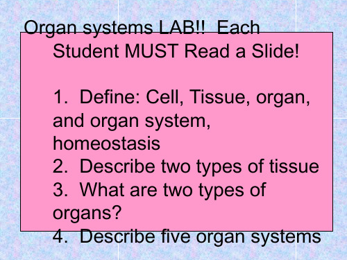
Organization of Vertebrate Body
Tissues are groups of cells that are similar in structure and function
In adult vertebrates, there are four primary tissues -Epithelial, connective, muscle and nerve
Q 4: Brain, heart and lungs are some of the important _______________ in a body.
organs tissues cells system
Q 5: Different tissues work together to form _________ .
system organ tissue
Q 2: Some tissues and organs work together like the members of the team. The parts that work together are called a _____________.
cell system group
Levels of Organization
• Organ Systems—Groups of organs that work together to perform a specific function.
– Examples:
• Digestive system • Circulatory system • Respiratory system • Nervous system • Muscular system • Skeletal system • Integumentary system (skin) • Vascular system in plants
外科急诊创伤(英文)-烧伤-精选文档148页

Leaugeay Webre BS, CCEMT-P, NREMT-P
Scenario
Paramedic is called to the scene of a structure fire. FD has removed a victim from the house. BSI Scene safe 1 patient A/C standby FD/ PD on scene Now what?
superficial partial thickness red, painful, blistered
deep partial thickness pale, mottled
Very painful Infection may evolve into 3rd degree
Burn Depth
Partial-Thickness Burn: 2nd Degree Burn
Function
Protection Regulation Prevention Sensory
Epidermis
Outer, thinner layer Consists of dead keratinized cells Protects
dehydration trauma light infection
ABCTransport decision? % BSA burned? Tx?
Objectives
Describe the structure and function of skin Discuss the types of burns. Explain the degrees of thermal burns. Discuss causes and treatments of inhalation injuries. Identify methods of approximating burn injuries. Describe and apply treatment modalities for the burn patient.
外周前庭疾病的诊断和治疗

收稿日期:2020 ̄08 ̄04基金项目:上海交通大学医工交叉重点项目(ZH2018ZDA11)ꎻ上海交通大学医学院附属新华医院院级临床研究培育基金项目(17CSK03ꎬ18JXO04)通信作者:杨军ꎮE ̄mail:yangjun@xinhuamed.com.cndoi:10.6040/j.issn.1673 ̄3770.1.2020.074述评外周前庭疾病的诊断和治疗杨军ꎬ郑贵亮上海交通大学医学院附属新华医院耳鼻咽喉头颈外科/上海交通大学医学院耳科学研究所/上海市耳鼻疾病转化医学重点实验室ꎬ上海200092㊀㊀杨军ꎬ上海交通大学医学院附属新华医院耳鼻咽喉头颈外科主任ꎬ博士生导师ꎬ上海市优秀学科带头人ꎬBarany协会会员ꎬ中华医学会耳鼻咽喉头颈外科分会耳科组委员ꎬ上海医学会耳鼻咽喉头颈外科分会副主任委员ꎮ中宣部 国家出版基金 项目评审委员会二审专家ꎮ«ActaOto ̄Laryngologica»«中华耳鼻咽喉头颈外科杂志»«山东大学耳鼻喉眼学报»等八种期刊编委㊁特邀审稿专家ꎮ临床专长为耳神经及侧颅底外科ꎬ在眩晕外科㊁耳神经外科㊁侧颅底肿瘤以及听觉植入方面具有丰富的临床经验ꎻ基础研究方向为耳蜗发育㊁听觉可塑性㊁耳蜗转导机制ꎮ作为项目负责人承担国家自然基金5项㊁973子课题1项㊁上海市科委重大项目1项ꎬ发表论文160余篇ꎮ作为主要完成人ꎬ获得国家科技进步二等奖㊁华夏医学科技奖二等奖㊁上海市科技进步奖一等奖㊁上海医学科技奖一等奖㊁中华医学科技奖三等奖㊁高等学校科学研究优秀成果奖科学技术进步奖一等奖等奖项ꎮ成功申办并将于2021年4月22~25日在上海举办第八届梅尼埃及内耳疾病国际研讨会ꎮ入选中国耳科医生百强榜名录和中国名医百强榜眩晕外科医生名录ꎮ摘要:眩晕是外周前庭疾病的主要表现之一ꎬ发病原因涉及多个学科ꎬ临床诊治较为困难ꎮ随着前庭功能检查技术的发展㊁对前庭疾病研究的不断深入ꎬ相关科研成果与日剧增并广泛应用于临床ꎮ近年来ꎬ随着前庭疾病国际分类标准制定和发布ꎬ各类前庭疾病诊断标准相继出台ꎬ治疗前庭疾病的药物㊁手术的相关规范日趋完善ꎬ加之前庭康复技术的飞速发展ꎬ使得对前庭疾病的诊疗越来越规范和精准ꎮ关键词:外周前庭疾病ꎻ眩晕ꎻ诊疗进展ꎻ前庭功能检查ꎻ前庭康复治疗中图分类号:R764.3㊀㊀㊀文献标志码:A㊀㊀㊀文章编号:1673 ̄3770(2020)05 ̄0001 ̄06引用格式:杨军ꎬ郑贵亮.外周前庭疾病的诊断和治疗[J].山东大学耳鼻喉眼学报ꎬ2020ꎬ34(5):1 ̄6.YANGJunꎬZHENGGuiliang.Diagnosisandmanagementofperipheralvestibulardiseases[J].JournalofOtolaryngologyandOphthalmologyofShan ̄dongUniversityꎬ2020ꎬ34(5):1 ̄6.DiagnosisandmanagementofperipheralvestibulardiseasesYANGJunꎬZHENGGuiliangDepartmentofOtolaryngology ̄Head&NeckSurgeryꎬXinhuaHospitalꎬShanghaiJiaotongUniversitySchoolofMedicine/EarInstituteꎬShanghaiJiaotongUniversitySchoolofMedicine/ShanghaiKeyLaboratoryofTranslationalMedicineonEarandNoseDiseasesꎬShanghai200092ꎬChinaAbstract:Vertigoisoneofthemostimportantsymptomofperipheralvestibulardiseaseswhicharedifficulttodifferentiallydiag ̄noseandmanagebecausemultipledisciplinesareinvolved.Thepremiseofeffectivemanagementisaccuratediagnosisofvestibulardiseases.Withthedevelopmentofvestibularfunctionexaminationtechnologyandthedeepeningofvestibulardiseaseresearchꎬgreatprogresshasbeenmadeinthediagnosisandmanagementofvestibulardiseases.Theestablishmentandpublicationofinternationalclassificationofvestibulardiseasesꎬtheintroductionofdiagnosticstandardsforvariousvestibulardiseasesintheworldꎬtheformula ̄tionofvestibulardiseasedrugsꎬsurgicalspecificationsandtherapiddevelopmentofvestibularrehabilitationtechnologymakethediagnosisandmanagementofvestibulardiseasesmoreandmorestandardizedandaccurate.Keywords:PeripheralvestibulardiseaseꎻVertigoꎻDiagnosisandmanagementꎻVestibularfunctionexaminationꎻVestibularrehabilitationtherapy㊀㊀眩晕作为多感官综合征ꎬ是耳鼻咽喉科门诊就诊的主要症状之一ꎬ涉及耳鼻咽喉科㊁神经内科㊁骨科㊁心内科㊁精神科㊁心理科等多个学科ꎬ临床诊治较为困难[1 ̄2]ꎮ众所周知ꎬ对于任何疾病的准确治疗ꎬ都是以准确的诊断为前提的ꎮ近年来ꎬ随着对前庭疾病研究的深入以及各种前庭检查技术的发展ꎬ在临床各学科的共同关注和努力下ꎬ前庭疾病的国际分类㊁诊疗规范和指南㊁专家共识等相继制定和推出ꎬ使得对前庭疾病的诊断和治疗越来越精准[3 ̄6]ꎮ本文主要从前庭疾病国际分类和规范㊁前庭功能检查技术进展㊁外周前庭疾病的药物和手术治疗㊁前庭康复等四个方面来对外周前庭疾病的诊疗进展进行述评ꎮ1㊀前庭疾病国际分类和规范为了提高临床质量ꎬ加强研究促进学科发展ꎬ世界卫生组织决定在国际疾病分类第11版(interna ̄tionalclassificationofdiseases11ꎬICD ̄11)中首次加入前庭疾病国际分类(internationalclassificationofvestibulardisordersꎬICVD)ꎮICVD的框架结构㊁定义和疾病诊断标准等陆续出台ꎬ于2018年正式发布了完整版本[3]ꎮ在ICVD的框架结构中ꎬ对前庭疾病㊁前庭症状㊁诊断标准等分别进行了界定和定义ꎮ前庭疾病除了包括累及前庭的内耳疾病外ꎬ也涵盖了前庭至脑的传导通路ꎬ包括脑干㊁小脑㊁相关皮层下结构以及前庭皮层的病变[4]ꎻ对于临床症状与前庭疾病类似ꎬ但原发于其他系统的疾病(如心脏㊁颈椎等)ꎬICVD主要集中于这些疾病的前庭表现ꎬ并未重新定义和分类这些非前庭原发疾病ꎮ前庭疾病的分类和定义对促进诊断标准的制定㊁临床医生及研究者的交流㊁疾病治疗及机制研究方面有着重要意义[3]ꎮICVD框架结构由相互关联的四个层面构成[3 ̄4]ꎬ包括症状和体征㊁综合征㊁功能失调和疾病㊁发病机制ꎮ在第一层中对前庭症状分别进行了定义ꎬ包括眩晕㊁头晕㊁前庭-视觉症状㊁姿势症状ꎻ在第二层中对常见的前庭综合征进行了定义ꎬ包括急性㊁发作性㊁慢性前庭综合征ꎻ在第三层中对前庭疾病分别进行了定义ꎬ并制定了前庭性偏头痛(vestibularmigraineꎬVM)㊁良性阵发性位置性眩晕(benignparoxysmalpositionalvertigoꎬBPPV)㊁梅尼埃病(Meniere̓sdiseaseꎬMD)以及持续性姿势知觉性头晕(persistentposturalperceptualdizzinessꎬPPPD)㊁前庭阵发症(vestibularparoxysmꎬVP)等疾病的诊断标准ꎮ我国也陆续制定出台了MD诊断和治疗指南㊁BPPV诊断和治疗指南㊁眩晕诊治多学科专家共识㊁前庭功能检查专家共识VM诊治专家共识等ꎮ这些前庭疾病的国际分类㊁诊断标准㊁诊疗指南㊁规范以及专家共识的出台ꎬ对提高我国眩晕/头晕疾病的整体诊治水平起到了极大的促进作用ꎮ2㊀前庭功能检查技术进展前庭功能检查是临床上辅助前庭疾病诊断的必要手段ꎬ通过前庭功能检查来对前庭系统的生理功能进行定性㊁定量评估ꎬ明确病变侧别㊁部位[5 ̄6]ꎮ随着前庭功能检查技术的飞速发展ꎬ除了传统的眼震电图㊁旋转试验外ꎬ前庭诱发肌源性电位(vestibu ̄lar ̄evokedmyogenicpotentialꎬVEMP)㊁视频头脉冲检查(videoheadimpulsivetestꎬvHIT)㊁前庭自旋转实验(vestibularautorotationtestꎬVAT)以及主观视觉垂直线检查(subjectivevisualverticalꎬSVV)等也在临床上逐步开展ꎬ能够分别对半规管㊁耳石器㊁前庭神经等进行定性㊁定侧㊁定量的检查ꎮ眼震电图检查包括扫视试验㊁视跟踪试验㊁视动试验㊁凝视试验㊁自发眼震试验㊁冷热试验等ꎬ通过评估视动系统㊁前庭视动系统的功能来反映前庭系统的功能ꎮ外周前庭病变引起的眼震多为水平㊁扭转性眼震ꎬ有疲劳现象ꎬ并被固视抑制ꎻ中枢性眼震则多为粗大㊁非水平性ꎬ如垂直性㊁钟摆型眼震等ꎬ不会被固视抑制ꎬ无疲劳现象ꎮ扫视试验异常多提示为脑干或小脑等中枢系统病变ꎮ视跟踪试验Ⅰ㊁Ⅱ型为正常或外周前庭病变ꎬⅢ㊁Ⅳ型多提示中枢前庭病变ꎮ视动试验异常多提示前庭中枢病变ꎬ也可见于部分外周前庭病变的急性期ꎮ凝视试验中ꎬ中枢病变表现为方向改变的眼震ꎬ而外周前庭病变则表现为方向固定的眼震ꎮ前庭有自身的频率特性ꎬ目前前庭频率的客观检查主要是通过用不同频率刺激半规管壶腹后ꎬ观察和记录眼动情况来判断前庭受损侧别和各半规管的功能ꎮ包括超低频率刺激的温度试验㊁低频的转椅试验以及中高频的VAT和HIT等[5 ̄9]ꎮ温度试验(检测频率0.003~0.008Hz)是通过冷热水或冷热气的灌注ꎬ比较两侧水平半规管的功能ꎬ是临床上评价一侧外周或中枢性前庭功能障碍的常用方法[8]ꎮ在温度试验检查中的眼震极盛期ꎬ进行固视抑制ꎬ如果固视抑制失败ꎬ则提示可能为中枢病变ꎮ旋转试验(检测频率0.01~0.64Hz)通过检查前庭水平半规管系统对一定加速度刺激的反应情况ꎬ定量评价前庭系统功能ꎻvHIT不仅用于检查单侧或双侧水平半规管功能ꎬ还可用于评估垂直半规管的功能状态[9]ꎻVEMP用于检查耳石器功能和中枢病变[10]ꎻSVV是针对椭圆囊病变的主观量化检查[6]ꎮVAT检测频率为0.5~6.0Hzꎬ接近于人体日常活动的频率ꎬ可用于检测水平和垂直方向的高频眼动反射ꎬ通过检测受检者以一定频率主动摆头时的眼动反应ꎬ来评价较高频率的前庭眼动反射(vestibularoculomotorreflexꎬVOR)状况ꎮ临床上通过计算增益㊁相位㊁非对称性等参数来进行分析[6]ꎮ增益降低常见于外周损害ꎬ增高可见于中枢性病变ꎻ外周或中枢病变均可引起相位异常ꎬ增大常提示双侧前庭功能的不对称ꎮvHIT通过检测受检者在快速㊁高频㊁被动头动时的眼动反射来评价前庭功能ꎮ一般认为其代表了较高频率的VORꎬ可反映单个半规管功能状况ꎬ并可分别检查水平㊁垂直半规管中的任意一个ꎬ检测频率为2~5Hzꎮ结合VOR增益值和出现扫视波情况来综合评判半规管功能ꎮ增益反映的是VOR眼动反应与头动反应之间的比值关系ꎬ为眼动与头动的速度比值或眼动曲线与头动曲线下面积比值ꎮ半规管功能受损时表现为VOR增益值降低及延后出现的扫视波ꎬ根据出现的时间分为隐性扫视波和显性扫视波[9]ꎮvHIT异常高度提示外周前庭病变ꎮ目前临床上还有一种头脉冲检查的补充模式 头脉冲抑制试验(suppressionheadimpulseparadigmꎬSHIMP)ꎮ检测方法与传统模式的不同处在于受检者全程凝视随头位同步移动的激光点ꎬ与vHIT检查中出现扫视波是前庭功能障碍的表现不同的是ꎬSHIMP检查中出现的扫视波则是剩余前庭功能的表现[6 ̄7]ꎮ震动诱发眼震试验的检查频率在100Hz左右ꎬ是检测单侧外周前庭功能障碍的有价值的方法ꎬ出现连续5个3ʎ/s以上的眼震为阳性ꎬ常提示双侧前庭功能的不对称[11]ꎮ前庭耳石器对强声或振动刺激会引起相应的肌电反应ꎬ包括球囊诱发的胸锁乳突肌肌源性电位(cervicalVEMPꎬcVEMP)和椭圆囊诱发的眼外肌肌源性电位(ocularVEMPꎬoVEMP)ꎬ分别用于评价球囊与前庭下神经和椭圆囊与前庭上神经通路的功能[10]ꎮ常用的指标是两侧波幅不对称比ꎬ其增大常提示一侧耳石器与前庭上/下神经通路的损伤ꎬ如梅尼埃病㊁前庭神经炎等ꎻ如波幅明显增大或刺激阈值明显降低常见于上半规管裂综合征ꎻ潜伏期延长则多见于迷路后或中枢病变[6ꎬ10]ꎮSVV检查主要用于评价双侧耳石器功能对称性ꎬ偏差一般<3度ꎬ超过此值常提示双侧耳石器功能不对称ꎬ外周或中枢病变均可引起[6]ꎮSVV在鉴别前庭外周与前庭中枢病变方面具有重要意义ꎬ外周前庭病变SVV检查一般偏向患侧ꎬ中枢病变中ꎬ累及低位脑干病变SVV检查偏向患侧ꎬ累及上位脑干则偏向健侧ꎮ需要强调的是ꎬ前庭功能检查技术只是发现问题的手段和方法之一ꎬ因单一前庭功能检查异常并不能明确表示其前庭功能一定存在异常ꎬ常常需要不同检查之间互相印证和补充ꎮ当患者双侧cVEMP都引不出ꎬ并不代表双侧前庭功能一定存在问题ꎬ因为正常人群中亦有部分引不出者ꎬ所以需要其他前庭功能检查来明确该患者是否存在真性前庭功能障碍ꎮ因此ꎬ临床工作中ꎬ医师需要全面了解各种检查技术的适应证范围㊁具备解读检查结果的能力ꎬ才能有针对性地选择合理的检查项目ꎬ根据检查结果并结合病史信息确定临床诊断ꎬ从而精准治疗外周前庭疾病ꎮ3㊀前庭疾病的药物及手术治疗理想情况下ꎬ前庭药物治疗的目标是通过特定和有针对性的分子作用ꎬ显著减轻眩晕症状ꎬ保护或修复病理条件下的前庭感觉网络ꎬ促进前庭代偿ꎬ最终达到提高患者生活质量的目的[12 ̄13]ꎮ但由于前庭疾病的病因和药理学靶点信息的缺乏ꎬ以及不能有效地将药物定向靶向投放等问题ꎬ目前尚无法实现这一理想状态ꎮ目前治疗前庭疾病的药物主要分为前庭抑制药㊁止吐药㊁促进前庭代偿药物ꎬ以及激素㊁扩张血管药物等ꎮ前庭抑制药是减少前庭失衡引起的眼球震颤的药物ꎮ传统的前庭抑制药由三大类药物组成ꎬ即抗胆碱能药㊁抗组胺药和苯二氮类[12 ̄13]ꎮ抗胆碱能药物和抗组胺药是前庭中枢抑制物ꎬ可抑制动物前庭核神经元的放电ꎬ并能降低人类眼球震颤的速度[14]ꎮ但所有常规用于治疗眩晕或运动病的抗胆碱药都有显著的不良反应ꎬ通常包括口干㊁瞳孔扩大和镇静等ꎮ苯二氮类药物是γ氨基丁酸受体(γ ̄aminobutyricacidreceptorꎬGABAR)调节剂ꎬ主要作用是抑制中枢前庭反应[10ꎬ12ꎬ15]ꎮ研究证实小剂量的苯二氮类药物对治疗眩晕非常有用ꎬ也有助于预防运动病[15]ꎬ不良反应主要包括药物依赖㊁记忆受损和跌倒风险增加[13 ̄14]ꎮ无论是抗胆碱能药㊁抗组胺药还是苯二氮类药物ꎬ都会影响前庭损害的代偿ꎬ因此均不适合长期使用ꎮ另外一类用于前庭抑制的药物是钙通道拮抗剂ꎬ最常用的是氟桂利嗪和尼莫地平ꎬ其作用机制是前庭暗细胞也含有钙通道ꎬ钙通道拮抗剂可能通过影响前庭暗细胞内钙通道活动ꎬ改变内淋巴中的离子浓度[16]ꎬ从而发挥抑制前庭的作用ꎮ而且钙通道拮抗剂通常也常具有抗胆碱能和/或抗组胺活性ꎬ如氟桂利嗪[17]ꎮ胃复安㊁异丙嗪㊁昂丹司琼是常用的止吐药物ꎮ止吐药的选择取决于对给药途径和不良反应的考虑ꎮ口服制剂用于轻度恶心ꎻ栓剂常用于因胃无力或呕吐而无法吸收口服药物的门诊患者ꎻ舌下给药㊁注射剂用于急诊室或住院患者ꎮ昂丹司琼是高效的止吐药ꎬ对表现为恶心的前庭障碍者也有效ꎬ但价格昂贵ꎮ虽然这类药物有较好的止吐效果ꎬ但有研究表明这些药物对预防运动病没有帮助[16]ꎮ前庭抑制剂都会影响前庭损伤的代偿ꎬ加速代偿的药物主要是前庭兴奋剂ꎮ倍他司汀是一种H1受体激动剂和H3受体拮抗剂ꎬ可以解除控制组胺释放的负反馈回路ꎬ促进大脑中组胺能神经递质的传递ꎮ也有人认为倍他司汀可能加速前庭代偿ꎬ但也有研究得出结论ꎬ没有足够的证据表明倍他司汀对梅尼埃病是否有影响[18]ꎮ另外一个治疗前庭疾病的药物是银杏叶制剂ꎮ银杏叶可以降低血液黏度ꎬ同时它也可能是一种抗氧化剂ꎮ一项银杏叶与倍他司汀的对照研究结果显示ꎬ银杏叶对眩晕的疗效与倍他司汀相似[19]ꎮ对于前庭疾病的患者ꎬ其具体的药物治疗方案需要针对病因 量身定制 ꎬ比如对于前庭神经炎ꎬ目前只建议短暂使用前庭抑制药ꎻ对于MDꎬ前庭抑制剂无论加或不加止吐药用来治疗急性发作ꎬ这些药物都不能纠正前庭不平衡引起的症状ꎬ只是抑制了失衡的临床表现(如眩晕和恶心)ꎮ而对于预防发作ꎬ通常的做法是建议饮食限盐(1~2g/d)和使用温和的利尿剂ꎬ如氢氯噻嗪ꎮ对于BPPVꎬ物理治疗(手法复位或转椅复位)是最有效的方法[20]ꎬ对复位后有头晕㊁平衡障碍等症状的ꎬ可以给予改善内耳微循环的药物ꎮ对于VMꎬ预防性药物(L ̄型钙通道拮抗剂㊁三环类抗抑郁药㊁β ̄受体阻滞剂)则是治疗该病的主要药物ꎮ对于药物无法控制的MDꎬ可以根据疾病的听力分期和严重程度选择鼓室内注射类固醇激素㊁庆大霉素ꎬ以及手术治疗[21]ꎮ手术包括内淋巴囊手术(减压㊁内淋巴管夹闭)㊁半规管阻塞㊁前庭神经切断㊁迷路切除等ꎮ上半规管裂以及中耳胆脂瘤引起的迷路瘘管的治疗也是以手术为主ꎬ上半规管裂手术包括经乳突径路㊁颅中窝径路的半规管修补术以及经鼓室的圆窗加固术ꎮ听神经瘤引起的眩晕也往往需要手术治疗ꎬ根据肿瘤大小和听力情况等可以选择经迷路径路㊁乙状窦后径路以及颅中窝径路等ꎮ小的局限于内听道内而且无实用听力者也可在耳内镜下经耳蜗手术切除ꎮ双侧前庭病目前缺乏有效药物治疗ꎬ物理治疗效果也不甚理想ꎬ目前最有前景的治疗手段可能是前庭植入[22]ꎮ日内瓦Maastrichtgroup的一项研究中ꎬ为13例双侧前庭功能低下的患者成功安装了前庭植入物ꎬ并开发了一种将运动传感器与系统耦合的特殊接口ꎬ以捕捉来自三轴陀螺仪的信号ꎬ并调制 基线电活动 ꎮ前庭植入可行性所需的最后一个里程碑是证明人工恢复的前庭反射足以减轻患者的症状ꎬ尤其是频繁出现的振动幻视ꎮ2016年ꎬGuinand等[23]测量了6例前庭植入患者的动/静态视力ꎬ在没有前庭植入物的情况下ꎬ他们的视力在动态条件下受损ꎬ而当前庭植入物打开时ꎬ视力恢复正常ꎮ鉴于这些令人鼓舞的研究成果ꎬ人工前庭植入可能在不久的将来就能够进入临床应用ꎮ4㊀前庭康复前庭康复治疗(vestibularrehabilitationtherapyꎬVRT)已经有超过70年的历史ꎬ是一项以运动为基础的系统训练方式ꎬ通过中枢代偿ꎬ前庭康复能够改善不平衡㊁跌倒㊁头晕㊁眩晕㊁运动敏感等前庭症状和恶心㊁焦虑等伴随症状ꎮ越来越多的证据支持前庭障碍患者进行前庭康复治疗ꎬ而目前已经涌现了很多更有效的干预措施ꎮ比如前庭康复的辅助设施㊁可穿戴的前庭康复辅助设备以及结合虚拟现实技术的前庭康复训练等[24 ̄26]ꎮ自20世纪90年代末以来ꎬ有关前庭病变患者治疗技术的证据显著增加ꎬ使得干预措施变得更加精细和有效ꎮ具有相似诊断的患者的身体表现和功能局限性通常会大不相同ꎮ尽管大多数VRT运动项目利用眼睛和头部运动ꎬ但运动类型及其处方是个性化的ꎬ针对的是患者的缺陷和症状ꎬ而不是针对特定的诊断ꎮ症状主诉可能包括不平衡(静态姿势和步态ꎬ尤其是与头部运动相关的)㊁跌倒和害怕跌倒㊁视力下降㊁头晕㊁焦虑㊁恶心㊁眩晕和运动敏感性ꎮ针对前庭损伤和功能受限的前庭康复技术是基于前庭适应㊁习服㊁感觉替代等多种机制发展而来的ꎬ前庭康复的练习方法包括促进凝视稳定练习㊁锻炼习服症状㊁练习改善平衡㊁步态及耐力等[27 ̄28]ꎮ适应训练或视觉 ̄前庭相互作用练习通过反复的前庭刺激(如头部运动)ꎬ以促进残余前庭系统的适应ꎮ适应性训练对治疗凝视不稳是有效的ꎬ也被证明可以改善平衡和减少头晕ꎮ习服训练则利用反复暴露于诱发症状的刺激来减少姿势变化引起的头晕ꎮ随着时间的推移ꎬ系统地暴露于轻微的㊁暂时的刺激后ꎬ头晕会逐渐减轻ꎬ可适用于没有明确诊断㊁但具有良性病因的位置性眩晕患者ꎬ治疗的主要目标是改善眩晕症状ꎬ对快速运动导致的异常前庭反应习服化ꎮ替代练习是利用其他感官刺激ꎬ如视觉或本体觉来替代前庭功能的缺失或下降ꎬ从而加强姿势控制和减少跌倒ꎮ该策略对双侧前庭功能减退和多感觉不平衡的患者尤其有效ꎮ在传统前庭康复技术基础上ꎬ近年来的一些新技术也逐渐应用到前庭功能障碍患者的康复治疗中ꎬ比如利用振动触觉和听觉反馈来增强平衡功能ꎬ以及虚拟现实技术在前庭康复中的应用等ꎮ研究发现ꎬ老年人和前庭障碍患者在家中利用振动触觉反馈练习可改善其平衡功能[29]ꎮ虚拟现实或其他使用沉浸式环境模拟的视动训练平衡也越来越多地应用于前庭康复训练中ꎬ也取得了良好的效果[30 ̄31]ꎬ而且相对于传统的前庭康复ꎬ多数患者报告说他们更喜欢虚拟训练[32]ꎮ随着游戏产业的发展ꎬ已开发出低成本的虚拟现实系统ꎬ如任天堂WiiFitPlusꎬ该系统还能够在平衡游戏期间向屏幕提供视觉反馈[33]ꎮ鉴于这是一种可以在家里进行的令人愉快的运动方式ꎬ市场上的Wii ̄Fit或其他较新的低成本虚拟现实设备可能是居家进行前庭康复训练的有用工具ꎮ当然ꎬ还需要进一步的相关研究来验证这些设备的训练效果ꎮ前庭康复运动对外周前庭功能障碍的疗效可靠ꎬ定制化的治疗方案侧重于减少头晕㊁振动幻视和姿势不稳的症状ꎬ并解决患者的功能缺陷ꎮ运动的目的是促进前庭功能障碍的中枢代偿ꎮ单侧前庭功能障碍患者往往表现良好ꎬ通常可以恢复全部或大部分日常生活活动ꎬ但双侧前庭功能丧失的患者仍然存在平衡障碍ꎮ对于双侧前庭病患者来说ꎬ人工前庭植入可能是最有效的治疗方式ꎮ前庭康复技术在过去几十年中不断发展ꎬ但国内前庭康复起步较晚ꎬ相关研究与国外差距较大ꎬ需要进一步重视[34]ꎮ5㊀结㊀语随着前庭生理研究的深入㊁前庭功能检查技术的不断发展和完善ꎬ以及各相关学科的重视和不断努力ꎬ对前庭疾病的认识不断深入ꎬ诊治水平不断提高ꎮ前庭疾病的精准诊治ꎬ除了进行准确的病因诊断外ꎬ还要兼顾症状控制㊁生活质量改善ꎬ要结合患者的个体差异ꎬ精准选择合适的治疗方式ꎬ综合药物治疗㊁手术治疗以及VRT等ꎮ需要在准确病因诊断的前提下ꎬ选择最适合的个体化治疗ꎬ以达到更好的症状控制㊁更好的生活质量ꎮ参考文献:[1]WaltherLE.Currentdiagnosticproceduresfordiagnosingvertigoanddizziness[J].GMSCurrTopOtorhinolaryn ̄golHeadNeckSurgꎬ2017ꎬ18(16):Doc02.doi:10.3205/cto000141.[2]StruppMꎬMandalàMꎬLópez ̄EscámezJA.Peripheralvestibulardisorders[J].CurrOpinNeurolꎬ2019ꎬ32(1):165 ̄173.doi:10.1097/wco.0000000000000649. [3]WorldHealthOrganizationꎬInternationalClassificationofDiseasesꎬ11theditionꎬhttps://icd.who.int/browse11/l ̄m/en#/http://id.who.int/icd/entity/1300772836. [4]BisdorffARꎬStaabJPꎬNewman ̄TokerDE.Overviewoftheinternationalclassificationofvestibulardisorders[J].NeurolClinꎬ2015ꎬ33(3):541 ̄50ꎬvii.doi:10.1016/j.ncl.2015.04.010.[5]中国医药教育协会眩晕专业委员会ꎬ中国康复医学会眩晕与康复专业委员会ꎬ中西医结合学会眩晕专业委员会ꎬ等.前庭功能检查专家共识(一)(2019)[J].中华耳科学杂志ꎬ2019ꎬ17(1):117 ̄123.doi:10.3969/j.issn.1672 ̄2922.2019.01.020.[6]中国医药教育协会眩晕专业委员会ꎬ中国康复医学会眩晕与康复专业委员会ꎬ中西医结合学会眩晕专业委员会ꎬ等.前庭功能检查专家共识(二)(2019)[J].中华耳科学杂志ꎬ2019ꎬ17(2):144 ̄149.doi:10.3969/j.issn.1672 ̄2922.2019.02.002.[7]MacDougallHGꎬMcGarvieLAꎬHalmagyiGMꎬetal.Anewsaccadicindicatorofperipheralvestibularfunctionbasedonthevideoheadimpulsetest[J].Neurologyꎬ2016ꎬ87(4):410 ̄418.doi:10.1212/wnl.0000000000002827. [8]ShepardNTꎬJacobsonGP.Thecaloricirrigationtest[M]//HandbookofClinicalNeurology.Amsterdam:Elsevierꎬ2016:119 ̄131.doi:10.1016/b978 ̄0 ̄444 ̄63437 ̄5.00009 ̄1.[9]AlhabibSFꎬSalibaI.Videoheadimpulsetest:areviewoftheliterature[J].EurArchOtorhinolaryngolꎬ2017ꎬ274(3):1215 ̄1222.doi:10.1007/s00405 ̄016 ̄4157 ̄4. [10]RosengrenSMꎬColebatchJGꎬYoungASꎬetal.Vestib ̄ularevokedmyogenicpotentialsinpractice:Methodsꎬpitfallsandclinicalapplications[J].ClinNeurophysiolPractꎬ2019ꎬ4:47 ̄68.doi:10.1016/j.cnp.2019.01.005. [11]ZamoraEGꎬAraújoPEꎬGuillénVPꎬetal.Parametersofskullvibration ̄inducednystagmusinnormalsubjects[J].EurArchOtorhinolaryngolꎬ2018ꎬ275(8):1955 ̄1961.doi:10.1007/s00405 ̄018 ̄5020 ̄6.[12]ChabbertC.Principlesofvestibularpharmacotherapy[M]//HandbookofClinicalNeurology.Amsterdam:Elsevierꎬ2016:207 ̄218.doi:10.1016/b978 ̄0 ̄444 ̄63437 ̄5.00014 ̄5.[13]StruppMꎬKremmydaOꎬBrandtT.Pharmacotherapyofvestibulardisordersandnystagmus[J].SeminNeurolꎬ2013ꎬ33(3):286 ̄296.doi:10.1055/s ̄0033 ̄1354594. [14]SotoEꎬVegaR.Neuropharmacologyofvestibularsys ̄temdisorders[J].CurrNeuropharmacolꎬ2010ꎬ8(1):26 ̄40.doi:10.2174/157015910790909511.[15]HuppertDꎬStruppMꎬMückterHꎬetal.Whichmedica ̄tiondoIneedtomanagedizzypatients?[J].ActaOto ̄laryngolꎬ2011ꎬ131(3):228 ̄241.doi:10.3109/00016489.2010.531052.[16]HainTCꎬUddinM.PharmacologicaltreatmentofVertigo[J].CNSDrugsꎬ2003ꎬ17(2):85 ̄100.doi:10.2165/00023210 ̄200317020 ̄00002.[17]LepchaAꎬAmalanathanSꎬAugustineAMꎬetal.Fluna ̄rizineintheprophylaxisofmigrainousvertigo:aran ̄domizedcontrolledtrial[J].EurArchOtorhinolaryngolꎬ2014ꎬ271(11):2931 ̄2936.doi:10.1007/s00405 ̄013 ̄2786 ̄4.[18]RamosAlcocerRꎬLedezmaRodríguezJGꎬNavasRomeroAꎬetal.UseofbetahistineinthetreatmentofperipheralVertigo[J].ActaOtolaryngolꎬ2015ꎬ135(12):1205 ̄1211.doi:10.3109/00016489.2015.1072873. [19]SokolovaLꎬHoerrRꎬMishchenkoT.Treatmentofver ̄tigo:arandomizedꎬdouble ̄blindtrialcomparingeffica ̄cyandsafetyofGinkgobilobaextractEGb761andbeta ̄histine[J].IntJOtolaryngolꎬ2014ꎬ2014:682439.doi:10.1155/2014/682439.[20]陈太生ꎬ王巍ꎬ徐开旭ꎬ等.良性阵发性位置性眩晕及其诊断治疗的思考[J].山东大学耳鼻喉眼学报ꎬ2019ꎬ33(5):1 ̄5.doi:10.6040/j.issn.1673 ̄3770.1.2019.053.CHENTaishengꎬWANGWeiꎬXUKaixuꎬetal.ThoughtsonbenignparoxysmalpositionalVertigoanditsdiagnosisandtreatment[J].JournalofOtolaryngologyandOphthalmologyofShandongUniversityꎬ2019ꎬ33(5):1 ̄5.doi:10.6040/j.issn.1673 ̄3770.1.2019.053. [21]LiuYPꎬYangJꎬDuanML.CurrentstatusonresearchesofMeniere̓sdisease:areview[J].ActaOtolaryngolꎬ2020ꎬ140(10):808 ̄812.doi:10.1080/00016489.2020.1776385.[22]GuyotJPꎬPerezFornosA.Milestonesinthedevelopmentofavestibularimplant[J].CurrOpinNeurolꎬ2019ꎬ32(1):145 ̄153.doi:10.1097/wco.0000000000000639. [23]GuinandNꎬvandeBergRꎬCavuscensSꎬetal.Thevideoheadimpulsetesttoassesstheefficacyofvestibu ̄larimplantsinhumans[J].FrontNeurolꎬ2017ꎬ8:600.doi:10.3389/fneur.2017.00600.[24]SulwaySꎬWhitneySL.Advancesinvestibularrehabili ̄tation[J].AdvOtorhinolaryngolꎬ2019ꎬ82:164 ̄169.doi:10.1159/000490285.[25]DunlapPMꎬHolmbergJMꎬWhitneySL.Vestibularrehabilitation:advancesinperipheralandcentralvestibu ̄lardisorders[J].CurrOpinNeurolꎬ2019ꎬ32(1):137 ̄144.doi:10.1097/wco.0000000000000632.[26]MeldrumDꎬJahnK.Gazestabilisationexercisesinves ̄tibularrehabilitation:reviewoftheevidenceandrecentclinicaladvances[J].JNeurolꎬ2019ꎬ266(suppl1):11 ̄18.doi:10.1007/s00415 ̄019 ̄09459 ̄x.[27]HallCDꎬHerdmanSJꎬWhitneySLꎬetal.Vestibularrehabilitationforperipheralvestibularhypofunction:anevidence ̄basedclinicalpracticeguideline:FROMTHEAMERICANPHYSICALTHERAPYASSOCIATIONNEUROLOGYSECTION[J].JNeurolPhysTherꎬ2016ꎬ40(2):124 ̄155.doi:10.1097/npt.0000000000000120. [28]KlattBNꎬCarenderWJꎬLinCCꎬetal.Aconceptualframeworkfortheprogressionofbalanceexercisesinpersonswithbalanceandvestibulardisorders[J].PhysMedRehabilIntꎬ2015ꎬ2(4):1044.[29]AllumJHJꎬHoneggerF.Vibro ̄tactileandauditorybal ̄ancebiofeedbackchangesmuscleactivitypatterns:possi ̄bleimplicationsforvestibularimplants[J].JVestibResꎬ2017ꎬ27(1):77 ̄87.doi:10.3233/ves ̄170601. [30]PavlouMꎬKanegaonkarRGꎬSwappDꎬetal.TheeffectofvirtualrealityonvisualVertigosymptomsinpatientswithperipheralvestibulardysfunction:apilotstudy[J].JVestibResꎬ2012ꎬ22(5ꎬ6):273 ̄281.doi:10.3233/ves ̄120462.[31]AlahmariKAꎬSpartoPJꎬMarchettiGFꎬetal.Compari ̄sonofvirtualrealitybasedtherapywithcustomizedves ̄tibularphysicaltherapyforthetreatmentofvestibulardis ̄orders[J].IEEETransNeuralSystRehabilEngꎬ2014ꎬ22(2):389 ̄399.doi:10.1109/tnsre.2013.2294904. [32]MeldrumDꎬHerdmanSꎬVanceRꎬetal.Effectivenessofconventionalversusvirtualreality ̄basedbalanceexer ̄cisesinvestibularrehabilitationforunilateralperipheralvestibularloss:resultsofarandomizedcontrolledtrial[J].ArchPhysMedRehabilitationꎬ2015ꎬ96(7):1319 ̄1328.e1.doi:10.1016/j.apmr.2015.02.032.[33]ClarkRAꎬBryantALꎬPuaYHꎬetal.Validityandreli ̄abilityoftheNintendoWiiBalanceBoardforassessmentofstandingbalance[J].GaitPostureꎬ2010ꎬ31(3):307 ̄310.doi:10.1016/j.gaitpost.2009.11.012. [34]杨军ꎬ郑贵亮.进一步重视前庭康复[J].临床耳鼻咽喉头颈外科杂志ꎬ2019ꎬ33(3):204 ̄206.doi:10.13201/j.issn.1001 ̄1781.2019.03.004.YANGJunꎬZHENGGuiliang.Furtheremphasisonves ̄tibularrehabilitation[J].JournalofClinicalOtorhinolar ̄yngologyHeadandNeckSurgeryꎬ2019ꎬ33(3):204 ̄206.doi:10.13201/j.issn.1001 ̄1781.2019.03.004.(编辑:李纬)。
Mesenchymal Stromal Cells New Directions
Mesenchymal Stromal Cells:New DirectionsArmand Keating1,*1Cell Therapy Program,Princess Margaret Hospital,Toronto,ON M5G2M9,Canada*Correspondence:armand.keating@uhn.on.caDOI10.1016/j.stem.2012.05.015Research into mesenchymal stromal/stem cells(MSCs)has been particularly exciting in the pastfive years. Our understanding of mechanisms of MSC-mediated tissue regeneration has undergone considerable evolu-tion.Recent investigation of the primary in situ counterpart of cultured MSCs has led to fresh insights into MSC physiology and its role in the immune system.At the same time,the clinical application of MSCs continues to increase markedly.Taken together,a reappraisal of the definition of MSCs,a review of current research directions,and a reassessment of the approach to clinical investigation are timely and prudent.IntroductionFew cell types have captivated so many biomedical researchers over the last10years as have mesenchymal stromal/stem cells (MSCs).PubMed,in2012,identifies over17,000references for ‘‘mesenchymal stem cells’’and more than4,500for‘‘mesen-chymal stromal cells.’’There have been several comprehensive recent reviews on MSCs(Uccelli et al.,2008;Bianco et al.,2008; Tolar et al.,2010;Ranganath et al.,2012).Hence,rather than cover all of the work in thisfield,in this perspective I will focus on some areas that have seen notable advance in the past 5years and others that warrant further investigation to improve our insight into the properties and potential of this intriguing cell population.First,a case can be made to revisit the nomenclature and defi-nition of MSCs,not as a semantic exercise,but to better define the direction of research.The availability of new molecular tools makes the need for rigorous definitions increasingly important. Moreover,the differences between MSC populations derived from different tissues are becoming more apparent,presenting an additional challenge to devising a universal definition.MSCs as currently defined are a phenomenon of in vitro culture, suggesting that extrapolating the function of these cells to activity in vivo must be done with caution.This limitation high-lights the need for direct in vivo studies with endogenous MSCs or an equivalent physiological population as an essential next step in establishing their true biological role.It is encour-aging in this regard that recent studies have employed trans-genic animal models to enable the tracking and assessment of MSC-like cells in vivo.The mechanisms underlying tissue regen-eration and immune modulation by therapeutic doses of MSCs also require further elucidation,particularly the extent to which the two processes intersect.The more recent appreciation that MSCs may not mediate tissue regeneration by direct cell replacement is also likely to redirect investigation into more fruit-ful directions.Finally,in view of the extraordinarily rapid and extensive use of MSCs clinically,a reappraisal of the approach to the development of clinical protocols based on confirmed laboratory and preclinical observations would be timely and helpful.BackgroundMSCs were initially identified as a subpopulation of bone marrow cells with osteogenic potential as shown by heterotopic trans-plantation and subsequently were confirmed to contain clonal, plastic adherent bone-marrow derived nonhematopoietic cells in the mouse and guinea pig(Friedenstein et al.,1968,1970, 1976).An in vitro colony assay developed by Friedenstein and coworkers to detect the clonogenic cell among this population (the colony-forming unit-fibroblast[CFU-F])was also adapted for human marrow(Castro-Malaspina et al.,1980).Subsequent studies in the1980s focused on the role of a similar population of bone marrow stromal cells derived from the adherent layers of long-term bone marrow cultures in supporting hematopoiesis (Dexter et al.,1977,reviewed in Clark and Keating,1995). Caplan’s proposal that these cells were mesenchymal‘‘stem’’cells(Caplan,1991)capable of differentiation to all cells of mesodermal lineage stimulated investigation into their role in mediating tissue regeneration.Although the multilineage differ-entiation potential of MSCs was later shown(Pittenger et al., 1999),in vivo demonstration that these cells possess the hall-mark stem cell characteristics of self-renewal and differentiation had not been accomplished.Confusion arising from the definition of the MSC population made comparisons among published studies in the1990s and 2000s problematic and led to the proposal of new terminology and criteria by the International Society for Cellular Therapy (ISCT)(Horwitz et al.,2005;Dominici et al.,2006).According to these widely adopted proposals,the cells were more appropri-ately considered mesenchymal stromal cells given that not all were stem cells(Horwitz et al.,2005).The minimum criteria for MSCs included plastic adherence and in vitro trilineage differen-tiation to adipogenic,chondrogenic,and osteogenic cells (Dominici et al.,2006).Additional requirements included cell surface expression of CD105(endoglin,SH2),CD73(ecto-50-nucleotidase),and CD90(Thy1)and the absence of the hemato-poietic markers,CD45,CD19,CD19or CD79,CD14or CD11b, and HLA-DR.A particular challenge for thefield has been the absence of a specific marker to define MSCs,although a large number of different determinants have been associated,albeit not exclusively,with them(reviewed by Lindner et al.,2010 for human MSCs),including CD271(low-affinity nerve growth factor receptor)(Jones et al.,2002)and CD146(Sacchetti et al.,2007).MSCs are also highly active metabolically,secreting not only components of the extracellular matrix(Wight et al., 1986)but also a vast array of cytokines(reviewed by Horwitz and Dominici,2008).More recent work has documented Cell Stem Cell10,June14,2012ª2012Elsevier Inc.709extensively the secretome and proteome of MSCs(Ranganath et al.,2012).In addition to bone marrow,MSC populations can be obtained readily from adipose tissue(Zuk et al.,2002)and also from a variety of tissues including placenta(In’t Anker et al.,2004), skin(Shih et al.,2005),umbilical cord blood(Erices et al., 2000),umbilical cord perivascular cells(Sarugaser et al.,2005), umbilical cord Wharton’s jelly(Wang et al.,2004),dental pulp (Gronthos et al.,2000),amnioticfluid(Nadri and Soleimani, 2007),synovial membrane(De Bari et al.,2001),and breast milk(Patki et al.,2010).Revisiting the Definition of MSCsThe minimum criteria for defining MSCs established earlier (Horwitz et al.,2005;Dominici et al.,2006)may now be unduly constraining for a number of reasons.First,the characteristics of MSCs may vary according to the source of tissue.In an effort to define an MSC-like product,scientific entrepreneurs and biotechnology companies have focused on differences in surface marker profile to optimize intellectual property protec-tion of relatively similar cell types.The recognition of species-specific differences in cell characteristics and generation of a variety of transcriptional and secretomic signatures for the cells also indicate diversity.Moreover,panels of reagents(especially antibodies)equivalent to those available for characterizing human MSCs are still not in place for a number of other species, so the criteria recommended by the ISCT(Dominici et al.,2006) may be difficult to meet.The challenge is to devise an appropriate definition without losing the benefit that the current criteria provide in enabling evaluation of different studies of similar,if not identical,cell populations.A major hurdle is the absence of a single character-istic or marker with which to define MSCs.Nonetheless, a re-evaluation is timely and will require consensus among leading investigators in thefield.In addition to standard methods of cell characterization of which surface marker profile and differentiation potential are the mainstays,the relative benefits of more advanced molecular tools including assess-ments of the cell transcriptome,proteome,and secretome (Ranganath et al.,2012)should be evaluated in creating this new definition.Moreover,the need to demonstrate trilineage differentiation,especially toward the chondrogenic lineage by MSCs derived from tissues other than bone marrow,also requires reassessment.It is possible that a global definition of MSCs may now be overly simplistic or unnecessary.Specific definitions of particular MSC subsets may suffice,provided that they accurately and reproducibly define the cells under study.For example,the so-called stromal vascular fraction(SVF)of adipose-derived cells represents a highly heterogenous cell population and contains cells that express CD90but not CD105until they become plastic adherent(Yoshimura et al.,2006).Nonetheless,the cells have been considered to be MSC like.This issue is of additional significance because SVF cells have been extensively applied in clinical settings,despite a paucity of reported trials.It is unclear whether these cell products are uniformly defined prior to clinical administration.Some general concepts of a new approach to the nomencla-ture,definition,and characterization of MSCs may provide a framework for discussion.The rationale is to help inform the investigation of these cells rather than to serve merely as a clas-sification:(1)The general population of MSCs should continue to beidentified as mesenchymal stromal cells,although this isnot an ideal term.(2)The term‘‘mesenchymal stem cell’’should be used tospecifically describe a cell with documented self-renewaland differentiation characteristics.(3)MSCs should be categorized as cultured or primary—thisis an important distinction(see below)because thecharacteristics are likely to be different and should avoidconfusion when comparisons are made between studies.(4)The source of MSCs should be specified(e.g.,adipose,BM,cord blood,etc.);differences in cell characteristicsare likely to be encountered.(5)Species should be identified—this information is notalways explicitly stated in the text of publications(exceptin the Methods section)and has led to confusion in thepast.(6)Minimum criteria for a surface marker profile need to berevisited and are likely to vary among species.(7)The need to document the in vitro differentiation potentialof the cells should be re-examined.(8)The in vitro clonogenic capacity of MSCs should beenumerated.(9)The reproducible representation of transcriptome,pro-teome,and secretome of MSCs should be evaluatedand the major factors influencing the signatures shouldbe identified and specified.(10)Consideration should be given to characterizing the cellsaccording to tissue specificity(e.g.,the differentiationpotential of human umbilical cord perivascular cells ismore extensive than for BM MSCs).Stem Cell Properties of Cultured MSCsDespite numerous reviews attesting to the stem cell nature of MSCs from their ability to undergo differentiation along at least three lineages,there appear to be only three studies that can lay claim to identifying stem cells among human cultured MSCs,on the basis of rigorous clonal analysis.Muraglia et al. (2000)showed by limiting cell dilution that clones arising from single cells of bone marrow stromal cultures displayed multiline-age differentiation potential and exhibited self-renewal.These authors proposed a hierarchical model in which there was sequential loss of lineage potential from the most primitive osteo-chondroadipogenic to osteo-chondrogenic,andfinally to osteogenic precursors.Notably,osteo-adipogenic and chon-dro-adipogenic precusors were not detected,nor were purely chondrogenic or adipogenic clones.Lee et al.(2010)conducted single-cell studies of GFP-marked human MSCs(using irradi-ated stromal feeder layers to facilitate growth)and demonstrated that a minor subpopulation with high proliferative potential exhibited differentiation along osteogenic,chondrogenic,and adipogenic lineages and could self-renew from colony replating assays.Analyzing the clonogenic differentiation capacity of another MSC population,human umbilical cord perivascular cells710Cell Stem Cell10,June14,2012ª2012Elsevier Inc.(HUCPVCs),Sarugaser et al.(2009)documented the self-renewal and multipotent capacity of an infrequent mesenchymal stem cell able to differentiate to myogenic,osteogenic,chondro-genic,adipogenic,andfibroblastic lineages and proposed a hierarchical stem cell lineage relationship for these cells.These examples highlight the differences in differentiation potential between cells obtained from different tissues.This is an impor-tant area of investigation because as in the case of hematopoi-etic stem cell lineage relationships,much can be learned from studies of MSC clones that may be lost by an investigation of a heterogeneous MSC population,even one enriched for clono-genic cells.Immunomodulatory Properties of Cultured MSCsAt this point,there is a considerable body of literature document-ing the pleotropic effects of MSCs on the immune system.MSCs act on both the adaptive and innate immune systems by sup-pressing T cells,suppressing dendritic cell maturation,reducing B cell activation and proliferation,inhibiting proliferation and cytotoxicity of NK cells,and promoting the generation of regulatory T cells via an IL-10mechanism.The role of MSCs in mediating these processes by affecting the expression of inflammatory cytokines is well established.This topic has been covered extensively in several reviews(Nauta and Fibbe,2007; Le Blanc and Ringde´n,2007;Uccelli et al.,2008;Tolar et al., 2010;Chen et al.,2011,among others),and I will therefore focus on drawing attention to a few key issues.One major area of MSC-mediated activity is T cell suppression (Yang et al.,2009).Several recent studies have identified path-ways that are involved,including downregulation of NF-k B signaling and cell cycle arrest at G0/G1(Jones et al.,2007; Choi et al.,2011).However,it is still somewhat unclear to what extent these pathways will have physiological significance. Some of the confusion in the literature in this area may be allevi-ated by the appreciation that there are major differences in the mechanisms of T cell suppression among species.For example, in humans and Rhesus monkeys,indoleamine2,3-dioxygenase (IDO)is predominantly involved in T cell suppression,whereas ni-tric oxide is the main mediator in mice(Ren et al.,2008;Ren et al., 2009).One emerging area of investigation involves studies of Toll-like receptors(TLRs)on MSCs and their contribution to immune modulation.These receptors respond to so-called danger signals consisting of molecules released by injured tissue or microbial invasion(e.g.,endotoxin,LPS,dsRNA,and heat shock proteins).At least ten human TLRs are known and are expressed on innate immune effector cells(Kawai and Akira, 2011).Surprisingly,functional TLR3and TLR4are abundantly expressed on human BM-derived MSCs.Ligation of these TLRs induces activation of proinflammatory signals and prevents the suppression of T cell proliferation,possibly by MSC-medi-ated downregulation of Notch ligand(Liotta et al.,2008; Tomchuck et al.,2008).MSC-associated TLR signaling appears to not only involve a direct immune stress response but also the promotion of MSC migration(with TLR3ligation).Interestingly, TLR3and TLR4stimulation does not appear to suppress IDO activity or PGE2levels that decrease inflammatory responses (Liotta et al.,2008)and raises important implications for the role of MSCs in host defense.These observations suggest that activation of the TLRs on MSCs may maintain antiviral host defense.TLR-mediated proinflammatory responses by MSCs could potentially have additional functional implications.On the basis of the divergent patterns of TLR3versus TLR4ligation in a short-term assay with respect to cytokine and chemokine secretion,cell migration capabilities,TGF-b secretion,and expression of the downstream effectors,SMAD3/SMAD7, Waterman et al.(2010)proposed a novel paradigm for MSC action.In their model,MSCs can polarize to a proinflammatory MSC1type(TLR4-primed)or an immunosuppressive MSC2 (TLR3-primed)phenotype,analogous to the action of M1versus M2monocyte/macrophages(Dayan et al.,2011).Thus,the clas-sical monocyte/macrophage responses to injury are reprised with the MSC1(response to acute injury)/MSC2(anti-inflamma-tory/healing)model(Figure1).It is also possible that through TLR signaling,MSCs play a pivotal role in both initiating the clearance of pathogens and promoting the repair of injured tissue,raising the possibility that MSCs could be employed clinically to augment host defense (Auletta et al.,2012).For future applications,the challenge will be to discover the key factors that contribute to achieving a balance that functions effectively in the best interests of the host.The next steps include confirmatory studies using different assays for further testing in animal models.In that regard,investigators will need to deal with the additional level of complexity from MSC-mediated augmentation of IL-10production by macro-phages via TLR4ligation(Ne´meth et al.,2009).As is true for most studies of MSCs,the bulk of these immune modulation experiments were conducted with cultured MSCs. Data generated in vivo from putatively equivalent primary MSCs(MSCs in situ)remain lacking.Unfortunately,an assess-ment of immune interactions of uncultured MSCs in vivo has the same limitations as those for other MSC studies:the low frequency of primary MSCs in vivo,a lack of appropriate animal models,and interspecies variation in mechanisms of action(Ren et al.,2009).Differing results may also be reconciled by taking into account opposing mechanisms to maintain immune homeo-stasis.Alternative explanations include differences in cell dose, assay methodology,and MSC source.Given these limitations, an attempt to extrapolate in vitro data by Uccelli et al.(2008)is laudable and possibly amenable to testing.These authors provide several intriguing potential explanations for effects in vivo.For example,the effect of infused cultured MSCs on NK and dendritic cells may result in potentially opposite interac-tions that eventually will be resolved by predominant microenvi-ronmental cytokine levels.Evolving Concepts of Tissue Regeneration by MSCs Over the past decade,there has been considerable evolution in our understanding of the mechanisms underlying tissue regener-ation from MSCs.Progress may have been limited to some extent by the concept of the mesenchymal‘‘stem’’cell and the implicit idea that the objective was cell‘‘replacement’’therapy. For example,the concept of transdifferentiation of hematopoi-etic progenitors into cardiac cells was difficult to dislodge, despite rigorous studies failing to support the idea(Murry et al.,2004;Balsam et al.,2004).It was interesting that this notion was displaced by the phenomenon of cell fusion,another Cell Stem Cell10,June14,2012ª2012Elsevier Inc.711biological process also unlikely to account for documented improvements in preclinical models of cell treatment of injured tissue (if only because of its very low frequency).However,the possibility that partial cellular reprogramming,leading to the acquisition of some characteristics of the desired lineage,could contribute to the tissue regeneration capacity of MSCs (Rose et al.,2008)remains to be investigated.A recent example of high throughput screening using human MSCs to identify small molecules that promote chondrogenic differentia-tion suggests an approach that may be more fruitful (Johnson et al.,2012).These investigators showed that the small molecule kartogenin induces chondrocyte differentiation of MSCs,protects articular cartilage in vitro,and promotes cartilage repair after intra-articular injection in an osteoarthritis animal model.Whether the administration of exogenous MSC-derived chron-drocytic cells will be superior to local treatment with the hetero-cyclic molecule alone is not yet known.Nonetheless,a more extensive drug discovery approach to identify molecules that mediate the differentiation/reprogramming of MSCs along mesodermal lineages is an exciting prospect.Other explanations for the varying degrees of efficacy medi-ated by MSCs have been extensively reviewed elsewhere and are often characterized as ‘‘paracrine’’effects.The cells are perceived to exert their effects by the release of factors that stimulate tissue recovery on many potential levels,including stimulation of endogenous stem/progenitor cells,suppression of apoptosis of vulnerable cells,remodeling of extracellular matrix,and stimulation of new blood vessel formation.Investi-gating MSCs as cytokine ‘‘factories’’will likely uncover new mechanisms and identify compounds that may in some cases supplant the cells themselves (Ranganath et al.,2012).For example,tumor necrosis factor-inducible gene 6protein (TSG6)is an immunosuppressive molecule produced by MSCsthat partially recapitulates the hemodynamic improvement after intravenous infusion of the cells following experimental acute myocardial infarction in mice (Lee et al.,2009).This study serves to further underscore the shift toward the importance of the immunomodulatory properties of MSCs in regenerating injured tissue.Another example is the association between cardiac improvement and an MSC-mediated switch in macrophages/monocytes infiltrating ischemic tissue from the M1to M2pheno-type (Dayan et al.,2011).Of interest,the switch was observed among circulating monocytes but not in the bone marrow,raising the possibility of a potentially useful distinction between more commonly accepted paracrine phenomena versus an allocrine effect produced by exogenous cells in a remote location.How MSCs communicate with endogenous cells requires further study and the contribution by which cell-cell contact mediates the biological effects needs further clarification.In this regard,exploring the role of exosomes,secreted vesicles potentially involved in intercellular communication may provide novel insights (Lai et al.,2011).Physiological Role of Primary In Situ MSCsThe study of culture-expanded MSCs is unlikely to help establish the physiological role of native in vivo cells.Progress in dealing with this limitation has initially been slow,partly because poten-tially useful experimental tools have been employed only recently and the frequency of putative native MSCs is very low.However,momentum is growing as the importance of these studies becomes more evident.McGonagle and others have shown that the in vivo counter-part of MSCs has the following immunophenotype:CD45Àor low,CD271+(Jones et al.,2002).More recent data show that the cells within this population have greater transcriptional activity than cultured MSCs or dermal fibroblasts,reflectingFigure 1.Proposed Immunomodulatory Mechanisms of Cultured MSCsMSC-mediated immune interactions shown here include a proposed polarization of MSCs into MSC1and MSC2cells as a result of activation of Toll-like receptors (based on work by Waterman et al.,2010).Activation of MSC-resident TLR4leads to a MSC1or M1type cell with a proinflammatory response,whereas activation of TLR3gives rise to a M2type MSC with an anti-inflammatory/immunosuppressive response.Overall outcome will depend on the balance between the cytokines/chemokines released into the microenvironment.712Cell Stem Cell 10,June 14,2012ª2012Elsevier Inc.broader differentiation potential and a marked increase in the transcription of osteogenic and Wnt-related genes(Churchman et al.,2012).CD105+cells can also be isolated in situ from human bone marrow and exhibit high levels of CFU-F activity,generate CD105+CD90+,and CD106+cells that undergo trilineage differ-entiation(adipogenic,chondorgenic and osteogenic lineages) after culture,and differentiate into osteoblasts in vivo in response to BMP-2(Aslan et al.,2006).Other evidence indicates that the human in situ MSC in vivo is CD146+,gives rise to CFU-F,and exhibits self-renewal in vivo. These cells are also capable of forming both bone and hetero-topic hematopoiesis-associated MSCs from single clones in immune-deficient murine experiments.The CD146+CD45Àcells are subendothelial and localize in vivo as adventitial reticular cells(Sacchetti et al.,2007).More recent work from another group has confirmed that CFU-F activity resides exclusively in the CD271+cell population enriched directly from human marrow cells and shown that both CD271+CD146+or CD271+CD146(À)cells can give rise to stromal clones that form bone ossicles and hematopoiesis-associated stromal cells (Tormin et al.,2011).The Frenette group has shown that a small proportion of MSCs are Nestin+,can self-renew in vivo,contain all the CFU-F activity of the bone marrow,and undergo osteogenic,chondrogenic,and adipogenic differentiation (Me´ndez-Ferrer et al.,2010).The relationship of these mesen-chymal stem cells and CXCL12-abundant reticular(CAR)cells (Sugiyama et al.,2006),which also have osteoprogenitor capacity,requires further investigation.However,short-term ablation of CAR cells in vivo impaired the ability of BM cells to undergo adipogenic and osteogenic differentiation(Omatsu et al.,2010).The Scadden group has further examined osteoline-age progenitors in the MSC pool.Their recent elegant study of bone maintenance and repair(Park et al.,2012)highlights the importance of genetic tools that better define the in vivo role of BM MSCs.They showed that a subset of Nestin+osteoline-age-restricted MSCs present in vivo are able to replace short-lived mature osteoblasts to maintain homeostasis and respond to bone injury(Park et al.,2012).Taking an innovative approach involving phage display and cell sorting,Daquinag et al.(2011)screened combinatorial libraries for peptides that target adipose stromal cells in vivo in the mouse based on the immunophenotype profile, CD34ÀCD31ÀCD45À.They found a cell surface marker,the N-terminally truncated proteoglycan,d-decorin highly ex-pressed on the cells in vivo and identified resistin,a known protein adipokine,as its endogenous ligand.They hypothesized that signaling by resistin via the d-decorin receptor regulates the fate of adipose stromal cells.Although observed almost in passing,the authors note that the d-decorin is localized on the cell surface that faces away from blood vessels,suggesting an opportunity to interact with extracellular matrix components.In addition,they found that culturing the stromal vascular fraction (SVF)of adipose cells under standard conditions for generating MSCs led to loss of cell surface d-decorin.These data under-score the challenges associated with identifying unique cell markers on cultured MSCs.Nonetheless,a similar approach for identifying an analogous receptor/ligand on bone marrow-derived MSCs may also yield valuable information regarding the nature and biology of the native MSC in vivo.Clinical ApplicationAt this point,there is extensive clinical activity involving MSCs, and many available treatments are outside the oversight of national regulatory bodies or clinical trial sites such as .Moreover,the outcomes of a large number of these treatments are not documented in peer-reviewed journals.Unfortunately,the rationale for the clinical application of MSCs,particularly in regenerative medicine,has lagged behind laboratory observations.It is important to optimize the design of MSC trials based on the most current preclinical obser-vations to maximize their scientific rigor.Several protocols involving systemic administration of MSCs to treat injured tissue are still in progress because of the notion of cell replacement therapy rather than on the more recently accepted paracrine and anti-inflammatory effects of these cells.The study outcomes are unlikely to be optimal if the major effect is actually an anti-inflammatory one and may arise from number of factors including inappropriate dose,scheduling,or route of administra-tion.Furthermore,the coadministration of anti-inflammatory agents may be a confounding factor.A second issue is the difficulty in fully evaluating completed clinical trials for which the results have not been formally pub-lished in international peer-reviewed journals.Valuable insights into trials design,patient selection,underlying rationale,and potential improvements would be gained by rigorous peer review.Nevertheless,the results of several phase II trials with MSCs show promise.Le Blanc’s phase II trial using MSCs to treat steroid-resistant aGvHD(Le Blanc et al.,2008)indicates that a multicenter randomized controlled trial should be conducted. Because several transplant centers already routinely employ MSCs for that indication on the basis of only the phase II data, the need for a randomized controlled trial seems quite urgent. MSCs were also tested for their ability to support kidney trans-plantation on the basis of the promising data treating aGvHD.In an open label prospective trial,159patients undergoing a living related donor kidney transplant were registered for randomiza-tion to receive IL-2receptor antibody induction therapy versus autologous BM-MSCs to assess rejection rate(Tan et al., 2012).Although patient and graft survival were similar,patients receiving MSCs had a lower incidence of acute rejection, decreased probability of opportunistic infections,and better kidney function1year later.In addition,preclinical data have suggested that MSCs may have a role in the management of acute myocardial infarction. An industry-sponsored randomized double-blind placebo-controlled dose escalation study of systemically administered MSCs after acute myocardial infarction in53patients provided safety and preliminary efficacy data(Hare et al.,2009).Adverse events were similar between the test and placebo groups over a6month period.Ventricular arrhythmias were reduced(p= 0.025)and pulmonary function improved(p=0.003)in patients receiving MSCs.In a subset analysis,patients with an anterior acute myocardial infarct had improved ventricular function(ejec-tion fraction)compared with the placebo cohort.These are encouraging data and a prospective randomized trial with clini-cally significant endpoints is awaited with interest.Well-designed clinical trials will be critical for determining whether MSCs can be effective in treating tissue injury or Cell Stem Cell10,June14,2012ª2012Elsevier Inc.713。
诱导多能干细胞在脊髓与周围神经疾病中的研究进展与前景
导分化 为运动神经元 , 而运动 神经元正 是 A L S 患 者脊髓 中受 到破坏 的细胞种 类 。威斯 康辛 大学 的研 究员们 从 1 名 患脊 髓 性肌萎缩 的患儿皮肤成 纤维细胞 也成功获得 了 i P S 细胞 , 并且能够在 体外增殖 , 能够 保持该疾病 的基 因表 型 , 在体外 诱导分化能够形成运动神经元 , 该神经元与其健康母亲来源 的运动神经元相 比, 在选 择性方面存在不 足[ 1 6 1 。这些研究 的 结果证实 , 疾病患者的体细胞可 以诱导分化产生近乎正常 的 i P S细胞 , 但 可能 在某些 方面 存在 不足 , 需 要进 一步进 行研 究 。该类研究可 以用 于建 立疾 病特异病理模型 , 用 于研究 疾 病发生机制 , 筛查新型药物和研发新疗法 。应用来 源于患者 的细胞制备 i P S 细胞 , 为在体外建立疾病特异性 、 患者特异性
能 细 胞 的技 术 , 通 过 这 种 技 术 获 得 的多 能 细 胞 有 与胚 胎 干 细
移植 治疗 的 目的。那 么疾 病患者 的体细胞诱导 重编程之 后 会 继续 表达疾 病 的相关基 因 , 还 是形成 正常 的 i P S 细胞 呢 ?
为此 , 国 内外 学 者 展 开 了 相关 研究 。 哈 佛 大 学 的 K e v i n E g g —
王胜 群 王 赛 ! 高忠礼
i P S 细胞研究 的初衷是 达到疾病 特异 、 患 者 特 异 的细 胞
诱导 多能干细胞 ( i n d u c e d p l u r i p o t e n t s t e m c e l l s ,i P S 细
脂肪干细胞治疗ED复习进程
脂肪干细胞治疗ED 学习—————好资料 精品资料 EBioMedicine 5(2016)204-210
研究论文 前列腺癌根治术后勃起功能障碍患者海绵体内注射自体脂肪干细胞的安全
性和潜在功能性研究:一个开放的I期临床试验
Martha Kirstine Haahr a,e,f,1, Charlotte Harken Jensen b,e,1, Navid Mohamadpour Toyserkani c,e,f, Ditte Caroline Andersen b,e,f, Per Damkier b,f, Jens Ahm Sørensen c,e,f, Lars Lund a,e,f, Søren Paludan Sheikh b,d,e,⁎
摘要 背景:前列腺癌是男性最常见的癌症,而前列腺癌根治术(RP)通常会导致勃起功能障碍
(ED),严重影响患者术后的生活质量。当前的干预,主要包括PDE-5抑制剂,但治疗效果乏善可陈。有文献指出,利用脂肪干细胞来治疗ED得到了潜在的满意结果。本研究中我们使用自体吸脂后新鲜分离的脂肪来源的再生细胞(ADRCs)进行了一个前瞻性的一期的单臂临床试验。 方法: 17名前列腺癌根治术后的ED患者,使用传统疗法,无法得到满意的效果。本研究为
前瞻性的一期的单臂临床试验。所有受试者在入组之前进行5-18个月都经历了前列腺癌根治术,随访到海绵体内注射6个月后。ADRCs根据其干细胞表面标记物进行分选。主要终点是细胞治疗的安全性和耐受性,而次要结果是勃起功能的改善。记录任何不良事件,而勃起功能使用IIEF-5评分。本研究注册ClinicalTrials.gov账号为:NCT02240823。 研究结果:在为期一个月的评估中,海绵体内注射ADRCs的耐受性良好,只有使用脂肪提取液引起的不良反应,而在其后的研究随访中均未见效果报道。在研究期间,总体而言,8人恢复了勃起功能并能完成性交。进行分层分析之后,非尿失禁患者(平均IIEF = 7(95% CI 5-12),11人中的8人恢复勃起功能(IIEF6个月 = 17(6-23)),(P = 0.0069)。相比之下,失禁患者没有恢复勃起功能(中位IIEF1/3/6 个月 = 5(95%CI 5-6); 平均差1(95%CI 0.85-1.18),P > 0.9999)。 解释: 新鲜分离的自体ADRCs海绵体内注射是一种安全的手术。并且具有改善IIEF评分
脂肪源性干细胞的临床转化研究
了证据 支 持 , 截至 2 0 1 3年 3月 , 在 美 国 国立 卫 生 院 官方 网站上 已注册 的 A D S C s临 床 试 验 项 目有 6 8 项, 包括骨缺损 、 脑卒 中、 克罗恩病 、 移植 物 抗 宿 主 病、 软组 织缺 损等 疾 病 的治 疗 , 涉及细胞培养 、 相 关 治疗 的安全 性 和有效 性等 方 面的 问题 。本 文就 近年
1 A D S C s 的 来源 和分 离
脂肪 治疗 联 合 会 ( I n t e r n a t i o n a l F e d e r a t i o n o f A d i p o s e T h e r a p e u t i c s , I F A T ) 和 国际细胞 治疗 协会 联合 会 ( I n — t e r n a t i o n a l S o c i e t y f o r C e l l u l a r T h e r a p y , I S C T ) 2 0 1 3年 联合 声 明认 为, S V F能 以 如 下 表 型 定 义 : C D 4 5 一
稳定增殖 , 且衰亡率低、 遗传背景稳定 、 免疫源性低 ,
同时具 有 与 骨 髓 来 源 间 充 质 干 细 胞 ( b o n e m a r r o w s t e m c e l l s , B MS C s ) 相 似 的 自我 更 新 与 多 向分 化 潜
- 1、下载文档前请自行甄别文档内容的完整性,平台不提供额外的编辑、内容补充、找答案等附加服务。
- 2、"仅部分预览"的文档,不可在线预览部分如存在完整性等问题,可反馈申请退款(可完整预览的文档不适用该条件!)。
- 3、如文档侵犯您的权益,请联系客服反馈,我们会尽快为您处理(人工客服工作时间:9:00-18:30)。
Current progress in use of adipose derived stem cells in peripheral nerve regeneration
Shomari DL Zack-Williams, Peter E Butler, Deepak M KalaskarShomari DL Zack-Williams, Peter E Butler, Deepak M Kalaskar, Centre for Nanotechnology and Regenerative Medicine, Division of Surgery and Interventional Science, University College London, WC1E 6BT London, United KingdomPeter E Butler, Department of Plastic and Reconstructive Surgery, Royal Free Hampstead NHS Trust Hospital, NW3 2QG London, United KingdomAuthor contributions: All authors have contributed equally to the manuscript. Open-Access: This article is an open-access article which was selected by an in-house editor and fully peer-reviewed by external reviewers. It is distributed in accordance with the Creative Commons Attribution Non Commercial (CC BY-NC 4.0) license, which permits others to distribute, remix, adapt, build upon this work non-commercially, and license their derivative works on different terms, provided the original work is properly cited and the use is non-commercial. See: http://creativecommons.org/licenses/by-nc/4.0/Correspondence to: Dr. Deepak M Kalaskar, Centre for Nanotechnology and Regenerative Medicine, Division of Surgery and Interventional Science, University College London, 9th Floor, Royal Free Campus, Rowland Hill Street, NW3 2PF London, United Kingdom. d.kalaskar@ucl.ac.ukTelephone: +44-020-77940500Received: July 28, 2014 Peer-review started: July 29, 2014First decision: August 14, 2014Revised: October 20, 2014 Accepted: October 28, 2014Article in press: December 16, 2014Published online: January 26, 2015
AbstractUnlike central nervous system neurons; those in the peripheral nervous system have the potential for full regeneration after injury. Following injury, recovery is controlled by schwann cells which replicate and modulate the subsequent immune response. The level of nerve recovery is strongly linked to the severity of the initial injury despite the significant advancements in imaging and surgical techniques. Multiple experimental models
have been used with varying successes to augment the natural regenerative processes which occur following nerve injury. Stem cell therapy in peripheral nerve injury may be an important future intervention to improve the best attainable clinical results. In particular adipose derived stem cells (ADSCs) are multipotent mesenchymal stem cells similar to bone marrow derived stem cells, which are thought to have neurotrophic properties and the ability to differentiate into multiple lineages. They are ubiquitous within adipose tissue; they can form many structures resembling the mature adult peripheral nervous system. Following early in vitro work; multiple small and large animal in vivo models have been used in conjunction with conduits, autografts and allografts to successfully bridge the peripheral nerve gap. Some of the ADSC related neuroprotective and regenerative properties have been elucidated however much work remains before a model can be used successfully in human peripheral nerve injury (PNI). This review aims to provide a detailed overview of progress made in the use of ADSC in PNI, with discussion on the role of a tissue engineered approach for PNI repair.
Key words: Peripheral nerve injury; Adipose derived
stem cells; Cell based therapies; Stem cells
© The Author(s) 2015. Published by Baishideng Publishing Group Inc. All rights reserved.
Core tip: Adipose derived stem cell differentiation is
an area of important active research at present. Since adipose tissue is ubiquitous throughout the body it is an ideal source of cells for regeneration of damaged body parts. In the peripheral nervous system there are currently significant limitations in the methods of treatment and subsequent rehabilitation. Adipose stem cells can express proteins which are similar to schwann cells and are termed schwann like cells. In this review we provide an update on the current methods used in peripheral nerve reconstruction using adipose stem cells.
REVIEWSubmit a Manuscript: http://www.wjgnet.com/esps/Help Desk: http://www.wjgnet.com/esps/helpdesk.aspxDOI: 10.4252/wjsc.v7.i1.51World J Stem Cells 2015 January 26; 7(1): 51-64ISSN 1948-0210 (online)© 2015 Baishideng Publishing Group Inc. All rights reserved.
51January 26, 2015|Volume 7|Issue 1|WJSC|www.wjgnet.com
