Chromosome Structure and Org
染色体核型检测和分类算法

染色体核型检测和分类算法English:Chromosome karyotyping is a genetic test used to analyze the structure and number of chromosomes in a person's cells. It plays a crucial role in the diagnosis of chromosomal disorders and genetic diseases. The process involves obtaining a sample of cells, typically from blood, and stimulating them to divide in the laboratory. Then, the cells are stopped during metaphase, a stage of cell division where the chromosomes are most condensed and visible. The stained chromosomes are examined under a microscope, and an image is captured for analysis.To classify the chromosomes, several criteria are considered. These include the size, position of the centromere, and banding pattern. The size of chromosomes can vary greatly, ranging from the largest, chromosome 1, to the smallest, chromosome 22. The centromere is a specialized region that enables the separation of chromosomes during cell division. Its position can be used to categorize chromosomes into metacentric, submetacentric, acrocentric, ortelocentric types. Additionally, banding patterns, obtained through staining techniques, allow for further subcategories.Different banding techniques can be employed to enhance visualization of the chromosomes. G-banding is one of the most commonly used methods, where the chromosomes are stained with Giemsa dye and observed under a microscope. The G-banding pattern reveals distinct bands on chromosomes, which aids in their identification. Other banding methods like C-banding, R-banding, and Q-banding provide additional information about specific regions of the chromosomes, such as the centromeres or heterochromatic regions, thus facilitating their classification.Automated algorithms have been developed to assist in chromosome classification and analysis. These algorithms use image processing and pattern recognition techniques to identify and measure the characteristics of the chromosomes. They can accurately detect abnormalities, such as deletions or translocations, and provide quantitative data on chromosome size, length, and morphology. These algorithms enhance the efficiency and accuracy ofchromosome karyotyping, allowing for a quicker and more precise diagnosis of chromosomal disorders.In summary, chromosome karyotyping is a valuable genetic test that plays a crucial role in diagnosing chromosomal disorders and genetic diseases. Through the analysis and classification of stained chromosomes, important information about chromosome structure and abnormalities can be obtained. The development of automated algorithms further improves the efficiency and accuracy of this process.Chinese translation:染色体核型检测是一种用于分析人体细胞中染色体的结构和数量的基因检测方法。
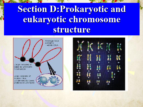
原核生物与真核生物染色体区别和识别
The importance of packing of DNA into chromosomes
Chromosome is a compact form of the DNA that readily fits inside the cell [?]
A Severe Problem of Packaging
1. Largest human chromosome: ~3 x 10⁸ bp How long is it?
3 x 108 bp x 3.4 Å/bp = 10 x 104um! 2. A typical cell = 10 um
Conclusion: chromosome in length/size of cell
Supercoiling of the genome
supercoil of the genome
unconstrained supercoil function: pass tensile forces among loops
constrained supercoil (bind proteins) 结构域被非特异结合蛋白如HU和H-NS等类 组蛋白缠绕,使一半超螺旋被束缚。
146bp的DNA片段和H2A、 H2B、H3和H4各二分子组 成,这种结构称为核心颗粒.
• 核心颗粒外观呈椭圆形,颗粒直径11nm,高 5.5nm.绕颗粒的DNA长度为50nm(146bp),连 接区DNA长度为20nm(约60bp).
11nm染色质纤维
The roles of H1
1. Stabilizes the point at which DNA enters and leaves the nucleosome core. Protect the further 20bp DNA from nuclease digestion.
遗传染色体常见异常报告中英文翻译
外周血:(Peripheral blood):外周血细胞培养染色体检查结果为男性核型,没有发现染色体数目或结构有明显的异常。
The chromosome detection showed male karyotype by culturing the peripheral blood lymphocytes and no obvious anomalies were found on chromosome number or structure.外周血细胞培养染色体检查结果为女性核型,没有发现染色体数目或结构有明显的异常。
The chromosome detection showed female karyotype by culturing the peripheral blood lymphocytes and no obvious anomalies were found on chromosome number or structure.外周血细胞培养染色体检查结果显示为男性核型,所分析的分裂相中均见Y染色体大于或等于18号染色体。
The chromosome detection showed male karyotype by culturing the peripheral blood lymphocytes and the phenomenon (Y chromosome ≥ chromosome 18) was found on all the analytical metaphase cells.外周血细胞培养染色体检查结果显示为男性核型,所分析的分裂相中均见Y染色体小于或等于22号染色体。
The chromosome detection showed male karyotype by culturing the peripheral blood lymphocytes and the phenomenon(Y chromosome≤ chromosome22)was found on all the analytical metaphase cells.外周血细胞培养染色体检查结果显示为男性核型,所分析的分裂相中均见Y染色体长臂异染色质区长度的增加。
果蝇唾腺多线染色体研究进展及其在遗传学教学中的应用-生物通

的研究成果后有人提出带是由转录惰性的基因组成, 化研究基因活性的变化;现在基于多线染色体的活
而间带则是转录活跃基因,特别是永久表达的看家 细胞成像技术结合蛋白质-DNA 的互作分析可以在
基因。最新的研究结果表明,在黑腹果蝇唾腺第四 染色体上,间带区包含了看家基因的启动子区域和 5′
全基因组规模上实时、直观地研究转录调控的机制 问题[15]。原核基因表达调控研究的模型在 20 世纪
第6期
李刚等: 果蝇唾腺多线染色体研究进展及其在遗传学教学中的应用
607
于带纹的遗传组织问题有很多假说,早期的假说认 看似染色体断裂现象的存在[13]。另外,利用该模型
为带和间带都是基因的载体,每条带或间带含有 1 ̄2 个基因;有的认为每条带包含多个结构基因,类似
研究发现复制的拷贝数可以通过抑制复制的起始和 延伸来实现,进而影响细胞和组织的分化[18]。除了
gaster)就成为遗传学研究的常用材料之一,但是由 于其染色体小,因而难于用于细胞学水平的观察[4]。
1881 年 Balbiani 发现双翅目昆虫唾腺有多线染色体 存在,但当时没有引起细胞遗传学家的注意[5];直
技术,发现异染色质区域存在组蛋白低乙酰化和 H3K9 的高甲基化,而常染色质则是高水平的组蛋白 乙酰化和 H3K4 的高甲基化[8];异染色质又分为结构 异染色质和兼性异染色质,前者如着丝粒、端粒等
教学实践中,循着前人研究的足迹展开教学不但有 普通光学显微镜可以直接观察到。通过对野生型和
助于学生对遗传学基础理论的掌握,而且还可以激 发学生学习遗传学的积极性[2]。本文一方面回顾了
突变型唾腺染色体带纹和蓬突的比较可以将不同染 色体分类并发现各种染色体畸变[7];结合组织化学
染色体文献

染色体文献(中英文版)英文文档:Chromosome LiteratureChromosomes are structures found in the nucleus of animal and plant cells that contain genetic information.They are composed of DNA and proteins and are responsible for carrying genes, which are the hereditary units that determine an organism"s traits.The study of chromosomes and their composition, organization, and behavior is known as cytogenetics.Over the years, numerous scientific studies have been conducted to explore the fascinating world of chromosomes.These studies have led to a better understanding of how genetic information is inherited, how chromosomes evolve, and how they can be affected by various factors such as radiation and chemicals.One of the most significant findings in the field of cytogenetics is the discovery of the structure of chromosomes.In 1953, James Watson and Francis Crick proposed the double-helix model of DNA, which revolutionized the way scientists thought about chromosomes and genetics.Their discovery laid the foundation for modern molecular biology and has been instrumental in unraveling the mysteries of life.Another important area of research in chromosome literature is thestudy of chromosomal abnormalities.Errors in the replication or division of chromosomes can lead to genetic disorders such as Down syndrome, cystic fibrosis, and many others.Understanding these abnormalities has crucial implications for the diagnosis, treatment, and prevention of genetic diseases.In recent years, advancements in technology have allowed scientists to study chromosomes at the atomic level.Techniques such as chromosome microscopy, fluorescence in situ hybridization (FISH), and polymerase chain reaction (PCR) have enabled researchers to explore the intricate details of chromosome structure and function.The field of chromosome literature is constantly evolving, with new discoveries and insights being published every day.Scientists around the world continue to delve into the complex world of chromosomes, seeking to unravel the remaining mysteries of genetics and improve our understanding of life itself.中文文档:染色体文献染色体是存在于动物和植物细胞核中的结构,负责携带遗传信息。
分子生物学(杨洋)第二章-染色体,染色质和核小体-chromosomes, chromatin, and the nucleosome

基因中平均的 内含子数目
• 两种因素会导致真核生物基因密度的降低: – 基因长度的增加 – 基因间隔区序列( intergenic sequences) 的增加
在更复杂的生物中基因间隔区序列数目的大量 增加是导致基因密度降低的另一个重要原因 基因间隔区( intergenic sequences) :基因组 上与蛋白质和结构RNA的表达无关的那部分 DNA
• 大多数真核生物细胞是二倍体(diploid), 一个染色体有两个拷贝 • 多倍体真核细胞(polyploid): 细胞中每 条染色体都超过两个拷贝
• 巨核细胞:特化的多倍体细胞,每条染 色体有128个拷贝,通过多倍体形式维持 高水平新陈代谢,以生产大量的血小板
单倍体 原核细
二倍体 真核细
单倍体 真核细
[Add a large layer of gene regulation]
OUTLINE
1. Chromosome sequence & diversity 3. The nucleosome
2. Chromosome duplication & segregation 4. Higher-order chromatin structure
1.2 Number of Chromosomes in an organism is characteristic(每个细胞都 有特定的染色体数目)
• 典型的原核生物细胞仅有一个完整的染色体拷贝 • 原核细胞中还经常携带一个或多个小的独立的环 状DNA—质粒(plasmid) • 质粒不是细菌生长所必需的,而是携带赋予细菌 良好特性的基因(如抗生素抗性基因) • 每个细胞中可以有多个完整拷贝的质粒存在
h4的n端尾巴乙酰化修饰影响了核小体整合进抑制状态的30onenterminaltailsaltersch加到组蛋白上而组蛋白去乙酰化酶可去除这种修饰组蛋白甲基转移酶将甲基加到组蛋白上而去甲基化酶则将甲基去除由于不同的修饰对核小体的功能有不同的影响因此不同的组蛋白乙酰转移酶或甲基转移酶对核小体的修饰会对染色质结构的很的多聚蛋白复合体的一部分其它亚基在引导这些酶到特定的dna区域上起重要作用引导这些修饰酶到特定的dna区域是调控真核染色体中基因表达核小体重塑的共同作用能够显著改变dna的易接近性n端尾巴的修饰能减少核小体束形成抑制性结构的能力产生能集合包括核小体重塑因子在内的蛋白随后的核小体重塑能够进一步增加核小体dna的易接近性允许dna结合蛋白接近它们的结合位点另外这些复合体能引起核小体的滑动或释放与适当的dna结合蛋白或dna序列结合导致dna特点位点色质重塑复合体来修饰邻近的核小体增加相关dna的易接近性?这导致第二个dna结合蛋白的结合它又引导一个组蛋白乙酰转移酶该酶通过修饰相邻核小体的n端尾巴改变了染色质构象使之从30nm纤丝结构变为更易接近的10nm纤丝结构30nm染色质纤丝dna结合蛋白1dna结合蛋白2组蛋白乙酰转移酶10nm染色质纤丝图741染色质重塑复合体和组蛋白修饰酶共同改蛋白乙酰转移酶结合也可以反过来的顺序而组蛋白甲基转移酶会导致形成更压缩的不易接esequencechromosomeduplicationhigherorderchromatinstructurechromatinst组的大小与生物体 的复杂性更密切相关
细胞生物学-细胞核与染色体

(一) 核被膜的结构(Structure of the Nuclear Envelope)
◆外核膜 ◆内核膜
◆核纤层
◆核周间隙 ◆核孔复合体
核纤层
核纤层由核纤肽(lamin)构成,是一类中间纤维。核纤层 的作用有以下两个方面: 1.保持核的形态 2.参与染色质和核的组装
(二)核被膜在细胞周期中的崩解与装配
核酸酶
DNA合成 RNA合成
切割DNA和/或RNA
DNA连接酶
Poly-A聚合酶 DNA甲基化酶 拓扑异构酶 螺旋去稳定酶
作用于染色质蛋白的酶
在DNA复制和修复时进行DNA连接
在mRNA3'添加poly A尾 DNA甲基化 将超螺旋DNA转变成松弛型 DNA解旋, 形成稳定的单链 蛋白质切割 组蛋白乙酰化和去乙酰化 组蛋白和非组蛋白的磷酸化 组蛋白甲基化
10nm
被动运输(passive transport)
小于10nm的分子自由出入
主动运输(active transport)
具有入核信号的蛋白的入核 RNA分子 核糖核蛋白颗粒出核
2. 通过核孔复合体的主动运输 生物大分子的核质分配如亲核蛋白的核输入,RNA分子及RNP颗粒的核输出,在 细胞核功能活性的控制中起非常重要的作用。
核定位信号(nuclear localization signals , NLS)
核蛋白输入机理
( 1 )亲核蛋白通过 NLS 识别 importinα ,与可溶性 NLS 受体 importinα/ improtinβ异二聚体结合,形成转运复合物;
(2)在importinβ 的介导下,转运复合物与核孔复合体的胞质纤
NLS只是亲核蛋白入核的一个必要条件,某种亲核蛋白是否被转运入核还受到 其它因素的影响。
性连锁遗传XXXX

基因型 BaBa TsTs Ba_ tsts baba Ts_ baba tsts
玉米的性别决定
性别
表型
♀♂
顶端长雄花序,叶腋长雌花序
♀
顶端和叶腋都长雌花序
♂
顶端长雄花序,叶腋不长花序
♀
顶端长雌花序,叶腋不长花序
环境与性别的决定
1 后螠: • 海底 雌虫 • 吻 雄虫 • 改变条件:间性 2、爬行类
Turner综合征
This condition occurs in about 1 in 2,500 female births worldwide, but is much more common among pregnancies that do not survive to term (miscarriages and stillbirths).
1905, Wilson和他的同事对蛛蝽属的昆虫进 行了广泛的研究,发现:
• 蛛蝽属的昆虫性别决定:
♂:1/2(6):1/2(7)
♀:(7)
• 1910年,Morgan,伴性遗传(性别—基因) • 1916,Bridges遗传的染色体学说的直接证据
染色体与性别决定 1、X0型性别决定
♀: A(X)+ A(X) →AA(XX) ♂: A(X) +A(0) → AA(X0) • 蚱蜢,蝗虫,蛛蝽属的昆虫。
根据已有知 识如何去解 释试验现象?
1.③F1白眼 雄蝇w0
2.③F1红眼 雌蝇wW
3.③P红眼雄 蝇自然状 态下W0
4.③P白眼雌 蝇ww
5.①F2白眼 雌蝇不可 能出现
6.为什么③P 红眼雄蝇 自W0? ——Morgan的假设
1) Mendel:基因成对存在,分别来自双亲 2) 红眼雄蝇自然状态下红眼基因(W0)不成对 3) 遗传的染色体学说:基因也应在性染色体上 4) 性别决定染色体学说:性染色体不匹配
- 1、下载文档前请自行甄别文档内容的完整性,平台不提供额外的编辑、内容补充、找答案等附加服务。
- 2、"仅部分预览"的文档,不可在线预览部分如存在完整性等问题,可反馈申请退款(可完整预览的文档不适用该条件!)。
- 3、如文档侵犯您的权益,请联系客服反馈,我们会尽快为您处理(人工客服工作时间:9:00-18:30)。
Junk DNA
During
the late 1960s papers began to appear that showed eukaryotic DNA contained large amounts of repetitive DNA that did not appear to code for proteins (ie, Britten and Kohne, 1968). By the early 1970s, the term Junk DNA had been coined to refer to this noncoding DNA (ie. Ohno, 1972).
Chromosome Structure
and
DNA Sequence Organization
An Overview
Eukaryotes Have Large Complex Geneomes
human genome is ≈ 3 x 10⁹ bp 3 x 109 bp x 0.34 nm/bp x 1 m/109 nm ≈ 1m Because humans are diploid, each nucleus contains 6 x 10⁹ bp or ≈ 2 m of DNA That is a lot to pack into a little nucleus! Eukaryotic DNA is highly packaged
Types of Junk DNA Nine different types of DNA were listed as junk DNA by Nowak (1994) These nine types can be grouped into three larger groups:
1Repetitive DNA sequences 2Untranslated parts of RNA transcripts (pre-mRNA) 3Other non-coding sequences
What is Junk DNA?
“Junk DNA” is DNA that does not code for proteins, this is the definition that we will use. The meaning of “junk DNA” has become restricted significantly in recent years as the functionality of much of what was once considered junk has become obvious. Most modern genetics texts avoid the term. Even when junk DNA is mentioned, it may be given significantly different definitions. For example, Lodish et al. (1995) called it “Extra DNA for which no function has been found.”
30 nm
Tight helical fiber
Looped 200 nm Domains
Protein scaffold
Hale Waihona Puke Packaging DNA
Nucleosomes
11 nm
30 nm
Tight helical fiber
Metaphase Chromosome 700 nm 200 nm Looped Domains
Evidence
Conservation
of protein (and DNA) sequences is commonly interpreted to indicate functionality Significant variation in non-coding DNA is evident between relatively closely related species and even within species (ie Zeyl and Green, 1992). Mutation of some non-coding DNA does not produce significant changes in phenotype (Nei, 1987).
11 nm Histone octomer
Histone proteins Nucleosome B DNA Helix 2 nm
Packaging DNA
Histone H1
Packaging DNA
Histone H1
Packaging DNA
“Beads on a string” 11 nm
Four
proteins: H2A, H2B, H3, and H4 H3 and H4 are arginine rich and highly conserved H2A and H2B are slightly enriched in lysine Both arginine and lysine are basic amino acids making the histone proteins both basic and positively charged The octomer is made of two copies of each protein
Nucleosomes
Nucleosome
- Nucle - kernel, some -
body The lowest DNA packaging level Can be thought of as like a length of thread wound around a spool, the thread representing DNA and the spool being histone proteins
2 nm
B DNA Helix
Protein scaffold
Highly Packaged DNA Cannot be Expressed
The most highly packaged form of DNA is “heterochromatin” Heterochromatin cannot be transcribed, therefore expression of genes is prevented Constitutive heterochromatin - Permanently unexpressed DNA e.g. satellite DNA Facultative heterochromatin - DNA that could be expressed if it was not packaged
Repeated sequences seem too short to code for proteins and are not known to be transcribed. Five major classes of repetitive DNA:
Repetitive DNA
1 Satellites - Up to 105 tandem repeated short DNA sequences, concentrated in heterochromatin at the ends (Telomeres) and centers (centromeres) of chromosomes. 2 Minisatellites - Similar to satellites, but found in clusters of fewer repeats, scattered throughout the genome 3 Microsatellites - Shorter still than minisatellites. 1 4 and 5 Short (300 bp) and Long (up to 7,000 bp) Interspersed Elements (SINEs and LINEs) - Units of DNA found distributed throughout the genome
The Histone Octomer
The Histone Octomer
A
fifth protein, H1, is part of the nucleosome, but seems to be outside the octomer H1 varies between tissue and organisms and seems to stick to the 19 bases attached to the end of the core sequence Ausio (2000) discusses data showing that, at least in fungi, survival is possible without H1 Lack of H1 does not impact cell viability but shortens the lifespan of the organism This raises the question of how H1 evolved in single celled organisms
The Fifth Histone, H1
Packaging DNA
Histone octomer Histone proteins B DNA Helix 2 nm
Packaging DNA
Histone octomer Histone proteins B DNA Helix 2 nm