美国伯乐电转化仪使用说明
伯乐显影仪的操作流程及注意事项

伯乐显影仪的操作流程及注意事项
操作流程:
1. 打开电源开关,待仪器启动并进入工作状态。
2. 将待显影的胶片放入伯乐显影仪的显影槽中,确保胶片完全浸没在显影液中。
3. 调节显影液的温度和浓度,一般根据胶片的要求进行调节。
确保显影液在适宜的温度和浓度范围内。
4. 设定显影时间,根据胶片的要求设置合适的显影时间。
5. 启动伯乐显影仪开始显影过程。
仪器会自动控制显影液的温度和搅拌速度。
6. 在显影过程中,可以通过观察显影仪的显示屏来了解显影的进程。
根据胶片要求,可以提前停止显影或继续延长显影时间。
7. 显影完成后,将胶片从显影槽中取出,并进行洗涤和定影等后续处理。
注意事项:
1. 在操作伯乐显影仪之前,需要了解所使用胶片的显影要求,包括温度、浓度和时间等参数,以便正确调节仪器。
2. 在放入胶片之前,确保显影槽干净,没有杂质和污染物。
可以用干净的布或棉签清洁显影槽表面。
3. 在调节显影液的温度和浓度时,可以使用温度计和浓度计等工具进行准确测量,以确保显影条件的准确性。
4. 在显影过程中,要定期检查显影液的温度和搅拌情况,确保显影液的稳定性和均匀性。
5. 注意个人安全防护,避免直接接触显影液或其他化学品。
使
用手套和护目镜等防护设备进行操作。
6. 在显影完成后,及时清洁和维护伯乐显影仪,确保仪器的正常运行和长久使用。
美国伯乐电转化仪使用说明
Gene pulser xcell TM Electroporation system quick guide产品名称: Gene Pulser Xcell 电穿孔系统产品型号: Gene Pulser Xcell 电穿孔系统产品展商: 众磊(北京)生物科技发展有限公司驻沪办简单介绍Gene Pulser Xcell 电穿孔系统Gene Pulser Xcell 电穿孔系统的详细介绍【介绍】电穿孔是功能强大的将核酸、蛋白及其它分子导入多种细胞的高效技术。
通过高强度的电场作用,瞬时提高细胞膜的通透性,从而吸收周围介质中的外源分子。
这种技术可以将核苷酸、DNA 与RNA 、蛋白、糖类、染料及病毒颗粒等导入原核和真核细胞内。
电转化相对其它物理和化学转化方法,是一种有价值和有效的替代方法。
Gene Pulser Xcell 系统的设计,基于Bio-Rad 15 年来在电转化技术上经验积累,它提供指数波和方波波型选择、系统配置选择及友好的用户界面。
主要特点指数波和方波波型确保所有细胞类型(原核及真核)均可获得最佳的电转化效果Bio-Rad 专利的* PulseTrac 电路和电弧保护设计,确保可重复性并保护样品模块化设计可根据研究需要选择系统用户友好的数字化界面,具有直观的编程以控制所有参数,包括附属模块的参数包括人工操作、预设规程、用户规程、一个优化规程及其它先进功能等程序选择指数波或方波脉冲选择Gene Pulser Xcell 系统可产生指数波和方波波型,使你选择最适合你细胞的波型与规程。
指数波和方波均能有效地用于电转化及电融合。
电穿孔波型对不同类型细胞的转化效率有很重要的影响。
左,指数衰变脉冲。
当一个充电至电压为V 0 的电容器放电到细胞,加在细胞上的电压随时间以指数方式下降。
从起始电压下降到V0 / e 所需的时间称为为时间常数τ, 一种方便的脉冲时间表达方式。
右,方波脉冲。
放电到样品后截断电容器脉冲可产生方波脉冲。
伯乐半干转膜仪说明书中文
伯乐半干转膜仪说明书中文伯乐半干转膜仪说明书一、产品概述\n伯乐半干转膜仪是一种用于分离液体和固体颗粒的实验室设备。
它采用半干转膜技术,通过膜的微孔将液体分离出来,从而实现固液分离的目的。
本仪器具有操作简便、效率高、分离效果好等特点,广泛应用于化学、生物、医药等领域。
二、产品结构\n伯乐半干转膜仪主要由以下部分组成:\n1. 转膜仪主机:包括电源开关、控制面板和显示屏等。
\n2. 转膜模块:包括转膜夹具和转膜杯等。
\n3. 液体收集容器:用于收集分离出的液体。
\n4. 固体残渣容器:用于收集分离出的固体残渣。
三、使用方法\n1. 将待处理的混合物倒入转膜杯中,注意不要超过杯口。
\n2. 将转膜杯放入转膜夹具中,并将夹具安装到主机上。
\n3. 打开电源开关,启动仪器。
\n4. 在控制面板上设置所需的分离参数,如转速、时间等。
\n5. 按下启动按钮,仪器开始工作。
\n6. 待分离完成后,关闭电源开关,停止仪器运行。
\n7. 将转膜杯从夹具中取出,将液体倒入液体收集容器中。
\n8. 将固体残渣从转膜杯中取出,放入固体残渣容器中。
四、注意事项\n1. 在操作过程中,请佩戴防护手套和护目镜等个人防护装备。
\n2. 请确保待处理的混合物不超过转膜杯的容量范围。
\n3. 在设置分离参数时,请根据实际需要进行调整,并确保参数合理可行。
\n4. 请勿将手指或其他物体接触到转膜模块内部,以免损坏设备或造成伤害。
\n5. 使用完毕后,请及时清洁仪器,并保持其干燥。
五、维护保养\n1.定期清洁仪器表面和内部零部件,以保持其正常运行和延长使用寿命。
\n2. 如发现任何故障或异常情况,请立即停止使用,并联系售后服务人员进行维修。
六、故障排除\n1. 仪器无法启动:请检查电源是否连接正常,电源开关是否打开。
\n2. 转膜杯无法安装:请检查转膜夹具是否正确安装,转膜杯是否损坏。
\n3. 分离效果不理想:请检查分离参数设置是否合理,转膜模块是否损坏。
美国Bio-rad伯乐Trans-Blot SD 半干转印槽使用手册

Trans-Blot®SD Semi-Dry Electrophoretic Transfer Cell InstructionManualCatalog Number170-3940NoteTo insure the best performance from the Trans-Blot SD semi-dry electrophoretic transfer cell, become fully acquainted with these operating instructions before using the cell to transfer samples. Bio-Rad recommends that you first read these instructions carefully. Then assemble and disassemble the cell completely without transferring sample. After these preliminary steps, you should be ready to transfer a sample.Bio-Rad also recommends that all Trans-Blot SD cell components and accessories be cleaned with a suitable laboratory cleaner (such as Bio-Rad Cleaning Concentrate, catalog number 161-0722) and rinsed thoroughly with distilled water, before use.Model___________________________________Catalog Number__________________________Date of Delivery___________________________Warranty Period__________________________Serial Number____________________________Invoice Number___________________________Purchase Order Number____________________WarrantyBio-Rad Laboratories warrants the Trans-Blot SD semi-dry electrophoretic transfer cell against defects in materials and workmanship for 1 year. If any defects occur in the instrument during this warranty period, Bio-Rad Laboratories will repair or replace the defective parts free. The following defects, however, are specifically excluded:1.Defects caused by improper operation.2.Repair or modification done by anyone other than Bio-Rad Laboratories or an authorizedagent.e of fittings or other spare parts supplied by anyone other than Bio-Rad Laboratories.4.Damage caused by accident or misuse.5.Damage caused by disaster.6.Corrosion due to use of improper solvent or sample.This warranty does not apply to parts listed below:1.Platinum plate electrode.For any inquiry or request for repair service, contact Bio-Rad Laboratories after con-firming the model and serial number of your instrument.T able of ContentsPage Section 1 Introduction (1)1.1 Specifications (1)Section 2 Equipment and Reagents (2)2.1 Equipment and Accessories (2)Instruments (4)2.2 RelatedReagents (4)2.3 ChemicalSection 3 Safety Instructions (5)Section 4 Trans-Blot SD Assembly (6)4.1 Preparation for Blotting (6)4.2 Assembly of the Unit for Standard Transfers (7)4.3 Assembly of the Unit for Acidic Transfers (10)Section 5 Buffer Formulation (10)Section 6 Examples of Specific Protocols (11)Blotting (11)6.1 SDS-Protein6.2 DNA Blotting (For acrylamide gels with DNA 250 bp to ~1 kb) (12)6.3 DNA & RNA Blotting (For agarose gels with DNA up to 23 kb,RNA up to 3.5 kb) (12)Section 7 Properties of Protein Blotting Media (12)Section 8 Troubleshooting Guide (13)8.1 PoorTransfer (13)8.2 Poor Binding to Nitrocellulose Membrane (14)8.3 High Background After Incubation with Antibody Probes; Nonspecificor Nonquantitative Detection (14)8.4 Poor Detection Sensitivity or No Reactivity (15)Section 9 References (15)Section 1IntroductionBlotting was first performed by Southern1in 1975 with the transfer of DNA from agarose gels to nitrocellulose membranes. Blotting has subsequently been applied to RNA2-4and protein5,6from both agarose and polyacrylamide gels. Membrane materials have been expanded to include PVDF for improved protein binding capacity. To overcome the inefficiency of capillary transfers, electric current has been adopted for eluting proteins from polyacrylamide gels, as first described by Towbin et al.7in 1979. Since that time, electrophoretic transfer has also been used for DNA and RNA blotting.8-14For blotting PCR fragments, plasmid and vector DNA, and RNA with the SD cell, use the Trans-Blot SD DNA blotting kit. DNA or RNA can be blotted from agarose gel to Zeta-Probe®GT membrane in only 10 minutes, without any gel pretreatments. The kit comes complete with DNA/RNA blotting accessories and a detailed instruction manual.Semi-dry blotting was first reported by Kyhse-Andersen in 1984.15Blotting was performed with plate electrodes in a horizontal configuration. The gel and nitrocellulose membrane were sandwiched between sheets of buffer-soaked filter paper, which served as the ion reservoir and replaced the buffer tank. The plate electrodes, separated only by the filter paper stack, provided high field strength (V/cm) across the gel, and very efficient, rapid transfers.The Trans-Blot semi-dry transfer cell incorporates the original concepts of semi-dry blotting along with innovative features for quick set-up and ease of use. The platinum-coated titanium and stainless steel electrode pair provides efficient, background-free blotting with trouble-free service.1.1 SpecificationsConstructionTrans-Blot SD body Molded polycarbonateAnode Platinum-coated titaniumCathode Stainless steelAnode platform Precision machined acrylicOverall size37 cm x 24 cm x 11 cmMaximum gel size25 cm x 18.5 cmCleaning Do not immerse the unit in liquid. Use special carewhen cleaning the anode plate to avoid scratchingor marring the platinum. Do not use abrasives orstrong detergents. The cathode plate (stainlesssteel) can be cleaned with a mild abrasive toremove salt that may deposit during normal opera-tion. The entire unit can also be periodically disas-sembled and cleaned with water to remove saltdeposits.Chemical compatibility The semi-dry blotter components are not compati-ble with chlorinated hydrocarbons (e.g., chloro-form), aromatic hydrocarbons (e.g., toluene,benzene), or acetone. Use of organic solventsvoids all warranties.4.Lengthy transfer times are not recommended.Do not leave this instrument unattended.Joule heat can be generated rapidly during semi-dry blotting. Transferring longer than2 hours can damage the unit.5.Power supply requirements. The Trans-Blot SD cell should only be used with themicroprocessor-controlled Model 200/2.0 power supply (catalog numbers 165-4761 and165-4762), or the Model 1000/500 power supply (catalog numbers 165-4710 and165-4711). Do not use the Model 250/2.5 power supply with this apparatus. The lowvoltage, high current operating conditions of the Trans-Blot SD cell are not compatiblewith the Model 250/2.5 power supply, and will cause the power supply to blow a fuse.6.Do not operate this instrument in ambient temperatures exceeding 50 °C.ImportantThis Bio-Rad instrument is designed and certified to meet IEC 1010-1* safety standards.Certified products are safe to use when operated in accordance with the instructtionmanual. This instrument should not be modified in any way. Alteration of this instrumentwill:•Void the manufacturer's warranty•Void the IEC1010-1 safety certification•Create a potential safety hazardBio-Rad is not responsible for any injury or damage caused by the use of this instrumentfor purposes other than for which it is intended or by modifications of the instrument notperformed by Bio-Rad or an authorized agent.*IEC 1010-1 is an internationally accepted electical safety standard for laboratory instruments.Section 4Trans-Blot SD AssemblyTo determine the optimum conditions for a particular sample, a time course of transfer should be performed. Since many factors affect transfer e.g.molecular weight, pI, and porosity of the gel, transferring for the full suggested time may not be necessary.4.1 Preparation for Blotting1.Prepare the transfer buffer. See Section 5 for buffer formulation.Note:Buffer preparation is extremely important. Do not adjust transfer buffer pH byaddition of acid or base unless specifically indicated in the instructions. Improperlyprepared buffer will cause excess heat generation and safety e only highquality, reagent grade methanol. Contaminated methanol can result in increased transferbuffer conductivity, as well as poor transfer of macromolecules.2.Following electrophoresis, equilibrate the gels in transfer buffer. Equilibration facilitatesthe removal of electrophoresis buffer salts and detergents. If the salts are not removed, theywill increase the conductivity of the transfer buffer and the amount of heat generatedduring the transfer. Also, low percentage gels (<12% acrylamide) will shrink in methanol-containing buffers. Equilibration allows the gel to adjust to its final size prior toelectrophoretic transfer. The length of time required for equilibration is dependent on thegel thickness. For example, 15 minutes for a 0.75 mm SDS-PAGE gel.Low molecular weight macromolecules (10,000 daltons) may diffuse out of gels more readily. One can allow adequate gel pre-equilibration by changing the pre-equilibration buffer several times during a relatively short pre-equilibration period. This will help to limit diffusion of low molecular weight macromolecules while providing efficient salt reduction.3.Cut the membrane to the dimensions of the gel. Wet the membrane by slowly sliding itat a 45° angle into transfer buffer and allowing it to soak for 15–30 minutes. Complete wetting of the membrane is important to insure proper binding. Abrupt wetting can lead to entrapment of air bubbles in the matrix. These air bubbles can block transfer of molecules. To avoid membrane contamination, always use forceps or wear gloves when handling membranes.4.Cut filter paper to the dimensions of the gel. Two pieces of extra thick filter paper (orfour pieces of thick or six pieces of thin filter paper) per gel are needed for each gel/mem-brane sandwich. Completely saturate the filter paper by soaking in transfer buffer.5.If more than one full-size gel is to be transferred at one time, cut a piece of dialysismembrane with the appropriate molecular weight cutoff to the dimensions of the gel.Completely wet the dialysis membrane in transfer buffer. Spectr/Por™ dialysis membrane is recommended for this use.4.2 Assembly of the Unit for Standard TransfersWear gloves for this procedure to avoid contamination of membranes.1.Remove the safety cover and the stainless steel cathode assembly.2.Place a pre-soaked sheet of extra thick filter paper onto the platinum anode. Roll a pipetor test tube over the surface of the filter paper (like a rolling pin) to exclude all air bubbles. If thick or thin filter paper is used, repeat with one or two more sheets of buffer-soaked filter paper.3.Place the pre-wetted blotting media on top of the filter paper. Roll out all air bubbles.4.Carefully place the equilibrated gel on top of the transfer membrane, aligning the gel onthe center of the membrane. Transfer will be incomplete if any portion of the gel is outside the blotting media. Roll out all air bubbles.5.Place the other sheet of pre-soaked filter paper on top of the gel, carefully removing airbubbles from between the gel and filter paper. If thick filter paper is used, place two sheets on top of the gel, and remove bubbles from between each layer. If thin filter paper is used, place three sheets on top of the gel, and remove bubbles from between each layer.6.If more than one full-size gel is to be transferred, place a sheet of pre-soaked dialysismembrane on top of the filter paper stack. Repeat the procedure from step 2. Up to four mini gels can be transferred at the same time by placing them side-by-side on the anode platform.7.Carefully place the cathode onto the stack. Press to engage the latches with the guideposts without disturbing the filter paper stack.8.Place the safety cover on the unit. Plug the unit into the power supply. Normal transferpolarity is cathode to anode, i.e., red wire to red outlet and black wire to black outlet on the power supply.Caution:Do not reverse polarity. This will result in damage to the stainless steel cathode.9.Turn on the power supply. Transfer mini gels for 15–30 minutes at 10–15 V. Large gelscan be transferred for 30 minutes to 1 hour at 15–25 V. Do not exceed 25V with this instrument. A current limit (3 mA/cm2for large gels; 5.5 mA/cm2for mini gels) is recommended to prevent excessive heating during the run. Under the strong fields developed by this apparatus, transfers may not always be quantitative. A certain quantity of protein may be transferred through the membrane and onto the filter paper below.The Model 200/2.0 power supply is capable of a 200 watt output. This means that unlessa current limit is set, uncontrolled conductivity changes may result in full power beingdelivered to the Trans-Blot SD cell. In this situation, the gel sandwich and electrodes will be exposed to excessive heat. This may result in a safety hazard. It is advisable to monitor resistance, power, and current during the run. Refer to the Model 200/2.0 Instruction Manual for setting current limits and run times, and monitoring these parameters.10.Following transfer, turn the power supply off, and disconnect the unit from the powersupply. Remove the safety cover and the cathode assembly. Discard the filter paper (and dialysis membrane, if used). The transfer efficiency can be monitored by staining the gel with Coomassie blue R-250 protein stain or with Bio-Rad's Silver Stain Kit. Alternatively, prestained molecular weight standards can be used, or a portion of the membrane can be stained for total protein with colloidal gold, Biotin Blot Total Protein Stain, or an anionic dye such as Amido Black. Zeta-Probe membrane can be stained with the Biotin-Blot Total Protein Stain.4.3 Assembly of the Unit for Acidic TransfersIf an acidic transfer buffer is used, the transfer direction will be from the anode to thecathode.1.Remove the safety cover and the stainless steel cathode assembly.2.Place a pre-soaked sheet of extra thick filter paper onto the platinum anode. Roll out allair bubbles. If thin filter paper is used, repeat with two more sheets of buffer-soakedfilter paper. If thick filter paper is used, repeat with one more sheet of buffer soakedfilter paper.3.Carefully place equilibrated gel on top of the filter paper, aligning the gel on the center ofthe membrane. Roll out all air bubbles.4.Place the pre-wetted blotting media on top of the gel. Roll out all air bubbles.5.Place another sheet of pre-soaked extra thick filter paper on top of the blotting membrane,carefully removing all air bubbles. If thin filter paper is used, place three sheets on top of themembrane, or if thick filter paper is used, place two sheets on top of the membrane.6.If more than one gel is to be transferred, place a sheet of pre-soaked dialysis membraneon top of the filter paper stack. Repeat the procedure from step 2.7.Carefully place the cathode assembly onto the stack. Press to engage the latches with theguide posts, without disturbing the filter paper stack.8.Place the safety cover on the unit. Plug the unit into the power supply, red wire to redoutlet and black wire to black outlet.Caution:Do not reverse polarity. This will damage the stainless steel cathode.9.Turn on the power supply. Transfer mini gels for 15–30 minutes at 10–15 V. Large gelscan be transferred for 30 minutes to 1 hour at 15–25 V. Do not exceed 25 V with thisinstrument. A current limit (3 mA/cm 2for large gels; 5.5 mA/cm 2for mini gels) isrecommended to prevent excessive heating during the run.Section 5Buffer FormulationThe following buffers are recommended for use with the Trans-Blot SD cell. For proteintransfers, the single buffer system of Bjerrum and Schafer-Nielsen 16provides more efficientelution than the original isotachophoretic system of Khyse-Andersen, which requires the useof three different buffers.15A carbonate buffer has also been shown to produce highefficiency transfers with improved antibody recognition.1.Bjerrum and Schafer-Nielsen transfer buffer for SDS-proteins using nitrocellulose (withmethanol) or Zeta-Probe membrane (without methanol):1648 mM Tris, 39 mM glycine, (20% methanol) pH 9.2Dissolve 5.82 g Tris and 2.93 g glycine [and 0.375 g SDS or 3.75 ml of 10% SDS] in ddH 2O (add 200 ml of methanol); adjust volume to 1 liter with dd H 2O.DO NOT ADD ACID OR BASE TO ADJUST pH.The buffer will range from pH 9.0to 9.4, depending on the quality of the Tris, glycine, dd H 2O, and methanol. Methanolshould be analytical reagent grade, because metallic contaminants in low grade methanol will plate on the electrodes.Note:Some pH electrodes will not perform a proper measurement for the pH of Trisbuffers. If the pH of the buffer is not correct, check the electrode to be sure it is designedto function with Tris buffers. If the pH electrode works properly with Tris buffers, and thepH is below 9.0, remake the buffer.2.SDS may be added to Buffer 1 to increase protein elution from the gel:48 mM Tris, 39 mM glycine, (20% methanol), 1.3 mM SDS (0.0375%), pH 9.2Dissolve 5.82 g Tris and 2.93 g glycine, and 0.0375 g SDS or 3.75 ml of 10% SDS in ddH 2O (add 200 ml of methanol); adjust the volume to 1 liter with dd H 2O.DO NOT ADD ACID OR BASE TO ADJUST pH.3.Towbin transfer buffer for SDS-proteins using nitrocellulose (with methanol) or Zeta-Probe membrane (without methanol):725 mM Tris, 192 mM glycine (20% methanol), pH 8.3Dissolve 3.03 g Tris and 14.4 g glycine in dd H 2O (add 200 ml of methanol); adjustvolume to 1 liter with dd H 2O.DO NOT ADD ACID OR BASE TO ADJUST pH.4.Dunn carbonate transfer buffer for SDS-proteins using nitrocellulose (with methanol) orZeta-Probe membrane (without methanol):1710 mM NaCHO 3, 3 mM Na 2CO 3(20% methanol), pH 9.9Dissolve 0.84 g NaHCO 3and 0.318 g Na 2CO 3(anhydrous) in dd H 2O (add 200 ml ofmethanol); adjust volume to 1 liter with dd H 2O.DO NOT ADD ACID OR BASE TO ADJUST pH.5.DNA transfer buffer for use with Zeta-Probe membrane:185x TBE stock solution (0.5 M Tris, 0.5 M boric acid, 10 mM EDTA in dd H 2O; adjustvolume to 1 liter with dd H 2O. Dilute to 0.5x TBE with dd H 2O for the working solution.DO NOT ADD ACID OR BASE TO ADJUST pH.6.5x dye buffer (20% Ficoll, 20 mM EDTA, 1% SDS, 0.2% bromophenol blue)Section 6Examples of Specific ProtocolsNote:In order to determine the optimum conditions for a particular sample, a time courseof transfer should be performed. Since many factors affect transfer, e.g., molecular weight,pI, porosity of the gel, it may not be necessary to transfer for the full time or to use highfield intensity transfer conditions. Final transfer conditions for any protein should bedetermined empirically.6.1 SDS-Protein BlottingStandard Blot to Nitrocellulose1. Equilibrate the gel in 500 ml of Towbin buffer (Section 5) for 15 minutes.2. Pre-chill buffer prior to transfer.3. Assemble the sandwich as described in Section4.2.4. Refer to Section 4.2, step 9 for transfer conditions with either large or small gels.6.2 DNA Blotting(For acrylamide gels with DNA 250 bp to ~1 kb)Electrophoresis Run on a Polyacrylamide Gel1.Prepare the stock electrophoresis 5x TBE buffer (Section 5). Dilute the stock to 1x.2.Mix 10–15 µl of the sample with 5 µl of 5x dye buffer, heat to 65 °C for 5 min and loadon a gel.3. A 5% PAGE gel can separate DNAs from about 250 to 1,000 bp.4.Run the gel in 1x TBE buffer at 100 V for 1–2 hours.Standard Blot to Zeta-Probe1.From the 5x TBE electrophoretic buffer, dilute the stock to 0.5x (Section 5) and pre-chill1 L of the buffer.2.Equilibrate the gel, extra thick blot paper, and Zeta-Probe membrane in 0.5x TBE bufferfor at least 15 minutes.Note: Zeta-Probe membrane will bind non-denatured nucleic acids. Therefore, denaturingis not mandatory before transferring. If non-denatured nucleic acids are transferred, theblotted Zeta-Probe membrane must be treated with NaOH prior to hybridization. Refer tothe Zeta-Probe membrane instruction manual.3.Assemble the sandwich as described in Section4.2.4.Run the transfer at 400 mA for 1 hour (voltage should not exceed 25 volts).5.After transfer, separate the membrane from the gel, and rinse the membrane briefly in0.5x TBE buffer.6.Fix the DNA to the membrane by placing the membrane on several pieces of blot papersaturated with 0.4 N NaOH for 10 minutes.7.Rinse the membrane in 2 x SSC for 10 minutes and bake at 80 °C for 1 hour (this isoptional if probing immediately). The membrane is now ready for hybridization. Refer tothe hybridization procedure in the Zeta-Probe blotting membrane instruction manual.6.3 DNA & RNA Blotting(For agarose gels with DNA up to 23 kb, RNA up to 3.5 kb) Refer to the Trans-Blot SD DNA blotting kit instruction manual for transfer protocol and conditions. DNA or RNA cannot be blotted from agarose gels without the use of the Trans-Blot SD DNA blotting kit.Section 7Properties of Protein Blotting MediaPVDF membrane is suitable for presenting transferred proteins for immuno detection (Immun-Blot PVDF) or analysis by Edman. It is resistant to tearing and chemicals. Immun-Blot PVDF is optimized for immunodevelopment with high protein binding capacity (160µg/cm2), but low nonspecific protein binding. This membrane material will resist tearing even when used in repeated stripping and reprobing applications. Sequi-blot PVDF has the highest protein binding capacity (170–200 µg/cm2) and gives outstanding performance in protein sequencing applications.Nitrocellulose membranes have been used extensively for protein binding and detec-tion.7,19-22They can easily be stained for total protein by a dye stain (Amido Black, Coomassie®blue, Ponceau S, Fast Green FCF, etc.22), or the more sensitive Colloidal Gold Total ProteinStain, and also allow either RIA, FIA, or EIA.7Nitrocellulose has a high binding capacity of 80–100 µg/cm2. Nonspecific protein binding sites are easily and rapidly blocked, avoidingsubsequent background problems. Low molecular weight proteins (esp. < 20,000 daltons) may be lost during post transfer washes, thus limiting detection sensitivity.21However, use ofglutaraldehyde fixation and a smaller pore size nitrocellulose membrane (0.2 µm) have been shown to be effective in eliminating this loss.22Large proteins (>100,000 daltons) denaturedby SDS may transfer poorly with the addition of alcohol to the transfer buffer. Alcohol increasesbinding of SDS-proteins to nitrocellulose, but decreases pore sizes in the gel. Elimination ofalcohol from SDS-protein transfers also results in considerably diminished binding to nitrocellulose. Under high field strengths of the Trans-Blot cell, proteins may be transferred through nitrocellulose without binding.The efficiency of binding can be increased by employing a smaller pore size nitrocellulose.23Zeta-Probe positively charged nylon membrane allows binding of SDS-protein complexes in the absence of alcohol.24,25This membrane binds proteins very tightly and is stable to post transfer washes. The binding capacity of Zeta-Probe membrane is ~480 µg/cm2.Reprobing, after stripping of prior probes, may be performed without significant loss of primary bound protein. Even small proteins appear to bind stably. Zeta-Probe membrane cannot be dye-stained, as destaining is impossible. Instead, the Biotin-Blot Total Protein Stain should be used on Zeta-Probe membrane. This assay uses NHS-Biotin (N-hydroxysuccin-imide-biotinate) to biotinylate all the proteins on the membrane surface, and a combination ofan avidin-horseradish peroxidase or avidin-alkaline phosphatase and a color development reagent to detect these biotinylated proteins.26,27The large capacity for molecules (480 µg/cm2) allows sensitive detection of small amounts of proteins in a complex mixture. This high capacity requires more stringent blocking conditions than nitrocellulose.25Zeta-Probe membranes can be effectively and economically blocked using a 5% solution of BLOTTO(non-fat dry milk)3,18,28Section 8Troubleshooting Guide8.1 Poor TransferA. Molecules remain in the gel matrix (as detected by Coomassie blue orsilver staining the gel)1.Transfer time is too short. Increase time of transfer.2.Charge to mass ratio is incorrect. Proteins near their isoelectric point at the pH of thebuffer will transfer poorly. Try a more basic or acidic transfer buffer to increase proteinmobility.3.Filter paper is too dry; insufficient buffer soaking the filter paper. Buffer is depleted earlyin the transfer. The filter paper should be fully saturated with buffer prior to transfer.Increase the number of sheets of filter paper, or use thicker filter paper.4.Power supply circuit tripped. Check the fuse.5.Gel percentage is too high. Reduce %T (total monomer) or %C(crosslinker). A 5% C(with bis as the crosslinker) will produce the smallest pore size gel. Decreasing from thisconcentration will increase pore size and increase transfer efficiency.6.Methanol in the transfer buffer is restricting elution of proteins from the gel. Eliminationof methanol results in increased transfer efficiency, but it also diminishes binding to nitrocellulose. Use PVDF.7.Protein is precipitating in the gel. Try using SDS in the transfer buffer. SDS can increasetransfer efficiency, but can also reduce binding efficiency to nitrocellulose and affect reactivity of some proteins with antibodies.B. Swirls or missing patterns on blot; diffuse transfers1.Contact between blot membrane and gel is poor. Air bubbles or excess moisture remainbetween the blot and gel. Use a test tube or pipet to roll over the membrane carefully in both directions until excess moisture and air bubbles are removed from between gel and membrane and complete contact is established. Use thicker filter paper in the gel/membrane sandwich.Make sure that there are no air bubbles trapped between the filter paper and the gel.2.The gel is not completely equilibrated in transfer buffer. Gel must be properly washed intransfer buffer to avoid shrinking or swelling during transfer. Increase time or number of washes.3.If multiple gels are being transferred simultaneously, cross-contamination may beoccurring. Use a smaller size pore dialysis membrane to separate gel/membrane sandwiches. Use PVDF to more completely bind small pieces.4.Power conditions are too high. Reduce the voltage. Check the buffer conductivity; improp-erly prepared buffer will result in excessive power delivered to the cell.8.2 Poor Binding to Nitrocellulose Membrane1.Proteins separated by SDS-PAGE require 20% methanol in the transfer buffer foroptimal protein binding. Make sure the buffer contains the proper amount of methanol.2.Proteins may be transferring through the nitrocellulose, driven by the high field strengthof the plate electrodes. Use Zeta-Probe membrane (higher binding capacity) or 0.2 micron nitrocellulose (smaller pore size). Transfer using the Trans-Blot cell or the Mini Trans-Blot cell with standard platinum wire electrodes.3.Protein >15,000 daltons may show diminished binding to 0.45 micron nitrocellulose, ormay be washed from the membrane during assays. Use Zeta-Probe membrane or0.2 micron nitrocellulose. To increase stability of binding, proteins can be cross-linked tonitrocellulose with glutaraldehyde.224.Proteins can be removed from nitrocellulose by SDS, NP-40, and several otherdetergents. Use Tween-20 detergent in wash and antibody incubation steps. Reduce or eliminate detergents from buffers. Try glutaraldehyde fixation.5.SDS in the transfer buffer will reduce binding efficiency of proteins. Use 20% methanolin the transfer buffer and equilibrate the gel in methanol buffer prior to transfer.8.3 High Background After Incubation with Antibody Probes; Nonspecific or Nonquantitative DetectionFor a complete troubleshooting guide to Immun-Blot assays, consult the Immun-Blot assay kit manual or the Zeta-Probe instruction manual. If using other detection kit, consult manual or contact manufacturer.。
伯乐半干转膜仪说明书中文
伯乐半干转膜仪说明书中文众所周知,伯乐半干转膜仪是一种常用的实验设备,广泛应用于化学、生物、医药等领域。
本文将为大家详细介绍伯乐半干转膜仪的使用方法和注意事项。
一、仪器概述伯乐半干转膜仪是一种用于分离和富集溶液中的物质的实验设备。
它采用半干转膜技术,通过半透膜将溶液分离为两个部分,达到富集物质的目的。
二、操作步骤1. 准备工作:将伯乐半干转膜仪放在水平台面上,确保仪器平稳。
2. 连接电源:将仪器的电源线插入电源插座,确认电源开关处于关闭状态。
3. 准备样品:将待处理的溶液倒入样品容器中,注意不要超过最大容量标记线。
4. 安装半透膜:将半透膜安装在转膜仪的转膜槽中,确保半透膜平整、无气泡。
5. 调整参数:根据需要,调整转膜仪的转速、温度等参数,以达到最佳分离效果。
6. 开始转膜:按下启动按钮,转膜仪开始工作,样品溶液将被分离为两个部分。
7. 收集物质:根据需求,将分离出的物质收集起来,可以通过出口管道或收集容器进行收集。
8. 清洗仪器:转膜结束后,将仪器内部进行清洗,保持仪器的干净和正常运转。
三、使用注意事项1. 操作前应仔细阅读伯乐半干转膜仪的说明书,了解仪器的使用方法和安全注意事项。
2. 在操作过程中,应佩戴适当的防护设备,如手套、眼镜等,以防溶液溅到身体或眼睛造成伤害。
3. 注意仪器的电源连接是否正确,以免引起电击或其他事故。
4. 样品溶液的浓度及pH值应符合仪器要求,避免对仪器造成损坏。
5. 在调整参数时,应根据实际情况选择适当的数值,避免因参数设置不当导致分离效果不佳。
6. 使用过程中应注意观察仪器运行情况,如发现异常应及时停止操作,并检查故障原因。
7. 转膜结束后,应及时清洗仪器,防止残留物质引起污染或堵塞。
8. 仪器长时间不用时,应进行适当的保养和维护,延长仪器的使用寿命。
伯乐半干转膜仪作为一种重要的实验设备,在科研和生产中发挥着重要作用。
通过合理的操作和注意事项,可以保证仪器的正常运行,同时获得准确的实验结果。
BIO-RAD电转化仪使用教程(自制)

BIO-RAD Gene Pulser Xcell Electroporation SystemBIO-RAD电转化仪使用教程(自制)cexoihtydx 20110902一电转仪示意图Figure 1 connecting the shockpad to the Gene Pulser Xcell main unit.Figure 2 Shockpod with cuvette.Figure 3 Gene Pulser Xcell front panel.二电转仪主界面在主界面中,我们经常会用到:4. Pre-set protocols和5. User protocolsPre-set protocols(预置方案)中,有Bacterial,Fungal和Mammalian三种预置方案。
下面简单介绍一下Bacterial中E. coli和Fungal中Pichia pastoris的电转化方案和注意事项。
三Electroporation of Bacterial Cells (E. coli)1 制备电转化感受态细胞1). Inoculate 5 ml of a fresh overnight E. coli culture into 500 ml of L-broth ina 2.8 L Fernbach flask.2). Grow the cells at 37°C shaking at 300 rpm to an OD600 of approximately 0.5–0.7. The best resultsare obtained with cells that are harvested at early- to mid-log phase; the appropriate cell densitydepends on the strain and growth conditions but should be about 4–5 x 10⁷cells/ml.3). Chill the cells on ice for ~20 min. For all subsequent steps, keep the cells as close to 0°C as possi-ble (in an ice/water bath) and chill all containers in ice before adding cells. Transfer the cells to asterile, cold 500 ml centrifuge bottle and centrifuge at 4000 xg for 15 minutes at 4°C.4). Carefully pour off and discard the supernatant. It is better to sacrifice yield by pouring off a fewcells than to leave any supernatant behind.5). Gently resuspend the pellet in 500 ml of ice-cold 10% glycerol. Centrifuge at 4000 xg for 15 minutes at 4°C; carefully pour off and discard the supernatant.6). Resuspend the pellet in 250 ml of ice-cold 10% glycerol. Centrifuge at 4000 xg for 15 minutesat4°C; carefully pour off and discard the supernatant.7). Resuspend the pellet in ~20 ml of ice-cold 10% glycerol. Transfer to a 30 ml sterile Oakridge tube.Centrifuge at 4000 xg for 15 minutes at 4°C; carefully pour off and discard the supernatant.8). Resuspend the cell pellet in a final volume of 1–2 ml of ice-cold 10% glycerol. The cell concentration should be about 1–3 x 10¹⁰cells/ml.9). This suspension may be frozen in aliquots on dry ice and stored at -70°C. The cells are stable forat least 6 months under these conditions.2 电转化转化参数:Pre-set protocols (E. coli)V oltage(V) 1800Capacitance(µF) 25Resistance(ohm) 200Cuvette(mm) 11). Thaw the cells on ice. For each sample to be electroporated: place a 1.5 ml microfuge tube onice, place either a 0.1 or 0.2 cm electroporation cuvette on ice, and place a 17 x 100 mm tubewith 1 ml of SOC at room temperature.2). To a cold, 1.5 ml polypropylene microfuge tube, add 20 µl of cell suspension if electroporating in 0.1 cmcuvettes, or 20–40 µl of cell suspension if electroporating in 0.2 cm cuvettes. Add 1 to 2 µlof DNA(DNA should be in a low ionic strength buffer such as water or TE). Mix well and incubateon ice for ~1 minute. (Note: it is best to mix the plasmids and cells in a microfuge tube since the narrow gap ofthe cuvettes prevents uniform mixing.)3). From the Home screen on Gene Pulser Xcell open the Pre-set Protocols screen, then the BacterialProtocol screen (press 4, then Enter twice). When using the 0.1 cm cuvettes, press Enter to open E. coli, 1mm cuvette Protocol Detail screen. When using the 0.2 cm cuvettes, press 2 then Enter, or 3 then Enter, to select the Protocol Detail screens for E. coli to pulse at 2.5 or 3.0 kV, respectively.4). Transfer the mixture of cells and DNA to a cold electroporation cuvette and tap the suspension to the bottom. Place the cuvette in the ShockPod. Push the chamber lid down to close.5). Pulse once.6). Remove the cuvette from the chamber and immediately add 1 ml of SOC medium to the cuvette.Quickly but gently resuspend the cells with a Pasteur pipette. (The period between applying the pulse and transferring the cells to out growth medium is crucial for recovering E. coli transformants (Dower et al., 1988). Delaying this transfer by even 1 minute causes a 3-fold drop in transformation.This decline continues to a 20-fold drop by 10 minutes.)7). Transfer the cell suspension to a 17 x 100 mm polypropylene tube and incubate at 37°C for 1 hour,shaking at 225 rpm.8). Check and record the pulse parameters. The time constant should be close to 5 milliseconds. Thefield strength can be calculated as actual volts (kV) / cuvette gap (cm).9). Plate on LB plates with antibiotic.3 溶液和试剂1). L-Broth: 10 g Tryptone peptone, 5 g Yeast extract, 5 g NaCl; dissolve in 1.0 L water.Autoclave.2). LB agar plates with selective antibiotic: prepare L broth as above, adding 15g of agar perliter. Autoclave. Cool to 55–60°C and add antibiotic. Pour 12–15 ml per 100 mm plate.3). 10% (v/v) Glycerol: 12.6 g glycerol (density = 1.26 g/cc) in 90 ml of water. Autoclave or filtersterilize.4). TE: 10 mM Tris-HCl pH 8.0, 1 mM EDTA.5). SOB: 2.0 g Tryptone peptone, 0.5 g Yeast extract, 0.2 ml 5 M NaCl, 0.25 ml 1 M KCl; dissolvein 90 ml water. Adjust pH to 7.0. Bring volume to 100 ml. Autoclave. Add 1.0 ml sterile 1 M MgCl2and 1.0 ml sterile 1 M MgSO4.6). SOC: to 100 ml SOB, add 2.0 ml sterile 1 M glucose (sterilize by filtration).4 注意事项1). 细菌电转化电压一般默认为1.8 KV即可,某些细菌可能会需要更高的电压,可以参考电转化仪器厂商的相关资料以及文献报道。
bio电转仪使用手册
bio电转仪使用手册一、简介Bio电转仪是一种用于测量生物电信号的仪器,主要用于医学研究和临床诊断。
本手册将详细介绍Bio电转仪的使用方法、操作步骤和注意事项,以帮助用户正确高效地操作该仪器。
二、仪器结构1. 主机:Bio电转仪的核心部件,包含高精度的信号采集和处理模块。
2. 探头:与被测对象直接接触,采集生物电信号。
3. 连接线:连接主机和探头,传输信号和供电。
4. 显示屏:显示实时测量结果和参数设置。
三、使用方法1. 准备工作在使用Bio电转仪之前,确保如下准备工作已完成:a. 确认仪器和电源正常工作。
b. 清洁探头,并检查是否存在损坏。
c. 将探头正确连接到主机,并确保连接牢固。
d. 检查是否有足够的传输线,以保证正常使用。
e. 打开仪器电源,并待显示屏正常启动后,进入下一步操作。
2. 调整参数a. 使用仪器自带的控制按钮或触摸屏,设置适当的参数。
参数设置包括采样频率、增益、滤波器等。
b. 根据测量对象的特点和需要,选择合适的参数。
如需测量心电图,可选择较高的采样频率和适当的增益。
3. 测量过程a. 将探头正确安放在被测对象的表面,确保电极与皮肤充分接触,同时保持稳定且不产生干扰。
b. 确认所有参数设置正确后,开始测量。
在测量过程中,保持被测对象的舒适和安静,避免产生干扰信号。
c. 实时观察显示屏上的测量结果,并进行记录或保存。
如有异常信号或测量错误,可尝试调整参数或重新测量。
四、注意事项1. 使用过程中,应遵守以下注意事项:a. 避免将仪器及配件放置在潮湿或易受污染的环境中。
b. 操作时需注意安全,避免电击和损伤。
c. 仪器和配件需定期进行清洁和维护,以确保正常使用和延长寿命。
d. 使用前务必阅读并理解本使用手册,并按照要求正确操作。
2. 避免干扰a. 在测量过程中,尽量避免接近大功率电源或其他强电磁源,以免产生干扰信号。
b. 当探头未使用时,可对其进行屏蔽,防止外部干扰。
3. 不当操作及异常情况处理a. 遵循正确的操作流程,避免不当触摸或擅自改变参数设置。
电融合仪操作手册
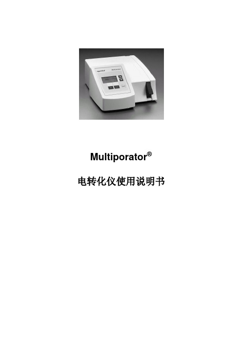
Multiporator®电转化仪使用说明书目录1 简介 (1)2 应用范围 (1)3 安全警示 (1)4 仪器介绍4.1 包装 (2)4.2 开始………………………………………………………………………………4.3 电击杯插槽……………………………………………………………………4.4 键区………………………………………………………………………………4.5 键及键组合………………………………………………………………………4.6 屏幕显示…………………………………………………………………………4.7 真核细胞和细菌/酵母的电转化………………………………………………4.7.1 连接外接电极(电转化/电融合)插件……………………………………4.8 细胞融合(可选择)……………………………………………………………4.8.1 优化细胞融合(镜下监测)………………………………………………4.8.2 清洗微融合杯………………………………………………………………4.8.3 螺旋融合杯…………………………………………………………………4.8.4 螺旋融合杯的上样及置入…………………………………………………4.8.5 融合及细胞悬液提取…………………………………………………………4.8.6 螺旋融合杯的清洗和消毒…………………………………………………4.9 打印连接/打印机(可选择)…………………………………………………5 操作模式5.1 真核细胞模式……………………………………………………………………5.2 细菌和酵母模式(可选择)……………………………………………………5.3 细胞融合模式(可选择)………………………………………………………5.3.1 功能:细胞排列………………………………………………………………5.3.2 细胞融合步骤…………………………………………………………………6 错误提示……………………………………………………………………………7 仪器的维护7.1 消毒………………………………………………………………………………7.2 清洗………………………………………………………………………………8 技术数据……………………………………………………………………………9 订购信息…………………………………………………………………………1 简介Multiporator®为真核细胞的电转染而设计,因为由有多种模式可选,也可进行细胞融合实验以及细菌和酵母的电转实验。
- 1、下载文档前请自行甄别文档内容的完整性,平台不提供额外的编辑、内容补充、找答案等附加服务。
- 2、"仅部分预览"的文档,不可在线预览部分如存在完整性等问题,可反馈申请退款(可完整预览的文档不适用该条件!)。
- 3、如文档侵犯您的权益,请联系客服反馈,我们会尽快为您处理(人工客服工作时间:9:00-18:30)。
Gene pulser xcell TM Electroporation system quick guide
产品名称: Gene Pulser Xcell 电穿孔系统
产品型号: Gene Pulser Xcell 电穿孔系统
产品展商: 众磊(北京)生物科技发展有限公司驻沪办
简单介绍
Gene Pulser Xcell 电穿孔系统
Gene Pulser Xcell 电穿孔系统的详细介绍
【介绍】
电穿孔是功能强大的将核酸、蛋白及其它分子导入多种细胞的高效技术。
通过高强度的电场作用,瞬时提高细胞膜的通透性,从而吸收周围介质中的外源分子。
这种技术可以将核苷酸、DNA 与RNA、蛋白、糖类、染料及病毒颗粒等导入原核和真核细胞内。
电转化相对其它物理和化学转化方法,是一种有价值和有效的替代方法。
Gene Pulser Xcell 系统的设计,基于Bio-Rad 15年来在电转化技术上经验积累,它提供指数波和方波波型选择、系统配置选择及友好的用户界面。
主要特点指数波和方波波型确保所有细胞类型(原核及真核)均可获得最佳的电转化效果Bio-Rad 专利的* PulseTrac 电路和电弧保护设计,确保可重复性并保护样品模块化设计可根据研究需要选择系统用户友好的数字化界面,具有直观的编程以控制所有参数,包括附属模块的参数包括人工操作、预设规程、用户规程、一个优化规程及其它先进功能等程序选择
指数波或方波脉冲选择
Gene Pulser Xcell 系统可产生指数波和方波波型,使你选择最适合你细胞的波型与规程。
指数波和方波均能有效地用于电转化及电融合。
电穿孔波型对不同类型细胞的转化效率有很重要的影响。
左,指数衰变脉冲。
当一个充电至电压为V 0 的电容器放电到细胞,加在细胞上的电压随时间以指数方式下降。
从起始电压下降到V0 / e 所需的时间称为为时间常数τ, 一种方便的脉冲时间表达方式。
右,方波脉冲。
放电到样品后截断电容器脉冲可产生方波脉冲。
脉冲时间为细胞被放电的时间。
所有方波设备都会产生细微的电压下降。
这种电压下降称为脉冲下垂,并以起始电压的百分比计量。
PulseTrac 电路和电弧保护提供可靠的、可重复的和安全的性能Bio-Rad 独创的专利微处理器控制电路能为任何规程提供可重复的结果,无论使用何种介质,它所产生的电压值可在全世界范围内测试。
PulseTrac 电路和电弧保护:
●确保电压准确地传送到电击杯
●把CE 模块中的低压电容器精确度从20% 提升到10%
●便于电容器再校正,以保持精确的脉冲指标,修正电容器随着时间而发生的偏移
●提供脉冲前样品电阻测量
●当脉冲或电路中断时,可安全地自动放电降
●低电弧危险,保护设备及样品
用户友好界面,便于编程和控制
主单元上的图形界面可控制包括任何连接的附属模块在内的所有功能。
界面由一个通过功能键和字母数字键盘
操作的屏幕组成。
根据屏幕提示进行简便而直观的编程。
屏幕用于编程和显示储存与设定的程序、运行参数以及波型。
规程包括:
●人工编程以输入或编辑方波或指数衰变脉冲的所有参数
●辅助编程需要的时间常数
●预设有适合常用细菌、真菌和哺乳动物细胞株的优化程序
●优化规程
●根据不同的波型选择,显示时间常数、实际电压、脉冲间隔和脉冲时间等参数
●储存和调用前100 个实验的脉冲参数
预优化规程,可快速选择程序这项功能为每个细胞株提供一组专用的预优化参数。
提供以下细胞株的预设程序
选择你所需要的系统
Gene Pulser Xcell 是由一个主单元、2 个附属模块(CE 与PC 模块)及ShockPod
电击槽组成的模块式电穿孔系统。
CE 模块与Gene Pulser Xcell主单元配合使用可电转化多数真核细胞(包括哺乳动物细胞和植物原生质体)。
PC模块建议用于细菌和真菌的电转化,或其它需要在小体积和高电阻样品上施加高电压脉冲的应用。
【技术指标】
Gene Pulser Xcell 完全系统
包括主单元、CE模块、PC模块
输出波型:指数衰变或方波
电压:10–3,000 V
电容10–500 V, 25–3,275 µF , 25 µF 增量500–3,000 V, 10, 25, 50 µF
电阻(并联)50–1,000 Ω ,50 Ω 增量,加无穷大
样品电阻10–2,500 V 内,最小20 Ω2,500–3,000 V 内,最小600 Ω
方波时间10–500 V: 持续时间0.05–10 ms ,0.05 ms 增量;
持续时间10–100 ms ,1 ms 增量,1–10 个脉冲,间隔0.1–10 s 500–3,000 V:
持续时间0.05-5 ms,0.05 ms 增量,1-2 个脉冲,最小间隔5s
Gene Pulser Xcell 真核系统
包括主单元、CE 模块;
输出和其他规格除了无并联电阻外,输出等与完全系统相同
Gene Pulser Xcell 微生物系统
包括主元件、PC 模块
输出波型:指数衰变或方波
电压:200–3,000 V
电容10, 25, 50 µF
电阻(并联)50–1,000 Ω 增量50 Ω ,加无穷大
样品电阻200–2,500 V 内,最小20 Ω
2,500–3,000 V 内,最小600 Ω
方波时间持续时间0.05–5 ms ,0.05 ms 增量,1-2 个脉冲,最小间隔5s
Gene Pulser Xcell 主单元
输出波型:指数衰变或方波
电压:200–3,000 V
放电电容10, 25, 50 µF
样品电阻200–2,500 V 内,最小20 Ω
2,500–3,000 V 内,最小600 Ω
方波时间持续时间0.05–5 ms ,0.05 ms 增量,1-2 个脉冲,最小间隔5s
常规
输入电压100–120 V AC 或220–240 V AC, 50/60 Hz
电源最大240 W (在短放电过程中)
工作环境温度0–35°C, 湿度0–95% (无冷凝水)
安全认证EN61010, EMC EN61326 A 级安全认证
大小(W x D x H) 主单元:31 x 30 x 14 cm
CE 模块:31 x 28 x 9 cm
PC 模块:31 x 28 x 5 cm
重量主单元: 6.6 公斤
CE 模块:3.1 公斤
PC 模块:1.9 公斤
使用说明
人工操作:
1.从最初的显示屏开始
按Enter键选择实验指数式衰减
按2键,之后按Enter键选择指数式衰减并且要指定一个时间常量
按3键,之后按Enter键来选择方波
2.使用上、下键来选择屏幕上各量的参数值。
当一个参数值是加亮的,使用键盘输入一个数值,然后按Enter键来接受这个数值
3. 当必要的参数输入完毕时,机器上的脉冲键就是可激活的了
4. 按下脉冲键来开始电击样品
5.按返回键返回详细设定显示界面,并且进行另一次的脉冲。
使用仪器设置好的程序
1.在最初的显示界面,按4键,然后按Enter键来浏览已经设置好的程序
2.按1-3键来选择样品是细菌、真菌或是哺乳动物细胞;按Enter键来选择组织类型和显示组织列表。
对
于细菌和哺乳动物细胞预先设置好的程序,使用左、右方向键来在两个屏之间触发
3.按数字键来设置参数来使之呈现高亮状态。
然后按Enter键来选择和显示程序详细的界面
4.按脉冲键来电击样品
5.按返回键来返回程序详细界面并且来进行下一次的脉冲
使用一个用户设置好的程序
1.在最初显示界面,按5键,之后按Enter键来打开用户程序菜单并且显示第一个用户界面。
用左、右
方向键来在两个界面间切换
2.按1-12键来使目标用户名高亮,按Enter键来选择名字并且显示第一个用户参数界面,使用左、右方
向键来在两个界面间切换
3.按1-12键来使目标程序名高亮,按Enter键来选择名字并且显示程序详细信息界面。
仪器上的脉冲键
现在是可被激活的
4.按脉冲键来电击样品
5.按返回键来返回程序详细界面并且来进行下一次的脉冲
BIO-RAD
Gene pulser xcell TM Electroporation system
quick guide
2009-4-5。