体外消化实验
模拟肠道消化实验报告
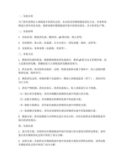
一、实验目的为了探究食物在人体肠道中的消化过程,本实验采用模拟肠道消化方法,对某种食物进行体外消化实验,观察食物在模拟肠道环境中的消化情况,并分析消化产物。
二、实验材料1. 实验试剂:模拟消化液、酶制剂、pH缓冲液、指示剂等;2. 实验器材:离心机、恒温箱、分光光度计、消化装置、烧杯、试管等;3. 实验样品:某种食物(如面粉、米饭等)。
三、实验方法1. 模拟消化液的制备:根据模拟肠道消化液成分,配制pH值为6.8的缓冲液,加入适量的消化酶,使酶浓度与人体肠道消化酶浓度相当。
2. 样品处理:将实验样品粉碎,过筛,称取适量样品置于烧杯中,加入适量的模拟消化液,搅拌均匀。
3. 模拟消化过程:将烧杯置于恒温箱中,模拟人体肠道温度(37℃),消化时间为2小时。
4. 消化产物检测:消化结束后,将消化液离心,取上清液进行以下检测:(1)蛋白质含量测定:采用双缩脲法检测消化液中的蛋白质含量;(2)淀粉含量测定:采用碘液法检测消化液中的淀粉含量;(3)脂肪含量测定:采用索氏抽提法检测消化液中的脂肪含量;(4)氨基酸含量测定:采用高效液相色谱法检测消化液中的氨基酸含量。
5. 数据分析:将实验数据与对照样品进行对比分析,评估实验样品在模拟肠道环境中的消化情况。
四、实验结果1. 蛋白质含量:实验样品在模拟肠道环境中的蛋白质含量较对照样品降低,说明蛋白质在模拟消化过程中得到了部分分解。
2. 淀粉含量:实验样品在模拟肠道环境中的淀粉含量较对照样品降低,说明淀粉在模拟消化过程中得到了部分分解。
3. 脂肪含量:实验样品在模拟肠道环境中的脂肪含量较对照样品降低,说明脂肪在模拟消化过程中得到了部分分解。
4. 氨基酸含量:实验样品在模拟肠道环境中的氨基酸含量较对照样品增加,说明蛋白质在模拟消化过程中被分解为氨基酸。
五、实验结论本实验采用模拟肠道消化方法,对某种食物进行了体外消化实验,结果表明:1. 实验样品在模拟肠道环境中的蛋白质、淀粉和脂肪含量均有所降低,说明这些营养成分在模拟消化过程中得到了部分分解;2. 实验样品在模拟肠道环境中的氨基酸含量增加,说明蛋白质在模拟消化过程中被分解为氨基酸。
肉饼体外消化实验报告

肉饼体外消化实验报告
实验目的:
验证肉饼在体外消化过程中的化学反应和分解过程。
实验原理:
体外消化是指将实验样品置于模拟人体胃液和胰液的溶液中,模拟人体消化过程,观察样品在消化液中的反应和分解过程。
胃液中含有胃蛋白酶,可以将蛋白质分解为肽和氨基酸,而胰液中含有胰蛋白酶和胰脂酶,可以进一步将肽和脂肪分解为氨基酸和脂肪酸。
本实验将肉饼置于模拟胃液和胰液中,观察肉饼的消化过程。
实验步骤:
1. 准备实验所需的材料,包括肉饼、模拟胃液、模拟胰液等。
2. 将肉饼置于模拟胃液中,放置一段时间,观察肉饼在胃液中的变化。
3. 将肉饼从胃液中取出,置于模拟胰液中,放置一段时间,观察肉饼在胰液中的变化。
4. 观察实验结果,记录下肉饼在胃液和胰液中的变化情况。
实验结果:
在模拟胃液中,肉饼经过一段时间的浸泡,变得柔软,并有一些褐色液体从肉饼中渗出。
在模拟胰液中,肉饼进一步分解,变得更为柔软,并有更多的褐色液体从肉饼中渗出。
实验讨论:
根据实验结果,可以得出肉饼在体外消化过程中的化学反应和
分解过程。
当肉饼置于胃液中时,胃蛋白酶开始将蛋白质分解为肽和氨基酸,导致肉饼变得柔软并有一些褐色液体渗出。
当肉饼进一步置于胰液中时,胰蛋白酶和胰脂酶进一步将肉饼中的肽和脂肪分解为氨基酸和脂肪酸,导致肉饼变得更为柔软并有更多的褐色液体渗出。
实验结论:
肉饼在体外消化过程中,经过胃液和胰液的作用,逐渐分解为氨基酸和脂肪酸。
体外模拟消化法优化生长猪饲粮非淀粉多糖酶谱
体外模拟消化法优化生长猪饲粮非淀粉多糖酶谱导读非淀粉多糖(NSP)作为一种抗营养因子,常影响畜禽饲粮营养物质的消化。
NSP 酶由于可降低NSP 的抗营养作用,越来越受到关注。
许多研究表明,NSP 酶能够降低NSP 的抗营养作用,提高营养物质利用率,改善肠道健康。
但是也有研究发现,NSP 酶对猪生产性能、营养物质利用率没有影响。
由于不同饲粮中NSP的含量和组成差异较大,而NSP 酶作为生物反应的催化剂,对底物具有专一性,因此,NSP 酶的合理配伍是充分发挥其对饲粮中NSP 降解作用的关键。
但是针对不同的饲粮,通过动物试验方法来筛选与之相适应的NSP 酶的配伍,不但工作量巨大,而且结果变异比较大,不能满足实际需求。
近年来,一些研究者通过体外模拟消化的方法来研究饲粮中添加NSP 酶的效果,为高效、快速的优化NSP 酶的配伍提供了一个可行的途径。
王恩玲等和何科林等使用胃蛋白酶-胰液素的体外2 步模拟消化法对NSP 酶在家禽饲粮中的作用效果进行了体外评定,取得了良好的促生产效果。
Narasimha 等使用体外消化法成功筛选出了纤维素酶、木聚糖酶和β-葡聚糖酶在几种饲料原料中的最佳组合。
但是,使用体外模拟消化法优化生长猪不同饲粮NSP 酶谱的研究目前还没有报道。
此外,胰液素是含有多种消化酶的复合物,不同批次之间组成有所不同,无法实现模拟肠液的重复以及测试重演性的需求。
本实验室前期在研究生长猪小肠液组成的基础上开发了猪模拟小肠液,并在猪饲粮的体外模拟消化上取得了较好的效果。
因此,本试验使用基于生长猪生理消化液组成依据的体外三角瓶2 步消化法,在玉米-豆粕型和玉米-杂粕型2 种不同类型的生长猪饲粮中使用6 种NSP 酶,探讨NSP 酶对不同类型饲粮的作用效果,探索优化猪饲粮中NSP酶配伍的方法,为NSP 酶在猪饲粮中的高效使用提供依据。
1材料与方法1.1 NSP 酶及酶活性测定选用纤维素酶、木聚糖酶、β-葡聚糖酶、β-甘露聚糖酶、α-半乳糖苷酶和果胶酶6 种NSP酶。
体外消化实验
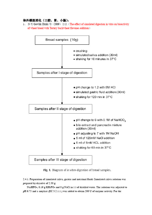
体外模拟消化(口腔、胃、小肠):1、参考Gawlik-Dziki等(2009)方法(The effect of simulated digestion in vitro on bioactivityof wheat bread with Tartary buckwheat flavones addition)2.4.1. Preparation of simulated saliva, gastric and intestinal fluids Simulated saliva solution was prepared by dissolve of 2.38 gNa2HPO4, 0.19 g KH2PO4 and 8 g NaCl in 1 l of distilled water. The solution was adjusted to pH 6.75 and a-amylase (EC 3.2.1.1.) was added to obtain 200 U of enzyme activity. For thegastric digestion, 0.32% pepsin (from porcine stomach mucosa, pepsin A, EC 3.4.23.1) dilution in 0.03 M NaCl, pH 1.2 was prepared. Simulated intestinal. juice was prepared by dilution of 0.05 g of pancreatin and 0.3 g of bile extract in 35 ml 0.1 M NaHCO3.2.4.1. Preparation of simulated saliva, gastric and intestinal fluidsSimulated saliva solution was prepared by dissolve of 2.38 g Na2HPO4, 0.19 g KH2PO4 and 8 g NaCl in 1 l of distilled water. The solution was adjusted to pH 6.75 and α-amylase (EC 3.2.1.1.) was added to obtain 200 U of enzyme activity. For the gastric digestion, 0.32% pepsin (from porcine stomach mucosa, pepsin A, EC 3.4.23.1) dilution in 0.03 M NaCl, pH 1.2 was prepared. Simulated intestinal juice was prepared by dilution of 0.05 g of pancreatin and 0.3 g of bile extract in 35 ml 0.1 M NaHCO3.2、参考Versantvoort等(2005)(Applicability of an in vitro digestion model in assessing thebioaccessibility of mycotoxins from food)和Hur等(2009)的方法(Influence of initial emulsifier type on microstructural changes occurring in emulsified lipids during in vitro digestion)Table 1Constituents and concentrations of the various synthetic juices of the in vitro digestion model representing fed conditionsSaliva 唾液Gastric juice 胃液Duodenal juice十二指肠液Bile juice 胆汁Inorganic solution 10 ml KCl 89.6g/l15.7 ml NaCl 175.3g/l40 ml NaCl175.3g/l30 ml NaCl175.3g/l10 ml KSCN20g/l3.0 ml NaH2PO4 88.8g/l40 ml NaHCO384.7 g/l68.3 ml NaHCO384.7 g/l10 ml NaH2PO488.8 g/l9.2 ml KCl 89.6 g/l10 ml KH2PO48g/l4.2 ml KCl 89.6 g/l 10 ml NaSO457g/l18 ml CaCl2· 2H2O22.2 g/l6.3 ml KCl89.6 g/l150 μl HCl 37%g/g1.7 ml NaCl175.3 g/l10 ml NH4Cl 30.6 g/l10 ml MgCl2 5g/l20 ml NaHCO384.7 g/l6.5 ml HCl 37%g/g180 μl HCl37%g/gOrganic solution 8 ml urea 25 g/l10 ml glucose 65 g/l 4 ml urea 25 g/l10 ml urea 25 g/l10 ml glucuronic acid(葡萄糖醛酸) 2g/l3.4 ml urea 25g/l10 ml glucosaminehydrochloride (盐酸氨基葡萄糖) 33g/lAdd to mixtureorganic+ inorganic solution290 mgα-amylase1g BSA9mlCaCl2· 2H2O22.2 g/l10ml CaCl2· 2H2O22.2 g/l15 mg uric acid 2.5 g pepsin1g BSA 1.8 g BSA25 mg mucin 3 g mucin9 g pancreatin30 g bile1.5 g lipasepH 6.8 ± 0.2 1.30 ± 0.028.1 ± 0.28.2 ± 0.2 The inorganic and organic solutions are augmented to 500ml with distilled water. After mixing of the inorganic and organic solutions, some further constituents are added and dissolved. If necessary, the pH of the juices is adjusted to the appropriate interval.另外,抗性淀粉含量测定:1、参考Li等(2008)方法(Characterization of maize amylose-extender (ae) mutant starches. PartI: Relationship between resistant starch contents and molecular structures)2.3. Resistant starch (RS) contentRS contents of the ae-mutant starch samples were determined using the AOAC Method 991.43 for total dietary fiber (AOAC, 2003) and Englyst’s method (1992) for comparison.For the AOAC 991.43 method, starch (1.0 g, dry-starch basis, dsb) was suspended in a Mes-tris buffer solution (0.05 M, 40 ml) and hydrolyzed with 500 u of a-amylase from Bacilluslicheniformis (Sigma Chemical, Cat. No. A3403) in a boiling water-bath for 30 min with stir. The sample was then digested with protease from Bacillus licheniformis (5 mg, Sigma Chemical, Cat. No. P3910) at 60 ºC in a shaker waterbath for 30 min. The sample dispersion was adjusted to pH 4.4–4.6 by adding HCl and then hydrolyzed with amyloglucosidase (300 U, Sigma chemical, Cat. No. A9913) at 60 ºC in a shaker water-bath for 30 min. The digested sample was filtered through a celite layer in a crucible and washed twice with 15 ml of 78% ethanol, twice with 15 ml of 100% ethanol and rinsed with 15 ml of acetone. The remaining sample was dried in an oven at 100 ºC overnight. The resistant starch content was calculated using the equation:(%)RS content = Remaining sample weight (g, dsb)/initial sample weight (g, dsb) ×100%For RS determined using Englyst’s method (1992), Starch (1.000 g, db) in 20 mL of sodium acetate buffer (0.1 M, pH 5.2) was cooked in a boiling water-bath for 30 min. The starch dispersion was cooled down to 37 ºC, mixed with an enzyme solution (5 mL) consisting of pancreatin extract and amyloglucosidase, and incubated in a water-bath at 37 ºC. The pancreatin extract was prepared as follows; 3.0 g of pancreatin (Sigma, Cat. No. P7545) was suspended in 20 mL deionized water, stirred for 10 min at room temperature, and centrifuged at 1500g for 10 min. The enzyme solution was prepared by mixing 13.5 ml supernatant of pancreatin extract, 210 U amyloglucosidase (Sigma, Cat. No. A7095), and 1.0 mL deionized water. The rapid digestible starch (RDS) was defined as the total starch digested within the first 20 min, and the slowly digestible starch (SDS) was the starch digested between 20 and 120 min (Englyst et al., 1992). The resistant starch content was calculated as follows:(%)RS content = 100%×(total starch – RDS - SDS) (g, dsb)/total starch (g, dsb)。
几种蛋白质原料体外消化率测定方法的比较
化的真实情况。 oe 和Eg ( 9) 蛋白 Bin g m 1 s u 1 在胃 酶- 9
胰蛋 白酶两步法 中利用标准过滤装置测得饲料蛋 白
质消化率与鼠和猪的真消化率十分接近, 他们认为这
种方法测得的蛋白质体外消化率经内源氮校正与回
用高, 而且对外界环境的要求较高, 季节、 温度、 光照
等都会影响消化率测定值。 体外消化法是利用精制的 消化酶或研究对象的消化道酶提取液在试管内进行
肠末端蛋白质表观消化率高度相关。 我国饲料原料品
种多, 营养成分含量差异大, 加工方式各异, 饲料原料
对不同鱼类的营养价值差异更大。 由于鱼类生活在水
的消化试验,其测定值可近似反映鱼对饲料的消化 率。此法能快速测定原料的相对利用率, 为营养师制
中, 测定鱼类饲料真消化率比 测定畜禽的更加困难。
所以寻找一种准确、 简便、 实用的消化率测定方法对 评价鱼类饲料的消化率有着十分重要的意义。 本试验
1 . 3 离体消化程 序 .2 2.
黄沦海等: 几种蛋白质原料体外消化率测定方法的比较
①胃蛋白酶处理
a 饲料样品2 分别置于20 . 称取0g . 5 份, 5 m 带盖 l 三角瓶中( 每个样品测2 个平行样) 。
b 称取17 蛋白 准确到00 ) .7 胃 酶( . 准确 7g .1 , 0g置
量一 消化后滤渣粗蛋白 含量) / 饲料样品 粗蛋白 含量
d . 将三角瓶置于( 1.℃台式恒温摇床中, 4 0) 0 1 以其 2 结 果 温度达到4℃时开始计时, 0 震荡3, h频率为8 次/i 0 / n 21 饲料的粗蛋白含量 m , .
《 门料工业)20 年第2 直第 2 朋 -05 6 0
j柳 . 9 1 >4率A鲤方法韵毙钗 rIMM磺10*外Nq jMis 4 4 01 JOl , wI g - Iw Ab & 1 j ,
体外模拟消化研究不同来源木聚糖酶对能量饲料原料降解的影响

不 同原 料小 麦 、 D S和美 国 玉 米 .进 行 体 外 消 化 模 拟 试 D G 验 .发 现 消 化 率 较 单 一 木 聚 糖 酶 配 制 的 复合 酶 分 别 提 高 3 . 3. 65 %、78 %和 6 .%, 蛋 白质 消 化 率 分 别 提 高 1 8 、 69 粗 . % 4
后用 05 o LH 1 p 调至 28 .m l C 将 H J .,加 3 L胃蛋 m
白 酶 液 .在 温 度 为 ( 9 1 c 3 ± )=培 养 2 m n 用 I 0 i,
1 5 o L N O 将 p 调 到 62 . ml aH 6 / H ..然后 加 人 3 mL
要 是玉 米 , 麦应 用较 少 , 其在 肉鸡 日粮 中 , 小 尤 其 中一 个 重要 原 因 就是 小 麦 含 有 较 多 的抗 营 养 因 子一 非淀 粉多糖 ( S ) N P
恒 温培 养 箱 、 温水 浴 锅 、 恒 离心 机 、 烘箱 、H p
计 、 析天平 、 量瓶 、 分 容 三角 烧 瓶 、 管 、 试 电炉 、 移 液枪 、 秒表 等 。
p . H6 2的磷 酸一 酸氢二 钠 缓 冲液 1mL 磷 0 ,分 别加
N P复 合 酶 制 剂 , S 温度 为 ( 9 1 o下 保 温 2 , 3_ ) + C h 然
马向东: 单位 同 第一 作 者 。
科 技 视 野
26
钢提广齑 00 21 年第2 期 2
S i d tl Ol c ㈨ … u … t ㈨
体外消化法评定饲料营养价值研究进展
体外消化法评定饲料营养价值研究进展对各种饲料的营养价值进行准确评定是配制日粮的关键。
众所周知,评定饲料的营养价值的方法主要有化学分析法、体内法和体外法。
化学分析法是评定饲料营养价值的一种常用方法,如总能、总磷、粗蛋白、粗纤维和粗脂肪等测定方法。
但利用概略养分分析所测得的饲料养分与动物消化吸收养分间存在很大差异,不足以准确地反映出饲料的实际营养价值。
利用动物试验能够比较准确地对饲料营养价值进行评定,但这些方法往往耗时、费力,很难在短时间内对大量的饲料样品进行评价分析,且生物学影响因素多,结果变异较大,重复性差,限制了其实用性。
因此,人们建立了体外法来研究饲料的营养价值。
随着技术的进步和动物消化生理的研究进展,利用动物体外消化模拟技术来研究饲料的营养价值也成为可能。
体外法具有简单、快捷、重演性好等特点,在饲料营养价值评定等方面占有重要地位。
本文就体外法在不同动物上的应用状况、应用中存在的问题及应用前景作以综述,以提高人们对体外法的认识和重视。
体外消化在猪上的应用由于猪个体较大,体外消化所需要的一些消化酶,如胃蛋白酶和胰酶以及小肠液等比较容易获得。
而且一次性获得数量也比较大。
因此,体外消化法在猪上的研究和应用较多。
猪体外消化法有pH法、单酶(比如胃蛋白酶)法、两步法(胃蛋白酶-胰酶法)、三步法(胃蛋白酶-胰酶-碳水化合物法)等,它们都能很好地预测猪饲料的体内消化情况。
利用NaOH不断中和蛋白质水解过程中所产生的H+,使pH保持恒定不变,并记录所用NaOH数量。
利用该方法所测定的31种植物性蛋白质饲料和11种动物性蛋白质饲料体外消化率与猪体内消化率存在显著相关(r=0.85和0.92)。
在对89个日粮样品进行评定时发现,用胃蛋白酶在 pH=1.5的条件下处理饲料样品,有机物质体外消化率与体内消化率相关系数很高(r=0.92),可以用来预测饲料在猪体内的消化率。
研究员对胃蛋白酶 -胰酶法进行了改进。
在分离已消化养分与残渣时,先利用三氯乙酸将可溶性肽和蛋白质沉淀出来,然后用测定纤维的标准过滤器过滤消化食糜液,分离已消化养分。
验证体外消化的方法与评价_概述及解释说明
验证体外消化的方法与评价概述及解释说明1. 引言1.1 概述本文旨在探讨体外消化的方法与评价,针对这一主题进行综述和解释。
随着生物科技的发展,体外消化作为一种重要的研究手段被广泛应用于生命科学领域,尤其在食品营养学和消化疾病研究中具有重要意义。
1.2 文章结构本文将按以下顺序组织:首先介绍整篇文章的大纲结构,并详述各个部分的内容安排。
然后进入正文部分,逐步展开对方法与评价的概述、验证重要性的解释以及最后得出的结论。
1.3 目的本文旨在向读者介绍体外消化方法与评价方面的知识,并深入探讨其在实验设计和结果可信度方面的重要性。
通过详细描述相关概念、技术和原理,希望能够给读者提供一个全面而清晰的认识,从而增强他们对于该领域的理解和应用能力。
以上是文章“1. 引言”部分所包含内容,请您根据需要进一步完善和调整。
2. 正文体外消化是一种常见的实验方法,用于模拟人体内消化过程,以便研究食物的消化和吸收情况。
该方法广泛应用于食品科学、药物开发和营养学等领域中。
在体外消化实验中,通常需要模拟口腔、胃和小肠三个部位的消化过程。
首先是口腔阶段,通过加入唾液来模拟咀嚼和混合食物的过程;接着是胃阶段,将酸性胃液加入样品中,模拟胃酸对食物的分解作用;最后是小肠阶段,添加碱性液体以调节pH值,并加入胰液来模拟小肠酶对食物的进一步分解。
对于每个阶段的体外消化实验,需要合理选择试剂配比和温度条件,并进行适当的时间控制。
为了评估消化效果,可以测量营养物质(如蛋白质、脂肪等)的降解率或者采用特定指标(如酶活性)来评估关键成分的变化。
此外,在进行实验前还需考虑是否需要添加辅酶、金属离子或其他辅助物质以提高消化效果。
同时,需要注意防止可能的实验干扰因素,如光照、氧化等。
体外消化实验的结果可用于研究食物的生物利用率、营养价值和口感特性。
通过模拟人体内消化过程,我们可以了解不同食物在机体中的代谢情况,并为开发更好的食品添加剂、制药工艺和营养配方提供参考依据。
- 1、下载文档前请自行甄别文档内容的完整性,平台不提供额外的编辑、内容补充、找答案等附加服务。
- 2、"仅部分预览"的文档,不可在线预览部分如存在完整性等问题,可反馈申请退款(可完整预览的文档不适用该条件!)。
- 3、如文档侵犯您的权益,请联系客服反馈,我们会尽快为您处理(人工客服工作时间:9:00-18:30)。
体外模拟消化(口腔、胃、小肠):1、参考Gawlik-Dziki等(2009)方法(The effect of simulated digestion in vitro on bioactivityof wheat bread with Tartary buckwheat flavones addition)2.4.1. Preparation of simulated saliva, gastric and intestinal fluids Simulated saliva solution was prepared by dissolve of 2.38 gNa2HPO4, 0.19 g KH2PO4 and 8 g NaCl in 1 l of distilled water. The solution was adjusted to pH 6.75 and a-amylase (EC 3.2.1.1.) was added to obtain 200 U of enzyme activity. For thegastric digestion, 0.32% pepsin (from porcine stomach mucosa, pepsin A, EC 3.4.23.1) dilution in 0.03 M NaCl, pH 1.2 was prepared. Simulated intestinal. juice was prepared by dilution of 0.05 g of pancreatin and 0.3 g of bile extract in 35 ml 0.1 M NaHCO3.2.4.1. Preparation of simulated saliva, gastric and intestinal fluidsSimulated saliva solution was prepared by dissolve of 2.38 g Na2HPO4, 0.19 g KH2PO4 and 8 g NaCl in 1 l of distilled water. The solution was adjusted to pH 6.75 and α-amylase (EC 3.2.1.1.) was added to obtain 200 U of enzyme activity. For the gastric digestion, 0.32% pepsin (from porcine stomach mucosa, pepsin A, EC 3.4.23.1) dilution in 0.03 M NaCl, pH 1.2 was prepared. Simulated intestinal juice was prepared by dilution of 0.05 g of pancreatin and 0.3 g of bile extract in 35 ml 0.1 M NaHCO3.2、参考Versantvoort等(2005)(Applicability of an in vitro digestion model in assessing thebioaccessibility of mycotoxins from food)和Hur等(2009)的方法(Influence of initial emulsifier type on microstructural changes occurring in emulsified lipids during in vitro digestion)Table 1Constituents and concentrations of the various synthetic juices of the in vitro digestion model representing fed conditionsSaliva 唾液Gastric juice 胃液Duodenal juice十二指肠液Bile juice 胆汁Inorganic solution 10 ml KCl 89.6g/l15.7 ml NaCl 175.3g/l40 ml NaCl175.3g/l30 ml NaCl175.3g/l10 ml KSCN20g/l3.0 ml NaH2PO4 88.8g/l40 ml NaHCO384.7 g/l68.3 ml NaHCO384.7 g/l10 ml NaH2PO488.8 g/l9.2 ml KCl 89.6 g/l10 ml KH2PO48g/l4.2 ml KCl 89.6 g/l 10 ml NaSO457g/l18 ml CaCl2· 2H2O22.2 g/l6.3 ml KCl89.6 g/l150 μl HCl 37%g/g1.7 ml NaCl175.3 g/l10 ml NH4Cl 30.6 g/l10 ml MgCl2 5g/l20 ml NaHCO384.7 g/l6.5 ml HCl 37%g/g180 μl HCl37%g/gOrganic solution 8 ml urea 25 g/l 10 ml glucose 65 g/l 4 ml urea 25 g/l 10 ml urea 25 g/l10 ml glucuronic acid(葡萄糖醛酸) 2g/l3.4 ml urea 25g/l10 ml glucosaminehydrochloride (盐酸氨基葡萄糖) 33g/lAdd to mixtureorganic+ inorganic solution290 mgα-amylase1g BSA9mlCaCl2· 2H2O22.2 g/l10ml CaCl2· 2H2O22.2 g/l15 mg uric acid 2.5 g pepsin 1g BSA 1.8 g BSA25 mg mucin 3 g mucin 9 g pancreatin 30 g bile1.5 g lipasepH 6.8 ± 0.2 1.30 ± 0.02 8.1 ± 0.2 8.2 ± 0.2 The inorganic and organic solutions are augmented to 500ml with distilled water. After mixing of the inorganic and organic solutions, some further constituents are added and dissolved. If necessary, the pH of the juices is adjusted to the appropriate interval.另外,抗性淀粉含量测定:1、参考Li等(2008)方法(Characterization of maize amylose-extender (ae) mutant starches. PartI: Relationship between resistant starch contents and molecular structures)2.3. Resistant starch (RS) contentRS contents of the ae-mutant starch samples were determined using the AOAC Method 991.43 for total dietary fiber (AOAC, 2003) and Englyst’s method (1992) for comparison.For the AOAC 991.43 method, starch (1.0 g, dry-starch basis, dsb) was suspended in a Mes-tris buffer solution (0.05 M, 40 ml) and hydrolyzed with 500 u of a-amylase from Bacilluslicheniformis (Sigma Chemical, Cat. No. A3403) in a boiling water-bath for 30 min with stir. The sample was then digested with protease from Bacillus licheniformis (5 mg, Sigma Chemical, Cat. No. P3910) at 60 ºC in a shaker waterbath for 30 min. The sample dispersion was adjusted to pH 4.4–4.6 by adding HCl and then hydrolyzed with amyloglucosidase (300 U, Sigma chemical, Cat. No. A9913) at 60 ºC in a shaker water-bath for 30 min. The digested sample was filtered through a celite layer in a crucible and washed twice with 15 ml of 78% ethanol, twice with 15 ml of 100% ethanol and rinsed with 15 ml of acetone. The remaining sample was dried in an oven at 100 ºC overnight. The resistant starch content was calculated using the equation:(%)RS content = Remaining sample weight (g, dsb)/initial sample weight (g, dsb) ×100%For RS determined using Englyst’s method (1992), Starch (1.000 g, db) in 20 mL of sodium acetate buffer (0.1 M, pH 5.2) was cooked in a boiling water-bath for 30 min. The starch dispersion was cooled down to 37 ºC, mixed with an enzyme solution (5 mL) consisting of pancreatin extract and amyloglucosidase, and incubated in a water-bath at 37 ºC. The pancreatin extract was prepared as follows; 3.0 g of pancreatin (Sigma, Cat. No. P7545) was suspended in 20 mL deionized water, stirred for 10 min at room temperature, and centrifuged at 1500g for 10 min. The enzyme solution was prepared by mixing 13.5 ml supernatant of pancreatin extract, 210 U amyloglucosidase (Sigma, Cat. No. A7095), and 1.0 mL deionized water. The rapid digestible starch (RDS) was defined as the total starch digested within the first 20 min, and the slowly digestible starch (SDS) was the starch digested between 20 and 120 min (Englyst et al., 1992). The resistant starch content was calculated as follows:(%)RS content = 100%×(total starch – RDS - SDS) (g, dsb)/total starch (g, dsb)澳大利亚NI 消化系统图片GI20。