医学英文文献 (1)
医学英文参考文献引用格式

医学英文参考文献引用格式在医学领域中,引用参考文献是非常重要的一部分。
正确的引用格式可以帮助读者理解您的研究来源和支持,同时也能够提高您的研究可信度。
以下是医学英文参考文献引用格式的基本要求和准则:1. 期刊文章:作者姓氏和名字缩写。
文章标题。
期刊名称缩写。
发表年份; 卷号(期号):页码。
例如:Hwang E, et al. The effects of acupuncture on high-risk patients with chronic gastritis. J Altern Complement Med. 2016;22(12):967-973.2. 书籍:作者姓氏和名字缩写。
书名。
版本。
出版地:出版者; 出版年份。
例如:Roberts LW, Louie AK, Sabath LD. Ethics in Psychiatry: A Review. 2nd ed. Washington, DC: American Psychiatric Press; 2013.3. 网络资源:作者姓氏和名字缩写。
文章标题。
网站名称。
发表年份。
访问日期。
可用网址。
例如:World Health Organization. WHO Director-General's speech to high-level pledging event for the humanitarian crisis in Yemen. World Health Organization. 2018. Accessed November 20, 2019.https://www.who.int/dg/speeches/2018/high-level-pledging-ev ent-yemen/en/4. 报告:组织名称。
报告标题。
出版日期。
可用网址。
例如:National Institutes of Health. Strategic Plan for NIH Obesity Research. February 2007.https:///about/strategic-plan.as px. Accessed November 20, 2019.总的来说,正确的参考文献引用格式不只是为了遵守学术规范,更是为了保证研究的可信度和可重复性。
医学影像学英文文献
医学影像学英文文献英文回答:Within the realm of medical imaging, sophisticated imaging techniques empower healthcare professionals with the ability to visualize and comprehend anatomical structures and physiological processes in the human body. These techniques are instrumental in diagnosing diseases, guiding therapeutic interventions, and monitoring treatment outcomes.Computed tomography (CT) and magnetic resonance imaging (MRI) are two cornerstone imaging modalities widely employed in medical practice. CT utilizes X-rays and advanced computational algorithms to generate cross-sectional images of the body, providing detailed depictions of bones, soft tissues, and blood vessels. MRI, on the other hand, harnesses the power of powerful magnets and radiofrequency waves to create intricate images that excel in showcasing soft tissue structures, including the brain,spinal cord, and internal organs.Positron emission tomography (PET) and single-photon emission computed tomography (SPECT) are nuclear medicine imaging techniques that involve the administration of radioactive tracers into the body. These tracers accumulate in specific organs or tissues, enabling the visualization and assessment of metabolic processes and disease activity. PET is particularly valuable in oncology, as it can detect the presence and extent of cancerous lesions.Ultrasound, also known as sonography, utilizes high-frequency sound waves to produce images of internal structures. It is a versatile technique commonly employed in obstetrics, cardiology, and abdominal imaging. Ultrasound offers real-time visualization, making it ideal for guiding procedures such as biopsies and injections.Interventional radiology is a specialized field that combines imaging guidance with minimally invasive procedures. Interventional radiologists utilize imaging techniques to precisely navigate catheters and otherinstruments through the body, enabling the diagnosis and treatment of conditions without the need for open surgery. This approach offers reduced invasiveness and faster recovery times compared to traditional surgical interventions.Medical imaging has revolutionized healthcare by providing invaluable insights into the human body. The ability to visualize anatomical structures andphysiological processes in exquisite detail has transformed the practice of medicine, leading to more accurate diagnoses, targeted treatments, and improved patient outcomes.中文回答:医学影像学是现代医学不可或缺的一部分,它利用各种成像技术对人体的解剖结构和生理过程进行可视化和理解,在疾病诊断、治疗方案制定和治疗效果评估中发挥着至关重要的作用。
《临床常见疾病:医学英语文献阅读》读书笔记模板

Section Two: Internal Disease第二部分内科疾病
21. Arrhythmia (Heart Rhythm Disorders)心律失常 22. Coronary Artery Disease (CAD)冠心病 23. Hypertension/High Blood Pressure高血压病 24. Heart Failure心力衰竭 25. Acute Respiratory Distress Syndrome (ARDS)急性呼吸窘迫综合征 26. Asthma哮喘 27. Chronic Obstructive Pulmonary Disease (COPD)慢性阻塞肺疾病 28. Pneumonia肺炎 29. Respiratory Failure呼吸衰竭
Section Five: Cancer第五部分恶性肿瘤
66. Breast Cancer乳腺癌 67. Esophagus Cancer食管癌 68. Liver Cancer肝癌 69. Stomach Cancer胃癌 70. Colorectal Cancer结直肠癌 71. Lung Cancer肺癌 72. Cervical Cancer宫颈癌 73. Ovarian Cancer卵巢癌 74. Bladder Cancer膀胱癌
Section Three: Obstetrical and Gynecological Disease第三 部分
59. Dysfunctional Uterine Bleeding功能失调性子宫出血
60. Ectopic Pregnancy异位妊娠
Section Four: Pediatric Disease第四部分儿科疾病
目录分析
前言 Introduction
医学英文翻译文献
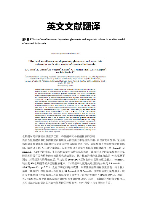
英文文献翻译第1 篇 Effects of sevoflurane on dopamine, glutamate and aspartate release in an vitro model of cerebral ischaemia七氟醚对离体脑缺血模型多巴胺、谷氨酸和天冬氨酸释放的影响兴奋性氨基酸和多巴胺的释放在脑缺血后神经损伤中起重要作用。
在当前的研究中,采用离体脑缺血模型观察七氟醚对大鼠皮质纹状体脑片中多巴胺、谷氨酸和天冬氨酸释放量的影响。
脑片以34℃人工脑脊液灌流,缺血发作以去除氧气和降低葡萄糖浓度(从4mmol/l至2mmol/l)≤30分钟模拟。
多巴胺释放量用伏特法原位监测,灌流样本中的谷氨酸和天冬氨酸浓度用带有荧光检测的高效液相色谱法测定。
脑片释放的神经递质在有或无4%七氟醚下测定。
对照组脑片诱导缺血后,平均延迟166s(n=5)后细胞外多巴胺浓度达最大77.0μmol/l。
缺血期4%七氟醚降低多巴胺释放速率,(对照组和七氟醚处理组脑片分别是6.9μmol/l/s和4.73μmol/l/s,p<0.05),没有影响它的起始或量。
兴奋性氨基酸的释放更缓慢。
每个脑片基础(缺血前)谷氨酸和天冬氨酸是94.8nmol/l和69.3nmol/l,没有明显被七氟醚减少。
缺血大大地增加了谷氨酸和天冬氨酸释放量(最大值分别是对照组的244%和489%)。
然而,4%七氟醚明显减少缺血诱导的谷氨酸和天冬氨酸释放量。
总结,七氟醚的神经保护作用与其可以减少缺血引起的兴奋性氨基酸的释放有关,较小程度上与多巴胺也有关。
第2篇The Influence of Mitochondrial K ATP-Channels in the Cardioprotection of Proconditioning and Postconditioning by Sevoflurane in the Rat In Vivo线粒体K ATP通道在离体大鼠七氟醚预处理和后处理中心肌保护作用中的影响挥发性麻醉药引起心肌预处理并也能在给予再灌注的开始保护心脏——一种实践目前被称为后处理。
英文医学数据库检索(1)讲解
记录标识(1)
[PubMed - in process] [ PubMed - as
supplied by publisher]
[PubMed – indexed 19 for Medline]
2. PubMed记录的字段
可供检索和显示的字段共60多个 (表4-1-1)
20
MEDLINE格式
Topic-Specific Queries Linkout(外部链接)
Clinical Trials
54
主页
55
Journals Database
56
Single Citation Matcher
57
举例
查找夏家辉教授发表在“Nat Genet, 1998;20(4):370-3”的关于神经性 耳聋基因的论文及相关信息
33
检索举例
查找入学前儿童的哮喘治疗的 文献(综述文献/全文)
child, preschool [mh] AND asthma/therapy [mh] 5348篇(756篇/920篇)
34
检索结果
child, preschool [mh] AND asthma/therapy [mh]
35
获取全文1
36
获取全文2
37
日期检索
格式:YYYY/MM/DD [date field] 2009/10/06 [edat] 2008 [dp] 2008:2010 [dp] 2009/01:2010/03 [edat]
38
•词语检索
自动词语匹配功能:输入未加任何限定 的检索词时,系统依次进行匹配、转换 和检索:MeSH转换表 刊名转换表 著者索引
pubmed(1)

Preview
检索下列各题,记录你的检索操作过程及命中文献数
1. 了解有关疯牛病(bovine spongiform encephalopathy ,BSE) 起源(origin)的情况,最早的报道在哪年,记录出处。 2. 2005年度诺贝尔医学奖授予了幽门螺杆菌的发现者Barry J. Marshall(1951-)和J. Robin Warren(1937-),请检索两人合 作发表的论文,记录最早的一篇文献题录。 3. 南方医院王继德教授在《Gastroenterology》发表了一篇有 关FHL2表达的文献,请找到这篇文献,并记录篇名和在 PubMed的被引情况 4. 请检索有关流感病毒H5N1人疫苗(vaccine)的临床试验 (clinical trial)文献。 5. RNA干扰(RNA INTERFERENCE )用于肝癌治疗的综述文献。
PubMed简介 收录70余个国家4900多种生物医学期刊的 题录与文摘,是世界上最权威的医学数据库。 现有1950年以来的1700多万篇文献的记录 (其中MEDLINE记录1400多万条)
目前每年增加60余万条文献记录
在全部记录中有76%的文献为英文;在 2000年以来的记录中90%的文献为英文; 收录中文刊500余种。
PubMed - indexed for MEDLINE
基本检索
检索某主题的文献 检索某一作者发表的文献 检索发表在某期刊上的文献
基本检索:检索某主题的文献
检索有关肺癌(lung cancer)的文献
Lung cancer
"lung neoplasms"[MeSH Terms] OR ("lung"[All Fields] AND "neoplasms"[All Fields]) OR "lung neoplasms"[All Fields] OR ("lung"[All Fields] AND "cancer"[All Fields]) OR "lung cancer"[All Fields]
医学英文文献汇报PPT
4
Discussion
Discussion
• PVOD确实是肺动脉高压的一种罕见病因,通常疾病晚期才能 诊断出来并且预后往往很差。PVOD的年发病率估计为0.1到 0.2例每百万人口,还不包括很多例误诊为其他病因的IPAH。 许多PVOD病例为特发性,但是这些被报道出来的疾病中被发 现常常与肺结缔组织疾病,艾滋病,骨髓移植,化学致病因 素及某些化疗药物有关。组织学检查的主要病变是小静脉内 层的纤维化。纤维化会引起肺静脉梗阻,导致肺毛细血管淤 血、肺间质和胸膜水肿、淋巴结肿大肺泡的含铁血黄素沉着。 肺动脉闭塞也发生在多达一半的PVOD患者。长期下去,那些 患有PAH,右心室功能障碍的病人,尽管经过药物治疗但仍然 会进展为心肺衰竭。
Case description
• 在这一点上,鉴于ILD的主要表现,以及病人的严重低氧血 症,CTA检查结果和肺功能测试,外院给她进行了支气管镜检 查和胸腔镜肺活检。而这一系列检查我们都不推荐。在麻醉 诱导后立即出现的无脉性电活动又使病情更加复杂化。这个 病人经历了不到5分钟心肺复苏后才恢复自主循环,整个过程 也才完成。随后,该患者出现低血压,并需要用多巴胺+米力 农等血管收缩药物来维持。初步肺活检结果与与特发性肺动 脉高压是一致的;活检的切片被送到外面的专业实验室(图1 示)。
• 该病人诊疗经过十分复杂,既需要血管升压药来治疗低血压,又有逐渐恶化的右 心室功能障碍和急性肾损伤。在移植评估过程中,她决定不想再继续接受那些试 图稳定其进行性多器官功能障碍的治疗,并改为安适疗法。在撤去支持治疗后数 小时她便去世了。
2
Background
Background
• 肺静脉闭塞性疾病(PVOD)是一种罕见的肺动脉高血压(PAH)病 因,其肺小静脉和微静脉纤维化,逐渐导致肺动脉高压、肺 间质、胸膜水肿和右心衰。
医学研究英语论文(renew)

1.1 生物医学论文的基本格式
根据国际医学杂志编辑委员会(ICMJE)2010年修订 的《生物医学期刊投稿的统一要求》,生物医学 科研论文包括: 标题页( Title Page) 摘要与关键词 (Abstract and Key Words) 引言 (Introduction) 材料 与方法 (Materials and Methods) 结果 (Results) 讨论 (Discussion) 致谢 (Acknowledgements) 参考文献 (References)
• • • • • • • •
• • • • •
图例 (Legends) 插图 (Figures) 表格 (Tables) 照片和说明 (Plates and Explanations) …
1.2 标题页 (Title page)
• 标题页是投稿论文的一部分,一般放在论文之前 单独成页。主要包含(根据《生物医学期刊投稿的 统一要求》) : • (1)论文标题(the title of the article); • (2)每位作者的姓名及其最高学位、所属单位 (the name by which each author is known, with his or her highest academic degree and institutional affiliation); • (3)研究工作的归属部门或单位名称(the name of the department and institution to which the work should be attributed); • (4)弃权者(若有)(disclaimers, if any);
(2) 突出研究重点
糖尿病:新的诊断标准 Diabetes : New diagnostic criteria
- 1、下载文档前请自行甄别文档内容的完整性,平台不提供额外的编辑、内容补充、找答案等附加服务。
- 2、"仅部分预览"的文档,不可在线预览部分如存在完整性等问题,可反馈申请退款(可完整预览的文档不适用该条件!)。
- 3、如文档侵犯您的权益,请联系客服反馈,我们会尽快为您处理(人工客服工作时间:9:00-18:30)。
Muscle-selective synaptic disassembly and reorganization in MuSK antibody positive MG miceAnna Rostedt Punga ⁎,1,Shuo Lin,Filippo Oliveri,Sarina Meinen,Markus A.RüeggDepartment of Neurobiology/Pharmacology,Biozentrum,University of Basel,Basel,Switzerlanda b s t r a c ta r t i c l e i n f o Article history:Received 23February 2011Revised 15April 2011Accepted 21April 2011Available online 30April 2011Keywords:Myasthenia Gravis MG MuSKNeuromuscular junction MasseterMuscle atrophy DenervationMuSK antibody seropositive (MuSK+)Myasthenia Gravis (MG)patients present a distinct selective fatigue,and sometimes atrophy,of bulbar,facial and neck muscles.Here,we study the mechanism underlying the focal muscle involvement in mice with MuSK+experimental autoimmune MG (EAMG).8week-old female wildtype C57BL6mice and transgenic mice,which express yellow fluorescence protein (YFP)in their motor neurons,were immunized with the extracellular domain of rat MuSK and compared with control mice.The soleus,EDL,sternomastoid,omohyoid,thoracic paraspinal and masseter muscles were examined for pre-and postsynaptic changes with whole mount immunostaining and confocal microscopy.Neuromuscular junction derangement was quanti fied and compared between muscles and correlated with transcript levels of MuSK and other postsynaptic genes.Correlating with the EAMG disease grade,the postsynaptic acetylcholine receptor (AChR)clusters were severely fragmented with a subsequent reduction also of the presynaptic nerve terminal area.Among the muscles analyzed,the thoracic paraspinal,sternomastoid and masseter muscles were more affected than the leg muscles.The masseter muscle was the most affected,leading to denervation and atrophy and this severity correlated with the lowest levels of MuSK mRNA.On the contrary,the soleus with high MuSK mRNA levels had less postsynaptic perturbation and more terminal nerve sprouting.We propose that low muscle-intrinsic MuSK levels render some muscles,such as the masseter,more vulnerable to the postsynaptic perturbation of MuSK antibodies with subsequent denervation and atrophy.These findings augment our understanding of the sometimes severe,facio-bulbar phenotype of MuSK+MG.©2011Elsevier Inc.All rights reserved.IntroductionAbout 40–70%of acetylcholine receptor (AChR)-antibody seronegative Myasthenia Gravis (MG)patients have antibodies against the muscle tyrosine kinase (MuSK)(Hoch et al.,2001;Sanders et al.,2003).In MuSK-antibody seropositive (MuSK+)MG patients,there is often selective involvement of bulbar-,neck-and facial muscles,as well as muscles that are usually asymptomatic in AChR-antibody seropositive (AChR+)MG,such as the paraspinal muscles (Sanders and Juel,2008).Contrary to conventional AChR+MG patients,the majority of MuSK+patients does not experience symptomatic relief from acetylcholine esterase inhibitors (AChEI)(Evoli et al.,2003)but instead may respond with pronounced nicotinic adverse effects,such as muscle fasciculations and cramps (Punga et al.,2006).Pronounced atrophy of facial muscles has also been described in MuSK+patients,although the concomitant treatment of corticosteroids in most cases has made it dif ficult tojudge whether the MuSK antibodies or the cortisone treatment is the cause of the atrophy (Farrugia et al.,2006).Muscle biopsy studies of the intercostal muscle and biceps brachii muscle from MuSK+patients have shown little AChR loss (Selcen et al.,2004;Shiraishi et al.,2005),however,the neuromuscular junction (NMJ)pathophysiology in the most affected facial or bulbar muscles has not been studied.Nevertheless,MuSK antibodies have been shown to be pathogenic in animals,both after immunization with the extracellular domain of the MuSK protein itself (Jha et al.,2006;Shigemoto et al.,2008;2006;Xu et al.,2006)and after passive transfer of sera from MuSK+MG patients (Cole et al.,2008;ter Beek et al.,2009).MuSK is essential to the process of NMJ formation,maintenance (Wang et al.,2006)and integrity,as perturbations in MuSK protein expression cause pronounced disassembly of the entire NMJ (Hesser et al.,2006;Kong et al.,2004).Other NMJ proteins that are essential for synaptogenesis include Dok-7,a downstream adaptor protein to MuSK (Okada et al.,2006),Lrp4,the co-receptor for neural agrin (Kim et al.,2008;Zhang et al.,2008),rapsyn and the AChR subunits.The effects of MuSK antibodies in-vivo on the gene expression of those synaptic proteins in the facial or bulbar muscles have not yet been established.Here,we hypothesized that low expression levels of MuSK may render some muscles more vulnerable to the effect of MuSK antibodies in the EAMG mouse model.We show that MuSK antibodiesExperimental Neurology 230(2011)207–217⁎Corresponding author at:Institute of Neuroscience,Department of Clinical Neuro-physiology,Uppsala University Hospital,Uppsala,Sweden.Fax:+4618556106.E-mail address:annarostedtpunga@ (A.R.Punga).1Present address:Department of Clinical Neurophysiology,Uppsala University Hospital,Uppsala,Sweden.0014-4886/$–see front matter ©2011Elsevier Inc.All rights reserved.doi:10.1016/j.expneurol.2011.04.018Contents lists available at ScienceDirectExperimental Neurologyj o u r n a l h o me p a g e :w w w.e l s e v i e r.c om /l o c a t e /y e x n rinduce severe fragmentation of the postsynaptic AChR clusters in particular in the masseter and thoracic paraspinal muscles,with less fragmentation in the limb muscles.The severe postsynaptic pertur-bation results in subsequent denervation of musclefibers,not previously described in EAMG or MG.We propose that one underlying mechanism for the severe involvement of the facial masseter muscle, with severely impaired NMJ architecture,atrophy and denervation,is its low intrinsic levels of MuSK.Moreover,muscles respond to the partial denervation caused by MuSK antibodies in two different ways: (1)terminal nerve sprouting in muscles with high intrinsic levels of MuSK(i.e.soleus,sternomastoid)and(2)no nerve sprouting in muscles with low intrinsic MuSK levels(i.e.masseter,omohyoid).MethodsProduction of recombinant rat MuSKpCEP-PU vector containing the His-tagged extracellular domain of recombinant rat MuSK(aa21-491;(Jones et al.,1999))was transfected(Lipofectamine2000;Invitrogen)into HEK293EBNA cells.The overexpressed protein was purified from the cell superna-tant over a Ni-NTA superflow column(Qiagen)and was subsequently dialyzed against PBS.Protein concentration was determined at OD 280nm and purity was ensured by SDS-PAGE.Experimental animalsC57BL6mice and mice expressing yellowfluorescence protein (YFP)in their motor neurons under Thy-1promoter(Feng et al., 2000)were originally supplied from Jackson Laboratories(Bar Harbor, Maine,US).For immunization,8week-old female mice were used.All mice were housed in the Animal Facility of Biozentrum,University of Basel,where they had free access to food and water in a room with controlled temperature and a12hour alternating light–dark cycle.All animal procedures complied with Swiss animal experimental regu-lations(ethical application approval no.2352)and EC Directive 86/609/EEC.ImmunizationThe immunization procedure has been described previously(Jha et al.,2006).Briefly,eleven C57BL6and seven Thy1-YFP female mice aged8weeks were anesthetized(Ketamine:111mg/kg and Xylazine: 22mg/kg)and immunized with10μg of MuSK emulsified in complete Freund's adjuvant(CFA,Difco laboratories,Detroit,Michigan,US) subcutaneously in the hind foot pads,at the base of the tail and dorsolateral on the back.At day28post-injection,immunization was repeated.A3rd immunization was given to mice that did not show any myasthenic weakness after56days.Control mice(8female mice) were immunized with PBS/CFA.Clinical and neurophysiological examinationMuscle weakness was graded every week,as described(Nakayashiki et al.,2000).Briefly,mice were exercised by20consecutive paw grips on a grid and were then placed on an upside-down grid.The time they could hold on to the grid reflected the grade of fatigue and muscle weakness.EAMG grades were as follows:grade0,no weakness;grade1, mild muscle fatigue after exercise;grade2,moderate muscle fatigue; and grade3,severe generalized weakness.Evaluation of the response to AChEIs was performed by i.p.injection of a mix of neostigmine bromide (0.0375mg/kg)and atropine sulfate(0.015mg/kg)in mice with EAMG grades2and3(Berman and Patrick,1980).Repetitive stimulation of the sciatic nerve and recording from the gastrocnemius muscle with monopolar needle electrodes was performed under anesthesia,in mice with EAMG grades2(n=2)and3(n=2),using a Saphire1L EMG machine(Medelec).Decrement was calculated as percent amplitude change between the1st and4th compound motor action potentials evoked by a train of10impulses where10%was considered as pathological.ELISASera were obtained from tail vein blood on day0(preimmune sera)and day35post-immunization.ELISA plates(Nunc MaxiSorp, Fisher Thermo Scientific,Rockford,IL,US)were coated with250ng/ ml of His-labeled rat MuSK(50μl/well),blocked with3%BSA/PBS and then incubated with a sera dilution row(1:3000–1:2,000,000).Pre-immune sera constituted negative and rabbit-anti-MuSK antibody (Scotton et al.,2006)positive controls.After washing,plates were incubated with secondary HRPO-conjugated goat-anti-mouse (1:2000)and goat anti-rabbit antibodies(1:2000;both from Jackson Immuno Research Laboratories,Westgrove,PA,US).HRPO activation by a TMB substrate was terminated with1N HCl after5min. Absorbance was read at450nm.Non-specific binding,determined by incubation of plates with pre-immune serum,was subtracted.The data were displayed as“half maximum MuSK immunoreactivity”,which represents the immunore-activity at a dilution of1:27,000,where the majority of sera obtained50% of maximum absorbance(in the linear range of the absorption at450nm).Western blotWestern blot of masseter muscles was conducted as described (Bentzinger et al.,2008).10μg of protein was resolved on a4–12%Nu-PAGE Bis–Tris gel(Invitrogen,Eugene,OR,US),transferred to nitrocellulose membrane,probed with rat monoclonal anti-NCAM (CD56;1:100;GeneTex)and rabbit polyclonal anti-pan-actin(1:1000; cell signaling)and then recognized with HRPO-conjugated antibodies (1:5000;Jackson Immuno Research Laboratories,Westgrove,PA,US). Quantitative RT-PCR analysisMouse muscle RNA was extracted and purified as previously described(Punga et al.,2011).RT-PCR reactions(triplicates)were carried out with Power SYBR Green PCR Master Mix reagent(Applied Biosystems,Warrington,UK).β-actin was used as endogenous control (Punga et al.,2011;Murphy et al.,2003;Yuzbasioglu et al.,2010).The following primer sets were used:MuSK:5′-GCCTTCAGCGGGACTGAG-3′and5′-GAGGCGTGGTGA-CAGG-3′Lrp4:5′-GGATGGCTGTACGCTGCCTA-3′and5′-TTGCCGTTGTCA-CAGTGGA-3′Dok-7:5′-CTCGGCAGTTACAGGAGGTTG-3′and5′-GCAATGC-CACTGTCAGAGGA-3′A C h Rα1:5′-G C C A T T A A C C C G G A A AG T G A C-3′a n d5′-CCCCGCTCTCCATGAAGTT-3′AChRε:5′-CTGTGAACTTTGCTGAG-3′and5′-GGAGATCAG-GAACTTGGTTG-3′AChRγsubunit:5′-AACGAGACTCGGATGTGGTC-3′and5′-GTCGCACCACTGCATCTCTA-3′Rapsyn:5′-AGGTTGGCAATAAGCTGAGCC-3′and5′-TGCTCTCACT-CAGGCAATGC-3′MuRF-1:5′-ACC TGC TGG TGG AAA ACA-3′and5′-AGG AGC AAG TAG GCA CCT CA-3′β-actin:5′-CAGCTTCTTTGCAGCTCCTT-3′and5′-GCAGCGA-TATCGTCATCCA-3′A C h E:5′-G G G C T C C T A C T T T C T G G T T T A C G-3′a n d5′-GGGCCCGGCTGATGAG-3′208 A.R.Punga et al./Experimental Neurology230(2011)207–217NMJ whole mount analysisAlexa Fluor555-conjugatedα-bungarotoxin(1μg/ml;Invitrogen) was injected into soleus,EDL,sternomastoid,omohyoid,masseter and the thoracic paraspinal muscles as described(Bezakova and Lomo, 2001).At least12images of each muscle per mouse(6MuSK-immunized YFP-transgenic mice and4CFA-immunized control mice) were collected with a confocal laser-scanning microscope(Leica TCS SPE).Laser gain and intensity were equal for all images.Quantification of pre-and postsynaptic area was performed in ImageJ(http://imagej. /ij/index.html).NMJs containing terminal nerve sprouts(processes with YFP expression)were counted in muscles fromfive MuSK+EAMG mice with disease grades1–3.At least275NMJs per muscle were analyzed. The number of postsynapse fragments per NMJ was counted using a fluorescence microscope(Leica DM5000B)in at least50NMJs per muscle deriving fromfive MuSK+EAMG grades1–2and from four control mice(Supplemental Fig.1).Fragmentation was classified as follows:1)normal pretzel-like NMJ;2)slight to moderate fragmen-tation;and3)severe fragmentation or absent postsynapse.The degree of postsynaptic perturbation per muscle was judged based on the percentage of NMJs belonging to each postsynaptic class and was further subdivided into number of postsynapse fragments per NMJ:1–3,4–6,7–9,10–12and more than12.Each NMJ was given the median score for that subgroup.The fragmentation score was obtained by taking the ratio of the score between the EAMG mice and control mice.Statistical analysisIndependent,2-sample t-test was performed for parametric data. For ordinal data(ELISA),the non-parametric test Spearman Rank Correlation was applied.A p-value b0.05was considered significant.Fig.1.(A)Development of EAMG after immunization with recombinant rat MuSK.Progress of clinical EAMG grade at the time points week4,5,7and10.*The mice with EAMG grade 3were sacrificed after week7(hence no new grade3mice week10)and the remaining mice were sacrificed andfinally evaluated at week10.(B)One mouse,representative of the most severely affected MuSK+EAMG mice,withflaccid paralysis and pronounced kyphosis.(C)Repetitive nerve stimulation performed in the same mouse.Stimulation of the sciatic nerve and recording of the gastrocnemius muscle demonstrated a35%decrement between the1st and4th compound motor action potentials at low frequency3Hz stimulation.(D)Correlation of clinical EAMG grade with MuSK antibody titer.Half of maximum MuSK immunoreactivity(1:27,000dilution)in sera from MuSK immunized mice was assessed by Elisa at450nm.R=0.483(Spearman's Rank Correlation);p b0.05.209A.R.Punga et al./Experimental Neurology230(2011)207–217ResultsMuSK+EAMG presents with prominent kyphosis,paralysis and weight lossOut of the18mice immunized with MuSK,thefinal EAMG grade 0was seen in3mice(17%),grade1in8mice(44%),grade2in4mice (22%)and grade3in3mice(17%)(Fig.1A).No difference in disease incidence was found between the groups of C57BL6mice and the YFP-transgenic mice.Based on this,the data were pooled in the current study.The most severe phenotype of EAMG(grade3)includedflaccid paralysis,pronounced kyphosis and weight loss(Fig.1B).In-vivo nerve stimulation at3Hz with recording from the gastrocnemius muscle revealed a decrement of10–40%in the MuSK+EAMG mice (Fig.1C),whereas the control mice had normal neuromuscular transmission(data not shown).MuSK immunoreactivity in sera(day 35)correlated with clinical severity(Fig.1D;Spearman Rank Correlation;R=0.483;p b0.05),although some mice developed measurable MuSK antibodies without showing obvious muscle weakness or fatigue.Bulbar symptoms underlying weight loss in MuSK+EAMG The MuSK immunized mice steadily decreased significantly in body weight after the2nd immunization(Fig.2A),in contrast to the control mice,and thefinal body weight of the MuSK+mice was significantly smaller(p b0.001;Fig.2B).This severe weight loss was slowed but not stopped after introduction of wet food(data not shown).Since the timeline of the weight drop also correlated with development of muscle fatigue,the weight loss was assumed to be indicative of chewing and swallowing difficulties,some of the cardinal symptoms of MuSK+MG.To determine whether loss of muscle weight significantly contributed to the overall lower body weight in the MuSK+EAMG mice,the weight offive different muscles was assessed.The masseter was the only muscle with a significant weight reduction(p b0.01;Fig.2C),implying that this muscle is atrophic and further indicating chewing difficulties.Adverse effects of AChEIs in MuSK+EAMGTo elucidate whether AChEIs have any beneficial effect on fatigue or weakness in MuSK+EAMG,a neostigmine test was performed in the mice with EAMG grade3(n=3).No apparent improvement in weakness at rest or exercise-induced fatigue was seen;instead the mice experienced shivering and constant twitching of the tail,trunk and limbs starting after approximately13min(Supplemental Video1).This effect wore off after40min and was interpreted as nicotinic side effects and neuromuscular hyperactivity,usually seen after an overdose of AChEIs.Thus,this intolerance towards AChEIs in MuSK+EAMG indicates an abnormal sensitivity to acetylcholine.Impairment of NMJs in different musclesBecause muscle groups in the bulbar/facial/back region are selectively involved in the MuSK+EAMG model,we next examined the morphological changes of NMJs in the thoracic paraspinal muscles, masseter,omohyoid and sternomastoid and compared them with those in two limb muscles(EDL and soleus).Typical features of postsynaptic impairment were a fainting of AChRfluorescence,areas lacking AChRs(holes)and disassembly of AChR clusters.To quantify these impairments,we classified NMJs into three classes as illustrated in Fig.3A.All muscles from MuSK+EAMG mice displayeddifferentFig.2.Weight loss in MuSK+EAMG.(A)The course of weight loss in two MuSK+mice with EAMG grade3compared to control(Ctrl CFA)mice(n=8).Initial body weight was comparable and weight loss started after the2nd immunization and weight kept dropping even though wet food was provided ad libitum.(B)Mean body weight was dramatically reduced in the MuSK immunized mice(MuSK+;n=18),compared to the control mice.Results shown as mean±SEM(gram);***p b0.001.(C)Muscles were weighed and compared between control mice(n=8)and MuSK+EAMG mice(n=18).Results displayed as mean muscle weight±SEM(mg).The only muscle which was significantly lighter in the MuSK+mice was the masseter.**p b0.01.210 A.R.Punga et al./Experimental Neurology230(2011)207–217degrees of NMJ impairment (Fig.3B).Moreover,we also observed that the postsynapses were often fragmented into two to three non-continuous fragments (see illustration in Fig.3A),which is in strong contrast to non-interrupted,pretzel-like shape of AChRs in non-immunized mice.This fragmentation was also quanti fied (Supplemental Fig.1)and expressed as “fragmentation score ”.Because fragmentation differs between muscles,this “fragmentation score ”was normalized to the control.As shown in Fig.3C,the fragmentation score varied between a minimum of 2.0in the soleus muscle and a maximum of 3.3in the masseter muscle.The postsynaptic labeling was markedly reduced in the MuSK+EAMG mice compared to control mice (Fig.4A).In the mild to moderately affected mice,AChR cluster area was reduced signi ficantly by 40–50%in all muscles (p b 0.01),except for the soleus,which had a remaining area of 85%(p b 0.05;Fig.4B).In the most severely affected mice,the postsynaptic area was also lost in the soleus muscle and less than 10%of the α-bungarotoxin staining remained in the masseter,sternomastoid and thoracic paraspinal muscles (p b 0.001).In the soleus,the nerve terminal area was unchanged in the severely affected mice,although in the other muscles the presynaptic area was reduced to about 80%in the mild to moderate cases and to 50%in the most severely affected mice (p b 0.01)(Fig.4C).Thus,both the pre-and postsynaptic data suggest that the masseter is the most affected muscle by MuSK antibodies whereas the soleus is the least affected.mRNA transcript expression of NMJ proteins in different muscles in MuSK+EAMGThe mRNA levels of MuSK differ signi ficantly between muscles,with the highest levels in the soleus muscle and lowest levels in the omohyoid muscle (Punga et al.,2011).Consequently,we additionally analyzed MuSK transcript levels in the masseter and these were even lower,with less than 20%of the mRNA levels detected in the soleus muscle (Supplemental Fig.2;p b 0.01).In the MuSK+EAMG mice,MuSK mRNA levels were signi ficantly reduced to 45%of control in the EDL (p b 0.05)and to 25%in the omohyoid muscle (p b 0.001;Fig.5A).Conversely,MuSK mRNA expression was signi ficantly increased in the masseter muscle (p b 0.01)and remained unchanged in the soleus and sternomastoid muscles.The expression of the AChR αsubunit was unchanged (Fig.5B),whereas the AChR εsubunit transcript was downregulated in the soleus and sternomastoid muscles and conversely a trend towards upregulation was seen in the masseter muscle (Fig.5C).The fetal AChR γsubunit mRNA was upregulated up to 1000-fold in the masseter (p b 0.001)and 5-fold in the soleus (p b 0.05;Fig.5D).The transcript levels of rapsyn,Lrp4and Dok-7were not signi ficantly altered (Fig.5E to G).Finally,the transcript levels of acetylcholine esterase (AChE)were signi ficantly down-regulated only in the sternomastoid and omohyoid muscles (p b 0.05;Fig.5H).Fig.3.Postsynaptic fragmentation of NMJs.(A)Examples from each postsynaptic classi fication.AChRs stained for alfa-bungarotoxin.(B)At least 275neuromuscular junctions (NMJs)per muscle were assessed in a total of 5MuSK+EAMG mice with moderate to severe disease.The appearance of NMJ postsynapses were divided into 3classes as indicated.Sol:soleus;EDL:extensor digitorum longus;STM:sternomastoid muscle;omo:omohyoid;mass:masseter.(C)Postsynapses were analyzed in at least 50NMJs per muscle in a total of 5MuSK+EAMG mice with slight to moderate disease grade and in 4control mice.Each NMJ in a certain subclass (for details see Supplemental Fig.1)was given the median score for that subclass.The fragmentation score for each muscle is the ratio in score between the EAMG mice and control mice.211A.R.Punga et al./Experimental Neurology 230(2011)207–217In summary,particularly the massive upregulation of MuSK transcripts and AChR γin conjunction with the overall trend for upregulation of mRNA for the other postsynaptic genes in the masseter muscle most likely re flects the severely disturbed neuro-muscular transmission in this muscle as a result of the MuSK antibodyattack.Fig.4.Pronounced reduction of postsynaptic and presynaptic area in different muscles in MuSK+EAMG.(A)Whole mount staining of single fiber layer bundles from the paraspinal muscle (ps),sternomastoid (STM),masseter (mass),omohyoid (omo),extensor digitorum longus (EDL)and soleus (sol)muscles from one control mouse immunized with CFA/PBS and one mouse with severe MuSK+EAMG.AChRs are visualized by alexa-555-bungarotoxin (red)and the motor nerve terminals by YFP expression (green).The AChRs are almost completely gone from the paraspinal muscles,STM and masseter.Confocal images of 100×magni fication,scale bar is 10μm.Quanti fication of (B)postsynapse (AChR clusters)and (C)presynaptic area in the different muscles;soleus (sol),extensor digitorum longus (EDL),omohyoid (omo),masseter (mass),sternomastoid (STM)and paraspinal muscles (ps).Results are given as %of ctrl area±SEM and at least 12NMJs in each muscle/mouse was measured in mice with EAMG grades 1–2(N =4),in mice with severe grade 3(N =2)and in control mice (N =4).**p b 0.01;#p b 0.001.212 A.R.Punga et al./Experimental Neurology 230(2011)207–217Denervation induced muscle atrophy in MuSK+EAMGThe masseter muscle lost weight and consequently we examined the mRNA levels of the atrophy marker muscle-speci fic RING finger protein 1(MuRF-1)and assessed signs of denervation.For this analysis we included the same muscles as previously examined,with the addition of thoracic paraspinal muscles due to the pronounced kyphosis of the MuSK+EAMG mice.MuRF-1mRNA levels were signi ficantly upregulated in the masseter muscle (p b 0.05;Fig.6A)and on the contrary MuRF-1transcript levels were strongly reduced in the soleus muscle (p b 0.01)in the MuSK+EAMG mice.Further,the protein levels of neural cell adhesion molecule (NCAM),a marker for denervation,were increased in the masseter muscle as detected by Western blot analysis.This is thus additional evidence of denervation in this particular muscle (Fig.6B).Absence of nerve sprouting may contribute to the severe denervation phenotypePresynaptic nerve terminals were visualized by transgenic YFP expression,allowing observation also of NMJs with absent post-synapses.In the masseter,the nerve terminals were still present although many postsynaptic AChRs were partially or completely lost.Intriguingly,no or little nerve sprouting was observed in these cases (Fig.7).This finding raised the possibility that postsynaptic perturbation in this muscle did not elicit any nerve sprouting response.To test this hypothesis,we examined ≥450NMJs in each of the muscles for such sprouting response.The quantitative examination of 4MuSK+EAMG mice revealed terminal nerve sprouting in approximately 20%of NMJs in the soleus and in 15%of endplates in the sternomastoid (Fig.7).On the contrary,theFig.5.mRNA levels of postsynaptic proteins in MuSK+EAMG.C:control mice immunized with CFA/PBS.MG:EAMG mice immunized with MuSK.mRNA levels in the control mice are set to 100%and levels in the EAMG mice are relative to the control levels.n=6control mice;n=6MuSK+EAMG mice.Genes of interest are relative to the house keeping gene β-actin.*p b 0.05;***p b 0.001.213A.R.Punga et al./Experimental Neurology 230(2011)207–217omohyoid,EDL,thoracic paraspinal muscles and masseter,showed terminal sprouting at only a few NMJs.In most samples no sprouting was observed,which differed signi ficantly from the soleus (p b 0.001)and the sternomastoid muscle (p b 0.05).Interestingly,the degree of nerve sprouting in each muscle correlates with their endogenous levels of MuSK,suggesting that MuSK levels regulate the nerve sprouting response and consequently reorganization of NMJs (Punga et al.,2011).DiscussionMuSK+MG patients often present with focal weakness of facial,bulbar,neck and respiratory muscles and sometimes also of paraspinal and upper esophageal muscles (Sanders and Juel,2008).The pathopysiological role of MuSK antibodies has been questioned based on the normal levels of MuSK and AChRs at the NMJ in cross-sections of the intercostal muscle and biceps brachii muscle of MuSK+MG patients (Selcen et al.,2004;Shiraishi et al.,2005).Further studies in mice have now shown that MuSK antibodies deplete MuSK from the NMJ,which results in disassembly of the postsynaptic apparatus and a reduced packing of AChRs (Jha et al.,2006;Shigemoto et al.,2006;Cole et al.,2010).Nevertheless,the exact mechanism of how MuSK antibodies affect muscles has so far not been revealed.Here we report that MuSK antibodies cause a severe pre-and postsynaptic disassembly in the facial,bulbar and paraspinal muscles but only a slight disruption in the limb muscles (i.e.soleus and EDL).The most affected muscle was the masseter and intriguingly this muscle expressed less than 20%of MuSK transcripts than the least affected soleus muscle.Considering that fast-twitch muscle fibers require larger depolarization to initiate contraction compared to slow-twitch fibers,we propose that the superfast twitch pattern of the masseter in combination with its critically low MuSK levels makes this muscle extra vulnerable to the synaptic perturbation following the MuSK antibody attack (Laszewski and Ruff,1985).Hence,over-expression of MuSK in vulnerable muscles could potentially alleviate the effects of the antibody-mediated attack against MuSK.Earlier studies of AChR+EAMG rats have been shown to respond to rapsyn-overexpression in the tibialis anterior muscle with no loss of AChRs and almost normal postsynaptic folds,whereas the NMJs of untreated muscles showed typical AChR loss and morphological damage (Losen et al.,2005).Nevertheless,we did not find any changes in rapsyn mRNA levels across the examined muscles.Additionally,our findings underline the importance of examining the clinically affected muscles in MuSK+MG in order to draw the right conclusions regarding NMJ morphology.In the MuSK+EAMG model,the mice exhibited obvious bulbar and thoracospinal muscle weakness,similar to the mouse model of congenital myasthenic syndrome with MuSK mutation (Chevessier et al.,2008).This implies a common clinical phenotype in mice with impaired MuSK function and corresponds well with the phenotype of human MuSK+MG.In contrast,we have noticed in parallel rounds with immunization of C57BL6mice with AChR from Torpedo californica that the AChR-antibody seropositive mice did not develop signi ficant weight loss,neck muscle weakness or kyphosis and also that their flaccid paralysis was reversible with AChEI treatment (Punga et al.,unpublished observations).The adverse effect of AChEIs in MuSK+EAMG resembles the neuromuscular hyperactivity reported in MuSK+MG patients (Punga et al.,2006).We hypothesize that the underlying reason for the hypersensitivity in the muscles to the additional amount of available ACh at the NMJ is a consequence of (1)the denervation of the muscle fibers with extensive AChR fragmentation and (2)the downregulation of AChE in some muscles (e.g.omohyoid and sternomastoid),which may result in AChEI overdose-like phenotype.The downregulation of AChE mRNA is most probably a direct consequence to the loss of MuSK,since MuSK binds collagen Q,an interaction that is thought to be largely responsible for the synaptic localization of AChE-collagen Q complex at the NMJ (Cartaud et al.,2004).The fragmentation and spatial dispersion of AChRs most likely inhibits the response to the increased ACh at the NMJ induced by AChEIs in the most clinically affected muscles.This study also investigated the possibility of MuSK antibodies to initiate a cascade that results in denervation-induced atrophy.MRI studies of newly diagnosed MuSK+patients have con firmed early muscle atrophy in the temporal,masseter and lingual muscles with fatty replacement (Zouvelou et al.,2009;Farrugia et al.,2006).We observed that,particularly in the masseter and paraspinal muscles,some NMJs were completely depleted of AChRs,and the subsequent loss of synaptic transmission most probably resulted in functional denervation.This explanation is further supported by the fact that pharmacological denervation arises after injection of botulinum toxin A (BotA),which also blocks the cholinergic synaptic transmission,and it is sometimes dif ficult to distinguish this from a surgical denervation due to the similarities in pattern,extent and time course (Drachman and Johnston,1975).Previous studies of passively induced AChR+EAMG in rats have shown an upregulation of the AChR ε,but not the γsubunits;thus suggesting that newly expressed AChRs in the case of antibody mediated AChR loss are of the adult type (Asher et al.,1993).However,our observed massive upregulation of AChR γtranscript in the masseter implies that,except for disturbed neuromuscular transmission,denervation is also ongoing since this usually correlates with the predominant expression of embryonic type receptors and re-expression of the fetal type occurs after experimental denervation (Witzemann et al.,1989).Levels of AChR γmRNA are normally extremely low in all muscles except for the extraocular muscles (Kaminski et al.,1996;MacLennan et al.,1997).Concomitantly with the very prominent increase in transcripts encoding AChR γ,MuSKFig.6.Atrophy-and denervation related markers in MuSK+EAMG mice.(A)mRNA levels of MuRF-1expressed as %of control in relation to β-actin.n=6control mice (C)and n=5MuSK+EAMG mice (MG).*p b 0.05.(B)Western blot of NCAM in the masseter muscle of one MuSK+EAMG mice with disease grade 3(MG)and in one CFA-immunized control mouse (Ctrl).NCAM was detected as 3bands at levels 120,140and 180kD and the loading control β-actin (45kD).An equal amount of protein was loaded.**p b 0.01.214 A.R.Punga et al./Experimental Neurology 230(2011)207–217。