T-5224_DataSheet_MedChemExpress
GW_4064_DataSheet_MedChemExpress

Inhibitors, Agonists, Screening Libraries Data SheetBIOLOGICAL ACTIVITY:GW 4064 is a potent FXR agonist with EC 50 of 65 nM.IC50 & Target: EC50: 65 nM (FXR)[1]In Vitro: Treatment with different concentrations of GW4064 (1, 2.5, 5, 10 μM) reduces the lipid accumulation in the cells.Concordantly, GW4064 treatment significantly represses oleic acid–induced CD36 protein levels in a dose–dependent manner. Taken together, these data indicate that prevention of hepatic lipid accumulation is likely due to an inhibition of Cd36 expression by long–term GW4064 treatment [2].In Vivo: GW4064 suppresses weight gain in C57BL/6 mice fed with either a high–fat diet (HFD) or high–fat, high–cholesterol diet.GW4064 treatment of mice on HFD significantly represses diet–induced hepatic steatosis as evidenced by lower triglyceride and free fatty acid level in the liver. GW4064 markedly reduces lipid transporter CD36 expression without affecting expression of genes that are directly involved in lipogenesis. GW4064 treatment attenuates hepatic inflammation while having no effect on whiteadipose tissue [2]. GW4064 (30 mg/kg) treatment results in substantial, statistically significant reductions in serum activities of ALT,AST, LDH, and ALP in the ANIT–treated rats. Serum bile acid levels are also significantly reduced by GW4064 treatment. Bilirubin levels are decreased in the GW4064–treated rats, but statistical significance is not achieved. Notably, GW4064 is much more effective in decreasing these markers of liver damage than TUDCA, which reduces only LDH levels [3].PROTOCOL (Extracted from published papers and Only for reference)Cell Assay: GW 4064 is dissolved in DMSO and stored, and then diluted with appropriate media before use [2].[2]Mouse liver cells (BNL CL.2) are maintained in a humidified incubator under 5% CO2 at 37°C in Dulbecco's Modified Eagle's Medium (DMEM)supplemented with 10% fetal bovine serum (FBS) and 1% Penicillin/Streptomycin. When cells are divided into six–well plates and reach ~90% confluence, sub–confluent cells are washed three times with phosphate buffered saline (PBS) and replaced with serum–free DMEM supplemented with 1% fatty acid–free BSA. Oleic acid (final concentration 500 μM) and GW4064 at variousconcentrations are added and incubated for 24 h. Cells are then fixed with 4% formaldehyde for Oil Red O staining or harvested for protein and western blot analysis [2].Animal Administration: GW 4064 is dissolved in DMSO and diluted (Mice)[2].GW 4064 is prepared in corn oil (Rat)[3].[2][3]Mice [2]Fifteen–week–old male C57BL/6 mice are fed a high–fat diet with or without additional 0.2% Cholesterol and received twice weekly injections of GW 4064 (50 mg/kg, intra–peritoneal) or carrier solution (DMSO) solution for 6 weeks. Animals are weighed weekly and their body composition is determined using EchoMRI–100TM from Echo Medical Systems.Rat [3]Animals. Adult male CRL:CD(SD)IGS rats weighing 300–350 g, are used. The rats receive a single analgesic dose of oxymorphoneProduct Name:GW 4064Cat. No.:HY-50108CAS No.:278779-30-9Molecular Formula:C 28H 22Cl 3NO 4Molecular Weight:542.84Target:FXR Pathway:Metabolic Enzyme/Protease Solubility:DMSO: ≥150 mg/mLfollowing surgery. Twenty–four hours after laparotomy, groups of rats (n=6) receive intraperitoneal injections once daily for 4 days. Bile duct–ligated (BDL) rats are treated with 5 mL/kg corn oil as vehicle, 30 mg/kg GW4064 in corn oil, or 15 mg/kg TUDCA in corn oil. Sham–operated animals received 5 mL/kg corn oil vehicle. Four hours after the final dose, serum and livers are collected for analysis.References:[1]. Akwabi–Ameyaw A, et al.Conformationally constrained farnesoid X receptor (FXR) agonists: Naphthoic acid–based analogs of GW 4064. Bioorg Med Chem Lett, 2008, 18(15), 4339–4343.[2]. Ma Y, et al. Synthetic FXR agonist GW4064 prevents diet–induced hepatic steatosis and insulin resistance. Pharm Res. 2013 May;30(5):1447–57.[3]. Liu Y, et al. Hepatoprotection by the farnesoid X receptor agonist GW4064 in rat models of intra– and extrahepatic cholestasis. J Clin Invest. 2003 Dec; 112(11):1678–87.Caution: Product has not been fully validated for medical applications. For research use only.Tel: 609-228-6898 Fax: 609-228-5909 E-mail: tech@Address: 1 Deer Park Dr, Suite Q, Monmouth Junction, NJ 08852, USA。
ML324_DataSheet_MedChemExpress
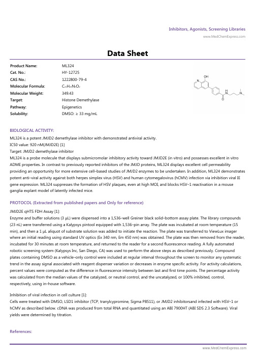
Inhibitors, Agonists, Screening Libraries Data SheetBIOLOGICAL ACTIVITY:ML324 is a potent JMJD2 demethylase inhibitor with demonstrated antiviral activity.IC50 value: 920 nM(JMJD2E) [1]Target: JMJD2 demethylase inhibitorML324 is a probe molecule that displays submicromolar inhibitory activity toward JMJD2E (in vitro) and possesses excellent in vitro ADME properties. In contrast to previously reported inhibitors of the JMJD proteins, ML324 displays excellent cell permeabilityproviding an opportunity for more extensive cell–based studies of JMJD2 enzymes to be undertaken. In addition, ML324 demonstrates potent anti–viral activity against both herpes simplex virus (HSV) and human cytomegalovirus (hCMV) infection via inhibition viral IE gene expression. ML324 suppresses the formation of HSV plaques, even at high MOI, and blocks HSV–1 reactivation in a mouse ganglia explant model of latently infected mice.PROTOCOL (Extracted from published papers and Only for reference)JMJD2E qHTS FDH Assay [1]:Enzyme and buffer solutions (3 μL) were dispensed into a 1,536–well Greiner black solid–bottom assay plate. The library compounds (23 nL) were transferred using a Kalypsys pintool equipped with 1,536–pin array. The plate was incubated at room temperature (15min), and then a 1 μL aliquot of substrate solution was added to initiate the reaction. The plate was transferred to ViewLux imager where an initial reading using standard UV optics (Ex 340 nm, Em 450 nm) was obtained. The plate was then removed from the reader,incubated for 30 minutes at room temperature, and returned to the reader for a second fluorescence reading. A fully automated robotic screening system (Kalypsys Inc, San Diego, CA) was used to perform the above steps as described previously. Compound plates containing DMSO as a vehicle–only control were included at regular interval throughout the screen to monitor any systematic trend in the assay signal associated with reagent dispenser variation or decreases in enzyme specific activity. For activity calculations,percent values were computed as the difference in fluorescence intensity between last and first time points. The percentage activity was calculated from the median values of the catalyzed, or neutral control, and the uncatalyzed, or 100% inhibited, control,respectively, using in–house software.Inhibition of viral infection in cell culture [1]:Cells were treated with DMSO, LSD1 inhibitor (TCP, tranylcypromine, Sigma P8511), or JMJD2 inhibitorsand infected with HSV–1 or hCMV as described below. cDNA was produced from total RNA and quantitated using an ABI 7900HT (ABI SDS 2.3 Software). Viral yields were determined by titration.References:Product Name:ML324Cat. No.:HY-12725CAS No.:1222800-79-4Molecular Formula:C 21H 23N 3O 2Molecular Weight:349.43Target:Histone Demethylase Pathway:Epigenetics Solubility:DMSO: ≥ 33 mg/mL[1]. Rai G, et al. Discovery of ML324, a JMJD2 demethylase inhibitor with demonstrated antiviral activity.Caution: Product has not been fully validated for medical applications. For research use only.Tel: 609-228-6898 Fax: 609-228-5909 E-mail: tech@Address: 1 Deer Park Dr, Suite Q, Monmouth Junction, NJ 08852, USA。
BI-847325_SDS_MedChemExpress

Inhibitors, Agonists, Screening LibrariesSafety Data Sheet Revision Date:Jun.-20-2017Print Date:Jun.-20-20171. PRODUCT AND COMPANY IDENTIFICATION1.1 Product identifierProduct name :BI-847325Catalog No. :HY-18955CAS No. :1207293-36-41.2 Relevant identified uses of the substance or mixture and uses advised againstIdentified uses :Laboratory chemicals, manufacture of substances.1.3 Details of the supplier of the safety data sheetCompany:MedChemExpress USATel:609-228-6898Fax:609-228-5909E-mail:sales@1.4 Emergency telephone numberEmergency Phone #:609-228-68982. HAZARDS IDENTIFICATION2.1 Classification of the substance or mixtureGHS Classification in accordance with 29 CFR 1910 (OSHA HCS)Acute toxicity, Oral (Category 4),H302Acute aquatic toxicity (Category 1),H400Chronic aquatic toxicity (Category 1),H4102.2 GHS Label elements, including precautionary statementsPictogramSignal word WarningHazard statement(s)H302 Harmful if swallowed.H410 Very toxic to aquatic life with long lasting effects.Precautionary statement(s)P264 Wash skin thoroughly after handling.P270 Do not eat, drink or smoke when using this product.P273 Avoid release to the environment.P301 + P312 IF SWALLOWED: Call a POISON CENTER or doctor/ physician if you feel unwell.P330 Rinse mouth.P391 Collect spillage.P501 Dispose of contents/ container to an approved waste disposal plant.2.3 Other hazardsNone.3. COMPOSITION/INFORMATION ON INGREDIENTS3.1 SubstancesSynonyms:BI847325; BI 847325Formula:C29H28N4O2Molecular Weight:464.56CAS No. :1207293-36-44. FIRST AID MEASURES4.1 Description of first aid measuresEye contactRemove any contact lenses, locate eye-wash station, and flush eyes immediately with large amounts of water. Separate eyelids with fingers to ensure adequate flushing. Promptly call a physician.Skin contactRinse skin thoroughly with large amounts of water. Remove contaminated clothing and shoes and call a physician.InhalationImmediately relocate self or casualty to fresh air. If breathing is difficult, give cardiopulmonary resuscitation (CPR). Avoid mouth-to-mouth resuscitation.IngestionWash out mouth with water; Do NOT induce vomiting; call a physician.4.2 Most important symptoms and effects, both acute and delayedThe most important known symptoms and effects are described in the labelling (see section 2.2).4.3 Indication of any immediate medical attention and special treatment neededTreat symptomatically.5. FIRE FIGHTING MEASURES5.1 Extinguishing mediaSuitable extinguishing mediaUse water spray, dry chemical, foam, and carbon dioxide fire extinguisher.5.2 Special hazards arising from the substance or mixtureDuring combustion, may emit irritant fumes.5.3 Advice for firefightersWear self-contained breathing apparatus and protective clothing.6. ACCIDENTAL RELEASE MEASURES6.1 Personal precautions, protective equipment and emergency proceduresUse full personal protective equipment. Avoid breathing vapors, mist, dust or gas. Ensure adequate ventilation. Evacuate personnel to safe areas.Refer to protective measures listed in sections 8.6.2 Environmental precautionsTry to prevent further leakage or spillage. Keep the product away from drains or water courses.6.3 Methods and materials for containment and cleaning upAbsorb solutions with finely-powdered liquid-binding material (diatomite, universal binders); Decontaminate surfaces and equipment by scrubbing with alcohol; Dispose of contaminated material according to Section 13.7. HANDLING AND STORAGE7.1 Precautions for safe handlingAvoid inhalation, contact with eyes and skin. Avoid dust and aerosol formation. Use only in areas with appropriate exhaust ventilation.7.2 Conditions for safe storage, including any incompatibilitiesKeep container tightly sealed in cool, well-ventilated area. Keep away from direct sunlight and sources of ignition.Recommended storage temperature:Powder-20°C 3 years4°C 2 yearsIn solvent-80°C 6 months-20°C 1 monthShipping at room temperature if less than 2 weeks.7.3 Specific end use(s)No data available.8. EXPOSURE CONTROLS/PERSONAL PROTECTION8.1 Control parametersComponents with workplace control parametersThis product contains no substances with occupational exposure limit values.8.2 Exposure controlsEngineering controlsEnsure adequate ventilation. Provide accessible safety shower and eye wash station.Personal protective equipmentEye protection Safety goggles with side-shields.Hand protection Protective gloves.Skin and body protection Impervious clothing.Respiratory protection Suitable respirator.Environmental exposure controls Keep the product away from drains, water courses or the soil. Cleanspillages in a safe way as soon as possible.9. PHYSICAL AND CHEMICAL PROPERTIES9.1 Information on basic physical and chemical propertiesAppearance Light yellow to yellow (Solid)Odor No data availableOdor threshold No data availablepH No data availableMelting/freezing point No data availableBoiling point/range No data availableFlash point No data availableEvaporation rate No data availableFlammability (solid, gas)No data availableUpper/lower flammability or explosive limits No data availableVapor pressure No data availableVapor density No data availableRelative density No data availableWater Solubility No data availablePartition coefficient No data availableAuto-ignition temperature No data availableDecomposition temperature No data availableViscosity No data availableExplosive properties No data availableOxidizing properties No data available9.2 Other safety informationNo data available.10. STABILITY AND REACTIVITY10.1 ReactivityNo data available.10.2 Chemical stabilityStable under recommended storage conditions.10.3 Possibility of hazardous reactionsNo data available.10.4 Conditions to avoidNo data available.10.5 Incompatible materialsStrong acids/alkalis, strong oxidising/reducing agents.10.6 Hazardous decomposition productsUnder fire conditions, may decompose and emit toxic fumes.Other decomposition products - no data available.11.TOXICOLOGICAL INFORMATION11.1 Information on toxicological effectsAcute toxicityClassified based on available data. For more details, see section 2Skin corrosion/irritationClassified based on available data. For more details, see section 2Serious eye damage/irritationClassified based on available data. For more details, see section 2Respiratory or skin sensitizationClassified based on available data. For more details, see section 2Germ cell mutagenicityClassified based on available data. For more details, see section 2CarcinogenicityIARC: No component of this product present at a level equal to or greater than 0.1% is identified as probable, possible or confirmed human carcinogen by IARC.ACGIH: No component of this product present at a level equal to or greater than 0.1% is identified as a potential or confirmed carcinogen by ACGIH.NTP: No component of this product present at a level equal to or greater than 0.1% is identified as a anticipated or confirmed carcinogen by NTP.OSHA: No component of this product present at a level equal to or greater than 0.1% is identified as a potential or confirmed carcinogen by OSHA.Reproductive toxicityClassified based on available data. For more details, see section 2Specific target organ toxicity - single exposureClassified based on available data. For more details, see section 2Specific target organ toxicity - repeated exposureClassified based on available data. For more details, see section 2Aspiration hazardClassified based on available data. For more details, see section 212. ECOLOGICAL INFORMATION12.1 ToxicityNo data available.12.2 Persistence and degradabilityNo data available.12.3 Bioaccumlative potentialNo data available.12.4 Mobility in soilNo data available.12.5 Results of PBT and vPvB assessmentPBT/vPvB assessment unavailable as chemical safety assessment not required or not conducted.12.6 Other adverse effectsNo data available.13. DISPOSAL CONSIDERATIONS13.1 Waste treatment methodsProductDispose substance in accordance with prevailing country, federal, state and local regulations.Contaminated packagingConduct recycling or disposal in accordance with prevailing country, federal, state and local regulations.14. TRANSPORT INFORMATIONDOT (US)This substance is considered to be non-hazardous for transport.IMDGUN number: 3077Class: 9Packing group: IIIEMS-No: F-A, S-FProper shipping name: ENVIRONMENTALLY HAZARDOUS SUBSTANCE, SOLID, N.O.S.Marine pollutant: Marine pollutantIATAUN number: 3077Class: 9Packing group: IIIProper shipping name: Environmentally hazardous substance, solid, n.o.s.15. REGULATORY INFORMATIONSARA 302 Components:No chemicals in this material are subject to the reporting requirements of SARA Title III, Section 302.SARA 313 Components:This material does not contain any chemical components with known CAS numbers that exceed the threshold (De Minimis) reporting levels established by SARA Title III, Section 313.SARA 311/312 Hazards:No SARA Hazards.Massachusetts Right To Know Components:No components are subject to the Massachusetts Right to Know Act.Pennsylvania Right To Know Components:No components are subject to the Pennsylvania Right to Know Act.New Jersey Right To Know Components:No components are subject to the New Jersey Right to Know Act.California Prop. 65 Components:This product does not contain any chemicals known to State of California to cause cancer, birth defects, or anyother reproductive harm.16. OTHER INFORMATIONCopyright 2017 MedChemExpress. The above information is correct to the best of our present knowledge but does not purport to be all inclusive and should be used only as a guide. The product is for research use only and for experienced personnel. It must only be handled by suitably qualified experienced scientists in appropriately equipped and authorized facilities. The burden of safe use of this material rests entirely with the user. MedChemExpress disclaims all liability for any damage resulting from handling or from contact with this product.Caution: Product has not been fully validated for medical applications. For research use only.Tel: 609-228-6898 Fax: 609-228-5909 E-mail: tech@Address: 1 Deer Park Dr, Suite Q, Monmouth Junction, NJ 08852, USA。
BFH772_DataSheet_MedChemExpress

Inhibitors, Agonists, Screening Libraries Data SheetBIOLOGICAL ACTIVITY:BFH772 is a potent oral VEGFR2 inhibitor, which is highly effective at targeting VEGFR2 kinase with an IC 50 value of 3 nM.IC50 & Target: IC50: 2.7±0.9 nM (hVEGFR2), 1.5±0.53 μM (mVEGFR2), 1.7±0.36 μM (hVEGFR1), 1.1±0.29 μM (hVEGFR3)[1]In Vitro: BFH772 is highly selective; apart from inhibiting VEGFR2 at 3 nM IC 50, it also targets B–RAF, RET, and TIE–2, albeit with atleast 40–fold lower potency. BFH772 is inactive (IC 50>10 μM; >2 μM for cKIT) against all other tyrosine specific– andserine/threonine–specific protein kinases tested. BFH772 inhibits VEGFR2 with IC 50 of 4.6±0.6 nM in CHO cells. BFH772 inhibits VEGFR2 with IC 50 of 3 nM in HUVEC cells. BFH772 inhibits the ligand induced autophosphorylation of RET, PDGFR, and KIT kinases,with IC 50 values ranging between 30 and 160 nM. BFH772 is selective (IC 50 values >0.5 μM) against the kinases of EGFR, ERBB2,INS–R, and IGF–1R and against the cytoplasmic BCR–ABL kinase. IC 50 of BFH772 (<0.01 nM, n=2) demonstrates that they abrogated VEGF induced proliferation at remarkably low nM concentrations [1].In Vivo: BFH772 at 3 mg/kg orally dosed once per day potently inhibits melanoma growth (by 54–90% for primary tumor and71–96% for metastasis growth) as depicted by treatment to control ratios. Dose–response curves of BFH772 at 0.3, 1, and 3 mg/kg demonstrate that even at the lowest concentrations, this naphthalene–1–carboxamide inhibits VEGF induced tissue weight and TIE–2 levels but only reaches statistical significance at 1 mg/kg and above [1].PROTOCOL (Extracted from published papers and Only for reference)Kinase Assay:[1]In vitro kinase assay is based on a filter binding assay, using the recombinant GST–fused kinase domainsexpressed in baculovirus and purified over glutathione–sepharose, γ–[33P]ATP as the phosphate donor, and poly(Glu:Tyr 4:1) peptide as the acceptor. Each GST–fused kinase is incubated under optimized buffer conditions [20 mM Tris–HCl buffer (pH 7.5), 1–3 mM MnCl 2, 3–10 mM MgCl 2, 3–8 μg/mL poly(Glu:Tyr 4:1), 0.25 mg/mL polyethylene glycol 20000, 8 μM ATP, 10 μM sodium vanadate, 1mM DTT] and 0.2 μCi γ–33P ATP in a total volume of 30 μL in the presence or absence of a test substance for 10 min at ambient temperature. The reaction is stopped by adding 10 mL of 250 mM EDTA. Using a 384–well filter system, half the volume istransferred onto an Immobilon–polyvinylidene difluoride membrane. The membrane is then washed extensively and dried, and scintillation counting is performed. IC 50s for compounds are calculated by linear regression analysis of the percentage inhibition [1].Cell Assay: BFH772 is dissolved in DMSO (10 mM) and stored, and then diluted with appropriate medium before use [1]. [1]DifferentBa/F3 cell lines rendered IL–3 independent by transduction with various constitutively active tyrosine kinases are grown in RPMI 1640 medium containing 10% fetal calf serum. For maintenance of parental Ba/F3 cells, the medium is additionally supplemented with 10 ng/mL interleukin–3 (IL–3). For proliferation assays, Ba/F3 cells are seeded on 96–well plates in triplicates at 10000 cells per well and incubated with various concentrations of compounds for 72 h followed by quantification of viable cells using a resazurin sodium salt dye reduction readout (commercially known as Alamar Blue assay). IC 50s are determined with the XLFit Excel Add–In using a four–parameter dose response model [1].Animal Administration: BFH772 is prepared in PEG200 100% (Mice)[1].Product Name:BFH772Cat. No.:HY-100419CAS No.:890128-81-1Molecular Formula:C 23H 16F 3N 3O 3Molecular Weight:439.39Target:VEGFR Pathway:Protein Tyrosine Kinase/RTK Solubility:DMSO: 7.75 mg/mLBFH772 is dissolved in N–methyl pyrrolidone/polyethylene glycol200 (30:70, v/v) (Rat)[1].[1]Mice[1]Female FVB mice weighing between 18 and 20 g are housed in groups of six. Porous chambers containing VEGF (2 μg/mL) in 0.5 mL of 0.8% w/v agar (containing heparin, 20 U/mL) are implanted subcutaneously in the flank of the mice (n=6 per group). VEGF induces the growth of vascularized tissue around the chamber. This response is dose–dependent and can be quantified by measuring the weight and TIE–2 levels of the tissue. Mice are treated either orally once daily with compounds or vehicle (PEG200 100%, 5 mL/kg) starting4–6 h before implantation of the chambers and continuing for 4 days. The animals are sacrificed for measurement of the vascularized tissues 24 h after the last dose. Tissue weight is taken and then a lysate prepared for TIE–2 ELISA analysis .Rat[1]Catheters are implanted into the femoral artery and vein of na?ve female rats strain OFA for BFH772, and BAW2881, or in the jugular vein and femoral artery in female Sprague–Dawley rats for compounds 4, 9, and 10. Animals are allowed to recover for 96 h and are housed in single cages with free access to food and water throughout the experiment. Female OFA rats received 2.5 mg/kg ofBAW2881 dissolved in ethanol/dimethylisosorbide/polyethylene glycol400/D5W (10/15/35/40 v/v) or 1 mg/kg of BFH772 dissolved in N–methyl pyrrolidone/polyethylene glycol200 (30:70, v/v) via injection into the femoral vein. D5W is glucose 5%/water (v/v). Oral administration: BAW2881 and BFH772 are formulated as a micronized suspension (dissolved/suspended in 0.5% carboxymethyl cellulose in distilled water) and administered by gavage to female OFA rats to deliver a dose of 25 mg/kg for BAW2881 or 3 mg/kg BFH772 (n=4 rats per group). For compounds 4, 9, and 10, female Sprague–Dawley rats at 8 weeks of age received an intraveno References:[1]. Bold G, et al. A Novel Potent Oral Series of VEGFR2 Inhibitors Abrogate Tumor Growth by Inhibiting Angiogenesis. J Med Chem. 2016 Jan 14;59(1):132–46.Caution: Product has not been fully validated for medical applications. For research use only.Tel: 609-228-6898 Fax: 609-228-5909 E-mail: tech@Address: 1 Deer Park Dr, Suite Q, Monmouth Junction, NJ 08852, USA。
EPZ015666_DataSheet_MedChemExpress
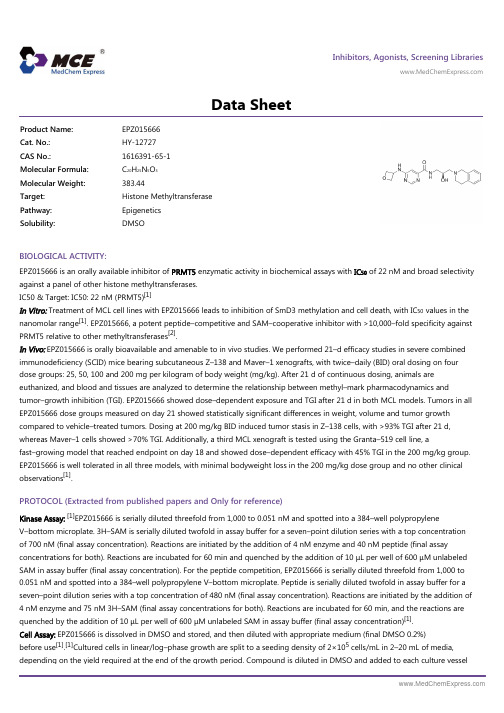
Inhibitors, Agonists, Screening Libraries Data SheetBIOLOGICAL ACTIVITY:EPZ015666 is an orally available inhibitor of PRMT5 enzymatic activity in biochemical assays with IC 50 of 22 nM and broad selectivity against a panel of other histone methyltransferases.IC50 & Target: IC50: 22 nM (PRMT5)[1]In Vitro: Treatment of MCL cell lines with EPZ015666 leads to inhibition of SmD3 methylation and cell death, with IC 50 values in the nanomolar range [1]. EPZ015666, a potent peptide–competitive and SAM–cooperative inhibitor with >10,000–fold specificity againstPRMT5 relative to other methyltransferases [2].In Vivo: EPZ015666 is orally bioavailable and amenable to in vivo studies. We performed 21–d efficacy studies in severe combined immunodeficiency (SCID) mice bearing subcutaneous Z–138 and Maver–1 xenografts, with twice–daily (BID) oral dosing on four dose groups: 25, 50, 100 and 200 mg per kilogram of body weight (mg/kg). After 21 d of continuous dosing, animals areeuthanized, and blood and tissues are analyzed to determine the relationship between methyl–mark pharmacodynamics andtumor–growth inhibition (TGI). EPZ015666 showed dose–dependent exposure and TGI after 21 d in both MCL models. Tumors in all EPZ015666 dose groups measured on day 21 showed statistically significant differences in weight, volume and tumor growth compared to vehicle–treated tumors. Dosing at 200 mg/kg BID induced tumor stasis in Z–138 cells, with >93% TGI after 21 d,whereas Maver–1 cells showed >70% TGI. Additionally, a third MCL xenograft is tested using the Granta–519 cell line, afast–growing model that reached endpoint on day 18 and showed dose–dependent efficacy with 45% TGI in the 200 mg/kg group.EPZ015666 is well tolerated in all three models, with minimal bodyweight loss in the 200 mg/kg dose group and no other clinical observations [1].PROTOCOL (Extracted from published papers and Only for reference)Kinase Assay:[1]EPZ015666 is serially diluted threefold from 1,000 to 0.051 nM and spotted into a 384–well polypropyleneV–bottom microplate. 3H–SAM is serially diluted twofold in assay buffer for a seven–point dilution series with a top concentration of 700 nM (final assay concentration). Reactions are initiated by the addition of 4 nM enzyme and 40 nM peptide (final assayconcentrations for both). Reactions are incubated for 60 min and quenched by the addition of 10 μL per well of 600 μM unlabeled SAM in assay buffer (final assay concentration). For the peptide competition, EPZ015666 is serially diluted threefold from 1,000 to 0.051 nM and spotted into a 384–well polypropylene V–bottom microplate. Peptide is serially diluted twofold in assay buffer for a seven–point dilution series with a top concentration of 480 nM (final assay concentration). Reactions are initiated by the addition of 4 nM enzyme and 75 nM 3H–SAM (final assay concentrations for both). Reactions are incubated for 60 min, and the reactions are quenched by the addition of 10 μL per well of 600 μM unlabeled SAM in assay buffer (final assay concentration)[1].Cell Assay: EPZ015666 is dissolved in DMSO and stored, and then diluted with appropriate medium (final DMSO 0.2%)before use [1].[1]Cultured cells in linear/log–phase growth are split to a seeding density of 2×10⁵ cells/mL in 2–20 mL of media,depending on the yield required at the end of the growth period. Compound is diluted in DMSO and added to each culture vesselProduct Name:EPZ015666Cat. No.:HY-12727CAS No.:1616391-65-1Molecular Formula:C 20H 25N 5O 3Molecular Weight:383.44Target:Histone Methyltransferase Pathway:Epigenetics Solubility:DMSOwith a final DMSO concentration of 0.2%. Cells are allowed to grow for 96 h undisturbed. At the conclusion of each treatment period, cells are harvested by centrifugation (5 min, 1,200 rpm), and cell pellets are rinsed once with PBS before being frozen on dry ice pending further processing. Long–term proliferation assays are performed on all MCL lines, with slight adjustments to initial seeding densities, depending on growth characteristics for each cell line. All assays are carried out for 12 d[1].Animal Administration: EPZ015666 is formulated in 20% N–N–dimethylacetamide in water (Mice)[1].[1]Mice[1]Male CD–1 mice (25–40 g; n=6, with 3 per time point) are treated with a single dose of EPZ015666 at 2 mg/kg by intravenoustail–vein injection and 10 mg/kg by oral gavage administration, with both doses formulated in 20% N–N–dimethylacetamide in water. Animals are fasted overnight and weighed before dose administration on the day of dosing. Approximately 30 μL ofblood are taken from animals by submandibular or retro–orbital bleeding at pre–specified time intervals (seven time points). For the last time point (24 h), samples are collected via cardiac puncture while the animals are under anesthesia (70% CO2:30% O2). Blood samples are transferred into K2–EDTA tubes and placed on wet ice before centrifugation at 4°C (3,000g, 15 min) to obtain plasma within 30 min after sample collection. Plasma samples are stored at -70±10°C before protein precipitation and LC–MS/MS analysis. We constructed standard calibration curves by analyzing a series of control plasma aliquots containing 100 ng/mL labetalol as an internal standard and 1–3,000 ng/mL EPZ015666. Four levels of quality control are also included in the analysis (3–2,400 ng/mL EPZ015666). Data are analyzed using Phoenix WinNonlin 6.2.1.References:[1]. Chan–Penebre E, et al. A selective inhibitor of PRMT5 with in vivo and in vitro potency in MCL models. Nat Chem Biol. 2015 Jun;11(6):432–7.[2]. Kryukov GV, et al. MTAP deletion confers enhanced dependency on the PRMT5 arginine methyltransferase in cancer cells. Science. 2016 Mar 11;351(6278):1214–8.Caution: Product has not been fully validated for medical applications. For research use only.Tel: 609-228-6898 Fax: 609-228-5909 E-mail: tech@Address: 1 Deer Park Dr, Suite Q, Monmouth Junction, NJ 08852, USA。
A-443654_DataSheet_MedChemExpress

Inhibitors, Agonists, Screening Libraries Data SheetBIOLOGICAL ACTIVITY:A–443654 is a potent small–molecule inhibitor of all three Akt serine/threonine kinases , with K i of 160 pM for Akt1.IC50 & Target: Ki: 160 pM (Akt1)In Vitro: A–443654 exhibits a K i of 160 pM, a 30,000–fold improvement in potency versus the initial lead molecule. A–443654 is 40–fold selective for Akt over PKA. A–443654 inhibits Akt1, Akt2, or Akt3 equally within cells. A–443654 reduces the P–GSK3 in a dose–responsive manner in all three cell lines. A–443654 inhibits the proliferation of tumor cells with EC 50 of 0.1 μM [1].A–443654–induced morphological changes occur very rapidly (within 2 to 4 h) in both 10A and 10CA1a cells, with 10CA1a cells more sensitive to A–443654 than the 10A cells. A–443654 alone at 2 μM causes the 10CA1a cells, but not the 10A cells, to detach from the plate after 12 h, whereas 1 μM of A–443654 causes 10CA1a cells to detach from the plate after 12 h. FACScan Analysis of rapamycin and A–443654 effects on DNA content in 10A and 10CA1a cells. In contrast, A–443654 at 2 and 5 μM decreases Bcl–2levels by 30 to 40% in the 10CA1a cells at 8h. The combination of rapamycin with 2 or 5 μM A–443654, however, markedlydecreases Bcl–2 protein levels by appr 40 to 50% in the 10A cells and by appr 70% in the 10CA1a cells, respectively [2]. A–443654demonstrates the greatest selective effect on the mutant cells compared to the WT cells with greater than 3.5 fold relative growth inhibition of the mutant cells [3].In Vivo: A–443654 (7.5 mg/kg/d, s.c.) inhibits tumor growth in the 3T3–Akt1 flank tumor model. A–443654 (50 mg/kg, s.c.) induces apoptosis in 3T3–Akt1 flank tumors. A–443654 (30 mg/kg, s.c.) leads to increased levels of phosphorylated Akt1 in MiaPaCa–2tumors [1].PROTOCOL (Extracted from published papers and Only for reference)Cell Assay:[1]The cells on 96–well plates are gently washed with 200 μL of PBS. Alamar Blue reagent is diluted 1:10 in normal growth media. The diluted Alamar Blue reagent (100 μL) is added to each well on the 96–well plates and incubated until the reaction is complete as per manufacturer's instructions. Analysis is done using an fmaxFluorescence Microplate Reader, set at the excitation wavelength of 544 nm and emission wavelength of 595 nm. Data are analyzed using SOFTmax PRO software provided by the manufacturer.Animal Administration: A–443654 is given s.c. in a vehicle of 0.2% HPMC [1]Immunocompromised male scid mice are 6 to 8weeks of age. The 3T3–Akt1 cell line is developed and characterized in our laboratory. The 1×10⁶ 3T3–Akt1 or 2×10⁶ MiaPaCa–2and PC–3 cells in 50% Matrigel are inoculated s.c. into the flank. For early treatment studies, mice are randomLy assigned totreatment groups and therapy is initiated the day after inoculation. Ten animals are assigned to each group, including controls. For established tumor studies, tumors are allowed to reach a designated size and mice are assigned to treatment groups of equal tumor size (n=10 mice per group). Tumor size is evaluated by twice weekly measurements with digital calipers. Tumor volume is estimated using the formula: V=L×W 2/2. A–443654 is given s.c. in a vehicle of 0.2% HPMC. A–674563 is given orally in a vehicle of 5%dextrose.Product Name:A–443654Cat. No.:HY-10425CAS No.:552325-16-3Molecular Formula:C 24H 23N 5O Molecular Weight:397.47Target:Akt Pathway:PI3K/Akt/mTOR Solubility:10 mM in DMSOReferences:[1]. Luo Y, et al. Potent and selective inhibitors of Akt kinases slow the progress of tumors in vivo. Mol Cancer Ther. 2005 Jun;4(6):977–86.[2]. Zheng J, et al. Rapamycin sensitizes Akt inhibition in malignant human breast epithelial cells. Cancer Lett. 2010 Oct 1;296(1):74–87.[3]. Gallia GL, et al. Inhibition of Akt inhibits growth of glioblastoma and glioblastoma stem–like cells. Mol Cancer Ther. 2009 Feb;8(2):386–93.Caution: Product has not been fully validated for medical applications. For research use only.Tel: 609-228-6898 Fax: 609-228-5909 E-mail: tech@Address: 1 Deer Park Dr, Suite Q, Monmouth Junction, NJ 08852, USA。
AS-252424_DataSheet_MedChemExpress
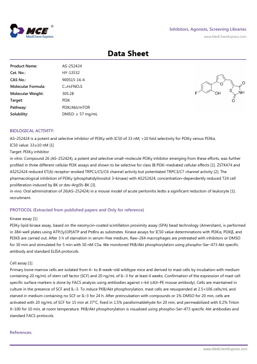
Inhibitors, Agonists, Screening Libraries Data SheetBIOLOGICAL ACTIVITY:AS–252424 is a potent and selective inhibitor of PI3Kγ with IC50 of 33 nM; >10 fold selectivity for PI3Kγ versus PI3Kα.IC50 value: 33±10 nM [1]Target: PI3Kγ inhibitorin vitro: Compound 26 (AS–252424), a potent and selective small–molecule PI3Kγ inhibitor emerging from these efforts, was further profiled in three different cellular PI3K assays and shown to be selective for class IB PI3K–mediated cellular effects [1]. ZSTK474 and AS252424 reduced ET(A) receptor–evoked TRPC1/C5/C6 channel activity but potentiated TRPC3/C7 channel activity [2]. Thepharmacological inhibition of PI3Kγ (phosphatidylinositol 3–kinase) with AS252424, concentration–dependently reduced T24 cell proliferation induced by BK or des–Arg(9)–BK [3].in vivo: Oral administration of 26(AS–252424) in a mouse model of acute peritonitis ledto a significant reduction of leukocyte [1].recruitment.PROTOCOL (Extracted from published papers and Only for reference)Kinase assay [1]PI3Kγ lipid kinase assay, based on the neomycin–coated scintillation proximity assay (SPA) bead technology (Amersham), is performed in 384–well plates using ATP/[γ33P]ATP and PtdIns as substrates. Kinase assays for IC50 value determinations with PI3Kα, PI3Kβ, and PI3Kδ are carried out. After 3 h of starvation in serum–free medium, Raw–264 macrophages are pretreated with inhibitors or DMSO for 30 min and stimulated for 5 min with 50 nM C5a. We monitored PKB/Akt phosphorylation using phospho–Ser–473 Akt specific antibody and standard ELISA protocols.Cell assay [1]Primary bone marrow cells are isolated from 4– to 8–week–old wildtype mice and derived to mast cells by incubation with medium containing 20 ng/mL of stem cell factor (SCF) and 20 ng/mL of IL–3 for at least 4 weeks. Confirmation of the expression of mast cell specific surface markers is done by FACS analysis using antibodies against c–kit (cKit–PE mouse antibody). Cells are maintained in culture in the presence of SCF and IL–3. To induce PKB/Akt phosphorylation, mast cells are resuspended at 2.5×10⁶ cells/mL and starved in medium containing no SCF or IL–3 for 24 h. After preincubation with compounds or 1% DMSO for 20 min, cells areactivated with 20 ng/mL of SCF for 15 min at 37°C, fixed in 1.5% paraformaldehyde for 20 min, and permeabilized with 0.2% Triton X–100 for 10 min, at room temperature. PKB/Akt phosphorylation is visualized using phospho–Ser–473 specific Akt antibodies and standard FACS protocols.References:Product Name:AS–252424Cat. No.:HY-13532CAS No.:900515-16-4Molecular Formula:C 14H 8FNO 4S Molecular Weight:305.28Target:PI3K Pathway:PI3K/Akt/mTOR Solubility:DMSO: ≥ 57 mg/mL[1]. Pomel V, et al. Furan–2–ylmethylene thiazolidinediones as novel, potent, and selective inhibitors of phosphoinositide 3–kinase gamma. J Med Chem. 2006 Jun 29;49(13):3857–71.[2]. Shi J, et al. Pharmacological profile of phosphatidylinositol 3–kinases and related phosphatidylinositols mediating endothelin(A) receptor–operated native TRPC channels in rabbit coronary artery myocytes. Br J Pharmacol. 2012 Aug;166(7):2161–75.[3]. Sgnaolin V, et al. Functional and molecular characterization of kinin B1 and B 2 receptors in human bladder cancer: implication of the PI3Kγ pathway. Invest New Drugs. 2013 Aug;31(4):812–22.Caution: Product has not been fully validated for medical applications. For research use only.Tel: 609-228-6898 Fax: 609-228-5909 E-mail: tech@Address: 1 Deer Park Dr, Suite Q, Monmouth Junction, NJ 08852, USA。
b-AP15_DataSheet_MedChemExpress

Inhibitors, Agonists, Screening Libraries Data SheetBIOLOGICAL ACTIVITY:b–AP15 is a specific inhibitor of the deubiquitinating enzymes UCHL5 and Usp14.IC50 & Target: UCHL5/Usp14[1]In Vitro: Purified 19S proteasomes (5 nM) are treated with indicated concentrations of b–AP15 and DUB activity is determined by detectionof Ub–AMC cleavage. The IC 50 value (2.1±0.411 μM) is determined from log concentration curves in Graph Pad Prism using non linear regression analysis. b–AP15 as a previously unidentified class of proteasome inhibitor that abrogates thedeubiquitinating activity of the 19S regulatory particle. b–AP15 inhibited the activity of two 19S regulatory–particle–associated deubiquitinases, ubiquitin C–terminal hydrolase 5 (UCHL5) and ubiquitin–specific peptidase 14 (USP14), resulting in accumulation of polyubiquitin. b–AP15 induced tumor cell apoptosis that is insensitive to TP53 status and overexpression of the apoptosisinhibitor BCL2[1]. The ability of b–AP15 is determined to inhibit proteasome deubiquitinase activity using Ub–AMC as the substrate.An IC 50 of 16.8±2.8 μM is observed [2]. b–AP15 is a specific USP14 and UCHL5 inhibitor, which blocks growth and induces apoptosis in MM cells [3].In Vivo: b–AP15 (2.5 mg/kg) inhibits tumor growth in syngenic mice models with less frequent administration schedules. We administered b–AP15 to C57BL/6J mice with Lewis lung carcinomas (LLCs) using a 2–d–on, 2–d–off schedule and to BALB/c mice with orthotopic breast carcinoma (4T1) using a 1–d–on, 3–d–off schedule. b–AP15 significantly inhibited tumor growth in both models, with T/C=0.16 (P≤0.01) for the C57BL/6J mice and T/C=0.25 (P≤0.001) for the BALB/c mice. A reduction in the number of pulmonary metastases also is observed in the group of mice with 4T1 breast carcinomas treated with b–AP15[1].PROTOCOL (Extracted from published papers and Only for reference)Kinase Assay:[1]For deubiquitinase inhibition assays, 19S regulatory particle (5 nM), 26S (5 nM) UCH–L1 (5 nM), UCH–L3 (0.3 nM),USP2CD (5 nM) USP7CD (5 nM) USP8CD (5 nM) or BAP1 (5 nM) is incubated with DMSO or b–AP15 and monitored the cleavage of ubiquitin–AMC (1,000 nM) using a Wallac VICTOR Multilabel counter or a Tecan Infinite M1000 equipped with 380 nm excitation and 460 nm emission filters [1].Cell Assay: b–AP15 is dissolved in DMSO and stored, and then diluted with appropriate medium before use [2]. [2]Cell viability is monitored by either the fluorometric microculture cytotoxicity assay or the MTT assay. For the MTT assay, cells are seeded into 96–well flat–bottomed plates overnight and exposed to drugs, using DMSO as the control. At the end of incubations, 10 μl of a stock solution of 5 mg/mL MTT is added into each well, and the plates are incubated 4 hours at 37°C. Formazan crystals are dissolved with 100 μL 10% SDS/10 mM HCl solution overnight at 37°C. Absorbance is measured using an enzyme–linkedimmunosorbent assay (ELISA) plate reader at 590 nm [2].Animal Administration: b–AP15 is dissolved it in Cremophor EL and polyethylene glycol 400 (1:1) by heating to reach aworking concentration of 2 mg/mL. Working stock is 1:10 diluted in 0.9% saline immediately before injection (Mice)[1].[1]Mice [1]For the squamous carcinoma model, 1×10⁶ FaDu cells are subcutaneously injected into the right rear flank of female SCIDProduct Name:b–AP15Cat. No.:HY-13989CAS No.:1009817-63-3Molecular Formula:C 22H 17N 3O 6Molecular Weight:419.39Target:Deubiquitinase Pathway:Cell Cycle/DNA Damage Solubility:DMSO: ≥ 44 mg/mLmice. Tumor growth is measured by the formula length×width2×0.44. When tumors have grown to a size of approximately 200 mm3 (defined as day 0), mice are randomized to receive either vehicle (n=10) or b–AP15 (n=15) at 5 mg per kg of body weight by daily subcutaneous injection. For the colon carcinoma model, we subcutaneously injected 2.5 × 10⁶ HCT–116 colon carcinoma cells overexpressing Bcl2 into the right flank of female nude mice. We treated mice with 5 mg of b–AP15 per kg of body weight by intraperitoneal injection. For the lung carcinoma model, we subcutaneously injected 2×10⁵ LLC cells into the right rear flank of female C57/B6 mice. When tumors had grown to a size of approximately 50 mm3 (defined as day 0), we randomized mice to receive either vehicle (n=4) or b–AP15 (n=4) at 5 mg per kg of body weight intraperitoneally, with a treatment cycle consisting of 2 d of treatment followed by 2 d of rest (2 d on, 2 d off) for 2 weeks.References:[1]. D'Arcy P, et al. Inhibition of proteasome deubiquitinating activity as a new cancer therapy. Nat Med. 2011 Nov 6;17(12):1636–40.[2]. Wang X, et al. The 19S Deubiquitinase Inhibitor b–AP15 is Enriched in Cells and Elicits Rapid Commitment to Cell Death. Mol Pharmacol. 2014 Jun;85(6):932–45.[3]. Tian Z, et al. A novel small molecule inhibitor of deubiquitylating enzyme USP14 and UCHL5 induces apoptosis in multiple myeloma and overcomes bortezomib resistance. Blood. 2014 Jan 30;123(5):706–16.Caution: Product has not been fully validated for medical applications. For research use only.Tel: 609-228-6898 Fax: 609-228-5909 E-mail: tech@Address: 1 Deer Park Dr, Suite Q, Monmouth Junction, NJ 08852, USA。
- 1、下载文档前请自行甄别文档内容的完整性,平台不提供额外的编辑、内容补充、找答案等附加服务。
- 2、"仅部分预览"的文档,不可在线预览部分如存在完整性等问题,可反馈申请退款(可完整预览的文档不适用该条件!)。
- 3、如文档侵犯您的权益,请联系客服反馈,我们会尽快为您处理(人工客服工作时间:9:00-18:30)。
Inhibitors, Agonists, Screening Libraries
Data Sheet
BIOLOGICAL ACTIVITY:
T–5224 is a selective inhibitor of c–Fos/activator protein (AP)–1 for rheumatoid arthritis therapy, and inhibits MMP activity with IC 50s of 10 nM for both MMP–3 and MMP–13.
IC50 & Target: IC50: 10 nM (MMP–3), 10 nM (MMP–13)
In Vitro: T–5224 (0–80 μM) significantly inhibits the invasion, migration, and MMP activity of HSC–3–M3 cells in a dose–dependent manner. There is no significant influence on HSC–3–M3 amd OSC–19 cells proliferation [4].
In Vivo: G2 is observed in rat and monkey liver microsomes as a major metabolite of T–5224, suggesting that G2 is not a
human–specific metabolite [1]. T–5224 (300 mg/kg, p.o.) inhibits the production of TNF–alpha and other downstream effectors in C57BL/6 mice [2]. Administration of T–5224 (300 mg/kg, p.o.) after intraperitoneal injection of LPS impartes appreciable protection
against acute elevations in serum levels of TNFα, HMGB1, ALT/AST as well as in liver tissue levels of MIP–1α and MCP–1, and reduces the lethality (27%)[3]. PROTOCOL (Extracted from published papers and Only for reference)
Cell Assay: T–5224 is dissolved in DMSO and diluted in DMEM.[4]HSC–3–M3 cells are starved for 24 h with DMEM containing 0.5%FBS. The top chamber of the cell invasion device is coated with 50 μL of 0.1 × basement membrane extract solution and incubataed overnight. HSC–3–M3 cells (5.0 × 10⁴ cells/well) are added to the top chamber with DMEM containing 0.5% FBS mixed with 0–80 μM
T–5224; DMEM with 10% FBS is added to the bottom chamber and incubated for 48 h. The bottom plate is read using a multilabel plate reader. The data are compared with the standard curve to determine the fraction of invaded cells.
Animal Administration: T–5224 is dissolved in a polyvinylpyrrolidone solution, and adjusted to a concentration of 30 mg/mL.[2]Mice in LPS group are administered orally with polyvinylpyrrolidone solution in the same volume of T–5224 solution immediately after LPS injection, while in the T–5224 group, mice are administered orally with T–5224 (300 mg/kg, p.o.) in the same manner. In the control group, mice receives polyvinylpyrrolidone solution orally soon after intraperitoneal saline injection. Blood samples are collected for each measurement at the optimal time.References:
[1]. Uchihashi S, et al. Metabolism of the c–Fos/activator protein–1 inhibitor T–5224 by multiple human UDP–glucuronosyltransferase isoforms. Drug Metab Dispos. 2011 May;39(5):803–13.
[2]. Miyazaki H, et al. The effects of a selective inhibitor of c–Fos/activator protein–1 on endotoxin–induced acute kidney injury in mice. BMC Nephrol. 2012Nov 23;13:153.
[3]. Izuta S, et al. T–5224, a selective inhibitor of c–Fos/activator protein–1, attenuates lipopolysaccharide–induced liver injury in mice. Biotechnol Lett. 2012Dec;34(12):2175–82.
Product Name:
T–5224Cat. No.:
HY-12270CAS No.:
530141-72-1Molecular Formula:
C 29H 27NO 8Molecular Weight:
517.53Target:
MMP Pathway:
Metabolic Enzyme/Protease Solubility:
DMSO: ≥ 31 mg/mL
[4]. Kamide D, et al. Selective activator protein–1 inhibitor T–5224 prevents lymph node metastasis in an oral cancer model. Cancer Sci. 2016 May; 107(5):666–73.
Caution: Product has not been fully validated for medical applications. For research use only.
Tel: 609-228-6898 Fax: 609-228-5909 E-mail: tech@
Address: 1 Deer Park Dr, Suite Q, Monmouth Junction, NJ 08852, USA。