zno纳米粒子英文论文
纳米氧化锌的制备及其应用
纳米氧化锌的制备及其应用学院:电子信息学院专业:电子科学与技术班级: 101 班学号: 1007010043学生姓名:杨晓玲2014年1月3日纳米氧化锌的制备及其应用电子信息学院杨晓玲 1007010043摘要纳米氧化锌作为一种功能材料,有着许多有益的性能和广泛的应用。
通过对纳米氧化锌的主要制备技术过程和工艺特点,介绍了纳米氧化锌在各个领域的应用。
关键词:纳米氧化锌,制备,应用Abstract Nanometer zinc oxide as a kind of functional material, has many good properties and wide application. Through the process of main preparation technology of nanometer zinc oxide and the technological characteristics, the author introduces the application of nanometer zinc oxide in various fields.Key words: nano zinc oxide, preparation, application一、前言近年来纳米材料因其独特的物理化学作用而被广为重视并逐步应用于各个领域,纳米氧化锌粒子作为联系宏观物体及微观粒子的桥梁其潜在的重要性毋庸置疑一些发达国家都投入大量资金开展预研究工作国内的许多科研院所、高等院校也组织科研力量开展纳米材料的研究工作。
纳米氧化锌是一种面向21 世纪的新型高功能精细无机产品其粒径介于1~100nm,由于具有纳米材料的结构特点和性质使得纳米氧化锌产生了表面效应及体积效应等从而使其在磁、光、电、敏感性等方面具有一般氧化锌产品无法比拟的特殊性能和新用途。
二、纳米氧化锌的结构分析采用沉淀法制备了纳米氧化锌粉体,利用 Rietveld方法[1]对所得样品的结构进行了精修,结果显示所得纳米氧化锌为六方结构,空间群为P63mc,其晶胞参数口=3.2533A,c=5.2129A,与氧化锌体相材料相比其晶胞参数明显增大。
纳米氧化锌抗菌性能及机制
氧化物抗菌材料的研究,人们最先关注的是以氧 化锌(ZnO)、氧化钙、氧化镁为代表的活性氧化 物,发现它们都具有良好的抗菌性,甚至较低浓 度的氧化物在无光条件下也显示出了优异的抗 菌性能。ZnO是一种宽禁带Ⅱ,Ⅵ族化合物半导 体材料,具有规整的六角形纤锌矿结构,本身为 白色,稳定性好,高温下不变色、不分解、价格 低廉、资源丰富,己成为无机抗菌剂研究的热点 之一。关于ZnO抗菌性能的研究[1],称ZnO的光催 化活性甚至强于二氧化钛,在很多方面,ZnO完 全可以作为二氧化钛的替代材料。二氧化钛在未 进行紫外光照射时是一种生物兼容性很好的材 料,但是经使用UVA进行照射后,又可以显示出 极强的细胞毒性[2-3]。因此,与二氧化钛相比ZnO 更具有实用价值。
1Key Laboratory for Magnetism and Magnetic Materials of the Ministry of Education, School of Physical Science and Technology, Lanzhou University, Lanzhou 730000, Gansu Province, China; 2School of Stomatology, Lanzhou University, Lanzhou 730000, Gansu Province, China
Hu Zhan-jiang1, Zhao Zhong1, Wang Xue-mei2
Abstract BACKGROUND: The zinc oxide has a good biocompatibility, security and long effectiveness, and can be used as a type of antibacterial material of active oxide category. OBJECTIVE: To summarize the antibacterial properties and mechanism of nano-zinc oxide (nano-ZnO). METHODS: A computer-based online search of related papers from December 1995 to February 2011 was performed in Elsevier (Science Direct) and Web of Science databases using the key words of “antibacterial properties of nano-ZnO” in English, and in CNKI and Wanfang databases using the key words of “antibacterial properties of nano-ZnO” in Chinese. Totally 75 literatures were selected. RESULTS AND CONCLUSION: The nano-ZnO has a strong bactericidal property in many fields. It can replace other materials of active oxide category based on its good biocompatibility, security and long effectiveness. The antibacterial properties and mechanism of nano-ZnO were summarized in this study from the sides of modified antibacterial properties and the effects of morphology and structure of nano-ZnO on antibacterial properties. However, more studies are in need to solve how to improve the utilization and antibacterial properties, and to expand the applications of nano-ZnO in antibacterial and other fields.
纳米ZnO抗菌材料

Controllable synthesis of highly efficient antimicrobial agent-Fe dopedsea urchin-like ZnO nanoparticlesJianzhong Ma a,c,n,Aiping Hui a,c,Junli Liu b,c,Yan Bao a,ca College of Resources and Environment,Shaanxi University of Science&Technology,Xi’an710021,PR Chinab College of Materials Science and Engineering,Shaanxi University of Science&Technology,Xi’an710021,PR Chinac Shaanxi Research Institute of Agricultural Products Processing Technology,Xi'an710021,PR Chinaa r t i c l e i n f oArticle history:Received12April2015Received in revised form4June2015Accepted10June2015Available online12June2015Keywords:ZnONanoparticlesDopingDepositionAntimicrobial activitya b s t r a c tIn this paper,Fe doped sea urchin-like ZnO nanoparticles were synthesized by a simple chemical de-position.The crystalline structure,element composition and morphology of the obtained Fe doped ZnOnanoparticles were characterized by X-ray diffraction(XRD),energy dispersive spectroscopy(EDS)andscanning electron microscopy(SEM).The effect of different[Fe]/[Zn]molar ratios on the antimicrobialproperties of the obtained nanoparticles was investigated in detail.The results indicated that doping Feions into ZnO lattice could improve the oriented growth of ZnO along(002)plane,form sea urchin-likestructure and enhance its antimicrobial activity.Antimicrobial assay demonstrated that Fe doped seaurchin-like ZnO with5%Fe content had the strongest antimicrobial activity against Candida albicans(94%)and Aspergillusflavus(81%).This strategy indicated that the inhibition rate of ZnO nanoparticlesincreased with the forming of sea urchin-like structure and the decrease of ZnO size due to the doping ofFe ions into ZnO lattice.&2015Elsevier B.V.All rights reserved.1.IntroductionWith the increasing concern and attentions about environ-mental protection,the research about antimicrobial material hasbeen widely studied in recent years[1,2].As a wide band semi-conductor,zinc oxide(ZnO)has excellent electrical,optical,pho-tocatalytic and antimicrobial properties[3,4].In particular,theantimicrobial performance of ZnO causes a wide range of concernsrecently,it is considered as an ideal antimicrobial agent due to itsbetter biocompatibility,stability and reusability than organic an-timicrobial agents[5–7].In the view of literatures,ZnO has betterantimicrobial activity to both gram-positive bacteria like Staphy-loccocus aureus and gram-negative bacteria like Escherichia coli,and even exhibits antimicrobial effect on spores[3,4,8,9].Besides,morphology,size,shape,stability,capping agents and structuraldefects of ZnO play a significant role in determining its anti-microbial performances[1,2,6,8-10].In particular,Xiaoling Xu et al.[8]have found that tetrapod-like ZnO whiskers had better anti-bacterial properties than nanosized and microsized ZnO nano-particles.However,the fabrication of tetrapod-like ZnO whiskersneeds strict conditions,such as high pressure,high temperatureand high quality for apparatus.Therefore,it is necessary forseeking a facile approach to fabricate ZnO nanoparticles with newtype acicular structure to further improve its antibacterial prop-erties.In the present work,Fe doped sea urchin-like ZnO wasprepared by a simple chemical deposition method.The effect of Fedoping dosage on the crystallization,morphology and anti-microbial activity of ZnO against Candida albicans and Aspergillusflavus was investigated in detail.At last,the antimicrobial me-chanism of Fe doped sea urchin-like ZnO was found.2.Experimental section2.1.Fabrication of Fe doped sea urchin-like ZnO nanoparticlesIn a typical synthesis of Fe doped sea urchin-like ZnO nano-particles,9.0g NaOH was dissolved into25mL deionized waterand stirred for1h,then4.5g Zn(NO3)2Á6H2O and Fe(NO3)3Á9H2Owith different[Fe]/[Zn]molar ratios(0%,1%,3%and5%)were addedinto the above solution,separately.At the same time, 2.8gCH3(CH2)11OSO3Na was added to a mixed solvent of50mL deionizedwater and250mL absolute ethanol.Hereafter,the above-mentionedtwo solutions were mixed together,and transferred to aflaskequipped with a reflux condenser and a digital mechanical stirrer,heated in the water bath at80°C for3h.At last,the solid powder wascollected by centrifugation,washed with deionized water and abso-lute ethanol,and then dried at60°C for6h in an oven.Contents lists available at ScienceDirectjournal homepage:/locate/matletMaterials Letters/10.1016/j.matlet.2015.06.0370167-577X/&2015Elsevier B.V.All rightsreserved.n Corresponding author.Tel.:þ8602986132559;fax:þ8602986132559.E-mail address:majz@(J.Ma).Materials Letters158(2015)420–4232.2.Structure characterizationThe products were characterized by X-ray diffractometer(XRD, Rigaku D/MAX-2200,Japan),field emission scanning electron mi-croscopy(SEM,Hitachi S-4800,Japan)equipped with energy dis-persive spectroscopy(EDS)elemental composition analyzer.2.3.Antimicrobial assayC.albicans and A.flavus cultures were kindly provided by Xi’an Microorganism Research Institution.The antimicrobial activity of various Fe doped ZnO and pure ZnO nanoparticles was evaluated by examining the growth density and numbers of bacterial colony with the traditional plating methods,as reported in our previous studies[9].3.Results and discussionThe XRD results of the obtained pure ZnO(0%)and Fe doped sea urchin-like ZnO are shown in Fig.1a.The diffraction peaks of the synthesized ZnO samples were quite similar to those of crys-talline wurtzite ZnO structure(JCPDS standard card36-1451).No others characteristic peaks of the impurities appeared.It was ob-served the peak location and shape has not changed with the doping of different Fe content.However,the doping of Fe into ZnO crystal structure could improve the peak intensities of(002) planes and enhance the crystallization of ZnO,when the XRD patterns of Fe doped ZnO patterns(Fig.1a)were compared.What’s more,EDS analysis results showed the surface composition of Zn: O:Fe was57.18:39.16:3.66,which implying that the doping con-centration of Fe was too low(Fig.1b).SEM images of pure ZnO and Fe doped ZnO samples are shown in Fig.1c.The morphologyof Fig.1.XRD patterns of Fe doped ZnO samples(a);EDS of5%Fe doped ZnO(b);SEM images of Fe doped ZnO samples with different[Fe]/[Zn]molar ratios(c).J.Ma et al./Materials Letters158(2015)420–423421pure ZnO was flower-like structure that has been reported in our previous work [7].Moreover,the diameter of pure ZnO rods was obviously larger than that of the doped ZnO.The dopant ion of Fe has an obvious effect on morphology of ZnO.With the increasing of Fe concentration,flower-like ZnO had transferred into sea urchin-like ZnO structure due to the clearly oriented growth of ZnO along (002)plane,which was proved by XRD,EDS and SEM patterns.When the content of Fe increased to 3%and 5%,the diameter of ZnO rods became more and more small,and uniform sea urchin-like structure ZnO appeared.3.1.Antimicrobial activityThe growth situation of C.albicans and A.flavus without treatment and treated by pure ZnO,Fe doped sea urchin-like ZnO products are shown in Fig.2.It can be seen from Fig.2a and b,numerous white and yellow spots appeared in the plate of C.al-bicans and A.flavus bacterial colony without treatment.However,the number of bacterium decreased signi ficantly with the treat-ment of the products.In particular,few C.albicans colony could be observed in the plate treated with 5%Fe doped sea urchin-like ZnO (Fig.2f and j).The inhibition rates of the samples against C.albicans and A.flavus were determined through counting the number of CFU according to the formula (inhibition rates ¼(N 0-N t )/N 0Â100%,where N 0is the number of C.albicans and A.flavus without the treatment of ZnO and Nt is the number of C.albicans and A.flavus treated by ZnO for 24h).The speci fic values were shown in Table 1.It can be seen that 5%Fe doped sea urchin-like ZnO had the strongest antimicrobial activity among the tested samples.The inhibition rates of 5%Fe doped ZnO samples against C.albicans and A.flavus were 94%and 81%,respectively.Fig.2.Control of C.albicans (a)and A.flavus (b);C.albicans treated by pure ZnO (c),1%(d),3%(e)and 5%(f);A.flavus treated by pure ZnO (g),1%(h),3%(i)and 5%(j).Table 1The inhibition rates of C.albicans and A.flavus treated by the as-obtained sample.SamplesC.albicans A.flavus Concentration (CFU/L)Inhibition rate (%)Concentration (CFU/L)Inhibition rate (%)Control 3.5(70.1)Â105 2.8(70.1)Â1050% 1.4(70.1)Â105609.6(70.1)Â104661% 6.0(70.1)Â104837.5(70.1)Â104733% 5.3(70.1)Â10485 6.8(70.1)Â104765%2.2(70.1)Â10494 5.4(70.1)Â10481J.Ma et al./Materials Letters 158(2015)420–423422The reason for the above antibacterial results could be ex-plained by Fig.3.As it known,the antimicrobial performance of ZnO nanoparticles were a complex reaction,comprising the gen-eration of reactive oxygen species and its impact on cellular membrane dysfunction,cellular internalization of nanoparticles,mechanical abrasive action of nanoparticles and the leaching of zinc ions from ZnO nanoparticles[1,7,11-13].The generation of reactive oxygen species is attributed to the production of photo-induced charge carriers and their interactions with oxygen and water molecules at the surface of ZnO nanoparticles.The anti-microbial activity of ZnO was mainly due to the production of hydroxyl radicals (ÁOH)[3-5,13].In this research,sea urchin-like ZnO particles had the best antimicrobial properties.That was be-cause the doping Fe ions into ZnO lattice could lead to the forming of urchin-like ZnO structure and displaying uniform dispersion.And the pointed end of ZnO rods had stronger mechinery damage and nanometer effect than undoped ZnO.When the weight and concentration of ZnO suspensions was similar,the smaller size sea urchin-like ZnO particles could diffuse and adhere to the surface of bacterial cell membrane more easily.That would lead to dena-turation of membrane proteins,change the permeability of membrane and further destroy bacterial cell membrane structure [6,8].Meanwhile,as can be seen from the schematic in Fig.3,black elliptical shape is TEM photograph of C.albicans cell.Herein,there are two TEM images of C.albicans cells,which were C.albicans cells without treatment and treated by ZnO nanoparticles.It was noted that there was an obvious collapse and disruption of cellular in-tegrity in the schematic.Moreover,the smaller size ZnO could also permeate into the bacterial cell and combine with intracellular DNA and RNA molecules to block the genome replication[1,6].4.ConclusionIn summary,Fe doped sea urchin-like ZnO nanoparticles were prepared by a simple chemical deposition method.The XRD,EDS,SEM and antimicrobial assay results show that the doping of Feions into ZnO lattice could promote the (002)planes growth of ZnO and form sea urchin-like structure.That greatly improved the antimicrobial activity of ZnO.5%Fe doped sea urchin-like ZnO nanoparticles had the strongest antimicrobial activity against C.albicans (94%)and A.flavus (81%)among all the tested ZnO samples.AcknowledgmentsThis investigation was supported by National Natural Science Foundation of China (21376145),Scienti fic Research Program Funded by Shaanxi Provincial Education Department (14JK1110),Key Scienti fic Research Group of Shaanxi Province (2013KCT-08).References[1]Y.Li,W.Zhang,J.F.Niu,Y.S.Chen,ACS Nano 6(2012)5164–5173.[2]R.Brayner,R.Ferrari-Iliou,N.Brivois,S.Djediat,M.F.Benedetti,F.Fiévet,NanoLett.6(2006)866–870.[3]M.G.Nair,M.Nirmala,K.Rekha,A.Anukaliani,Mater.Lett.65(2011)1797–1800.[4]A.J.Cai,A.Y.Guo,Y.F.Chang,Y.F.Sun,S.T.Xing,Z.C.Ma,Mater.Lett.111(2013)158–160.[5]W.W.He,H.K.Kim,W.G.Wamer,D.Melka,J.H.Callahan,J.J.J.Yin,Am.Chem.Soc.136(2014)750–757.[6]J.L.Liu,J.Z.Ma,Y.Bao,J.Wang,Z.F.Zhu,H.R.Tang,L.M.Zhang,Compos.Sci.Technol.98(2014)64–71.[7]J.Z.Ma,J.L.Liu,Y.Bao,Z.F.Zhu,H.Liu,Cryst.Res.Technol.48(2013)251–260.[8]X.L.Xu,D.Chen,Z.G.Yi,M.Jiang,L.Wang,Z.W.Zhou,et al.,Langmuir 29(2013)5573–5580.[9]J.Z.Ma,J.L.Liu,Y.Bao,Z.F.Zhu,X.F.Wang,J.Zhang,Ceram.Int.39(2013)2803–2810.[10]D.H.Jin,D.Kim,SeoY,H.Park,HuhYD,Mater.Lett.115(2014)205–207.[11]K.R.Raghupathi,R.T.Koodali,A.C.Manna,Langmuir 27(2011)4020–4028.[12]S.H.Cho,S.H.Jung,K.H.Lee,J.Phys.Chem.C 112(2008)12769–12776.[13]N.M.Franklin,N.J.Rogers,S.C.Apte,G.E.Batley,G.E.Gadd,P.S.Casey,Environ.Sci.Technol.41(2007)8484–8490.Fig.3.The antimicrobial mechanism of Fe doped sea urchin-like ZnO nanoparticles.J.Ma et al./Materials Letters 158(2015)420–423423。
有关纳米的英语作文题目400字
有关纳米的英语作文题目400字Nanotechnology: A Promising Frontier with Limitless Possibilities.Nanotechnology, the manipulation of matter on an atomic and molecular scale, has emerged as a transformative field with vast potential to revolutionize various aspects of modern society. This revolutionary technology promises advancements in medicine, engineering, energy production, and countless other industries. In this essay, we will explore the fascinating world of nanotechnology, its current applications, and its boundless future prospects.The World of the Infinitesimally Small.The prefix "nano" is derived from the Greek word for "dwarf," reflecting the incredibly small scale at which nanotechnology operates. One nanometer (nm) is a billionth of a meter, approximately the size of a few atoms arranged side by side. At this minuscule scale, materials exhibitunique properties that differ significantly from their bulk counterparts. This extraordinary phenomenon forms the foundation for the remarkable applications of nanotechnology.Medical Marvels.Nanotechnology holds immense promise forrevolutionizing healthcare. Nanoparticles, designed with specific properties and functionalities, can be engineered to deliver drugs and therapies directly to diseased cells, minimizing side effects and improving treatment efficacy. Additionally, nanotechnology enables the development of highly sensitive biosensors capable of detecting trace amounts of biomarkers, allowing for early diagnosis and precise patient monitoring.Engineering Innovations.In the field of engineering, nanotechnology has ushered in a new era of lightweight, durable, and multifunctional materials. Carbon nanotubes, for instance, areexceptionally strong and possess excellent electrical conductivity, making them ideal for use in aerospace, automotive, and electronics applications. Nanomaterials also find applications in water purification, energy storage, and the development of self-cleaning surfaces.Energy Revolution.Nanotechnology is playing a pivotal role in addressing global energy challenges. The development of highlyefficient solar cells based on nanomaterials promises to harness renewable energy sources more effectively. Additionally, nanotechnology enables the creation of advanced batteries with increased storage capacity and rapid charging capabilities, paving the way for the widespread adoption of electric vehicles.Beyond Current Horizons.While nanotechnology has already made significant contributions to modern society, its full potential remains largely untapped. Future advancements in this field areanticipated to reshape industries and create groundbreaking applications. Some potential frontiers include:Nanorobotics: Microscopic robots that can navigate the human body, perform surgeries, and deliver targeted therapies.Biomimetic Materials: Materials that mimic the structures and functions of biological systems, offering new possibilities for tissue engineering and regenerative medicine.Quantum Computing: The use of nanomaterials to create quantum computers, which possess vastly superior computational power compared to traditional computers.Ethical Considerations.As with any powerful technology, nanotechnology raises ethical questions that must be carefully considered. Potential concerns include environmental impacts, health risks, and privacy issues. Responsible development andcomprehensive regulations are essential to ensure that nanotechnology is harnessed for the benefit of humanity without compromising safety and societal values.Conclusion.Nanotechnology stands at the cusp of a transformative era, offering boundless possibilities for advancements in various fields. Its potential to revolutionize healthcare, engineering, energy production, and countless other industries is truly remarkable. As we continue to explore the world of the infinitesimally small, we embark on a journey of scientific discovery and technological innovation that promises to shape the future of our world in ways we can only begin to imagine.。
211171506_生物合成氧化锌纳米颗粒材料及其抗菌应用
化工进展Chemical Industry and Engineering Progress2023 年第 42 卷第 4 期生物合成氧化锌纳米颗粒材料及其抗菌应用司银芳1,2,3,胡语婕1,2,3,张凡2,4,董浩2,3,5,佘跃惠1,2,3(1 长江大学石油工程学院,湖北 武汉 430100;2 非常规油气湖北省协同创新中心,湖北 武汉 430100;3湖北省钻采工程重点实验室,湖北 武汉 430100;4 中国地质大学(北京)能源学院,北京 100083;5长江大学化学与环境工程学院,湖北 荆州 434023)摘要:随着环境污染加剧与抗生素的广泛使用,各种威胁人类健康的疾病逐渐爆发,病原菌对抗生素的耐药性问题也愈发严重。
这促使许多研究都集中在对绿色环保、抗菌活性强、不易产生耐药性的新型抗菌剂的探索上,并且纳米技术已被证明可作为对抗病原菌的有效手段。
氧化锌纳米颗粒材料具有优异的抗菌抑菌性能,有望作为新型金属离子抗菌材料而被广泛应用。
与传统物理化学方法相比,氧化锌纳米颗粒的生物方法具有操作简单、安全性高、对环境污染小等优势,已成为纳米合成技术发展的新趋势。
本文首先综述了利用植物、藻类、微生物等提取物进行氧化锌纳米颗粒的生物合成方法与合成机理,总结了氧化锌纳米颗粒的抗菌机制,讨论了氧化锌纳米材料在医药行业、纺织工业、食品行业、农业等相关领域的抗菌应用,最后进一步展望了含有氧化锌的创新型多金属复合型纳米颗粒的相关研究与应用前景,为氧化锌纳米技术发展提供了新思路。
关键词:氧化锌纳米颗粒;生物合成;抗菌机制;抑菌应用;多金属纳米材料中图分类号:TQ132.4+1;TB383.1 文献标志码:A 文章编号:1000-6613(2023)04-2013-11Biosynthesis of zinc oxide nanoparticles and its application to antibacterialSI Yinfang 1,2,3,HU Yujie 1,2,3,ZHANG Fan 2,4,DONG Hao 2,3,5,SHE Yuehui 1,2,3(1 College of Petroleum Engineering, Yangtze University, Wuhan 430100, Hubei, China; 2 Hubei Cooperative Innovation Center of Unconventional Oil and Gas, Wuhan 430100, Hubei, China; 3 Key Laboratory of Drilling and Production Engineering for Oil and Gas ,Hubei Province ,Wuhan 430100, Hubei, China; 4 College of Energy, China University of Geosciences (Beijing),Beijing 100083, China; 5 College of Chemistry & Environmental Engineering, Yangtze University, Jingzhou 434023, Hubei, China)Abstract: With the aggravation of environmental pollution and the widespread use of antibiotics, various diseases threatening human health have gradually broken out, and the problem of antibiotic resistance of pathogenic bacteria has become increasingly serious. This has prompted many researches to explore new antibacterial agents that are environmentally friendly, have strong antibacterial activity, and do not easily produce drug resistance. Nanotechnology has been proved to be an effective means to fight against pathogens. Zinc oxide nanoparticles have excellent antibacterial properties and are expected to be widelyused as new metal ion antibacterial materials. Compared with traditional physical and chemical methods, the biological method of zinc oxide nanoparticles has the advantages of simple operation, high safety, and less environmental pollution. It has become a new trend in the development of nano synthesis technology.综述与专论DOI :10.16085/j.issn.1000-6613.2022-1026收稿日期:2022-06-02;修改稿日期:2022-08-11。
英文ZnO光催化写作
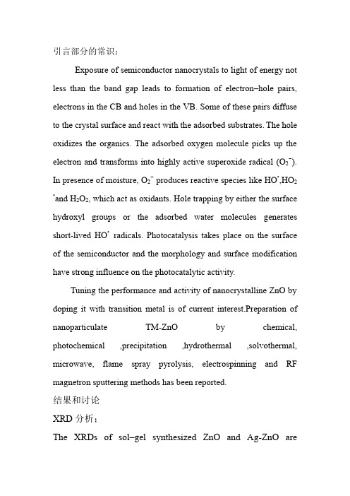
引言部分的常识:Exposure of semiconductor nanocrystals to light of energy not less than the band gap leads to formation of electron–hole pairs, electrons in the CB and holes in the VB.Some of these pairs diffuse to the crystal surface and react with the adsorbed substrates.The hole oxidizes the organics. The adsorbed oxygen molecule picks up the electron and transforms into highly active superoxide radical (O2-). In presence ofmoisture, O2-produces reactivespecieslike HO•,HO2•and H2O2, whichactasoxidants.Holetrapping by eitherthesurfacehydroxylgroupsortheadsorbed water moleculesgeneratesshort-livedHO•radicals. Photocatalysis takesplaceonthesurfaceofthesemiconductor andthemorphologyandsurfacemodificationhave strong influenceonthephotocatalyticactivity.Tuning the performance and activity of nanocrystallineZnO by doping it with transition metal is of current interest.Preparationof nanoparticulate TM-ZnO by chemical, photochemical ,precipitation ,hydrothermal ,solvothermal, microwave, flame spray pyrolysis, electrospinning and RF magnetron sputtering methods has been reported.结果和讨论XRD分析;The XRDs of sol–gel synthesized ZnO and Ag-ZnO are displayed in Fig.1. They reveal the crystal structures ofthe undopedaswellasthedopedoxidesasprimitive hexagonal withcrystalconstants a and b as 3.249Åand c as 5.205 Å.The diffractograms match with the standard JCPDS pattern of zincite (89–7102).Agcanbeincorporated intoZnOlatticeasasubstituentforZn2+or asan interstitial atom. IfthesilverissubstitutedforZn2+, a corresponding peakshiftwouldbeexpectedintheXRD. Lack of such shifts in the recorded XRD indicates the segregation of Ag particles in the grain boundaries of ZnO or only an insignificant quantity has been incorporated at the substitutional Zn site.the silver particles preferentially choose to segregate around the ZnO grain boundaries.Theaverage crystal sizesofthepreparedAg-ZnOandZnOhavebeen obtained fromthehalf-widthofthefullmaximum (HWFM) ofthemostintensepeaksoftherespective crystals usingtheScherrerequation,比较一下掺杂后的ZnO与纯净ZnO 的晶粒大小。
Sn掺杂ZnO纳米针的结构及其生长机制(英文)

S n掺 杂 Z O 纳米针 的结 构及 其 生长机 制 n
・
干
—
一
未
●、 、 、
庄 惠照
薛成 山
李俊林
20 1) 5 04
徐
鹏
( 山东师范大学半导体研究所, 济南
摘要 : 利用包括磁控溅射和热氧化 的两步法在 S(]) i 1衬底上制备 了 s 1 n掺杂 Z O纳米针. n 首先用磁 控溅 射法 在 S(l) i 1衬底上制备 S :n薄膜 , 1 nZ 然后在 6 0℃的 Ar 氛中对 薄膜进行热 氧化 , 5 气 制备 出 s n掺杂 Z O纳米针. n
m a ner n s te ng s se .S do e Zn n no e dls we e t e r g to put r y tm i n— p d O a n e e r h n g own b i p e t e a i to f t e a — y sm l r loxda n o s h m i h
fE , ihrs l i a s si l t nm coc p ( R E , n r y i es e - y(DX s et so y a d M)hg — ou o t m s ne c o irso y H T M)e eg s r v r E ) p c oc p ,n e t n r i o er n dp i X a r p oou n se c P )se toc p . h eut v a a eZ O a o e de o e t .% , tmi ai) h tlm e c n e( L p c so y T ers l r e t t n n n n e lsd p dwi 25 i r se l h t h h ao crt o
纳米氧化锌论文:纳米氧化锌对人冠状动脉内皮细胞的影响及其机制

纳米氧化锌论文:纳米氧化锌对人冠状动脉内皮细胞的影响及其机制【中文摘要】背景纳米氧化锌(Zinc Oxide Nanoparticles, ZnO-NPs)是目前世界上产量较高的纳米材料之一,可通过呼吸道吸入引起肺部炎症;此外,ZnO-NPs粒子还可能通过肺气血屏障进入血液循环,进一步穿透血脑、血眼及血睾等多种生物屏障系统,对身体主要脏器造成损害。
流行病学及毒理学研究发现ZnO-NPs可诱导人类心脏微血管内皮细胞渗透性改变及炎性反应,造成内皮细胞损伤,甚至动脉粥样硬化(Atherosclerosis, AS)。
ZnO-NPs对心血管内皮细胞损伤尤其是AS改变的作用机制目前还不清楚。
研究发现AS发生时血管内皮细胞凋亡增加,血红素氧合酶-1(Heme Oxygenase-1,HO-1)及血小板内皮细胞粘附分子-1(Platelet endothelial cell adhesion molecules-1, PECAM-1)表达均明显增加。
为此,该研究利用人肺泡上皮细胞A549和冠状动脉内皮细胞(Human Coronary Artery Endothelial Cell, HCAEC)共培养系统,模仿肺部气血屏障,探讨ZnO-NPs是否会引起HCAEC损伤及HO-1和PECAM-1表达水平的变化;然后分别用基因转录抑制剂放线菌素D(Actinomycin D, Act D)以及胞噬作用抑制剂细胞松弛素B(Cytochalasin B, CB)抑制A549细胞蛋白的合成及ZnO-NPs对HCAEC直接刺激,来抑制ZnO-NPs对HCAEC 的间接及直接作用。
初步探讨其对HCAEC的影响及其作用机制,为阐明ZnO-NPs颗粒的动脉内皮细胞毒性及其机制、制定有效的防治措施和卫生标准提供分子生物学依据。
利用A549/HCAEC共培养模型以及HCAEC直接作用模型,测定国产ZnO-NPs (30 nm)对HCAEC的损伤作用以及对细胞HO-1和PECAM-1蛋白表达的影响及其可能机制。
- 1、下载文档前请自行甄别文档内容的完整性,平台不提供额外的编辑、内容补充、找答案等附加服务。
- 2、"仅部分预览"的文档,不可在线预览部分如存在完整性等问题,可反馈申请退款(可完整预览的文档不适用该条件!)。
- 3、如文档侵犯您的权益,请联系客服反馈,我们会尽快为您处理(人工客服工作时间:9:00-18:30)。
Rapid synthesis of blue emitting ZnO nanoparticles forfluorescentapplicationsLeta T.Jule a,n,Francis B.Dejene a,Kittessa T.Roro b,Zelalem N.Urgessa c,Johannes R.Botha ca Physics Department,University of the Free State,Private Bag X13,Phuthaditjhaba9866,South Africab CSIR-Energy center,Council for Scientific and Industrial Research,PO Box395,Pretoria,South Africac Department of Physics,Nelson Mandela Metropolitan University,P.O.Box77000,Port Elizabeth6031,South Africaa r t i c l e i n f oArticle history:Received4May2016Received in revised form8June2016Accepted9June2016Available online11June2016Keywords:ZnOBlue emissionThermal decompositionZinc acetate dehydrateFluorescent applicationDonor–acceptor pair(DAP)a b s t r a c tZnO nanoparticles(NPs),with size∼16–20nm were produced using simple,cost effective and rapidsynthesis method.In this method zinc salt(typically zinc acetate dehydrate)is directly annealed in air ata temperature from°200C to°500C for2h to form ZnO(NPs).This synthesis method,only requires zincprecursor to produce NPs that can emit visible emission without external doping.X-ray diffraction(XRD)patterns confirm the prepared ZnO NPs is polycrystalline structure with wurtzite phase.The observedvariation in scanning electron microscopy(SEM)images showed spherical shape of the ZnO NPs.It wasfound that the NPs exhibited the estimated direct bandgap(E g)of3.28eV,3.29eV,3.33eV and3.39eVfor a decomposition temperature of500,400,300and°200C.Energy dispersive X-ray(EDX)analysisshowed that carbon is the only impurity at lower temperature which was most likely originated from theacetate group.The photoluminescence(PL)spectra of ZnO NPs showed the appearance of a blue emis-sion,attributed to Zn interstitials,whose intensity reduces with increase in decomposition temperatureand the underlying mechanism are discussed.For the samples prepared at°200C and300°C a tem-perature dependent PL was studied and found out that there are about three transition lines at∼3.01eV,∼3.21eV and∼3.33eV,which are ascribed to zinc vacancy(V zn),donor–acceptor pairs(DAP)and excitonsbound to structural defects respectively.It is hoped that ZnO NPs produced using this method would beideal for blue light emittingfluorescent application as it is catalyst free growth,uses simple equipment,less hazardous and easy to control particle size and morphologies by scalable temperature.&2016Elsevier B.V.All rights reserved.1.IntroductionSemiconductor nanostructures are promising candidates forfuture electronic and photonic devices.Nanostructures based onwide bandgap semiconductors such as GaN and ZnO are of parti-cular interest because of their applications in short wavelengthlight emitting devices andfield emission devices[1,2].ZnO is apotential competitor of GaN for blue and UV emission and char-acterized by direct band gap of3.37eV at room temperature withlarge exciton binding energy of60meV compared to GaN(28meV)[3–5].In addition to the UV emission ZnO is also known to emit in thevisible region[6].The photoluminescence spectrum of ZnO isnormally composed of two parts:excitonic near band edge emis-sion with energy around the band gap of ZnO and defect relateddeep level emission in the visible range.ZnO is n-type semi-conductor due to oxygen vacancies,impurities like Al,H and in-terstitial Zn ions which act as donors in ZnO lattice.These nativedefects are believed to be responsible for visible photo-luminescence.The UV emission is due to excitonic related re-combination[7,8].The exact mechanism for deep level emission isstill controversial although intrinsic defects such as oxygen va-cancies,oxygen interstitial,zinc vacancies and extrinsic impuritiesare all considered as a possible origin[9–12].However,under-standing the origin of photoluminescence in ZnO nanoparticlesand improving the emission efficiency is still a major challenge.Synthesis and characterization of zinc oxide(ZnO)nano-particles has found widespread interest during past few years dueto their unique electro optical properties,which can be employedin devices such as ultraviolet(UV)light-emitting diodes(LEDs)and blue luminescent devices[13].Searching of new methodology to synthesize ZnO NPs with auniform morphology,size and reproducibility is of greatContents lists available at ScienceDirectjournal homepage:/locate/physbPhysica B/10.1016/j.physb.2016.06.0080921-4526/&2016Elsevier B.V.All rightsreserved.n Corresponding author.E-mail address:JuleLT@ufs.ac.za(L.T.Jule).Physica B497(2016)71–77importance both for fundamental studies and practicaltion.In the past,several crystal-growth technology, explored,among which are sol-gel,spray pyrolysis,por transport,vapor-phase growth,chemical bath (CBD),direct precipitation methods and hydrothermalwhich also had the additional motivation of dopingeffort to obtain p-type material[1,14,15].However,theseinvolve a strictly controlled synthesis environment, procedures,high-temperature synthesis processes and equipment.Despite extensive research over the pastsome fundamental properties of theluminescence in the ZnO synthesized by thermalare not fully understood and rarely reported.In this paper,we therefore report on ZnO NPs thatvisible region without the requirement of additional temperature-dependent PL for samples of ZnOtemperature of°200C and°300C were discussed withderlying mechanism.Moreover,the structural and optical prop-erties of ZnO NPs synthesized via thermal decomposition method has been studied.2.Experiment2.1.Sample preparationThe zinc acetate dihydrate(Zn(CH3COOH)2Á2H2O))>99%,purity purchased from Sigma Aldrich,were used as precursor without further purification to prepare ZnO NPs.In order to determine possible decomposition temperature of zinc acetate dihydrate into ZnO NPs,samples were annealed at various temperature for2h. Four sets of5g of Zn(CH3COOH)2Á2H2O was put into sample holders,the crucible and annealed at a temperature of200,300, 400and°500C for2h in muffle furnace in air to produce the ZnO NPs.2.2.CharacterizationsThe crystal structures of the samples were determined with a Bruker AXS D8ADVANCE Discover diffractometer(XRD),with Cu Ka(1.5418)radiation.Surface morphologies and elemental com-positions were studied using a scanning electron microscope(SEM) (Shimadzu model ZU SSX-550Superscan)equipped with EDX.The optical absorption measurements were carried out in the200–600nm wavelength range using a Perkin Elmer UV/Vis Lambda20 powder Spectrophotometer.Luminescence measurements were done using a photoluminescent(PL)laser system 5.0(Photon systems,USA),which uses a He–Cd laser with an excitation wa-velength of248.6nm.3.Results and discussion3.1.Structural analysisFig.1shows the XRD pattern of ZnO NPs synthesized by ther-mal decomposition method for various annealing temperatures. XRD pattern of ZnO NPs exhibits various peaks which could be indexed according to ZnO diffraction peak(JCPDS card no.36-1451).The presence of various diffraction peaks reveals hexagonal wurtzite phase of ZnO which suggests polycrystalline nature of ZnO.The diffraction peaks at scattering angleθ()2of31.74°,34.43°, 36.25°and47.5°belongs to100,002,101and102diffraction planes from ZnO,and the other peak marked with asterisks* corresponds to zinc acetate.It can be clearly seen that by increasing the growth temperature from°200C to°500C ZnO peaks became prominent and prevailed.No other impurities are observed except for zinc acetate related peaks in the samples prepared at lower temperature,but fully decomposes for the higher temperature.The estimated lattice constants slightly in-creased as the decomposition temperature increased from°200C to°500C as summarized in Table1.The increase of the lattice parameter of ZnO NPs with increase in temperature was calculated using the equation:=+()⎛⎝⎜⎜⎞⎠⎟⎟d a c143111 101222where d is the interplanar distance,a and c are the lattice parameters.Fig.2shows the variation of FWHM as measured from dif-fraction plane101and particle size of ZnO NPs synthesized at various decomposition temperatures for annealing time of2h.The FWHM,which is an indication of the crystalline quality of the prepared ZnO NPs,decreases significantly as the decomposition temperature increases.Fig.3depicts variation of lattice parameter, a and c with increasing annealing temperature respectively con-firming the increase in grain size at higher decomposition temperature.The average crystallite size of prepared ZnO NPs can be esti-mated from the FWHM of the101diffraction peak estimated using the Debye Scherrer's equation:λβθ=() D0.9cos2 where D is the crystallite size,λis the wavelength λ(=)0.15402nm of radiation used,θis the Bragg angle andβis the full-width at half-maximum measured in degrees.The estimated average particle sizes were16.3,17.8,19.6and19.7nm2degree))ΘFig.1.XRD pattern of ZnO nanoparticles synthesized at various temperatures for 2h.Table1Measured properties of ZnO nanoparticles at various temperature.Temp.(°C)Particle diameterR070.5(nm)Lattice parameter,a(Å)Lattice parameter,c(Å)20016.3 3.245 5.29930017.8 3.247 5.30240019.6 3.249 5.30550019.7 3.250 5.307L.T.Jule et al./Physica B497(2016)71–77 72corresponding to the 200,300,400and °500C decomposition temperatures.The estimated particle sizes increases with anneal-ing temperature and it has highest value at of °500C due to nar-rowing of the diffraction peak.According to Ostwald ripening the increase in the particle size is due to the merging of the smaller particles into larger ones as suggested by Nanda et al.[16]and is a result of potential energy difference between small and large particles and can occur through solid state diffusion.The intensity ratio,()()I I 002101of the peaks presenting ()002and (101)at °500C is equalto 0.53which is higher than the corresponding standard value of 0.44of bulk hexagonal wurtzite ZnO [17]suggesting the prepared ZnO structure is preferred the (002)orientation.3.2.Morphological analysisFig.4A –C illustrates the SEM images of ZnO NPs prepared at a temperature of 300,400and °500C respectively.The SEM images clearly indicate that the surface morphology of the ZnO NPs de-pending on the synthesis temperatures.It is interesting to observe the formation of spherical ZnO NPs which is fully noted when the decomposition temperature was increased to °500C (see the inset in Fig.4C).It is observed that as decomposition temperature increases,the structural defects(dis-location)for the ZnO NPs prepared at a temperature of °200C reduces as compared to °500C causing uniform ZnO NPs which are purely spherical in shape.Fig.4D –F shows chemical stoichiometryof the ZnO nanoparticles prepared at a temperature of 300,400and °500C .The results presented shows that the prepared mate-rial contains C,O and Zn elements.The decrease in the C con-centration with increasing synthesis temperature is suggested to be as a result of ef ficient evaporation of the acetate-group.4.UV –visible spectrophotometer analysisFig.5shows room-temperature UV –visible absorption spec-trum of ZnO nanoparticles synthesized for various decomposition temperatures.The prepared ZnO NPs have band edge absorption peaks at 362.2nm,389.4nm,429.72nm and 455.32nm for sam-ples prepared at a temperature of 200,300,400and °500C re-spectively.These absorption peaks conform to the well-known intrinsic band gap absorption of ZnO.The absorption edge around 389.4nm was assigned to intrinsic band-gap absorption of ZnO due to the electron transitions from the valence band to the con-duction band →O Zn p d 23[18]and the other possible explanation are given in terms of structural defects associated with Zn inter-stitial as reported in Refs.[6,19].It is observed from the spectra that the absorbance of the samples reduces slightly with increase in decomposition temperature and the absorption edge slightly shifts to lower energy.Furthermore this red shift indicated the shrinkage effect in the band gap energy as a function of tem-perature.They obtained ZnO which exhibited a high absorption band in the UV region λ(<)380nm .Fig.6represents the relationship αν()h 2versus the photon en-ergy ν()h from which the optical band gap of the nanoparticle was determined by extrapolating the linear part of the spectrum.The characteristic's change of the band gap with increase in size of the ZnO nanostructure has been studied by the observation of the red shift in photoluminescence peak position.Thus photo-luminescence is useful for the study of quantum con finement of electrons.The direct bandgap energy of the synthesized ZnO na-noparticles was estimated using Tauc's plots relation [20].ZnO nanoparticles synthesized have estimated band gap values of 3.28eV,3.29eV,3.33eV and 3.39eV for samples prepared at a temperature of 500,400,300and °200C respectively.Thus,the estimated band gap energy of the ZnO nanoparticles was found to decrease with the increase in the decomposition temperature and similar results were obtained by Kathalingam et al.[21].It is clear that when the particle size increases,the electronic states are not discrete as a result the band gap energy reduces and the oscillator strength decreases [5,22].5.Photoluminescence analysisFig.7shows the PL spectra of ZnO NPs prepared at various annealing temperatures.In the PL spectrum,four emission bands,including band edge emission at 398.3nm (3.11eV),402.8nm (3.07eV),406.9nm (3.04eV)and 409.6nm (3.02eV)for ZnO prepared at a temperature of 500,400,300and °200C respectively were observed.The visible emission in ZnO is due to different intrinsic defects such as oxygen vacancies (V o ),zinc vacancies (V Zn ),oxygen interstitials (O i ),zinc interstitials (Zn i )and oxygen antisites (O Zn )[7,23].Band edge emission centered at around 398.3nm should be attributed to the recombination of excitons and V Zn [24,25].However the origin of violet emissions centered at 3.07eV(402.79nm),406.9nm (3.04eV)and 409.6nm (3.02eV)are ascribed to an electron transition from a shallow donor level of neutral Zn i to the top level of the valence band [26,27,9].The broad deep level emission that started from UV to the visible region 360–480nm with the maximum peak at 409.64nm for the sample prepared at °200C was observed.The decrease in PL intensity withTemperature (C )P a r t i c l e s i z e (n m )F W H M ()Fig.2.Variation of FWHM of (101)X-ray diffraction peaks and estimated particle sizes plotted against decompositiontemperature.Temperature (0C )L a t t i c e p a r a m e t e r a ,(A n g .)L a t t i c e p a r a m e t e r c ,(A )Fig.3.Variation of lattice parameter,a and c as a function of temperature.L.T.Jule et al./Physica B 497(2016)71–7773increase in decomposition temperature can be attributed to for-mation of better ZnO stoichiometry and near surface band bending caused by surface impurities [15,28,29].In samples of ZnO pre-pared at higher decomposition temperatures,the increase in grain size will reduce the relative contribution from recombination near the grains,resulting in strongly reduced violet emission.On the other hand,as the decomposition temperature increases more and more ZnO NPs with less deep defects form and as the result the transition line for the emission will decrease causing the PL in-tensity to decrease [25].The other possible explanation can be given as the existence of high dislocation density (defects)at the lower decomposition temperature.The dislocation density (δ),which represents the amount of defects in the sample is de fined as the length of dislocation lines per unit volume of the crystal andisFig.4.SEM micrograph and EDX spectrum of ZnO nanoparticles at:(A)°300C .(B)°400C .(C)°500C for 2h,and D,E and F are the corresponding EDX spectra.L.T.Jule et al./Physica B 497(2016)71–7774calculated using the equation [23]:δ=()D 132where D is the crystallite size.Calculating the dislocation density (δ)of ZnO NPs synthesized at °200C and °500C using Table 1and Eq.(3)it has value of ×−25.76104()−nm 2and ×−37.63104()−nm 2respectively,suggesting high dislocation density at °200C and the lattice imperfection decrease with increase in particle size.Moreover,defect density decreases with synthesis temperature in the present study.For samples synthesized at low temperature,a number of lattice defects which can act as radiative recombination centers are suggested.The reduction of these defect related ra-diative centers after annealing at °500C in air is likely related to formation better stoichiometric ZnO NPs.This hypothesis can be supported by the fact that surface defects strongly depend on morphology (SEM),with suppression of the emission as discussed under PL section.6.Temperature dependent PLRelative changes in state population with temperature provide evidence that PL peaks originate in the same part of the sample and that carriers are free to move between the available states.This feature can be useful because thermal quenching can hide sparse low-energy states and thermal broadening can obscure important details in the spectrum,low temperature PL experi-ments were conducted for samples prepared at 200and °300C .Fig.8and 9A depicts the temperature dependent PL spectra of synthesized ZnO NPs prepared at °200C and °300C respectively studied at a temperature of 300K,273K,173K,123K and 73K.The broadness and increase in PL emission intensity were ob-served with decreasing temperature as shown in Fig.8.It is well known that the donor –acceptor pair transition energy decreases along with the band gap energy when the temperature is in-creased causing the PL intensity to decrease.The experimental data for the temperature dependence of PL band intensity can be fitted by the following expression [14]:()=+()−βI T I Ae 14E 0a where I o is the peak intensity at temperature T ¼0K,A is assumed to be constant,E a is the activation energy of the thermal quenching process,and Kb is the Boltzmann's constant.Fig.9B is a plot of the integrated intensity of the 3.21eV transition versus reciprocal temperature for the ZnO NPs prepared at a temperature of °300C in the luminescence as one step quenching process andA b s o r b a n c e (a r b .u n i t s )Wavelength (nm)Fig.5.UV –vis absorbance spectra of ZnO nanoparticles synthesized at different annealing temperatures.(h v 101(e V /c m hv (eV))αFig.6.The optical absorption energy band gap estimated using Tauc's plot relationfor ZnO nanoparticles synthesized at different annealing temperatures.P L i n t e n s i t y (a r b .u n i t s )Wavelength (nm)Fig.7.PL emission of ZnO nanoparticles synthesized at various temperatures.P L I n t e n s i t y (a r b .u n i t s )Energy (eV)Fig.8.Temperature dependent PL emission of ZnO nanoparticles prepared at °200C .L.T.Jule et al./Physica B 497(2016)71–7775fitted by the Eq.(4).The best fit,demonstrated by the solid curve has been achieved with parameters =I 10200,=E 12.9meV a .A range of experimental values have been reported for the quench-ing of the dominant bound exciton for ZnO nanorods as 13.2meV and 13.1meV for bulk ZnO [11,25,26].Haynes'empirical rule E a ¼a þb E D can be used to calculate thedonor binding energy E D ,using the value obtained for =E 12.9meV a .Different values have been reported for the con-stants a and b in the above relation.In our opinion,the most ac-curate values have been reported by Meyer et al.[11],namely =−a 3.8meV and b ¼ing these values,E D was cal-culated to be 45.8meV.This agrees with the value reported in Ref.[25].In the prepared ZnO NPs,it seems entirely plausible that hydrogen (from precursor)is the dominant donor.Fig.8shows temperature-dependent PL for ZnO NPs prepared at a temperature of °200C ,for which five distinct deep-level emission peaks at 3.01eV,3.03eV,3.04eV,3.08eV and 3.16eV were observed which was ascribed to native defects.The tem-perature-dependent PL spectra shown in Fig.9A for sample of ZnO NPs prepared at °300C clearly depicts thermal redistribution among states compared to the ZnO sample prepared at °200C .The transition line at 3.01eV were attributed to native defect (V zn )and the near-band-edge (NBE)emission peak at 3.21eV were assigned to DAP.The peak labeled in 3.21eV gains strength with decreasing temperature,in contrast,peak labeled 3.01eV is strong at low temperature because carriers are trapped at these sites and do not have enough thermal energy to escape,but it disappears at room temperature(300K)because the states are sparse relative to the intrinsic bands.With increasing temperature,the DAP emission energy was shifted to the low energy side because carriers on DAP with small donor –acceptor distance are released into the band [14,18].The transition at 3.33eV is ascribed to excitons bound to structural defects.This transition line at 3.33eV was previously observed in various ZnO samples and tentatively ascribed to donor bound excitons (DX),acceptor bound excitons,transitions of in-trinsic point defects and excitons bound to extended structural defects.The most detail study of this transition was reported by Yalishev et al.[12]and Wagner et al.[3].In their work,the 3.33eV emission line was attributed to recombination of excitons bound to extended structural donor defect complexes which disappear at temperature of 10K.Study done by Urgessa et al.[26]on ZnO nanorods growth for temperature dependent PL also observe dominance of donor-bound exciton as possible reason for this emission.Fig.10illustrates the Commission Internationale de l'Eclairage (CIE)chromaticity diagram of ZnO NPs prepared at various growth temperatures calculated using photoluminescence data and color calculator software.The coordinates were shown deep blue region with the increase in annealing temperature indicating growth temperature play a major role in tuning the emission color of the ZnO NPs.7.ConclusionIn conclusion,the luminescent properties of ZnO NPs prepared from zinc precursor at various decomposition temperatures have been investigated.The samples of ZnO NPs synthesized at a tem-perature from 200–500°C exhibits broad visible emission.ZnO NPs with crystallite size of 16–20nm and hexagonal wurtzite structure was successfully produced.A linear increase in lattice parameters and grain size with temperature was observed.It was demonstrated that the morphology and band gap energy of ZnO NPs can be tuned with annealing temperature.The temperature dependent PL spectra of ZnO NPs prepared at a temperature of °300C shows three transition lines at 3.01eV,3.21eV and 3.33eV which are ascribed to zinc vacancy (V zn ),donor –acceptor pairs (DAP)and excitons bound to structural defects respectively.The activation and binding energy for the transition at 3.21eV were calculated to be about 12.9meV and 45.8meV respectively.De-pending on the hydrogen concentration in the precursor and these energy values,hydrogen was suggested to be the possible impurityP L I n t e n s i t y (a r b .un i t s )Energy (eV)AL o g i n t e g r a t e d i n t e n s i t y (a r b .u n i t s )100/T (K -1)Fig.9.(A).Temperature dependent PL emission of ZnO nanoparticles prepared at °300C and (B).shows thermally activated luminescence quenching of the 3.21eV emission for the ZnO NPs prepared at a temperature of °300C .Fig.10.CIE diagram for temperature dependent PL sample and ZnO prepared at various measuring temperature.L.T.Jule et al./Physica B 497(2016)71–7776acting as donor in our material.It is believed that ZnO NPs pro-duced using this method would be quick and cost effective synthesis method for blue light emittingfluorescent applications. AcknowledgmentsThis work was supportedfinancially by University of the Free State through directorate of research and the authors kindly ac-knowledge University of the Free State,Physics department for the characterization of the samples.References[1]Y.U.Ozgur,Y.I.Alivov,C.Liu,A.Teke,M.A.Reshchikov,S.Doan,V.Avrutin,S.-J.Cho,H.Morko,J.Appl.Phys.98(4)(2005)041301,/10.1063/1.1992666.[2]Y.G.Wang,u,H.W.Lee,S.F.Yu,B.K.Tay,X.H.Zhang,H.H.Hng,J.Appl.Phys.94(2003)354–358,/10.1063/1.1577819.[3]M.R.Wagner,G.Callsen,J.S.Reparaz,J.-H.Schulze,R.Kirste,M.Cobet,I.A.Ostapenko,S.Rodt,C.Nenstiel,M.Kaiser,A.Hoffmann,A.V.Rodina,M.R.Phillips,utenschläger,S.Eisermann,B.K.Meyer,Phys.Rev.B84(2011) 035313,/10.1103/PhysRevB.84.035313.[4]K.Rainey,J.Chess,J.Eixenberger,D.A.Tenne,C.B.Hanna,A.Punnoose,J.Appl.Phys.115(2014)17727,/10.1063/1.4867596.[5]L.Koao,F.Dejene,H.Swart,Mater.Sci.Semicond.Process.27(2014)33–40,/10.1016/j.mssp.2014.06.009.[6]T.Malevu,R.Ocaya,Int.J.Electrochem.Sci.10(2015)1752–1761〈www.elec〉.[7]K.Suzuki,M.Inoguchi,K.Fujita,S.Murai,K.Tanaka,N.Tanaka,A.Ando,H.Takagi,J.Appl.Phys.107(12)(2010)124311,/10.1063/1.3425783.[8]S.Lin,H.He,Z.Ye,B.Zhao,J.Huang,J.Appl.Phys.104(11)(2008)114307,http:///10.1063/1.3033560.[9]C.H.Chia,i,T.C.Han,J.W.Chiou,Y.M.Hu,W.C.Chou,Appl.Phys.Lett.96(8)(2010)081903,/10.1063/1.3327338.[10]B.Madavali,H.-S.Kim,S.-J.Hong,Mater.Lett.132(2014)342–345,http://dx./10.1016/j.matlet.2014.06.111.[11]B.K.Meyer,H.Alves,D.M.Hofmann,W.Kriegseis,D.Forster,F.Bertram,J.Christen,A.Hoffmann,M.Straburg,M.Dworzak,U.Haboeck,A.V.Rodina,Phys.Status Solidi B241(2)(2004)231–260,/10.1002/pssb.200301962.[12]V.S.Yalishev,Y.S.Kim,X.L.Deng,B.H.Park,S.U.Yuldashev,J.Appl.Phys.112(1)(2012)013528,/10.1063/1.4733952.[13]D.C.Look,E.R.Heller,Y.-F.Yao,C.C.Yang,Appl.Phys.Lett.106(15)(2015)152102,/10.1063/1.4917561.[14]W.S.H.Y.H.P.Y.G.K.S.K.M.S.K.J.H.J.J.L.Y.K.Giwoong Nam,Sang-heon Lee,J.-Y.Leem,Bull.Korean Chem.Soc.34(15)(2013)95–98,/10.5012/bkcs.2013.34.1.95.[15]X.Fan,J.Lian,L.Zhao,Y.Liu,Appl.Surf.Sci.252(2)(2005)420–424,http://dx./10.1016/j.apsusc.2005.01.018.[16]K.K.Nanda,F.E.Kruis,H.Fissan,Evaporation of free pbs nanoparticles:evi-dence of the kelvin effect,Phys.Rev.Lett.89(2002)256103,/10.1103/PhysRevLett.89.256103.[17]F.V.Molefe,L.F.Koao,B.F.Dejene,H.C.Swart,Opt.Mater.46(2015)292–298,/10.1016/j.optmat.2015.04.034.[18]B.K.Meyer,J.Sann,S.Eisermann,utenschlaeger,M.R.Wagner,M.Kaiser,G.Callsen,J.S.Reparaz,A.Hoffmann,Phys.Rev.B82(2010)115207,http://dx./10.1103/PhysRevB.82.115207.[19]J.Tao,W.Gong,Z.Yan,D.Duan,Y.Zeng,J.Wang,Mater.Sci.Semicond.Process.27(2014)452–460,/10.1016/j.mssp.2014.07.026.[20]J.Tauc,R.Grigorovici,A.Vancu,Phys.Status Solidi B15(2)(1966)627–637,/10.1002/pssb.19660150224.[21]M.K.J.E.Y.C.J.R.A.Kathalingam,N.Ambika,Mater.Sci.Pol.32(2014)555–564,/10.2478/s13536-014-0227-8.[22]A.K.Kole,P.Kumbhakar,T.Ganguly,J.Appl.Phys.115(22)(2014)224306,/10.1063/1.4883244.[23]F.L.R.H.W.F.H.Q.X.C.K.C.Saleem,M.,Intl.J.Phy.Sci.,vol.7,2012,pp.2971–2979./10.5897/IJPS12.219.[24]K.Vanheusden,W.L.Warren,C.H.Seager,D.R.Tallant,J.A.Voigt,B.E.Gnade,J.Appl.Phys.79(10)(1996)7983–7990,/10.1063/1.362349. [25]V.Khranovskyy,R.Yakimova,F.Karlsson,A.S.Syed,P.-O.Holtz,Z.N.Urgessa,O.S.Oluwafemi,J.R.Botha,Phys.B:Condens.Matter407(10)(2012)1538–1542, /10.1016/j.physb.2011.09.080.[26]Z.N.Urgessa,J.R.Botha,M.O.Eriksson,C.M.Mbulanga,S.R.Dobson,S.R.TankioDjiokap,K.F.Karlsson,V.Khranovskyy,R.Yakimova,P.-O.Holtz,J.Appl.Phys.116(12)(2014)123506,/10.1063/1.4896488.[27]V.A.Fonoberov,K.A.Alim,A.A.Balandin,F.Xiu,J.Liu,Phys.Rev.B73(2006)165317,/10.1103/PhysRevB.73.165317.[28]M.Schirra,R.Schneider,A.Reiser,G.M.Prinz,M.Feneberg,J.Biskupek,U.Kaiser,C.E.Krill,K.Thonke,R.Sauer,Phys.Rev.B77(2008)125215,http: ///10.1103/PhysRevB.77.125215.[29]K.T.Roro,J.K.Dangbegnon,S.Sivaraya,A.W.R.Leitch,J.R.Botha,J.Appl.Phys.103(5)(2008)053516,/10.1063/1.2873872.L.T.Jule et al./Physica B497(2016)71–7777。
