Indirect ELISA 1
间接elisa原理

间接elisa原理一、引言酶联免疫吸附法(Enzyme-Linked Immunosorbent Assay,简称ELISA)是一种广泛应用于生物学、医学等领域的分析方法。
其中,间接ELISA是其中一种常用的方法。
它通过特定的抗体与抗原相互作用,利用酶做标记来检测样品中是否存在目标物质。
本文将详细介绍间接ELISA的原理。
二、实验步骤1.涂布:将待测物质(如蛋白质)溶液加入到微孔板中,使其在孔壁上均匀地吸附。
2.阻断:在孔壁上添加非特异性蛋白(如牛血清白蛋白),以避免后续步骤中非特异性结合。
3.加入第一层抗体:加入与待测物质相对应的第一层抗体,并与待测物质结合形成复合物。
4.加入第二层抗体:加入与第一层抗体相对应的酶标记二抗,并与第一层抗体结合形成复合物。
5.加入底物:加入底物,使酶催化反应产生可检测的信号。
6.测定:利用光密度计等设备测定底物反应产生的吸光度,从而判断待测物质是否存在。
三、原理解析1.抗原-抗体反应间接ELISA是基于抗原-抗体反应的原理。
在实验中,待测物质(如蛋白质)被吸附到微孔板上,并与第一层特异性抗体结合形成复合物。
此时,如果样品中存在与待测物质相对应的第二层抗体,则第二层抗体会与复合物结合形成更大的复合物。
这种特异性的结合反应是ELISA检测方法的核心。
2.酶标记在间接ELISA中,酶标记是检测结果可视化的关键。
在实验中,第二层抗体被标记上酶(如辣根过氧化物酶),当底物被加入时,酶催化底物产生可检测的信号(如荧光、发光、颜色变化等)。
这种信号可以通过吸光度计等仪器来检测和量化。
3.非特异性结合在实验中,非特异性结合是需要避免的。
为了避免非特异性结合,实验中会加入一定量的阻断剂(如牛血清白蛋白),以防止非特异性蛋白质与抗体结合。
4.优缺点间接ELISA具有以下优点:(1)灵敏度高:ELISA可以检测极低浓度的物质,通常可以检测到纳克/毫升级别的物质。
(2)特异性强:ELISA可以区分不同种类的物质,因为它是基于抗原-抗体反应的原理。
间接竞争elisa实验原理
间接竞争elisa实验原理
ELISA(enzyme-linked immunosorbent assay)实验原理是生物分析技术中最常用的发光免疫检测技术,它主要采用发光双抗原技术,来测定酶标物质或抗原抗体,它将酶作为检测和显示
发光反应的桥梁,组成一个特殊的免疫系统,其基本思想是利用
活性试剂将目标物质与酶或抗原抗体结合,然后再经由发光催化反应检测。
ELISA实验于1970年由Engvall 及 Perlman 提出,其用抗原抗体及酶组成一个特殊的免疫系统,被检测的物质(抗原或抗体)结
合在特殊酶上,发光试剂(如酵素标记物、二抗原、发光试剂等)连称后,在受体结合后诱导发光,由发光信号最终获得定量结果。
ELISA实验一般用于抗原、抗体的定量检测,具有可灵敏度高、
响应时间短、占用空间小、工作人员较少等优点,且分子量大小
的测定范围较广,可应用于血清学、免疫学、病毒学、细胞生理
学和药物代谢等多个领域。
由于采用的是一种直接的试验方法,
可以迅速测量出抗原或抗体的含量,优于间接酶连结免疫实验方法,尤其适合于临床检测领域。
简述间接elisa法的基本原理

简述间接elisa法的基本原理
间接酶联免疫吸附试验(indirect enzyme-linked immunosorbent assay,简称indirect ELISA)是一种常用的免疫学检测技术,广泛应用于医学、生物学、农业、环境等领域。
本文将简述间接ELISA法的基本原理。
基本原理
间接ELISA法是利用酶标记的二抗来检测样品中的特定抗原或抗体。
其基本原理如下:
1. 将要测定的抗原或抗体与特异性一抗结合,形成免疫复合物。
2. 将未结合的一抗洗脱,将酶标记的二抗加入,与免疫复合物结合。
3. 加入底物,使酶标记的二抗发生化学反应,生成可测量的产物。
4. 通过检测产物的光学密度,来确定样品中的特定抗原或抗体的浓度。
优点和缺点
间接ELISA法的优点是灵敏度高、特异性强、操作简单、成本低、适用范围广。
其缺点是需要进行多次洗涤,操作时间较长,对于复杂的样品矩阵,可能会产生假阳性或假阴性的结果。
应用领域
间接ELISA法广泛应用于血清学检测、病原体检测、免疫学研究、药物研发等领域。
例如,在临床应用中,间接ELISA法可以用于检测病毒、细菌、真菌等感染性疾病的诊断;在药物研发中,可以用于筛选药物的效力和剂量。
总之,间接ELISA法是一种简单、快捷、敏感、特异的免疫学检测技术,对于研究各种生物分子的表达、诊断疾病、药物研发等领域具有重要意义。
ELISA六种方法类型及原理

ELISA六种方法类型及原理ELISA(酶联免疫吸附测定)是一种常用的实验技术,用于测定样品中特定抗原或抗体的存在和浓度。
它的原理基于抗原和抗体之间的专一结合,利用酶标记的抗体或抗原来检测并定量目标物。
ELISA有多种不同的方法类型,以下将对其中六种方法类型及其原理进行详细介绍。
1.直接ELISA:直接ELISA是最简单和最常用的ELISA方法,适用于寻找目标抗原。
在这种方法中,被测抗原直接吸附在固相或表面上,然后与特异性酶标记的抗原特异性结合。
最后,通过酶标记的底物的反应来定量测定目标物的浓度。
2.间接ELISA:间接ELISA也是常用的方法,适用于寻找目标抗体。
首先,将被测抗原吸附在固相或表面上,然后加入待测抗体。
之后,特异性结合的第二抗体(酶标记的)被加入用于识别和检测第一抗体。
最后通过酶标记的底物的反应来定量测定目标物的浓度。
3.竞争ELISA:竞争ELISA用于检测样品中的特定抗原或抗体。
在这种方法中,特异性酶标记的抗原或抗体与待测样品中的抗原或抗体竞争结合。
通过测定酶标记物的信号强度,可以确定待测样品中目标物的含量。
4.间接竞争ELISA:间接竞争ELISA是一种用于定量测定目标抗原的方法。
首先,在固相或表面上吸附被测抗原,然后加入特异性抗体。
该抗体与样品中的目标物竞争结合。
接着,再加入另一特异性抗体,该抗体与前面结合的抗体有竞争关系。
最后通过测定酶标记物的信号强度,可以确定目标物的浓度。
5.间接夹心ELISA:间接夹心ELISA用于寻找样品中的特定抗体。
首先,在固相或表面上吸附被测抗原,然后加入待测抗体。
随后,特异性酶标记的第二抗体被加入,用于识别和检测待测抗体。
最后通过测定酶标记物的信号强度,可以定量测定目标抗体的浓度。
6.双抗体ELISA:双抗体ELISA常用于寻找特定抗原。
首先,在固相或表面上吸附被测特异性抗体,然后加入样品。
目标抗原与抗体特异性结合。
接着,酶标记的第二抗体被加入,该抗体与目标抗原结合。
间接法Elisa
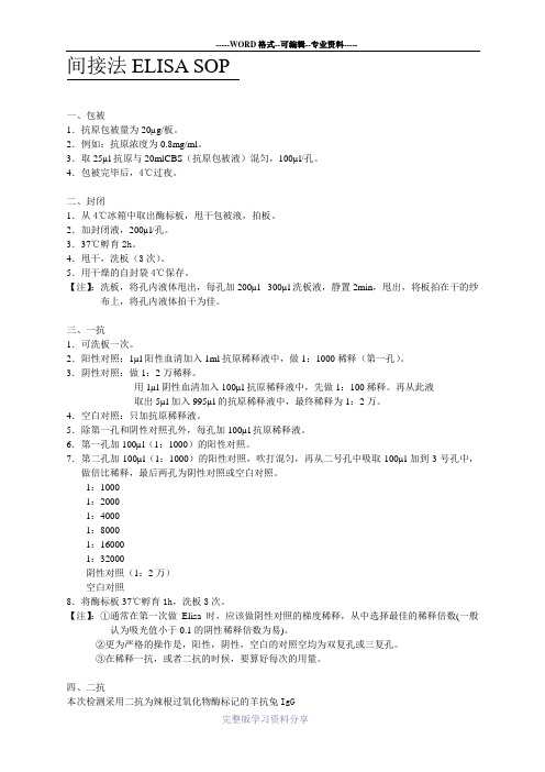
间接法ELISA SOP一、包被1.抗原包被量为20µg/板。
2.例如:抗原浓度为0.8mg/ml。
3.取25µl抗原与20mlCBS(抗原包被液)混匀,100µl/孔。
4.包被完毕后,4℃过夜。
二、封闭1.从4℃冰箱中取出酶标板,甩干包被液,拍板。
2.加封闭液,200µl/孔。
3.37℃孵育2h。
4.甩干,洗板(3次)。
5.用干燥的自封袋4℃保存。
【注】:洗板,将孔内液体甩出,每孔加200µl -300µl洗板液,静置2min,甩出,将板拍在干的纱布上,将孔内液体拍干为佳。
三、一抗1.可洗板一次。
2.阳性对照:1µl阳性血清加入1ml抗原稀释液中,做1:1000稀释(第一孔)。
3.阴性对照:做1:2万稀释。
用1µl阴性血清加入100µl抗原稀释液中,先做1:100稀释。
再从此液取出5µl加入995µl的抗原稀释液中,最终稀释为1:2万。
4.空白对照:只加抗原稀释液。
5.除第一孔和阴性对照孔外,每孔加100µl抗原稀释液。
6.第一孔加100µl(1:1000)的阳性对照。
7.第二孔加100µl(1:1000)的阳性对照,吹打混匀,再从二号孔中吸取100µl加到3号孔中,做倍比稀释,最后两孔为阴性对照或空白对照。
1:10001:20001:40001:80001:160001:32000阴性对照(1:2万)空白对照8.将酶标板37℃孵育1h,洗板3次。
【注】:①通常在第一次做Elisa时,应该做阴性对照的梯度稀释,从中选择最佳的稀释倍数(一般认为吸光值小于0.1的阴性稀释倍数为易)。
②更为严格的操作是,阳性,阴性,空白的对照空均为双复孔或三复孔。
③在稀释一抗,或者二抗的时候,要算好每次的用量。
四、二抗本次检测采用二抗为辣根过氧化物酶标记的羊抗兔IgG1.用0.01MPBS做1:5000稀释(现用现配)。
不同品牌ELISA试剂盒对口蹄疫抗体检测的试验

不同品牌 ELISA试剂盒对口蹄疫抗体检测的试验摘要:用不同品牌的口蹄疫O型ELISA试剂盒,对免疫口蹄疫疫苗后的猪群进行口蹄疫抗体检测,发现检测结果阳性率各不相同。
结果表明不同品牌试剂盒因制备方法、包被抗原不同对猪群的抗体检测结果不同,所以,建议在实践中用口蹄疫ELISA试剂盒检测抗体效价应根据情况有选择使用。
关键词:试剂盒;抗体效价检测;阳性率;有选择使用;口蹄疫(Foot-and-Mouth Disease,FMD)是由口蹄疫病毒(Foot-and-mouth disease virus)引起的偶蹄动物烈性传染病,是世界卫生组织(OIE)规定必报的烈性传染病,我国将其排在一类传染病之首。
口蹄疫病毒共有七个血清型,分别为O、A、C、Asia1、SAT1、SAT2、SAT3型。
每个血清型又有许多不同的亚型,各血清型之间没有交叉保护,同一血清型的各亚型之间仅有部分交叉保护,这使得对口蹄疫的防控更加困难。
目前我国主要流行O型和A型口蹄疫。
目前,我国防控口蹄疫主要采取疫苗免疫与扑杀相结合的综合防治措施,对所有易感动物猪、牛、羊进行口蹄疫灭活疫苗免疫接种。
但是在给动物注射疫苗后能否预防口蹄疫的发生一直的广大养殖户迫切需要知道的问题。
所以免疫后的效果评价就显得特别重要。
目前主要有以下几种检测方法:1.免疫动物攻毒实验;这一方法是判断疫苗免疫效果的金标准,但因需要在生物安全三级实验室内进行,一般养殖企业和研究单位都没法实施。
2.免疫动物血清中和实验;这一方法相对准确些,但同样也受一定条件限制,一般的研究检测单位也不完全具备检测条件。
3.免疫动物血清ELISA抗体检测;这一方法的准确性比前两种方法相对较差,但也是一般检测单位都容易实施的检测方法,因此也被广泛用于疫苗免疫效果的评价。
但由于这一方法的存在的一定的缺陷,评价结果也一直存在争议。
本文所研究的就是使用第三种方法对疫苗免疫效果进行检测,根据检测结果进一步分析这一方法的准确性,对使用单位给予一些指导建议。
间接ELISA检测方法

间接ELISA检测⽅法ARTICLEDevelopment of an indirect ELISA based on a truncated S protein of the porcine epidemic diarrhea virusYufeng Li,Fangyuan Zheng,Baochao Fan,Hassan Mushtaq Muhammad,Yao Zou,and Ping JiangAbstract:Porcine epidemic diarrhea (PED)is a highly contagious,enteric disease of swine caused by the porcine epidemic diarrhea virus (PEDV).To ?nd a suitable ELISA method to assess the infection of PEDV and the effective-ness of vaccines,we developed and evaluated an indirect enzyme-linked immunosorbent assay (iELISA)based on a truncated recombinant spike (S)protein expressed in Escherichia coli .The parameters of the iELISA were opti-mized,and the cutoff value determined as 0.259by analyzing optical density (OD)values of 80PEDV negative sera con?rmed by westernblot.Repeatability tests revealed that the coef?cients of variation of positive sera within and between runs were both less than 10%.Cross-reactivity assays demonstrated that iELISA was PEDV-speci?c.A virus neutralization test with sera of 7different OD values showed a positive correlation between the OD values and virus neutralization.The results suggest this iELISA is speci?c,sensitive,and repeatable.Further studies should focus on the relationship between OD values of sera and its virus neutralization.Key words:indirect ELISA,PEDV,truncated S protein,virus neutralization.Résumé:La diarrhée épidémique porcine (DEP)est une maladie entérique du porc hautement contagieuse qui estcausée par le virus de la diarrhée épidémique porcine (PEDV).A?n de concevoir une méthode ELISA apte a`évaluer l’infection au PEDV et l’ef?cacitédes vaccins,on a élaboréet mis a`l’essai un test enzymatique indirect par immunoadsorption (iELISA)basésur une forme recombinante et tronquée de la protéine de spicule (S)expriméechez Escherichia coli .On a optimiséles paramètres de l’iELISA et établi la valeur seuil a`0,259en vertu d’une analyse des valeurs de DO de 80sérums a`PEDV négatif con?rmépar immunobuvardage de type western.Les tests de répétabilitéont révéléque les coef?cients de variation des sérums positifs intraessais et interessais se situaient tous deux sous la barre des 10%.Les essais de réactivitécroisée ont démontréque l’iELISA était spéci?que aux PEDV.Un test de neutralisation du virus d’après 7valeurs de DO a mis en évidence une corrélation positive entre les valeurs de DO et la neutralisation virale.Les résultats indiquent que cet iELISA est spéci?que,sensible etreproductible.Les analyses ultérieures devront s’attarder a`la relation entre les valeurs de DO des sérums et la neutralisation virale correspondante.[Traduit par la Rédaction]Mots-clés :ELISA indirect,PEDV,protéine S tronquée,neutralisation virale.IntroductionPorcine epidemic diarrhea virus (PEDV)is an envel-oped virus possessing an approximately 28kb,positive-sense,single-stranded RNA genome with a 5=cap and a 3=polyadenylated tail (Pensaert and Debouck 1978).The virus possesses glycosylated spike (S),Poll (P1),en-velope (E),glycosylated membrane (M),and an unglyco-sylated RNA-binding nucleocapsid (N)proteins (Song and Park 2012).The S protein of PEDV is a type I mem-brane glycoprotein composed of 1383amino acids and is predicted to contain a signal peptide (1–24aa),a large extracellular region,a single transmembrane domain (1334–1356aa),and a short cytoplasmic tail (Lee et al.2010).The S protein plays important roles in induction of neutralizing antibodies,speci?c receptor binding,and cell membrane fusion (Chang et al.2002;Sun et al.2008).The S1-GST protein (S1D,aa 636–789)is essential for in-duction of neutralizing antibodies (Sun et al.2007).The N-terminal region of the S protein is especially impor-tant for the receptor binding,and the 25–225aa region is indispensable to bind with amino peptidase N (receptor of PEDV)(Lee 2011).To date,most of the reported PEDV vaccines belong to attenuated vaccines and subunit vac-cines (Kweon et al.1999;Kang et al.2005;Song et al.2007;Received 8April 2015.Revision received 31July 2015.Accepted 4August 2015.Y.Li,F.Zheng,B.Fan,Y.Zou,and P.Jiang.Key Laboratory of Bacteriology,Ministry of Agriculture,College of Veterinary Medicine,Nanjing Agricultural University,Nanjing 210095,People’s Republic of China.H.M.Muhammad.Department of Epidemiology and Public Health,Faculty of Veterinary Science,University of Veterinary and Animal Sciences,Lahore,Pakistan.Correspondence author:Yufeng Li (e-mail:yufengli@/doc/597e2535d5bbfd0a785673aa.html ).811C a n . J . M i c r o b i o l .D o w n l o a d e d f r o m w w w .n r c r e s e a r c h p r e s s .c o m b y F o s h a n u n i v e r s i t y o n 11/03/15F o r p e r s o n a l u s e o n l y .Meng et al.2013).To date,M protein-based enzyme-linked immunosorbent assay (ELISA)and double anti-body sandwich ELISA have been developed to detect PEDV (Sozzi et al.2010;Ren etal.2011).N protein and viral protein have been used as coating antigens to estab-lish ELISA methods to detect antibodies against PEDV (Oh et al.2005;Hou et al.2007).Viral proteins have been used as a coating antigen of PEDV ELISA,and ELISA has been compared with virus neutralization.An overall agreement of 84.2%was generated between the serum neutralization and ELISA (Oh et al.2005).In the present study,an indirect ELISA (iELISA)based on a truncated recombinant protein S (tSc,N-terminal region 25–225aa of PEDV)was developed and assay conditions were optimized.The relationship between the iELISA results and virus neutralization titers was assessed.Materials and methodsSera and antibodyEighty-six sow sera were obtained from farms with the outbreaks of porcine epidemic diarrhea (PED)and health farms without clinical signs of PED.Among of them,6and 80sera were con?rmed as positive and negative,respectively,with western blot (WB)and immuno?uo-rescence (IFA)and were then stored at –80°C.The blood was drawn from the precaval vein of anesthetized pigs.Animal experiments were approved by the Institutional Animal Care and Ethics Committee of Nanjing Agricul-tural University (Approval No.IACECNAU20100902).Cloning and sequencing of the recombinant tSc protein geneThe tSc gene was ampli?ed from plasmids pFast-S (constructed by our laboratory)using a forward primer (5=-ATAGGATCCAGGTGCCAGTCTACT-3=)and a reverse primer (5=-GCGCTCGAGTTAAGGTTCATAGTAAAT-3=)(positions 20682–21284;DR13:GenBank accession No.JQ023162.1)containing restriction enzyme sites Bam HI and Xho I,underlined).The PCR product was cloned into the prokaryotic expression vector pET-28a(+)on corresponding restriction enzyme sites.This vector was designated as pET-28a/tSc,and its sequence was con-?rmed by sequencing analysis by Invitrogen (Shanghai,China).Expression and puri?cation of tSc proteinThe recombinant plasmid was transformed into Escherichia coli BL-21.The tSc protein was induced by addi-tion of 1mmol/L isopropyl-?-D -thiogalactopyranoside (IPTG).Four hours after IPTG induction,the cells were harvested and lysed by sonication.According to the man-ufacturer’s instructions,under the native condition,the recombinant protein was puri?ed by af?nity chro-matography with Ni-nitrilotriacetic acid agarose.The expressed protein was analyzed by sodium dodecyl sulfate –polyacrylamide gel electrophoresis (SDS–PAGE)with Coomassie brilliant blue R250and con?rmed by WBanalysis using His Tag monoclonal antibody (Boster,China)and PEDV polyclonal antibody prepared by immu-nizing rabbits with tSc protein that containing tSc.The puri?ed tSc protein was separated by SDS–PAGE using 12%separating gel and 5%stacking gel.Gels were stained with Coomassie brilliant blue R250,followed by immunoblotting.Brie?y,after transferring the proteins to nitrocellulose membrane,the membrane blocking was performed with 10%skimmed milk and incubated overnight at 4°C.The membranes were incubated with His Tag monoclonal antibody and PEDV polyclonal anti-body in blocking solution at 1:5000and 1:500dilution,respectively,for 2h at room temperature.After 1h of incubation,the membrane was extensively washed with PBS containing 0.05%Tween 20(PBST)and incubated for 45min with goat anti-mouse IgG (Boster,Wuhan).After washing,the membranes were incubated with horserad-ish peroxidase (HRP)-tagged goat anti-mouse IgG second-ary antibody (Boster,Wuhan),and the colorimetric reaction was developed using chemiluminescence lumi-nal reagents (Super Signal West Pico TrialKit,Pierce).iELISAA microtiter plate (96-well;Nunc,Nunclon,China)was coated with 100?L of tSc in 0.05mol/L bicarbonate–carbonate buffer (pH 9.6)and incubated for 2h at 37°C.Following 3washes with PBST,the plates were blocked for 2h at 37°C with 5%skimmed milk in PBST and then incubated for 1h at 37°C with 100?L serum samples in 5%skimmed milk in PBST.All samples were analyzed in triplicate.After5washes with PBST,the plates were fur-ther incubated with 100?L of HRP-conjugated protein A in PBST at 37°C.Then,the plates were washed again,and the colorimetric reaction was developed using 100?L of chromogenic substrates (0.4mol/L tetramethylbenzidine and 1mmol/L H 2O 2in 100mmol/L acetate buffer,pH 5.6)at 37°C.Color development was stopped with 2mol/L H 2SO 4,and the optical density at 450nm (OD 450)was recorded using an ELISA plate reader (BioTek,USA).Optimization of tSc iELISA working conditionsBased on the procedure described above,the optimal antigen concentration and sera dilutions were deter-mined through standard checkerboard titration proce-dures with triplicates.Brie?y,the tSc protein was coated on 96-well microtiter plates in serial 2-fold dilutions from 8?g to 250ng/well.Correspondingly,standard swine PEDV-positive serum and PEDV-negative control serum were also diluted in serial 2-fold dilutions from 1:25to 1:800for optimization.After the antigen and anti-serum dilutions were analyzed,HRP-labeled protein A was added to the plate with dilutions from 1:5000to1:20000to determine the optimal conjugate dilution.The conditions that gave the highest OD 450ratio between the positive and negative serum (P/N value)and an OD 450value for positive serum close to 1.0(with the OD 450value812Can.J.Microbiol.Vol.61,2015C a n . J . M i c r o b i o l .D o w n l o a d e d f r o m w w w .n r c r e s e a r c h p r e s s .c o m b y F o s h a n u n i v e r s i t y o n 11/03/15F o r p e r s o n a l u s e o n l y .of negative serum ≤0.2)were scored as optimal working conditions.After the conditions mentioned above were deter-mined,the coating conditions were optimized from at 37°C for 2h to 4°C for overnight.Then,the blocking buffers with 1%BSA,1%gelatin,5%and10%skimmed milk were used and blocked for 1,2,and 3h.The dilu-tions and incubation time of serum sample and HRP-conjugate staphylococal protein A were optimized with 15,30,45,60,90,or120min.The reactions were stopped and optimized by assessing 5,10,15,and 20min.Validation of tSc iELISATo establish a negative–positive cutoff value for this assay,80negative serum samples collected from healthy pig farms without clinical signs of PED (con?rmed by WB)were tested in duplicate using the tSc iELISA.The cutoff value of the ELISA was calculated from the mean OD values of the 80sera negative samples plus 2or 3standard deviations (SDs).This calculation gives 95%or 99%con? dence,respectively,that all negative values will fall within the de?ned range.To determine the speci?city of the tSc iELISA,positive sera against porcine circovirus type 2(PCV2),classical swine fever virus (CSFV),pseudorabies virus (PRV),foot-and-mouth disease virus (FMDV),porcine reproductive and respiratory syndrome virus (PRRSV),and transmissi-ble gastroenteritis virus (TGEV)were tested using the tSc iELISA protocol.Each sample was tested in triplicate,and the mean OD values were calculated.At the same time,6positive and another 30negative sera con?rmed by WB and IFA were detected by tSc iELISA to determine the sensitivity of this assay.Reproducibility experimentsEvaluation of assay reproducibility within and between runs was performed as previously proposed (Jacobson 1998).Six ?eld serum samples (5WB-positive samples and 1WB-negative sample)were selected for reproduc-ibility experiments.For intra-assay reproducibility,3replicates of each serum sample were assigned to the same plate.For inter-assay (between-run)reproducibil-ity,3replicates of each sample were run on different plates.The mean OD values,standard deviation (SD),and coef?cient of variation (CV)were calculated.Neutralization testVero-E6cells (1×10⁵cells/well)were seeded in a 96-well cell culture plate,and plates were incubated overnight at 37°C with 5%CO 2until a con?uent monolayer formed.Seven serum samples with different OD values (1.531,1.294,0.980,0.784,0.661,0.378,and 0.078)were heated at 56°C for 30min and used for testing.Sera were diluted 2-fold and incubated with 200TCID 50of strain DR13at 37°C for 1h.After incubation,100?L of the serum+virus mixture was transferred from each well of the incuba-tion plate to a 96-well cell culture plate containing Vero-E6cells.After 1h of incubation at 37°C,the serum+virus mixtures were replaced by complete Dul-becco’s Modi?ed Eagle Medium with 10%fetal calf se-rum.The plates were incubated at 37°C with 5%CO 2for 5days,and the numbers of wells with cells with cyto-pathic effect were counted under an inverted micro-scope and neutralization titers were evaluated.StatisticsPASW Statistics for Windows,Version 18(SPSS Inc.,Chicago,Illinois,USA),was used to analyze standard de-viation,CV,and correlation analysis.ResultsExpression,puri?cation,and identi?cation of tSc proteinSDS–PAGE and Coomassie brilliant blue staining dem-onstrated the successful expression of the tSc protein in E.coli (Fig.1a ).Compared with the expression of induced E.coli /pET-28a and E.coli culture,tSc with a molecular mass of approximately 28kDa was observed in induced E.coli /pET-28a-Sc (Fig.1a ).The tSc protein was expressed in large amounts and puri?ed using nickel af?nity chro-matography.SDS–PAGE indicated that the protein was of high purity (Fig.1a ).In addition,WB showed that the tSc protein could be detected with an anti-His antibody and PEDV-positive sera (Fig.1b ). Fig.1.Identi?cation of the truncated recombinant protein S (tSc)protein by SDS–PAGE (a )and western blot (b )by His tag monoclonal antibody and porcine epidemic diarrhea virus (PEDV)polyclonal antibody.(a )Lanes:M,unstained protein molecular mass marker;1,pET-28a-Sc induced with IPTG;2,pET-28a-Sc not induced with IPTG;3,supernatant of cell lysates;4,precipitation of cell lysates;5,puri?ed Sc protein.(b )Cell lysates were incubated with His tag monoclonal antibody (lanes 1and 2)and PEDV polyclonal antibody (lanes 3and 4).Lanes:1and 3,pET-28a-Sc induced with IPTG;2and 4,pET-28a-Sc not induced withIPTG.Li et al.813C a n . J . M i c r o b i o l .D o w n l o a d e d f r o m w w w .n r c r e s e a r c h p r e s s .c o m b y F o s h a n u n i v e r s i t y o n 11/03/15F o r p e r s o n a l u s e o n l y .Optimization of iELISA working conditionsUsing a checkerboard ELISA,the optimal antigen concentration and serum sample dilution were set at 1?g/well and 1:100,respectively,based on the following standards:the OD 450value of positive serum was close to 1.0,the OD 450ratio between positive and negative serum (P/N value)was highest,and the background was /doc/597e2535d5bbfd0a785673aa.html ing the same standards,the optimal blocking condi-tion was at 37°C for 2h,and the optimal dilution of the conjugate was de?ned as 1:20000.After the above-mentioned conditions were determined,it was found that 5%skimmed milk in PBST was the optimal block-ing buffer,and the best blocking time was 2h at 37°C.For the optimal exposure time of serum samples and conjugate,it was determined that incubations of 60and 45min for the serum samples and the conjugate,respectively,were suf?cient and time-saving in the tSc ELISA,as the P/N value was relatively stable and high.Finally,the optimal stopping time was determined to be 15min (Fig.2).Cutoff value of iELISAThe OD values of 80negative serum samples varied from a minimum of 0.052to a maximum of 0.201.Thus,samples with OD values of ≤0.223were considered neg-ative,and those with values of≥0.259were considered positive (Supplemental Table 11).After the cutoff was determined,the speci?city of the tSc iELISA was evaluated by testing the reactivity of antibodies against other porcine viruses.The OD values of standard positive sera againstPCV2,CSFV,PRV,FMDV,TGEV,and PRRSV are shown in Table 1.The OD values of all anti-sera were smaller than the cutoff value.These data revealed that there is no cross-reactivity between the PEDV tSc antigen and antibodies against other por-cine viruses,proving that the tSc antigen is speci?c for antibodies against PEDV.Diagnostic sensitivity tests showed that 6positive sera and another 30negative sera identi?ed by WB and IFA were still positive and negative in ELISA results.Repeatability of the tSc iELISAThe repeatability of tSc iELISA was determined by comparing the OD value of each ?eld serum sample.As shown in Table 2,the intra-assay CV of 5positive serum samples ranged from 0.019to0.094,whereas the inter-assay CV of these positive serum samples ranged be-tween 0.038and 0.088.These data indicate that the tSc iELISA is repeatable and yields low and acceptable varia-tion (DeSilva et al.2003).Neutralization testsThe results showed that 6positive sera had different levels of virus neutralization activity.Interestingly,OD values are positively correlated with virus neutralization activity (Fig.3).More positive sera are needed to further con?rm the relationship between OD values and virus neutralization titers.DiscussionIn China,PEDV was ?rst con?rmed in 1984,and since 2010,it has caused enteric disease with a devastat-ing impact on swine industry.This disease is character-ized by high morbidity and mortality in preweaning piglets,causing serious economic losses to the swine industry in China (Gao et al.2013;Li et al.2014).On the basis of the gene sequence of PEDV,many methods had been developed to detect the infection of PEDV,such as RT-PCR (Ishikawa et al.1997),duplex RT-PCR (Kim et al.2001),immunohistochemistry,in situ hy-bridization (Kim and Chae 2002),and RT-LAMP (Ren and Li 2011).Recombinant N protein had been used to establish an ELISA method to detect antibodies against PEDV (Hou et al.2007).To date,the recombi-nant S protein as a coating antigen for the ELISA method has not been reported.1Supplementary data are available with the article through the journal Web site at /doc/597e2535d5bbfd0a785673aa.html /doi/suppl/10.1139/cjm-2015-0213.Table 1.Speci?city of the truncated S pro-tein indirect ELISA to antibodies against some swine viruses.Virus antisera X ¯±SD PEDV1.037±0.012PRV 0.054±0.030PCV20.046±0.016FMDV 0.066±0.028PRRSV 0.055±0.035CSFV 0.047±0.023TGEV0.086±0.013Negative serum0.063±0.008Note:Data are the mean ±standard deviation of 3replicate measurements.PEDV,porcine epi-demic diarrhea virus;PRV,pseudorabies virus;PCV2,positive sera against porcine circovirus type2;FMDV,foot-and-mouth disease virus;PRRSV,porcine reproductive and respiratory syndrome vi-rus;CSFV,classical swine fever virus;TGEV,trans-missible gastroenteritis virus.Table 2.Intra-assay and inter-assay repeatability of the in-direct ELISA.Serum sample Intra-assay variability Inter-assay variability X¯±SD CV X¯±SD CV 1 1.095±0.0210.019 1.087±0.0510.0472 1.066±0.0770.0720.987±0.0560.0573 1.076±0.0750.070 1.100±0.0970.08840.955±0.0560.0590.912±0.0350.03850.730±0.0640.0880.690±0.0660.096Negative0.096±0.0090.0940.100±0.0090.090Note:Data are the mean ±standard deviation of 3replications.Li et al.815C a n . J . M i c r o b i o l .D o w n l o a d e d f r o m w w w .n r c r e s e a r c h p r e s s .c o m b y F o s h a n u n i v e r s i t y o n 11/03/15F o r p e r s o n a l u s e o n l y .Recent studies reported that the N-terminal region (25–225amino acids)of PEDV is associated with the binding of the virus to the receptor aminopeptidase N (Lee 2011).So,we postulated that antibody against this protein fragment may be associated with virus neu-tralization.In this study,the tSc protein (25–225ami-no acids)was expressed and an iELISA method was constructed.Cross-reactivity assays revealed that the tSc iELISA was PEDV-speci?c,and repeatability tests demonstrated that the assay is repeatable.Thus,the tSc iELISA is simpler to produce and perform,time-saving,and may be more suitable for large-scale surveys of PEDV /doc/597e2535d5bbfd0a785673aa.html bining virus neutral-ization results with 7different OD values sera,we ?nd that the OD values obtained by iELISA established by tSc are positively correlated with virus neutralization.The preliminary results showed that the sensitivity of this assay is 100%;however,more positive sera samples should be used to further con?rm the sensitivity.ConclusionThe tSc iELISA established in the present study was repeatable and speci?c for PEDV antibody detection,simple and economical to produce and perform,and time-saving.The present report may facilitate the devel-opment of a reliable tool for the large-scale detection of PEDV neutralization antibodies and assess the effective-ness of vaccines.AcknowledgementsWe thank Jiang Ping for helpful comments.The au-thors declare no potential con?icts of interest with re-spect to the research,authorship,and (or)publication of this article.This study was supported by public welfare grants from the Ministry of Agriculture,the People’s Re-public of China (201203039),and Priority Academic Pro-gram Development (PAPD)of Jiangsu Higher Education Institutions. ReferencesChang,S.H.,Bae,J.L.,Kang,T.J.,Kim,J.,Chung,G.H.,Lim,C.W.,et al.2002.Identi?cation of the epitope region capable of inducing neutralizing antibodies against the porcine epi-demic diarrheavirus.Mol.Cells,14(2):295–299.PMID:12442904.DeSilva,B.,Smith,W.,Weiner,R.,Kelley,M.,Smolec,J.,Lee,B.,et al.2003.Recommendations for the bioanalytical method validation of ligand-binding assays to support pharmacoki-netic assessments of macromolecules.Pharm.Res.20(11):1885–1900.doi:10.1023/B:PHAM.0000003390.51761.3d .PMID:14661937.Gao,Y.,Kou,Q.,Ge,X.,Zhou,L.,Guo,X.,and Yang,H.2013.Phylogenetic analysis of porcine epidemic diarrhea virus ?eld strains prevailing recently in China.Arch.Virol.158(3):711–715.doi:10.1007/s00705-012-1541-2.PMID:23151819.Hou,X.L.,Yu,L.Y.,and Liu,J.2007.Development and evaluation of enzyme-linked immunosorbent assay based on recombi-nant nucleocapsid protein for detection of porcine epidemic diarrhea (PEDV)antibodies.Vet.Microbiol.123(1–3):86–92.doi:10.1016/j.vetmic.2007.02.014.PMID:17368968.Ishikawa,K.,Sekiguchi,H.,Ogino,T.,and Suzuki,S.1997.Direct and rapid detection of porcine epidemic diarrhea virus by RT-PCR.J.Virol.Methods,69(1–2):191–195.doi:10.1016/S0166-0934(97)00157-2.PMID:9504764.Jacobson,R.H.1998.Validation of serological assays for diagno-sis of infectious diseases.Rev.Sci.Tech.17(2):469–526.PMID:9713892.Kang,T.J.,Kim,Y.S.,Jang,Y.S.,and Yang,M.S.2005.Expression of the synthetic neutralizing epitope gene of porcine epi-demic diarrhea virus in tobacco plants without nicotine.Vac-cine,23(17–18):2294–2297.doi:10.1016/j.vaccine.2005.01.027.PMID:15755614.Kim,O.,and Chae,/doc/597e2535d5bbfd0a785673aa.html parison of reverse transcription polymerase chain reaction,immunohistochemistry,and in situ hybridization for the detection of porcine epidemic di-arrhea virus in pigs.Can.J.Vet.Res.66(2):112–116.PMID:11989732.Kim,S.Y.,Song,D.S.,and Park,B.K.2001.Differential detection of transmissible gastroenteritis virus and porcine epidemic diarrhea virus by duplex RT-PCR.J.Vet.Diagn.Invest.13(6):516–520.doi:10.1177/104063870101300611.PMID:11724144.Kweon,C.H.,Kwon,B.J.,Lee,J.G.,Kwon,G.O.,and Kang,Y.B.1999.Derivation of attenuated porcine epidemic diarrheaFig.3.Virus neutralization titers and optical density (OD)values of truncated recombinant protein S indirect enzyme-linked immunosorbent assay for 7sera.816Can.J.Microbiol.Vol.61,2015C a n . J . M i c r o b i o l .D o w n l o a d e d f r o m w w w .n r c r e s e a r c h p r e s s .c o m b y F o s h a n u n i v e r s i t y o n 11/03/15F o r p e r s o n a l u s e o n l y .virus (PEDV)as vaccine candidate.Vaccine,17(20–21):2546–2553.doi:10.1016/S0264-410X(99)00059-6.PMID:10418901.Lee,D.K.2011.The N-terminal region of the porcine epidemic diarrhea virus spike protein is important for the receptor binding.Korean J.Microbiol.Biotechnol.39(2):140–145.Lee,D.K.,Park,C.K.,Kim,S.H.,and Lee,C.2010.Heterogeneity in spike protein genes of porcine epidemic diarrhea viruses isolated in Korea.Virus Res.149(2):175–182.doi:10.1016/j.virusres.2010.01.015.PMID:20132850.Li,R.,Qiao,S.,Yang,Y.,Su,Y.,Zhao,P.,Zhou,E.,and Zhang,G.2014.Phylogenetic analysis of porcine epidemic diarrhea vi-rus (PEDV)?eld strains in central China based on the ORF3gene and the main neutralization epitopes.Arch.Virol.159:1057–1065.doi:10.1007/s00705-013-1929-7.PMID:24292967.Meng,F.,Ren,Y.,Suo,S.,Sun,X.,Li,X.,Li,P.,et al.2013.Evalu-ation on the ef?cacy and immunogenicity of recombinant DNA plasmids expressing spike genes from porcine trans-missible gastroenteritis virus and porcine epidemic diar-rhea virus.PLoSOne,8(3):e57468.doi:10.1371/journal.pone.0057468.PMID:23526943.Oh,J.S.,Song,D.S.,Yang,J.S.,Song,J.Y.,Moon,H.J.,Kim,T.Y.,and Park,/doc/597e2535d5bbfd0a785673aa.html parison of an enzyme-linked immu-nosorbent assay with serum neutralization test for serodiag-nosis of porcine epidemic diarrhea virus infection.J.Vet.Sci.6(4):349–352.PMID:16294000.Pensaert,M.,and Debouck,P.1978.A new coronavirus-like par-ticle associated with diarrhea in swine.Arch.Virol.58:243–247.doi:10.1007/BF01317606.PMID:83132.Ren,X.,and Li,P.2011.Development of reverse transcription loop-mediated isothermal ampli?cation for rapid detection of porcine epidemic diarrhea virus.Virus Genes,42(2):229–235.doi:10.1007/s11262-011-0570-3.PMID:21286798. Ren,X.,Suo,S.,and Jang,Y.S.2011.Development of a porcine epidemic diarrhea virus M protein-based ELISA for virus de-tection.Biotechnol.Lett.33(2):215–220.doi:10.1007/s10529-010-0420-8.PMID:20882317.Song,D.,and Park,B.2012.Porcine epidemic diarrhoea virus:a comprehensive review of molecular epidemiology,diagno-sis,and vaccines.Virus Genes,44(2):167–175.doi:10.1007/s11262-012-0713-1.PMID:22270324.Song,D.S.,Oh,J.S.,Kang,B.K.,Yang,J.S.,Moon,H.J.,Yoo,H.S.,et al.2007.Oral ef?cacy of Vero cell attenuated porcine epi-demic diarrhea virus DR13strain.Res.Vet.Sci.82(1):134–140.doi:10.1016/j.rvsc.2006.03.007.PMID:16730762.Sozzi,E.,Luppi,A.,Lelli,D.,Martin,A.M.,Canelli,E.,Brocchi,E.,et /doc/597e2535d5bbfd0a785673aa.html parison of enzyme-linked immunosorbent assay and RT-PCR for the detection of porcine epidemic diar-rhoea virus.Res.Vet.Sci.88(1):166–168.doi:10.1016/j.rvsc.2009.05.009.PMID:19501378.Sun,D.B.,Feng,L.,Shi,H.Y.,Chen,J.F.,Liu,S.W.,Chen,H.Y.,and Wang,Y.F.2007.Spike protein region (aa 636789)of porcine epidemic diarrhea virus is essential for induction of neutral-izing antibodies.Acta Virol.51(3):149–156.PMID:18076304.Sun,D.,Feng,L.,Shi,H.,Chen,J.,Cui,X.,Chen,H.,et al.2008.Identi?cation of 2novel B cell epitopes on porcine epidemic diarrhea virus spike protein.Vet.Microbiol.131(1–2):73–81.doi:10.1016/j.vetmic.2008.02.022.PMID:18400422.Li et al.817C a n . J . M i c r o b i o l .D o w n l o a d e d f r o m w w w .n r c r e s e a r c h p r e s s .c o m b y F o s h a n u n i v e r s i t y o n 11/03/15F o r p e r s o n a l u s e o n l y .。
elisa间接法原理
elisa间接法原理
Elisa(酶联免疫吸附试验)是一种常用的免疫学实验技术,它可以用来检测和定量分析目标物质(如抗体、抗原、蛋白质等)的存在和浓度。
Elisa间接法是其中一种常见的Elisa技术。
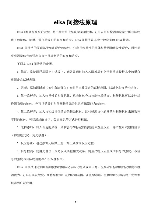
Elisa间接法的原理基于免疫反应的特性。
它利用特异性的抗体与待测物质发生反应,通过观察或测量信号的强度来确定目标物质的存在和浓度。
下面是Elisa间接法的步骤:
1. 修复:将待测样品固定在试板上,通常是通过加入乙醛或其他化学物质来使样品中的蛋白质固定在试板表面。
2. 阻断:添加阻断剂(如牛血清蛋白)来封闭未被固定的试板表面,以减少非特异性结合。
3. 第一次孵育:加入特异性的初级抗体,这些抗体会与待测物质结合。
初级抗体可以是针对待测物质的抗体,也可以是其他与待测物质无关但具有识别能力的抗体。
4. 第二次孵育:加入与初级抗体结合的辅助抗体。
这些辅助抗体通常是与初级抗体来源物种不同的抗体,可以通过酶标记、荧光标记等方式进行标记。
5. 底物添加:加入合适的底物,底物会与酶标记的辅助抗体发生反应,并产生可观察的信号(如颜色变化、荧光强度)。
6. 反应停止:通过添加反应停止剂,终止底物的反应过程。
7. 信号检测:使用光谱仪、荧光仪或其他相关设备,测量底物反应生成的信号的强度,该信号的强度与目标物质的存在和浓度相关。
Elisa间接法通过利用辅助抗体的酶标记或标记物来放大信号,提高对目标物质的灵敏度和检测能力。
它具有高灵敏度、高特异性和广泛的应用范围,在医学诊断、生物学研究和药物开发等领域得到广泛应用。
1。
- 1、下载文档前请自行甄别文档内容的完整性,平台不提供额外的编辑、内容补充、找答案等附加服务。
- 2、"仅部分预览"的文档,不可在线预览部分如存在完整性等问题,可反馈申请退款(可完整预览的文档不适用该条件!)。
- 3、如文档侵犯您的权益,请联系客服反馈,我们会尽快为您处理(人工客服工作时间:9:00-18:30)。
THE PROCEDURE OF INDIRECT ELISA PROCEDURE
A. Antigen Coating
1.Prepare an antigen solution at the appropriate concentration in
carbonate-bicarbonate buffer (coating buffer), e.g. human IgG (0.025mg/ml) in coating buffer (antigen solution).
2.Pipette 0.2 ml of the above solution to each well of the microtiter plate.
3.Incubate at 37 o C for 30 min., or incubate overnight at 4 o C.
4.Remove the coating solution. Wash three times with PBS-T.
5.Blocking step: 0.5% BSA-PBS (0.2ml/well) at 37 o C for 1 h.
B. Primary Antibody Reaction
1. Dilute the primary (first) antibodies in PBS-T, e.g. Rabbit-anti-human IgG
antiserum.
2.Add 0.2 ml of the diluted first antibody to each well. Negative control should
be included.
3.Incubate at room temperature for 2 h (or 37 o C for 1 h).
4.Wash three times with PBS-T.
C. Application of Secondary Antibody
1.Dilute the peroxidase conjugated secondary antibody in PBS-T, e.g.
Goat-anti-rabbit IgG-HRP (1:10 000). Add 0.2 ml of this solution to each well.
2.Incubate at room temperature for 1.5 h (or37 o C for 45min).
3.Wash three times with PBS-T.
D. Substrate Preparation
During the last incubation and immediately before use, dissolve o-Phenylenediamine in Citric acid-sodium hydrogen phosphate buffer, add 30% H2O2 before use.
E. Development
1. Add 0.2 ml of the freshly prepared substrate to each well.
2. Orange-yellow color should develop in positive wells after 5 min.
3. Stop reaction with 50 l per well of preferred stopping reagent and read at
490nm in a microplate reader.
REAGENTS
1.Coating buffer (Carbonate-Bicarbonate buffer, pH 9.6) :
Na2CO30.15g,
NaHCO30.293g, add H2O to 100ml (pH 9.6).
2.Phosphate buffered saline (PBS):
NaCl 8g,
KCl 0.2g,
KH2PO40.24g,
Na2HPO4. 12 H2O 2.9g, add H2O to 1000ml (pH 7.4).
3.Washing buffer (PBS-T): PBS + 0.05% Tween 20.
4.Blocking solution: 0.5% BSA in PBS.
5.First antibody: Rabbit-ani-human IgG antiserum.
6.Peroxidase conjugated secondary antibody: Goat-anti-rabbit IgG-HRP
(1:10 000).
7.Substrate: substrate buffer(fresh):
0.1mol/L Citric acid (2.1g/100ml), 6.1ml.
0.2mol/L Na2HPO4. 12 H2O(7.163g/100ml), 6.4ml.
add H2O 12.5ml.
dissolve 8mg o-Phenylenediamine,
and add 40 l of 30% H2O2 before use.
8.Stopping reagent: 2mol/L H2SO4.。