Relationship between blood glucose
空腹血糖水平与胰岛素抵抗关系的分析

(FPG)的 升 高 ,体 重 指数 (BMI)、腰 围(WC)、三 酰 甘 油 (TG)、空 腹 胰 岛 素 (FINS)、2 h PG 逐 步上 升 ,与 FPG均具 有 统计 学 意 义 (P<0.05),但 6.1 mmol/L ̄<FPG<7.0 mmol/L组 与 5.6 mmol/L≤FPG<6.1
Yancheng City No.1 People s Hospital in Jiangsu Province from January 2008 to January 2009 were selected as the
study subjects.The subjects were divided into four groups according to the fasting blood sugar leve1.Basic physical examination and the oral glucose tolerance test were conducted and the 2-hour plasma glucose (2 h PC)was men- sured.The homeostasis model assessment insulin resistance index (HOMA-IR)and the homeostasis model assessment islet beta-cell function(HOMA-B)were calculated using the homeostasis model assessment(HOMA).The relationship
稳态模 式 评 估 法 (HOMA)计 算 稳态模 式 评 估 胰 岛素抵 抗 指数 (HOMA—IR)、胰 岛 B一细 胞功 能稳 态 模 型评 估
血药浓度和药效的关系英文作文
血药浓度和药效的关系英文作文The relationship between blood concentration and drug effectiveness is a crucial aspect of pharmacology and therapeutics. Blood concentration, often referred to as plasma concentration or serum concentration, refers to the amount of a drug present in the bloodstream at a given time. This concentration is directly linked to the drug's effectiveness, as it determines how much of the drugreaches its target site and how potent its therapeuticaction will be.Drug absorption, distribution, metabolism, andexcretion (ADME) are the four primary factors thatinfluence blood concentration. The rate and extent of drug absorption from the site of administration determine how quickly the drug enters the bloodstream. Distributionrefers to how the drug spreads throughout the body,reaching its target tissues and organs. Metabolism, the chemical transformation of the drug within the body, can either increase or decrease its blood concentration, depending on whether the metabolism leads to inactivationor activation of the drug. Excretion, the elimination ofthe drug from the body, is the final step in the ADME process, and it also affects blood concentration.Blood concentration is typically measured in micrograms per milliliter (μg/mL) or nanomoles per liter (nmol/L), depending on the drug and its properties. The optimal blood concentration for a drug is typically determined through clinical trials and pharmacokinetic studies, which aim to establish the dose-response relationship and identify the minimal effective concentration (MEC) and the maximal tolerable concentration (MTC).Drug effectiveness is influenced by both the blood concentration and the pharmacokinetic properties of the drug. A drug with a high affinity for its target site and a low clearance rate from the body is likely to be more effective at lower blood concentrations. Conversely, a drug with low affinity and high clearance may require higher blood concentrations to achieve the desired therapeutic effect.In addition to blood concentration, other factors such as the duration of drug exposure, the route of administration, and the patient's physiological status canalso affect drug effectiveness. For example, drugs administered intravenously typically achieve higher and faster blood concentrations than those administered orally. Similarly, patients with liver or kidney dysfunction may experience altered drug metabolism and excretion, leadingto changes in blood concentration and effectiveness.In summary, the relationship between bloodconcentration and drug effectiveness is complex and multifaceted. Understanding this relationship is crucialfor optimizing drug dosing, achieving desired therapeutic effects, and minimizing adverse effects. Future research in the field of pharmacology and therapeutics will continue to elucidate the intricacies of this relationship, leading to improved patient outcomes and more effective drug therapies. **血药浓度与药效的关系**血药浓度与药效之间的关系是药理学和治疗学中的关键方面。
探讨T2DM患者血糖及胰岛素敏感性与夜间血皮质醇水平的关系

(The Affiliated Hospital of North China University of Science and Technology, Hebei, Tangshan)
mass index (BMI) divided into normal body mass index group (BMI < 25) 64 cases, the increase body mass index group (BMI≥25) in 82 cases. Results The increase body mass index group of cortisol levels(F), glycosylated hemoglobin (HbA1c) and insulin resistance index (HOMA-IR) were higher than the normal body mass index group ,P<0.05, the difference was statistically significant; The increase body mass index group, insulin sensitivity index (ISI) is lower than the normal body mass index group ,P<0.05,The difference was statistically significant; HbA1c was positive correlated with BMI, FPG, 2hPG, 0 point cortisol (P<0.05); HbA1c was negatively correlated with FIns, 2hIns, 2hC-P (P<0.05); BMI was positive correlated with HbA1c, 0 positive cortisol (P<0.05); BMI was negatively correlated with ISI (P<0.01); Conclusion The increased blood glucose level and decreased insulin sensitivity are associated with the increased cortisol levels at night on patients of the increase body mass index with type 2 diabetes. KEY WORDS: Diabetes mellitus; Type 2; Cortisol; Blood glucose; Insulin sensitivity
血清胱抑素及骨保护素水平与2型糖尿病患者并发非酒精性脂肪肝的相关性分析
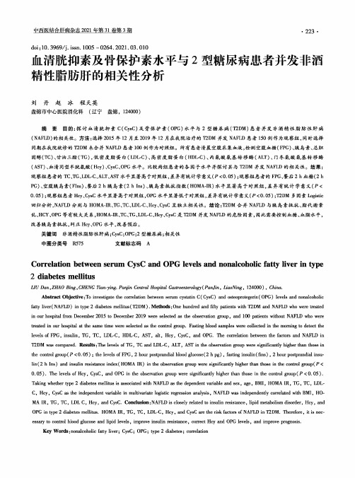
doi:10.3969/j.issn.1005-0264.2021.03.010血清胱抑素及骨保护素水平与2型糖尿病患者并发非酒精性脂肪肝的相关性分析刘丹赵冰程天英盘锦市中心医院消化科(辽宁盘锦,124000)摘要目的:探讨血清胱抑素C(CysC)及骨保护素(OPG)水平与2型糖尿病(T2DM)患者并发非酒精性脂肪性肝病(NAFLD)的相关性。
方法:选择2015年12月至2019年12月在我院治疗的T2DM并发NAFLD患者150例作为观察组,同时选择同期在我院就诊的T2DM未合并NAFLD患者100例作为对照组。
所有患者清晨空腹采集血液,检测空腹血糖(FPG)、胰岛素、总胆固醇(TC)、甘油三酯(TG),低密度脂蛋白(LDL-C)、高密度脂蛋白(HDL-C)、丙氨酸氨基转移酶(ALT)、门冬氨酸氨基转移酶(AST)、血清同型半胱氨酸(Hcy)、CysC、OPG水平。
比较两组患者的各因子水平并探讨其与T2DM并发NAFLD的相关性。
结果:观察组患者的TC、TG、LDL-C、ALT、AST水平显著高于对照组,差异有统计学意义(P<0.05);观察组患者的FPG、餐后2h血糖(2h PG)、空腹胰岛素(Fins)、餐后2h胰.岛素(2h Ins)、胰岛素抵抗指数(HOMA-IR)水平显著高于对照组,差异有统计学意义(P< 0.05);观察组患者Hcy、CysC水平显著高于对照组,OPG水平显著低于对照组,差异有统计学意义(P<0.05);T2DM多因素Logistic 回归分析,NAFLD分别与HOMA-IR、TG、TC、LDL-C、Hcy、CysC呈独立相关性。
结论:T2DM合并NAFLD与胰.岛素抵抗、脂代谢紊乱、HCY、OPG等有较大关系,HOMA-IR、TC、TG、LDL-C、Hcy、CysC是T2DM并发NAFLD的危险因素,因此需要控制血糖、血脂水平,改善胰岛素抵抗,纠正Hcy、OPG水平,改善预后。
2型糖尿病患者血糖控制水平与血清肿瘤标志物相关性研究

2型糖尿病患者血糖控制水平与血清肿瘤标志物相关性研究胡吉萍,朱丽丽,杜丙梅淄博市张店区妇幼保健院检验科,山东淄博255000[摘要]目的分析2型糖尿病患者血糖控制水平与血清肿瘤标志物相关性,为2型糖尿病合并恶性肿瘤患者的诊治提供依据。
方法选取2022年1—12月淄博市张店区妇幼保健院与淄博市中心医院诊断为2型糖尿病合并恶性肿瘤的患者200例作为研究对象,根据糖化血红蛋白的水平分为两组,观察组为血糖控制不佳糖化血红蛋白≥7.0%者102例,对照组为控糖佳糖化血红蛋白<7.0%者98例。
对比两组患者肿瘤标志物水平,Pearson相关性分析糖化血红蛋白与肿瘤标志物的相关性。
结果观察组血清神经元特异性烯醇化酶、癌胚抗原、糖链抗原19-9明显高于对照组,差异有统计学意义(P<0.05)。
糖化血红蛋白与血清神经元特异性烯醇化酶、癌胚抗原、糖链抗原19-9呈正相关(r=0.784、0.712、0.714,P<0.05)。
结论2型糖尿病患者血糖控制水平与血清肿瘤标志物水平呈正相关。
[关键词] 2型糖尿病;血糖控制水平;血清肿瘤标志物;相关性研究[中图分类号] R4 [文献标识码] A [文章编号] 1672-4062(2023)08(a)-0186-04Study on the Correlation between Blood Glucose Control Level and Serum Tumor Markers in Patients with Type 2 Diabetes MellitusHU Jiping, ZHU Lili, DU BingmeiDepartment of Laboratory, Zibo Zhangdian District Maternal and Child Health Hospital, Zibo, Shandong Province, 255000 China[Abstract] Objective To analyze the correlation between blood glucose control level and serum tumor markers in pa⁃tients with type 2 diabetes mellitus, and to provide a basis for the diagnosis and treatment of patients with type 2 dia⁃betes mellitus combined with malignant tumors. Methods From January to December 2022, 200 patients diagnosed as type 2 diabetes with malignant tumor in Zibo Zhangdian District Maternal and Child Health Hospita and Zibo Central Hospital were selected as the study subjects. According to the level of glycosylated hemoglobin, they were divided into two groups. The observation group was 102 patients with poor blood glucose control HbA1c≥7.0%, and the control group was 98 patients with good glucose control HbA1c<7.0%. Compared the levels of tumor markers between two groups of patients, and analyzed the correlation between HbA1c and tumor markers through Pearson correlation analy⁃sis. Results The NSE, CEA, and CA19-9 levels in the observation group were significantly higher than those in the control group, the difference was statistically significant (P<0.05). HbA1c was positively correlated with NSE, CEA, and CA19-9 (r=0.784, 0.712, 0.714, P<0.05). Conclusion The level of blood glucose control in patients with type 2 diabetes mellitus was positively correlated with the level of serum tumor markers.[Key words] Type 2 diabetes mellitus; Blood glucose control level; Serum tumor markers; Correlation study目前肿瘤标志物已成为恶性肿瘤筛查的一项主要检查[1]。
2型糖尿病患者血糖水平与肿瘤标志物CEA、CA199、AFP的相关性分析
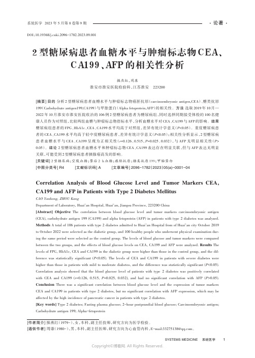
DOI:10.19368/ki.2096-1782.2023.09.0012型糖尿病患者血糖水平与肿瘤标志物CEA、CA199、AFP的相关性分析操燕红,周康淮安市淮安医院检验科,江苏淮安223200[摘要]目的分析2型糖尿病患者血糖水平与肿瘤标志物癌胚抗原(carcinoembryonic antigen,CEA)、糖类抗原199(Carbohydrate antigen199,CA199)与甲胎蛋白(Alpha fetoprotein,AFP)的相关性。
方法选取2019年10月—2022年10月淮安市淮安医院收治的106例2型糖尿病患者为糖尿病组,同时选择同期接受体检的100名健康人员作为对照组,比较两组血糖与肿瘤标志物指标水平,分析血糖水平对CEA、CA199与AFP的影响。
结果糖尿病组患者的FPG、HbA1c、CEA、CA199水平均高于对照组,差异有统计学意义(P<0.05)。
重度糖尿病患者的CEA、CA199水平均高于轻中度糖尿病患者,差异有统计学意义(P<0.05);相关性分析显示,2型糖尿病患者血糖水平与CEA、CA199呈现为正相关性(r=0.126、0.515,P=0.025、0.032),与AFP无明显相关性(P> 0.05)。
结论 2型糖尿病患者血糖水平和肿瘤标志物CEA、CA199表达存在明显关联,但与AFP表达无明显关联,可能受到2型糖尿病患者胰腺癌高发的影响。
[关键词]2型糖尿病;空腹血糖;餐后2 h血糖;癌胚抗原;糖类抗原199;甲胎蛋白[中图分类号]R4 [文献标识码]A [文章编号]2096-1782(2023)05(a)-0001-04Correlation Analysis of Blood Glucose Level and Tumor Markers CEA, CA199 and AFP in Patients with Type 2 Diabetes MellitusCAO Yanhong, ZHOU KangDepartment of Laboratory, Huai´an Hospital, Huai´an, Jiangsu Province, 223200 China[Abstract] Objective The correlation between blood glucose level and tumor markers carcinoembryonic antigen (CEA), carbohydrate antigen 199 (CA199) and alpha fetoprotein (AFP) in patients with type 2 diabetes was analyzed.Methods A total of 106 patients with type 2 diabetes admitted to Huai´an Hospital from of Huai´an city October 2019 to October 2022 were selected as the diabetic group, and 100 healthy people who underwent physical examination dur‐ing the same period were selected as the control group. The levels of blood glucose and tumor markers were compared between the two groups, and the effects of blood glucose levels on CEA, CA199 and AFP were analyzed. Results The levels of FPG, HbA1c, CEA and CA199 in the diabetic group were higher than those in the control group, and the dif‐ference was statistically significant (P<0.05). The levels of CEA and CA199 in patients with severe diabetes were higher than those in patients with mild to moderate diabetes, and the difference was statistically significant (P<0.05). Correlation analysis showed that the blood glucose level of patients with type 2 diabetes was positively correlated with CEA and CA199 (r=0.126, 0.515, P=0.025, 0.032), and had no significant correlation with AFP (P>0.05).Conclusion There was a significant correlation between blood glucose level and the expression of tumor markers CEA and CA199 in patients with type 2 diabetes, but no significant correlation with AFP expression, which may be affected by the high incidence of pancreatic cancer in patients with type 2 diabetes.[Key words] Type 2 diabetes; Fasting plasma glucose; 2-hour postprandial blood glucose; Carcinoembryonic antigen; Carbohydrate antigen 199; Alpha-fetoprotein[作者简介] 操燕红(1979-),女,本科,副主任技师,研究方向为医学检验。
为什么清晨反而比晚睡前的血糖水平高

为什么清晨反而比晚睡前的血糖水平高?将近半年多了,有两位朋友一直在问我,为什么她们清晨的空腹血糖比晚睡前还高?照理,一夜过去,又没吃东西,到早晨血糖应该更低才对呀。
我推想是内分泌系统在临晨发生小小兵乱,苦于没有证据,不可妄言。
今天发现了一篇好文章,答案找到了!既然已经帮朋友翻译过来了,就贴在这儿,供糖尿病和准糖尿病患友参考。
早晨空腹血糖高的原因有三个, 1.黎明现象, 这是身体自然的反应,是为了预备身体将要开始新的一天的工作所需要的能量,正常的人会分泌相应量的胰岛素来控制此时的血糖水平,有糖尿病的人,控制能力底了,所以血糖升高。
2。
Somogyi 效应,这是一位医生发现的现象,所以以他的名字命名。
当你半夜血糖水平降得太低时,身体为了保护自己,通过分泌其他激素而提高血糖水平,如果你睡觉前吃一点小点,就不会有这个问题。
3. waning insulin (胰岛素缺乏),通常发生在已经在用胰岛素治疗糖尿病的患者身上。
怎样分辨你是那一种原因造成的?以下是简易的方法。
1。
分别测定临晨3点,早晨的空腹血糖,如果3点时血糖正常,而清晨血糖较高,你可能是黎明现象。
2。
如果3点时血糖偏低,而清晨血糖较高,那么就是Somogyi 效应。
3。
如果3点时血糖比上床睡觉的时候,到早晨还持续高,说明是胰岛素缺乏。
原文如下:Rocky Morning Highs?With a little sleuthing you can identify—and fight—the causes of those rises in waking blood glucoseBy Terri D'Arrigo September 2008 Other Monitoring, BG and A1CSometimes diabetes doesn't make a whole lot of sense. Take those mornings when you wake up with blood glucose that's higher than it was when you went to sleep. You'd think that not eating for those seven or eight hours would give you lower blood glucose. But in fact, there are three reasons your blood glucose may be higher in the morning: the dawn phenomenon, the Somogyi effect, or waning insulin.The Dawn PhenomenonThe dawn phenomenon is a natural rise in blood glucose between the hours of 4:00 a.m. and 8:00 a.m., and it occurs because of hormonal changes in the body. "The body does several things to get ready for the day," says David S. Schade, MD, professor of medicine and chief of the Division of Endocrinology at the University of New Mexico School of Medicine in Albuquerque. "The body releases hormones like cortisol and growth hormone and the blood glucose rises. People without diabetes just secrete more insulin to handle the blood glucose, but for people with diabetes, the rise in blood glucose can be substantial."Schade notes that the effects of dawn phenomenon vary in each person, and your blood glucose may be higher on some mornings than on others. "You can do the same exercise and eat the same thing every day and have different blood glucose [levels] on different mornings because of dawn phenomenon," he says. "That makes it a little problematic." He adds that the scientific community is still figuring out the relationship between the release of these hormones and the rise in blood glucose. However, one thing scientists do know is that the liver produces glucose as part of the dawn phenomenon.Treatment for dawn phenomenon depends on how you treat your diabetes, says Stuart T. Haines, PharmD, BCPS, professor and pharmacotherapy specialist at the University of Maryland School of Pharmacy in Baltimore. If you take insulin, you may be able to adjust your dosing so that peak action occurs closer to the morning rise in your blood glucose. If you have type 2, diabetes pills provide options as well, he says: "You can add metformin to reduce the liver's glucose production."The Somogyi EffectThe Somogyi effect, named for researcher Michael Somogyi, PhD, who studied and first described it, is your body's response to a low that you had while you were sleeping. "This happens after low blood glucose induced by excess insulin, alcohol consumption, or not having had enough food," says Haines. "You have a low, and to counter that, your body responds in a rigorous way and cranks out a bunch of hormones, like glucagon." The body responds to those hormones by raising blood glucose—sometimes too much.You would treat this the opposite way of how you would treat dawn phenomenon, says Schade. "You could have a snack before you go to sleep or reduce your insulin infusion at night. If you take NPH, you can switch to an insulin that won't dip you down at 3:00 a.m." But wouldn't a low wake you up? Not necessarily, says Mindy Saenz, RD, LDN, CDE, clinical dietitian and diabetes educator in the Division of Endocrinology at the Brody School of Medicine at East Carolina University in Greenville, N.C. "You can sleep right through them," she says. "Nighttime lows are the most dangerous." She adds that it's a good idea to check your blood glucose if you wake up sweating or with headaches, as those are signs of a low.Waning InsulinThe dawn phenomenon and the Somogyi effect are pretty complicated hormonal stuff, but sometimes the simplest explanation is the correct one, Saenz says. Sometimes yourinsulin just runs out or wears off. Then it's a matter of you and your doctor adjusting your insulin regimen accordingly. "If it's insulin waning, you could look at splitting your basal insulin or taking it at a different time of the day," she says. "If you take NPH at supper, you could move it closer to bedtime."Before you and your doctor can adjust your diet or medications to handle high morning blood glucose, you have to know which of the three potential causes is the culprit. Experts agree that there's one simple, if somewhat inconvenient, way of figuring out what that might be: Check your blood glucose at 3:00 a.m. for several nights in a row. "You need to see where your blood glucose is at bedtime, at 3:00 a.m., and in the morning," says Saenz. She explains it this way:∙ If your blood glucose is fairly even between bedtime and 3:00 a.m., but then rises between 3:00 a.m. and morning, chances are you're experiencing dawn phenomenon.∙ If your blood glucose is low at 3:00 a.m., you're most likely experiencing the Somogyi effect.∙ If your blood glucose is higher at 3:00 a.m. than at bedtime and higher still in the morning, your insulin is probably waning.A continuous blood glucose monitor can go a long way toward helping you nab the perpetrator. These monitors record your blood glucose every few minutes around the clock, and they have alarms to alert you to highs and lows. The problem is that these monitors are expensive, and insurance coverage for them is hard to come by, says Schade. "We're all trying to get insurance to cover them, and some plans will, butMedicare won't," he says. "Some insurance companies will cover them if your doctor fills out a special form indicating why one would help you, but it's sporadic so check with your insurance company and see what the criteria are."If you have diabetes, chances are you'll experience the occasional high morning blood glucose. That's not something to fret about too much. But if it happens regularly, then it's time to call your doctor. "You should also suspect a problem when your morning blood glucose is the highest of the day, and when it is consistently high for the rest of the day after that," says Haines. Schade agrees. "If it's significant, it should be dealt with because those who have high blood glucose in the morning tend to have high blood glucose all day," he says. "It's important to recognize that and adopt a strategy to control it."。
GLUCOSEHOMEOSTASISAnOverview:葡萄糖稳态的概述

Fig. 1: Variations in blood glucose and blood insulin levels correlated with periods of eating and fasting;
HOW DOES THE BODY NORMALLY DISPOSES OF HIGH LEVEL OF GLUCOSE IN BLOOD AFTER A MEAL?
• During prolonged fasting: • Blood glucose level usually decreases only slightly, but remains within normal range, • Brain and RBC are still actively metabolizing glucose, thus the blood glucose utilized must be replenished;
REGULATION OF BLOOD GLUCOSE DURING FASTING
How is Blood Glucose level regulated during fasting?
• Blood glucose level should normally remains constant, even if no food is consumed within 24-hour period;
• Increase in blood glucose level after a meal is immediately followed by increase in Blood Insulin level;
• Fig 1: Schematic representation of relationship between Blood Glucose and Insulin level in blood during periods of eating and fasting;
- 1、下载文档前请自行甄别文档内容的完整性,平台不提供额外的编辑、内容补充、找答案等附加服务。
- 2、"仅部分预览"的文档,不可在线预览部分如存在完整性等问题,可反馈申请退款(可完整预览的文档不适用该条件!)。
- 3、如文档侵犯您的权益,请联系客服反馈,我们会尽快为您处理(人工客服工作时间:9:00-18:30)。
Abstract.–OBJECTIVE:To evaluate the rela-tionship between blood glucose fluctuation and macrovascular dysfunction.PATIENTS AND METHODS:Eighty-eight type 2diabetes mellitus (T2DM)patients with or without coronary heart disease (CHD)and 30healthy con-trol subjects were recruited.Glycosylated hemo-globin A1c (HbA1c),fasting insulin (FIns),and C-reaction protein (CRP)and some other general clinical variables were measured.A 72-hour con-tinuous glucose monitoring (CGM)and brachial artery endothelium-dependent flow-mediated dila-tion (FMD)assessment were performed.The glu-cose excursion,MAGE (mean amplitude of glycemic excursions),LAGE (largest amplitude of glycemic excursions),MPPGE (mean postprandial glycemic excursions),MODD (absolute means of daily differences),and IAUC70(incremental area under the curve below 70mg/dl)during the CGM were analyzed.Correlations between the various variables were analyzed.RESULTS:Enhanced blood glucose fluctuation was observed in T2DM patients with CHD as compared to other participants.And blood glu-cose fluctuation was correlated with FMD,CRP and HOMA-IR.CONCLUSIONS:Blood glucose fluctuation is an important factor that affects inflammatory re-sponse and possibly induces CHD in T2DM pa-tients.Key Words:Type 2diabetes mellitus,Coronary heart disease,Continuous glucose monitoring system,Macrovascu-lar endothelial dysfunction,C-reactive protein.IntroductionCoronary heart disease (CHD)is one of the most common complications of Type 2diabetes mellitus (T2DM).Evidence-based medicine stud-Eur opean Rev iew for Med ical and Pharmacol ogical Sci ences Relationship between blood glucosefluctuation and macrovascular endothelial dysfunction in type 2diabetic patients with coronary heart diseaseX.-G.ZHANG,Y .-Q.ZHANG 1,D.-K.ZHAO,J.-X.WU,J.ZHAO,X.-M.JIAO,B.CHEN,X.-F .LVDepartment of Endocrinology,General Hospital of Beijing Military Area,Beijing,PR China 1China National Institute of Standardization,Beijing,PR ChinaCorresponding Author:Xiaofeng Lv,MD;e-mail:xiaofenglv7966@3593ies have shown that control of glycosylated hemo-globin A1c (HbA1c)may reduce the occurrence of microvascular complications significantly but not cardiovascular diseases.This suggests that the prevalence of macrovascular complications may not be evaluated by measuring HbA1c alone 1.It has been reported that blood glucose fluctuation is significantly associated with complications of T2DM 2-5.Research demonstrated by Jiao et al in-dicated that blood glucose fluctuation influence lower-extremity vascular disease in type 2diabetes and related with diabetic macroangiopathy.Mean amplitude of glycemic excursion (MAGE),firstly proposed in 1970by Service et al 5,changes the overall level of blood glucose independently.As a result,MAGE is constantly used to assess glycemic variability 7,8.It has been demonstrated that an intermittent exposure to high glucose induces more pro-nounced metabolic changes and cytotoxicity than a constant exposure 8.This is because intermittent high glucose is more effective in triggering the generation of nitrotyrosine,activating the expres-sion of protein kinase C (PKC)and inducing the expression of adhesion molecules (ICAM-1,VCAM-1and E-selectin)than constant high glu-cose 9,10.Additionally,intermittent high glucose is capable of enhancing oxidative stress,inducing cell apoptosis as well as reducing the synthesis of vascular relaxing factor (e.g.NO)as demonstrat-ed in cultured cells 12-14.Changes in vessel wall shear stress induced by increased blood flow may result in NO release from vascular endothelial cells.NO subsequently activates guanylate cyclase in smooth muscle cells,leading to an elevation in cyclic guanosine monophosphate (cGMP).Eventually,brachial artery flow-mediated endothelium-dependent va-2014;18:3593-3600sodilation(FMD)occurs in the smooth muscle. Previous studies have demonstrated that brachial artery FMD is closely related to vasodilation of the coronary artery and the injury severity of the brachial artery is closely related to vasodilation of the atherosclerotic carotid and the atherosclerotic coronary,these suggesting that brachial artery FMD may be used as an indirect indicator of the coronary and systemic vascular functions15,16.In the present study,we observed that blood glucose fluctuation was more pronounced in T2DM patients with CHD.Glycemic fluctuation may be one of the factors that influence brachial artery FMD.An increase in C reactive protein (CRP)concentration associated with glucose fluctuation may result in the decline of brachial artery FMD.Patients and Methods SubjectsType2diabetes mellitus(T2DM)patients aged between50and70years were consecutive-ly admitted to the General Hospital of Beijing Military Area December2010to November 2011.The study protocol including screening, treatment,and data collection were approved by the Institutional Ethics Committee.Written in-formed consent was obtained from all subjects. The provisions of the Declaration of Helsinki were strictly followed.Inclusion and Exclusion Criteria Coronary arteriography was adopted if the ex-amination performed in the last2months.Coro-nary heart disease was accordingly diagnosed if the left main artery showed≥30%stenosis or at least one branch of three major coronary arteries showed≥50%stenosis.The diagnosis of T2DM was adopted according to the diagnostic criteria of American Diabetes Association(ADA)in2010. Patients should have regular diet,exercise and medication,stable hypoglycemic scheme in the last three months and no extreme blood biochemi-cal indicators of hepatic and renal function.The exclusion criteria were:(1)acute complication of diabetes in the last six months;(2)post-menopausal women taking estrogen;(3)eye dis-ease caused by hypertension,thromboangioitis obliterans or Takayasu arteritis(caused by the non-diabetic vascular disease)and other diseases that could affect vascular endothelial function;(4) acute coronary syndrome or acute brain damage in the last month;(5)hepatic or renal dysfunction;(6)MI(myocardial infarction),unstable angina, stroke or a transient ischaemic attack;and(7)evi-dence of severe hepatic or renal disease. Demographic Data CollectionDemographic data and information on medica-tions prior to admission were recorded.After fasting for12hours,3-5ml venous blood was sampled,mixed and placed at room temperature. Hepatic and renal function parameters and lipid metabolism indicators were determined with an automatic biochemical analyzer(Dxc600,Beck-man,Fremont,CA,USA).Fasting plasma glu-cose(FPG)was measured by glucose oxidase method.Affinity chromatography detection was carried out on a glycated hemoglobin analyzer (D10,Bio-Rad,Hercules,CA,USA)to deter-mine HbA1c concentration.Radioimmunoassay was used for fasting insulin(FIns)determination. C-reaction protein(CRP)in the plasma was mea-sured using an enhanced turbidimetric im-munoassay.Non-anticoagulant blood was cen-trifuged at3,500rpm for10min and the same in-dicators in the serum were detected for calibra-tion and quality control.Insulin resistance index (IR)was calculated by the Homeostasis Model as previously described17:HOMA-IR=Fins (µIU/mL)×FPG(mmol/L)/22.5.Coronary an-giography was performed by coronary angiogra-phy equipment(General Electric,USA). Continuous Glucose Monitoring Continuous glucose monitoring(CGM)was performed at least one week after coronary an-giography.A CGM system(CGMS)sensor (Medtronic,Northridge,CA,USA)was inserted into all participants by the same specialized nurse at8:00-9:00AM on the first day of hospi-talization.First CGMS calibration by finger stick blood glucose was performed1h after the procedure initialization.Subsequently,calibra-tion was performed four times daily for each subject.The interval between two calibrations was not exceeding8hours.Events such as eat-ing,exercise,taking hypoglycemic drugs and low blood glucose reactions that might affect blood glucose fluctuation were digitally record-ed.If no abnormal CGMS situation was ob-served,CGM was performed for72consecutive hours.Data on mean blood glucose(MBG), standard deviation of blood glucose(SDBG), mean amplitude of glycemic excursions (MAGE),largest amplitude of glycemic excur-X.-G.Zhang,Y.-Q.Zhang,D.-K.Zhao,J.-X.Wu,J.Zhao,X.-M.Jiao,B.Chen,X.-F.Lv 3594Blood glucose fluctuation in T2DM with CHD3595were allocated to T2DM2group.Thirty (15male,15female)healthy individuals were in-cluded as control subjects (NC group).Clinical Characteristics,CGM,FMD and CRPIn the NC group,blood glucose was within the normal range with a minor fluctuation.In con-trast,significant blood glucose fluctuations were observed in the two (T2DM1and T2DM2)dis-ease groups.When the two disease groups were compared,a larger degree of blood glucose fluc-tuation was detected in the T2DM2group than that in T2DM1group (p <0.05).Showed in Table I are the clinical data of subjects in the 3groups.There was no signifi-cant difference (p >0.05)either in the average age (or in the use of hypoglycemic drugs be-tween the 3groups.No extreme blood pressure was measured in any of the pared with the NC group,T2DM1and T2DM2groups had significantly higher levels of SDP,LDL-C,FBG,HbA1c,HOMA-IR,MAGE,LAGE,MPPGE,MODD,IAUC70and CRP (p <0.05),and LDL-C,FBG,HbA1c,HOMA-IR,MAGE,LAGE,MODD and CRP (p <0.01).In contrast,levels of LDL-C and FMD were sig-nificantly lower in the two disease groups than in the NC group (p <0.01).Compared with pa-tients in the T2DM1group,those in the T2DM2group had higher levels MAGE,LAGE,MPPGE,MODD,IAUC70,CRP and lower level of FMD (p <0.01).A larger degree of blood glucose fluctuation was observed in the T2DM2group than in the T2DM1group and the NC group.Negative Correlation Between FDM and LAGE,MPPGE,MODD and IAUC70in T2DM2PatientsFDM decreased significantly in T2DM2pa-tients.In these patients,FDM was negatively cor-related with MAGE (p =0.003),LAGE (p =0.029),MPPGE (p =0.033),MODD (p =0.025)and IAUC70(p =0.042)(Table II).Inversely,CRP was positively correlated with MAGE (p =0.002),LAGE (p =0.001),MPPGE (p =0.010),MODD (p =0.020)and IAUC70(p =0.182);HOMA-IR was positively correlated with MAGE (p =0.025),LAGE (p =0.013),MPPGE and MODD (p =0.045).No significant correlation between HbA1c and the blood glu-cose fluctuation was detected (Table II).Taken together,these observations suggest that bloodsions (LAGE),mean postprandial glycemic ex-cursions (MPPGE),absolute means of daily dif-ferences (MODD),incremental area under the curve below 70mg/dl (IAUC 70),endothelium-dependent flow-mediated dilation (FMD),and C-reaction protein (CRP)were extracted and analyzed using the CGMS system solutions software (MMT-7310Version 3.0C 3.0.128).Brachial Artery FMD ExaminationBrachial artery FMD examination was carried out following fasting or low-fat diet for 8-12h during the course of CGM.Patients were told not to drink coffee or tea at least 2h before the exam-ination.Vasoactive drugs,antihypertensive drugs,nitrates and statins drugs were also avoided for at least3days before the test.The test was per-formed in a quiet environment at a comfortable temperature (18to 24°C).BP (blood pressure)was monitored during a 20min supine rest.The process of pressurization was performed by a spe-cialized nurse using an electric pressure pump.Ultrasonic examination was performed using a high-resolution ultrasound system (ProSound α10,Aloka,Tokjo,Japan)with a 13-Hz probe,which was placed 2-5cm above the elbow to de-tect brachial artery.The depth of investigation was adjusted so that the boundaries of the vessel lumen and vessel wall were clearly distinguish-able in the longitudinal section image.The base-line value of the internal diameter of the brachial artery was recorded and referred to as D 0(mm).Reactive hyperemia was induced by rapid pres-surization to 300mmHg for 5min.The peak val-ue of the internal diameter of the brachial artery after releasing was recorded and referred to as D 1(mm).FMD =(D1-D0)/D0×100%.Statistical AnalysisStatistical analysis was performed using SPSS 16.0software (SPSS Inc.,Chicago,IL,USA).Data were expressed as mean ±standard devia-tion and analyzed by Student’s t -test,one-way ANOV A,Pearson correlation analysis or multi-variate regression analysis.Results were consid-ered significant when p <0.05.ResultsA total of 88T2DM patients were enrolled in the study.Of them,36(16male,20female)did not have CHD and were allocated to T2DM1group and 52(26male,26female)had CHD and3596glucose fluctuation might be a result of changes in FMD,CRP and HOMA-IR in T2DM patients with CHD.Relationship Among Multi-Variables in T2DM2PatientsWhen FMD was considered as the dependent variable,it could be calculated using the follow-ing regression equation:FMD =11.217-0.369MAGE-0.346(HOMA-IR)-0.447SBP (Table III).When CRP was chosen as the dependent variable,the regression equation was as follows:CRP =3.527+0.566LAGE (Table IV).These further suggest that FMD was strongly correlated with MAGE,HOMA-IR and SBP while CRP was strongly correlated with LAGE.DiscussionThe effect of blood glucose fluctuation on the function of vascular endothelial cells has become a subject of extensive research in recent years.It is reported that postprandial hyperglycemia is anX.-G.Zhang,Y .-Q.Zhang,D.-K.Zhao,J.-X.Wu,J.Zhao,X.-M.Jiao,B.Chen,X.-F .LvTable I.Clinical characteristics,CGMS parameters and FMD in the three study groups.aMeasured by continuous glucose monitoring system;b Measured by high-resolution ultrasound system.*p <0.05;**p <0.01:compared with NC group;#p <0.05;##p <0.01:compared with T2DM1group.Table II.Correlations between the indicated variables in T2DM2group.aMeasured by continuous glucose monitoring system;b Measured by high-resolution ultrasound system.independent predictor of cardiovascular events and death in diabetic patients 18.Moreover,it has been shown that postprandial hyperglycemia is a risk factor for patients with or without diagnosed by diabetes 19.In our study,brachial artery FMD in patients with T2DM was significantly de-creased compared with healthy control subjects.Correlation analysis showed that FMD was cor-related with TG,SBP,HbA1c and blood glucose fluctuation.This suggests that decreased brachial artery FMD may be associated with aberrant glu-cose and lipid metabolism in patients with T2DM.Macrovascular complications occurred earlier in the T2DM2group than in the T2DM1group.Blood glucose fluctuation parameters (MAGE,LAGE,MODD,MPPGE and IAUC 70)in T2DM2patients were remarkably increased as compared with those in T2DM1patients.In other words,when the course of the disease,blood glu-cose,blood pressure,cholesterol and other fac-tors were controlled,increased blood glucose fluctuation became an important factor causes the decrease of brachial artery FMD in T2DM patients with CHD.Vascular dysfunction may result from blood glucose fluctuation,but the underlying mecha-nism remains unclear.Many studies indicated that oxidative stress play an important role in the process of vascular dysfunction caused by blood glucose fluctuation.Quagliaro et al 11have report-ed that temporary hyperglycemia accelerates the damage of vascular endothelial cells,and pro-motes apoptosis and oxidative DNA damage as compared with persistent hyperglycemia.Fur-thermore,in vivo studies have demonstrated that rapidly elevated glucose concentration may acti-vate P65subunit of NF-kB of arterial endothelial cells in non-diabetic rats,and elevated glucose concentration induce the expression of monocyte chemoattractant protein-1(MCP-1)and vascular cell adhesion molecule-l (VCAM-1).Inhibition of mitochondrial superoxide ion may attenuate the aberrant expression of these factors 20.In addi-tion to vascular endothelial cells,a variety of oth-er cell types including renal cortex fibroblast cells and human peripheral blood mononuclear cells can also be injured after intermittent high glucose exposure.Through analyzing changes in the intracellular nitrotyrosine and 8-hydroxy de-oxyguanosine (8-OHdG)expression and activity of Bcl-2and caspase-3,Piconi et al 21found in-termittent elevated blood glucose could induce endothelial cell apoptosis through reactive oxy-gen species (ROS)-associated oxidative stress.These observations indicate that oxidative stress is involved in blood glucose fluctuation.Previous clinical studies 22have reported that the excretion rate of 8-iso-prostaglandin F2alpha (8-isoPGF2α)is highly correlated with MAGE (r =0.86,p <0.001)but not the average blood glu-cose,fasting blood glucose or HbA1c.MAGE is considered to be the “gold standard”indicator of blood glucose fluctuation and 8-isoPGF2α(a peroxidation product)in vivo.The correlation be-tween these two factors suggest that oxidative stress may be an important mechanism underly-ing the blood glucose fluctuation-associated vas-cular injury.In the present study,MAGE was the variable that showed the strongest correlation with brachial artery FMD in T2DM2patients.It3597Blood glucose fluctuation in T2DM with CHDTable III.Results of multivariate regression analysis of factors affecting FMD in T2DM2 group.Table IV .Results of multiple regression analysis of factors affecting CRP in T2DM2 group.is possible that blood glucose fluctuation causes vascular endothelial injury through decreasing both NO synthesis and FMD. MAGE includes all valid blood glucose fluctuation indicators and, therefore, is closely related to endothelial func-tion in FMD. Parameters such as LAGE, MODD, MPPGE and IAUC70 were also correlated with FMD in the linear regression analysis but failed to perform the multivariate regression analysis. This might be due to the incomprehensive repre-sentativeness of these parameters to blood glu-cose fluctuation.Another potential mechanism is the inflamma-tion hypothesis. It is believed that type 2 diabetes is a chronic inflammatory disease. Our study found that CRP was significantly higher in T2DM patients as compared to healthy control subjects. This observation is consistent with re-sults from previous studies23-25. CRP can cause vascular endothelial dysfunction in a variety of ways26,27. To date, it remains inconclusive regard-ing whether blood glucose fluctuation plays a role in increasing inflammation in T2DM pa-tients. Tanaka et al28reported that postprandial hyperglycemia could increase IL-L and TNF-αin peripheral blood. And excessive secretion of IL-1 and TNF-αcan increase CRP synthesis29, which indicating that blood glucose fluctuation may lead to increased inflammatory factors such as IL-l, TNF-αand CRP. In this study, the CRP level was apparently increased in T2DM patients, particularly those with CHD as compared with control subjects. Correlation analysis showed that CRP was correlated with MAGE, LAGE, MPPGE, and MODD. The maximal correlation coefficient was obtained between LAGE and CRP, indicating that an increase in CRP may be related to excessive blood glucose drift in T2DM patients with CHD. LAGE was the maximal acute fluctuation during the entire observation period (24 hours) and CRP is an acute phase pro-tein. Our findings showed that increased blood glucose fluctuation could decrease the brachial artery FMD and elevate CRP in T2DM patients with CHD. This indicates that elevated CRP may be one of the mechanisms responsible for vascu-lar endothelial dysfunction in T2DM patients with CHD.In addition to blood glucose fluctuation, HOMA-IR was another important factor that af-fected the brachial artery FMD in T2DM patients with CHD. Insulin-dependent phosphatidylinosi-tol 3 kinase (PI-3K) signal transduction pathway regulates the expression of endothelial nitric ox-ide synthase (eNOS) gene30, thereby regulating NO production. IR leads to injuries of endothe-lial cells in many ways while damaged endothe-lial cells may exacerbate IR by changing the dis-tribution and function of insulin receptors on the cell surface.Furthermore, this study showed that in T2DM patients with CHD, systolic blood pressure was one of the important factors that affect brachial artery FMD in addition to blood glucose fluctua-tions. Woodman et al31found that there was a significant increase in CRP and von willebrand facotr (vWF) in T2DM patients with hyperten-sion. And von Willebrand Factor (vWF) is an im-portant indicator of vascular endothelial function. This indicates an endothelial dysfunction in T2DM patients with hypertension. The United Kingdom Prospective Diabetes Study (UKPDS) has demonstrated that a strict control of blood pressure may reduce the incidence of vascular complications in T2DM patients32. Elevated sys-tolic blood pressure is commonly seen in elderly patients. An increase in blood pressure may lead to injury of vascular endothelial cells of the af-fected vessels through a flow-associated mechan-ical mechanism. In addition, high blood pressure may cause endothelial cell dysfunction through insufficient L-arginine (NO precursors), NO in-activation induced by increased superoxide anion generation, activation of vascular renin-an-giotensin-aldosterone system (RAS), and imbal-ance of NO/ET-1 synthesized by endothelial cells33. The relationship between systolic blood pressure and brachial artery FMD in our study might result from a mechanical stimulation and the interaction of FMD with the vascular en-dothelial cells.ConclusionsThis study demonstrated that blood glucose fluctuation increased significantly in T2DM pa-tients with CHD as compared with those without CHD. Blood glucose fluctuation was found to be an important factor that affected the brachial artery FMD, possibly through a CRP elevation-associat-ed mechanism. Moreover, IR and systolic blood pressure were also important factors that could af-fect brachial artery FMD. All these observations suggest that vascular endothelial dysfunction in T2DM patients with CHD may be protected through an effective control of blood pressure and extenuation of blood glucose fluctuation.X.-G. Zhang, Y.-Q. Zhang, D.-K. Zhao, J.-X. Wu, J. Zhao, X.-M. Jiao, B. Chen, X.-F. Lv3598–––––––––––––––––-––––Conflict of InterestThe Authors declare that there are no conflicts of interest.References1)T EMELKOVA-K URKTSCHIEV TS, K OEHLER C, H ENKEL E,L EONHARDT W, F UECKER K, H ANEFELD M.Postchal-lenge plasma glucose and glycemic spikes are more strongly associated with atherosclerosis than fasting glucose or HbA1c level. Diabetes Care 2000; 23: 1830-1834.2)A ZUMA K, K AWAMORI R, T OYOFUKU Y, K ITAHARA Y, S ATOF, S HIMIZU T, M IURA K, M INE T, T ANAKA Y, M ITSUMATA M, W ATADA H. Repetitive fluctuations in blood glucose enhance monocyte adhesion to the endothelium of rat thoracic aorta. Arterioscler Thromb Vasc Bi-ol 2006; 26: 2275-2280.3)T ORIMOTO K, O KADA Y, M ORI H, T ANAKA Y. Relation-ship between fluctuations in glucose levels mea-sured by continuous glucose monitoring and vas-cular endothelial dysfunction in type 2 diabetes mellitus. Cardiovasc Diabetol 2013; 12: 1.4)DCCT R ESEARCH G ROUP.The relationship ofglycemic exposure (HbA1c) to the risk of develop-ment and progression of retinopathy in the dia-betes control and complications trial. Diabetes 1995; 44: 968-983.5)J IAO X M, Z HANG X G, X U X U, Y I C, B IN C, C HENG QP, G ONG Q Q, L V X F. Blood glucose fluctuation aggravates lower extremity vascular disease in type 2 diabetes. Eur Rev Med Pharmacol Sci 2014; 18: 2025-2030.6)S ERVICE FJ, M OLNAR GD, R OSEVEAR JW, A CKERMAN E,G ATEWOOD LC, T AYLOR WF. Mean amplitude ofglycemic excursions, a measure of diabetic insta-bility. Diabetes 1970; 19: 644-655.7)B LANES MG, O UBAHA M, R AUTUREAU Y, G RATTON JP.Phosphorylation of tyrosine 801 of vascular en-dothelial growth factor receptor-2 is necessary for Akt-dependent endothelial nitric-oxide syn-thase activation and nitric oxide release from en-dothelial cells. J Biol Chem 2007; 282: 10660-10669.8)R ODBARD D. Interpretation of continuous glucosemonitoring data: glycemic variability and quality of glycemic control. Diabetes Technol Ther 2009;11(Suppl 1): S55-67.9)Q UAGLIARO L, P ICONI L, A SSALONI R, M ARTINELLI L,M OTZ E, C ERIELLO A. Intermittent high glucose en-hances apoptosis related to oxidative stress in human umbilical vein endothelial cells: the role of protein kinase C and NAD(P)H-oxidase activation.Diabetes 2003; 52: 2795-2804.10)P ICONI L, Q UAGLIARO L, D A R OS R, A SSALONI R,G IUGLIANO D, E SPOSITO K, S ZABO C, C ERIELLO A.Inter-mittent high glucose enhances ICAM-1, VCAM-1, E-selectin and interleukin-6 expression in human umbilical endothelial cells in culture: the role of poly(ADP-ribose) polymerase. J Thromb Haemost 2004; 2: 1453-1459.11)Q UAGLIARO L, P ICONI L, A SSALONI R, D A R OS R, M AIERA, Z UODAR G, C ERIELLO A. Intermittent high glucose enhances ICAM-1, VCAM-1 and E-selectin ex-pression in human umbilical vein endothelial cells in culture: the distinct role of protein kinase C and mitochondrial superoxide production. Atheroscle-rosis 2005; 183: 259-267.12)S HI XL, R EN YZ, W U J. Intermittent high glucose en-hances apoptosis in INS-1 cells. Exp Diabetes Res 2011; 2011: 754673.13)M ASUMOTO A, T AKAMOTO N, M ASUYAMA H, A KAHORI Y,I NOUE S, H IRAMATSU Y. Effects of intermittent highglucose on BeWo choriocarcinoma cells in culture.J Obstet Gynaecol Res 2011; 37: 1365-1375. 14)L IAO J, L EI M, C HEN X, L IU F. Effect of intermittenthigh glucose on synthesis of nitric oxide in human umbilical vein endothelial cells and its mecha-nism. Zhong Nan Da Xue Xue Bao Yi Xue Ban 2010; 35: 295-300.15)A NDERSON TJ, U EHATA A, G ERHARD MD, M EREDITH IT,K NAB S, D ELAGRANGE D, L IEBERMAN EH, G ANZ P, C REA-GER MA, Y EUNG AC,et a L. Close relation of en-dothelial function in the human coronary and pe-ripheral circulations. J Am Coll Cardiol 1995; 26: 1235-1241.16)Z HOU J, L I H, R AN X, Y ANG W, L I Q, P ENG Y, L I Y, G AOX, L UAN X, W ANG W, J IA W. Reference values for continuous glucose monitoring in Chinese sub-jects. Diabetes Care 2009; 32: 1188-1193.17)V AGUE J, V AGUE P, T RAMONI M, V IALETTES B, M ERCIERP. Obesity and diabetes. Acta Diabetol Lat 1980;17: 87-99.18)G AO W, Q IAO Q, T UOMILEHTO J.Post-challenge hy-perglycaemia rather than fasting hyperglycaemia is an independent risk factor of cardiovascular disease events. Clin Lab 2004; 50: 609-615.19)C ERIELLO A. Postprandial hyperglycemia and car-diovascular complications of diabetes. Journ An-nu Diabetol Hotel Dieu 2006: 75-78.20)E L-O STA A, B RASACCHIO D, Y AO D, P OCAI A, J ONES PL,R OEDER RG, C OOPER ME, B ROWNLEE M. Transient high glucose causes persistent epigenetic changes and altered gene expression during sub-sequent normoglycemia. J Exp Med 2008; 205: 2409-2417.21)P ICONI L, Q UAGLIARO L, A SSALONI R, D A R OS R, M AIERA, Z UODAR G, C ERIELLO A. Constant and intermittent high glucose enhances endothelial cell apoptosis through mitochondrial superoxide overproduction.Diabetes Metab Res Rev 2006; 22: 198-203.22)M ONNIER L, M AS E, G INET C, M ICHEL F, V ILLON L,C RISTOL JP, C OLETTE C. Activation of oxidative stressby acute glucose fluctuations compared with sus-tained chronic hyperglycemia in patients with type2 diabetes. JAMA 2006; 295: 1681-1687.23)R ODRIGUEZ-M ORAN M, G UERRERO-R OMERO F.In-creased levels of C-reactive protein in noncon-trolled type II diabetic subjects. J Diabetes Com-plications 1999; 13: 211-215.24)A RNALICH F, H ERNANZ A, L OPEZ-M ADERUELO D, P ENAJM, C AMACHO J, M ADERO R, V AZQUEZ JJ, M ONTIEL C.3599Blood glucose fluctuation in T2 DM with CHD。