P19 of Tomato Bushy Stunt Virus Suppresses RNA Silencing Induced by Short Hairpin RNA in Mammal
番茄褪绿病毒侵染黄瓜的首次报道

番茄褪绿病毒侵染黄瓜的首次报道番茄褪绿病毒(Tomato chlorosis virus, ToCV)是一种广泛存在于番茄和柑橘等植物中的病毒,但过去很少有报道称ToCV能侵染黄瓜。
近期,我国某地发现黄瓜出现了明显的黄化症状,并通过病毒学鉴定发现该病毒为ToCV,这是目前国际上首次报道ToCV侵染黄瓜。
该病毒属于菜豆柄锈病毒科(Tombusviridae)、番茄丛病毒属(Crinitivirus)、为单股正链RNA病毒,其寄主范围和病原性较为复杂。
迄今为止,国内已经有不少的报道称ToCV感染了番茄、茄子、辣椒、豇豆、黄瓜和柑橘等多种作物,导致植株生长迟缓、叶片黄化、气孔关闭、果实变形、产量降低等症状,对农业生产造成了严重危害。
近年来,ToCV在中国的分布范围呈现出扩大的趋势。
不仅在传统的ToCV流行地如华南、西南等地区,也开始向东北地区等新的地理环境扩散。
黄瓜自古以来一直被誉为夏季的王者,适应性强、生长快、香甜爽口,深受消费者的青睐。
然而,在黄瓜生产过程中,由ToCV引起的黄化症状和减产问题却令种植户头痛不已。
目前,防治ToCV的主要措施有以下几点:1.科学旋种。
因为ToCV主要通过种子和昆虫传播,因此在生产过程中应注意旋作防范。
2.物理防治。
通过保持种植环境的卫生、治理虫源,可以有效减少ToCV的传播。
3.化学防治。
选用适当的农药,控制ToCV的传播。
4.选择抗病品种。
通过选择ToCV抗性强的品种,可以有效降低ToCV对作物的危害。
综上所述,随着ToCV不断向新的寄主和地理环境扩散,相关的研究和防治措施也日益重要。
目前,已经有不少科学家在研究ToCV的生物学、传播机制和防治措施等方面取得了一定的进展,相信在不久的将来,ToCV的危害将得到有效的控制。
马铃薯X病毒外壳蛋白基因的原核表达及多克隆抗体的制备-精品文档

马铃薯X病毒外壳蛋白基因的原核表达及多克隆抗体的制备是马铃薯最常见的病毒之一,是马铃薯X病毒属(Potexviruses)的典型成员,1931年由Smith在马铃薯上发现,目前在世界各地均有发现。
PVX可侵染16科240余种植物,侵染茄科(Solanaceae)种类最多[1-3]。
感染PVX病毒的马铃薯可能表现出的症状有萎缩、斑驳、叶子尺寸变小和花叶等,有时也会在块茎上出现坏死斑,表现的症状及轻重和PVX株系及所侵染马铃薯的品种有关[4]。
PVX为(+)ssRNA线状病毒,基因组长度约为6.4 kb,共有5个开放阅读框架(open reading frame,简称ORF),近3′端的ORF编码25 ku病毒外壳蛋白(简称CP)[1,5-6]。
马铃薯作为世界性重要的农业资源和多种工业加工原料,受到世界各国的高度重视。
马铃薯是贵州省仅次于水稻、玉米居第3位的粮食作物[7]。
郑世玲等对贵州省马铃薯种植区的部分样品进行病毒的血清学检测,发现大部分样品为2种或多种病毒共同侵染[8]。
颜谦等对贵州省海拔500~2 500 m 之间马铃薯产区的调查结果显示,PVX危害有逐年上升的趋势[9]。
病毒的侵染除直接引起马铃薯病毒病外,更重要的是会导致种质退化,引起产量的急剧下降[9]。
目前,通过脱毒技术去除PVX病毒是防治该病毒最为有效的手段,脱毒效果需要通过间接酶联免疫吸附试验(enzyme linked immunsorbent assay,简称ELISA)等检测方能确认。
笔者所在实验室已从贵州省感染PVX的马铃薯中分离出PVX-GZ,经鉴定该分离株属于PVX的X3株系[10]。
本研究采用RT-PCR等技术克隆PVX-GZ CP基因,构建原核表达载体,使其在大肠杆菌中大量表达,用获得的重组蛋白免疫家兔,制备出高效、灵敏、特异的抗血清,为PVX血清学检测试剂盒的研发奠定了良好的基础。
1材料与方法1.1试验材料植物总RNA提取试剂(RNAiso Plus)、一步RT-PCR试剂盒、BamHⅠ和HindⅢ限制酶、大肠杆菌DH5α菌株和pMD18-T克隆载体均购自宝生物工程(大连)XX公司;BL21(DE3)菌株和pET32a (+)载体、马铃薯X病毒、马铃薯Y病毒(potato virus Y,简称PVY)、马铃薯S病毒(potato virus S,简称PVS)、建兰花叶病毒(cymbidium mosaic virus,简称CyMV)、齿兰环斑病毒(odontoglossum ringspot virus,简称ORSV)和辣椒轻斑驳病毒(pepper mild mottle virus,简称PMMoV)由笔者所在实验室保存提供;PVX(普通烟)、PVY(昆诺藜)、PVS(昆诺藜)、CyMV(曼陀罗)、ORSV(苋色藜)、PMMoV(苋色藜)病叶取自笔者所在实验室防虫温室;碱性磷酸酶标记的羊抗兔免疫球蛋白G(简称IgG)及显色底物碱性磷酸酯显色试剂盒(BCIP/NBT)购自碧云天(Beyotime)生物技术研究所;ELISA 显色底物对-硝基苯磷酸二钠(简称pNPP)购自索来宝(Solarbio)公司;清洁级3月龄新西兰大白兔购自贵阳医学院实验动物中心,并由其隔离饲养。
瓜类作物新病毒——新德里番茄曲叶病毒

中国瓜菜2023,36(8):1-4编者按:目前,我国多地瓜类种植中出现了一种新病害,严重危害瓜类作物的安全生产。
最近(2023年7月)在河南省安阳市滑县发生的甜瓜病害(约4万亩)经中国农业科学院郑州果树研究所古勤生团队鉴定其病原为新德里番茄曲叶病毒,由于该病害发生与危害极为严重,因此被视作瓜类作物的“超级”病毒。
现由古勤生研究员联袂多地瓜类及植保专家团队对该超级病毒进行介绍,希望引起各级政府、种业界和种植者的高度重视,警惕该病害发生,避免遭受重大损失。
收稿日期:2023-01-16;修回日期:2023-07-28基金项目:国家现代农业产业技术体系(CARS-25);中国农业科学院科技创新工程(CAAS-ASTIP-2022-ZFRI-09)作者简介:古勤生,男,研究员,主要从事西甜瓜病害绿色防控研究。
E-mail :******************共同第一作者:严蕾艳,女,副研究员,主要从事瓜类作物病害研究。
E-mail :*****************通信作者:彭斌,男,副研究员,主要从事西甜瓜病害研究。
E-mail :***************吴会杰,女,副研究员,主要从事西甜瓜病害绿色防控研究。
E-mail :****************2022年秋季在我国发现了一种危害瓜类作物的新病害,经鉴定其病原为新德里番茄曲叶病毒(tomato leaf curl New Delhi virus,ToLCNDV )[1-2],由于该病害发生与危害极为严重,因此该病毒被视作瓜类作物的“超级”病毒。
笔者在本文中介绍该病害的症状、病毒的基本特征、病害的分布与危害、病瓜类作物新病毒——新德里番茄曲叶病毒古勤生1,严蕾艳2,刘莉铭1,陶小荣3,包卫红4,方辉5,徐锦华6,李菊芬7,康保珊1,王少丽8,王康9,彭斌1,吴会杰1(1.中国农业科学院郑州果树研究所·河南省果树瓜类生物学重点实验室郑州450009;2.宁波市农业科学研究院蔬菜研究所浙江宁波315040;3.南京农业大学植物保护学院·植物免疫重点实验室南京210095;4.江苏省海门市农业科学研究所江苏海门226111;5.宁波微萌种业有限公司浙江宁波315101;6.江苏省农业科学院蔬菜研究所·江苏省高效园艺作物遗传改良重点实验室南京210014;7.上海市农业科学院园艺研究所上海201106;8.中国农业科学院蔬菜花卉研究所北京100081;9.江苏沿江地区农业科学研究所江苏南通210023)摘要:2022年在我国大陆首次发现新德里番茄曲叶病毒(tomato leaf curl New Delhi virus,ToLCNDV )侵染瓜类作物,造成毁灭性的危害。
3种番茄斑萎病毒属病毒侵染寄主植物的细胞病理特征

摘
要: 应用超 薄切片 电镜观察, 3 对 种番茄斑萎病毒属 ( o v u) Ts i s 病毒侵染寄主细胞病理特征进行 了比较分析 。番茄 环纹斑点 o p r
病毒 ( o t zn tso i s q V) Tmao oaep 中病毒粒体 呈块状聚集于内质 网池 中, 果实细胞 中呈块状 、 管状 或单个 粒体散布 于细胞质 中, 细胞核较完整, 叶绿体空泡化; 风仙花坏死斑病毒(m ai snc t o v u, N V) 染的蝴蝶兰 叶部细胞 Ip tn er is t i s I S 侵 e o cp r 和亚细胞 结构消 失, 病毒粒体分布较少, 聚集于细胞质 中的 内质 网池 内; 生黄斑病 毒( rud u eo o v u ,G S 侵 染 的 花 Go nn t l w s t i s Y V) y l p r 辣椒果实的亚细胞结构较完整, 病毒粒体 呈块状聚集于细胞质 的囊腔 内。研 究结果表 明, 不同种类 的 Tsoi s 毒侵染寄 主植 o vu 病 p r 物后, 病毒粒体在寄主细胞 内的分布 、 其 聚集方式及对亚细胞结构 的影响具有 明显差异, 可作为诊 断的依据。 关键词 : 番茄环纹斑 点病毒 ; 凤仙花坏死斑病毒 ; 花生黄斑病毒 ; 细胞病理
西
12 52
南
农
业
学
报
21 2 00年 3卷 5期
V . 3 0 2 J Nn 5
S uh s Chn o ra fA c l rlS in e o twet iaJu lo n ut a ce cs u
文 章 编 号 :0 1 4 2 (0 0 0 10 — 89 2 1 )5—12 0 5 2— 3
la l’ bell rc rs fm ai sncocp t rs(NS )if t P aa a s m bl i p erd N Vprce mr e c lS u cl a s ute Ip tn e rt v u I V - e e hln p ia a i ds pae.I S atls e f e s u rt u o e is i o n cd eo s i a s i a
番茄褪绿病毒侵染黄瓜的首次报道

番茄褪绿病毒侵染黄瓜的首次报道作者:王天旗史晓斌郑立敏来源:《植物保护》2020年第02期摘要番茄褪綠病毒Tomato chlorosis virus(ToCV)是严重危害世界经济作物的一种病毒,寄主范围广泛。
田间调查发现黄瓜Cucumis sativus表现出叶片黄化、脉间褪绿的疑似番茄褪绿病毒感病症状,同时叶片背面聚集了大量烟粉虱。
采用RT-PCR方法对样品叶片和烟粉虱进行检测,ToCV感染率为65%,且发病叶片上烟粉虱携带ToCV。
为进一步确定黄瓜是否为番茄褪绿病毒的新寄主,室内利用农杆菌侵染性克隆接种健康黄瓜,结果显示:接种30 d的黄瓜新生叶片出现褪绿症状。
采用ToCV HSP70基因的引物对田间黄瓜叶片、烟粉虱和室内黄瓜新生叶片进行RT-PCR,扩增出约450 bp的条带,在NCBI上BLAST显示与KC887999.1的同源性最高,为99%。
这些数据表明黄瓜是番茄褪绿病毒的寄主。
这是ToCV感染黄瓜的首次报道。
关键词番茄褪绿病毒; 黄瓜; 烟粉虱; RT-PCR中图分类号: S 436.421 文献标识码: A DOI: 10.16688/j.zwbh.2019100Abstract Tomato chlorosis virus (ToCV) causes severe damage to crops worldwide, and it infects a wide range of plant hosts. In a field investigation, we found leaf etiolation, interveinal chlorosis of suspected symptoms of tomato chlorosis virus disease in cucumber (Cucumis sativus); meanwhile a large number of Bemisia tabaci gathered on the back of cucumber leaves. The leaves and B.tabaci were tested by RT-PCR. The results showed that the ToCV infection rate was 65%, and B.tabaci on the infected leaves carried ToCV. In order to further determine whether cucumber is the new host of tomato chlorosis virus, healthy cucumbers were inoculated with agrobacterium-infected clones indoors. The results showed that chlorosis appeared in new leaves of cucumbers 30 days after inoculation. The primers of ToCV HSP70 gene were used to perform RT-PCR on cucumber leaves,whiteflies and new leaves of indoor cucumber, and a band of 450 bp was amplified. BLAST on NCBI showed the highest homology (99%) with KC887999.1. These data indicate that cucumber is the host of tomato chlorosis virus. This is the first report of ToCV infection in cucumbers.Key words Tomato chlorosis virus; Cucumis sativus; Bemisia tabaci; RT-PCR番茄褪绿病毒Tomato chlorosis virus(ToCV)属于长线形病毒科Closteroviridae,毛形病毒属Crinivirus,于1998年在美国佛罗里达州的温室番茄中首次报道[1]。
番茄褪绿病毒侵染黄瓜的首次报道

番茄褪绿病毒侵染黄瓜的首次报道
番茄褪绿病毒(Tomato chlorosis virus, ToCV)是一种病毒,最初在西班牙的番茄
植株中被发现并报道。
随后,在全球范围内被发现侵染其他植物,如瓜类、豆科和菜豆等。
但是,对于ToCV在黄瓜中的影响,却一直没有得到足够的了解。
为此,本研究旨在调查ToCV在黄瓜中的传播和病理学特性。
通过在黄瓜植株中进行ToCV嫁接,我们发现ToCV可通过嫁接在黄瓜茎上进行有效传播,并在接种后4到6周内表现出不同程度的病症,如叶片脱水、萎缩和黄化。
病叶片经过检测后,发现有明显的ToCV RNA存在,证实ToCV在黄瓜中的传播。
在进一步的研究中,我们对ToCV在黄瓜中的病理学特性进行了深入研究。
在细胞水平上,ToCV在黄瓜叶片上形成不同形态和大小的包涵体,这些包涵体内有ToCV抗原的表达。
此外,ToCV感染黄瓜细胞后,可导致黄瓜植株的生长受阻,根系长度和总重量都减少,而叶片表面积明显增加,表明ToCV感染会导致黄瓜的营养吸收和生长受到影响。
总的来说,我们的研究是首个报道ToCV感染黄瓜的病理学研究。
我们的发现表明,ToCV可以在黄瓜中传播,导致病症的出现,影响黄瓜的营养吸收和生长。
这对于黄瓜种植业的管理和防控具有重要意义,在保证黄瓜健康生长的同时,也对ToCV和相关病毒的研究提供了新的研究方向。
表达马铃薯X病毒和Y病毒双价外壳蛋白基因马铃薯植株的抗病性

表达马铃薯X病毒和Y病毒双价外壳蛋白基因马铃薯植株的抗病性张鹤龄;宋艳茹;彭学贤;李天然;孟清;崔晓江;侯林林;张莉枝;马庆虎【期刊名称】《病毒学报》【年(卷),期】1996(12)4【摘要】表达马铃薯X病毒(PVX)和马铃薯Y病毒(PVY)双价外壳蛋白(CP)基因的马铃薯虎头和克新4号,用机械摩擦同时接种PCX和PVY后,通过症状观察,植株中PVX和PVY的ELISA检测结果表明,转基因虎头和克新4号的多数株系的平均病毒含量均明显低于未转基因的对照植株。
不同时期病毒测定结果表明,许多株系病毒积累缓慢,延迟麦病。
说明转PVX、PVY双价CP基因的马铃薯,对PVX和PVY复合侵染发生不同程度的抗性和保护作用。
接种PVX和PVY后,在转基因株系中出现感病植株,但同时也出现高抗PVX或PVY,以及同时高抗PVX、PVY的转基因单株,进一步对这些抗性单株的无性系进行抗病毒鉴定,可望选育出高抗PVX和PVY两种病毒复合侵染的转基因马铃薯。
【总页数】7页(P360-366)【关键词】转基因马铃薯;马铃薯;X病毒;Y病毒;外壳蛋白【作者】张鹤龄;宋艳茹;彭学贤;李天然;孟清;崔晓江;侯林林;张莉枝;马庆虎【作者单位】中国科学院微生物研究所,中国科学院植物研究所,内蒙古大学生物系【正文语种】中文【中图分类】S532.034;S432.41【相关文献】1.马铃薯X病毒25 kD运动蛋白基因和外壳蛋白基因介导的抗病性研究 [J], 刘晓玲;宋云枝;刘红梅;温孚江;朱常香;白庆荣2.马铃薯Y病毒外壳蛋白基因在转基因马铃薯中的表达 [J], 曹晓风;杨美珠3.马铃薯X病毒外壳蛋白基因在转基因烟草植株中的表达及抗病 [J], 王春香;杨美珠4.马铃薯病毒外壳蛋白融合基因转化马铃薯及其抗病性分析 [J], 胡新元;贾小霞;刘石;陈晓艳;黄伟;齐恩芳5.表达马铃薯Y病毒外壳蛋白的转基因烟草的抗病性研究 [J], 项瑜;杨兰英;周雪荣;崔晓江;彭学贤因版权原因,仅展示原文概要,查看原文内容请购买。
番茄丛矮病毒p19蛋白抑制哺乳动物细胞内短双链RNA诱导的RNA干扰
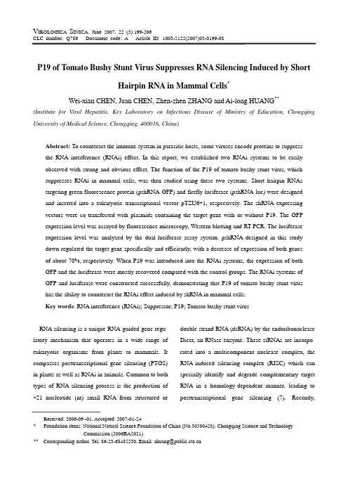
V IROLOGICA S INICA, June 2007, 22 (3):199-206Received: 2006-09 -01, Accepted: 2007-01-24* Foundation items: National Natural Science Foundation of China (No.30500428); Chongqing Science and Technology Commission (2006BA5021)** Corresponding author. Tel: 86-23-68485230, Email: ahuang@200 V IROLOGICA S INICA V ol.22, No 3microRNA (miRNA), another type of small RNA which is derived from intrinsic hairpin RNA precursor, was also highlighted as an important type of RNAi. miRNAs often combine the target gene in the part of 3’ untranslated region by in a complementary but inexact manner which attenuates the translational activity, and may also lead to degradation of target RNA (17).RNAi is postulated to bean ancient immune mecha- nism used by cells to impede the actions of viruses, transgenes, and transposons. It plays an important role in defending invading pathogens while maintaining normal functions of cell development and apoptosis. Consistent with the notion that RNAi is a natural antiviral mechanism, miRNAs related to certain viruses in cells and siRNA derived from viruses in infection process were recently identified. Furthermore, some viruses were found to be able to encode proteins to suppress RNA silencing (9). Thus, viruses can antagonize the cell immune response at the gene level and enhance their ability to survive.In this article, we designed short hairpin RNA (shRNA) targeted to the enhanced green fluorescence protein (EGFP) and luciferase genes expressed by vector in mammal cells and determined their abilities to down-regulate the target genes. Efficient screening systems of RNAi suppressor were established. Using this system, we further demonstrated the function of P19 of tomato bushy stunt virus to antagonist RNAi induced by shRNA in mammal cells.MATERIALS AND METHODSpshRNA constructionsOligonucleotides were synthesized by Shanghai Bioasia Corporation. Sequences corresponding to the siRNA hairpin targets were as follows: shRNA-EGFP (5’-TCGAGGCTGACCCTGAAGTTCATCGAGTAC TGGA TGAACTTCAGGGTCAGCTTTTT-3’)targeting the EGFP gene at sites 526nt-546nt, shRNA-Luc (5’-T CGAGAAGTGTTGTTCCATTCCATTTCAAGAGA ATGGAATGGAACAACACTTTTTTTTT-3’)targeting the luciferase gene at sites 485nt-426nt, and the cor- responding reverse sequences were also synthesized. After annealing, oligonucleotides were cloned into pTZU6+1 with SalⅠand XbaⅠrestriction sites. The constructs were identified by SalⅠdigestion and further confirmed by DNA sequencing analysis. Plasmid expressing P19 fused with his tagpSG5mp19, which contains the DNA sequence of p19 and expresses the P19 protein in mammalian cells, was provided by Prof. Charle (Institut de Biologie Moleculaire des Plantes, France). To detect the protein of P19 expediently, we constructed a plasmid ex- pressing the P19 fused with a his tag. We obtained the DNA sequence from pSG5mp19 by PCR with the primers 5'-AGTCTCGAGACCATGGAACGAGCTA T-3' and 5'-GACGGATCCCTCGCTTTCTTTTTCGA -3'. The sequence was then inserted into the pcDAN- 3.1-myc-his (-) between restriction sites Xho I and Bam H I. After transfecting the plasmids into HepG2 cells, the mRNA expression of P19 and P19-his were confirmed by RT-PCR. Human glyceraldehydes-3- phosphate dehydrogenase (hGAPDH) was detected at the same time as a positive control. Primers for hGAP- DH were 5'-GGCTCTCCAGAACATCA T-3 and 5'-CA CCTGGTGCTCAGTGTA-3'. The protein of P19-his was also confirmed by the immunofluorescent method. The rabbit antibody pointing to the his tag was purc- hased from santan cruz. The second antibody labeled with FITC was from Beijing Zhongshan Corporation.CHEN et al. P19 of T B S Virus Suppresses RNA Silencing Induced by shRNA 201Cell line stably expressing GFPHepG2 cells were cultured at 37℃, 5% CO2 in medium 1640 supplemented with 10% FBS. pEGFP- N1 (promega) was transfected into cells with lipofectamine (invitrogen) according to the manufac- turer’s instructions. 24hr after transfection, G418 (500μg/ml) was added into the culture medium, and the green fluorescence of cells were observed by fluores- cence microscopy every day. Cells producing strong green fluorescence were harvested and individually seeded into 96 well plates, guaranteeing only one cell in each well. Cells producing strong green fluores- cence were amplified. To confirm the integration of the GFP gene into the cell genome, the genomic DNA of HepG2 cells which expressing GFP was extracted by the phenol-chloroform method and then used as the template for PCR to detect GFP. The primers used in this test were as follows: forward primer (5'-GATGG TACCCTA TGGTGAGCAAGGGC-3'), reverse primer (5'-GACAGTACTGCTTGTACAGCTCGTCCA-3'). The genome DNA of hepG2 was used as a control. plasmids transfectionTo study the effect of P19 on the GFP RNAi system, 4 groups of various plasmids were transfected by lipofectamine into the cells of HepG2-GFP as follows: 1) pSG5mp19+pshRNA-GFP; 2) pcDNAp19-his+psh- RNA-GFP; 3) pcDNA3.1-myc-his(-) +pshRNA-GFP;4) pcDNA3.1-myc-his(-)+pshRNA-Luc. To analyze the influence of P19 on luciferase RNAi system, 4 groups of different plasmids were transfected into cells as follows: 1) pSG5mp19+pshRNA-Luc; 2) pc- DNAp19-his+pshRNA-Luc; 3) pcDNA3.1- mychis(-) +pshRNA-LUC; 4) pcDNA3.1-myc-his(-)+pshRNA- GFP. In each group, pGL3 and pRL TK were also transfected. The former was used as reporting gene and the latter was used to normalize the transfection efficiency.Western blottingThe green fluorescence of different groups was observed by fluorescence microscopy every day post- transfection. At 72hr post-transfection, proteins of cell lysis were separated by SDS-PAGE and transferred by electroblotting onto a polyvinyllidene difluoride mem- brane. A rabbit monoclonal antibody directed against eGFP (BD) was used and identified by a second HRP- conjugated antibody (Beijing Zhongshan) through enhanced chemiluminescence (Amersham). Signals were detected with genesnap and quantified with the genetool software. At the same time, actin protein was detected as a control in the same manner with a goat antibody directing actin and a second antibody directing goat IgG (both from Beijing Zhongshan). Semi-quantative RT-PCRTotal RNA was extracted from cultured cells post- transfection with the RNAeasy kit (Qiagen) and then the RNA was digested with DNaseⅠto exclude DNA contamination. To quantify the RNA from EGFP, the hGAPDH was amplified at the same time as a control. Primers for EGFP in the tests were as follows: forward (5'-GCAGCACGACTTCTTCAA -3'), reverse (5'-GT CCATG CCGAGAGTGAT-3'). The PCR products were analyzed by gel electrophoresis and the band was quantified with the genetools software. Luciferase assay48h after transfection, cells were lysed by 1×luciferase passive lysis buffer (Promega) and cen- trifuged at 12 000g for 15 sec, the liquid was used to detect luciferase activity by a multi-function enzyme analysizer (Gene corp). The relative activity of firefly luciferase was counted by normalizing to renal luciferase.202 V IROLOGICA S INICA V ol.22, No 3The reporter values represented averages ±1 SD from at least three independent transfections.RESULTSThe cell line of HepG2-GFP expressing GFP stably To construct a RNA interference model in mammal cells, we established the cell line stably expressing GFP (the cell line was named HepG2-GFP) by G418 selection culture. We obtained a cell line with strong green fluorescence observed by fluorescence micro- scopy after one-month culture. The insertion of EGFP into genome DNA was then confirmed by PCR. We detected the fragment of EGFP with the genome DNA as the template (Fig.1. lane 1) whereas the same fragment did not appear in the control test (Fig.1). P19 and P19-his expressed in mammal cells The tomato bushy stunt virus is a type of plant virus. To confirm its expression in mammal cells, we detected at the mRNA and protein levels by different methods. The mRNA of P19 were detected in cells of HepG2-GFP and HepG2 transfected with the Psg- 5mp19 or pcDNAp19-his respectively by RT-PCR. We furthermore detected the P19-his protein by an antibody targeting the his tag using the immu- nofluenscent method and P19-his proteins wereobserved to be primarily located in the plasma.Fig. 1. EGFP gene was detected in the genome DNA of HepG2.GFP by PCR. 1, DNA fragment for EGFP was amplified from the genome DNA in HepG2.GFP; 2, Genome DNA of HepG2 was used as negative control; 3, DNA marker.P19 recovered the expression of GFP down- regulated by shRNAWe next designed the siRNA targeting EGFP tosuppress its expression. Here, a vector expressing strategy and a vector with RNA Ⅲ promoter (U6) was chosen. The vector pTZU6+1 can drive the trans- cription of short hairpin RNA precisely, which would be transferred into a functional type of siRNA in mammal cells by the Dicer. When the HepG2-GFP was transfected with the plasmid pshRNA-GFP, the fluorescence was reduced significantly compared with the control group, which was transfected with the plasmid pshRNA-Luc (Fig. 2.A). This was consistent with the results from western blotting (Fig. 2.B). Analysis with the Genetools software indicated the amount of GFP decreased by 70%. To determine the influence on mRNA levels, we further detected mRNA by a semi-quantative RT-PCR test and found that pshRNA-GFP lead a decrease in mRNA level of EGFP (Fig. 2.C) by 78%. Therefore it could be concluded that the shRNA down-regulated the expres- sion of EGFP and the down-regulation was a con- sequence of the degradation of mRNA. On the basis of the successful RNAi system described above, we studied the ability of P19 to suppress the RNAi effect in mammalian cells. When P19 and shRNAGFP were co-expressed in HepG2.GFP, we observed the phenol menon that the fluorescence recovered to a significant intensity compared with those cells without P19. The effect on efficiency on GFP expression was further evaluated by western blot for protein levels and by RT-PCR for mRNA levels. In these tests, P19recovered the GFP protein expression as well as mRNA expression (Fig. 3) increasing the expression of GFP by 80% and mRNA levels by 70% compared to the controls.CHEN et al. P19 of T B S Virus Suppresses RNA Silencing Induced by shRNA 203Fig. 2.P19 counteract the effect of shRNA on GFP expression. A: fluorescence observed by fluorescent microscope. ShRNA-GFP down-regulate fluorescence intensity whereas the P19 recovered the fluorescence in hepG2.GFP. B: The GFP levels were observed decreased by the shRNA-GFP and increased when P19 was introduced. C: mRNA was found decreased when shRNA existed whereas P19 recovered the mRNA level of GFP.Fig. 3. P19 expressed in mammal cells detected in the level of protein and mRNA. A: P19-his was observed by immunnofluorescent method in hepG2 cells transfected with pCDNAp19-his. Cells transfected with pCDNA3.1-myc-his was used as negative control (NC). P19-his was observed located in the plasma mostly. B: mRNA of P19 was also detected in the cells transfected with pSG5mp19 or pCDNAp19-his, respectively. mRNA extracted from Cells transfected with pCDNA3.1-myc-his was used as negative control. A fragment of hGAPDH gene was amplified in all three samples while fragment of P19 was only amplified from cells transfected with pSG5mp19 and pCDNAp19-his.P19 rescued the expression of luciferase in RNAi systemTo further understand the shRNA interference efficiency, we designed siRNA to target firefly luciferase. When the cells were transfected with pshRNA-Luc and the reporter vector, pGL3 as control (Promega), which expresses firefly luciferase under the control of SV40 promoter, the luciferase activity204 V IROLOGICA S INICA V ol.22, No 3Fig. 4. P19 rescued the luciferase expression in the RNAi system. The relative luciferase activity was counted by firefly luciferase activity devided by rena luciferase activity and the value of control group was standardized to 100. when shRNA-Luc was introduced, the firefly luciferase decreased significantly. When P19 was added into the RNAi system, luciferase activity recovered to a high level.reduced by 70% (Fig. 4) compared with the control group. It showed that the shRNA-Luc designed could down-regulate the expression of luciferase gene efficiently. When it was studied by the luciferase RNAi system, P19 was also found to be able to recover the luciferase activity significantly. Compared with the control group, when P19 was introduced into the cells, the relative luciferase activity increased to about 80%. The his tag did not impair the function of P19. Results from the luciferase RNAi system coincided with those from the GFP RNAi system.DISCUSSIONSRNAi is an ancient immune surveillance mechanism on gene level. It was shown to act as an efficient antiviral system in plant and insect cells and might also played an antiviral role in mammal cells (2,11). To counteract the antiviral effect of RNAi and enhance their existing ability, many plant and insect viruses express different RNAi suppressor proteins (14). These proteins always play important roles in the virus infection process and are important pathogens (12). The first identified RNAi suppressor, HC-pro of tobacco etch potyvirus (TEV), was found when researchers studied the co-infection phenomenon in plants (8). Later, some other RNAi suppressors encoded by plant viruses were discovered and the mechanism of RNAi inhibition became better under- stood (15). Furthermore, several animal viruses such as flock house virus, influenza virus and reovirus were also found to encode proteins having the same effect as an RNAi suppressor (19,10). Recently, HIV-1 and PFV-1 (primate foamy virus type 1, a retrovirus similar to HIV)were found to be able to produce such RNAi suppressors too (1,3). Interestingly, HIV-1 can produce a siRNA in the infected cells to down- regulate its Env expression while a cellular miRNA was verified to target the sequence of PFV-1 and could restrict the accumulation of PFV-1. These reports indicated that RNAi mechanism may also play an important role in vertebrate cells and the RNAi suppressor exists as an counteraction strategy to this antiviral mechanism.Similar to the phenomenon of RNAi, RNAi suppressor was firstly studied in the field of plant research. Nowadays, we have known that RNAi suppressors could take effect at different steps in the RNAi pathway (14). HC-pro, δ3 factor of reovirus, and NS1 of the influenza virus countact RNAi by binding long dsRNA and reduce production of siRNA. Tat of HIV-1 can also limit the production of siRNA by influencing the activities of Dicer. Some suppres- sors such as P19 can bind the siRNAs and prohibit them into RISC (4). Other suppressors may also act at various steps. For example, some of them may influence the activities of members of RISC, and some of them may limit the transduction of systematic silencing signals in cells (18). In summary, althoughCHEN et al. P19 of T B S Virus Suppresses RNA Silencing Induced by shRNA 205much of the mechanism of RNAi suppressing is still to be studied, we can concluded that different suppressors may have different interference methods and show different abilities to inhibit RNA silence.To date, miRNA was regarded as a kind of siRNA and considered part of the RNAi process by some researchers although single strain miRNA has some differences to short double strain RNA. miRNAs were also produced by Dicer and incorporated into RISC at last. miRNA and siRNA crossed partly in their pathway at least. If a protein could influence RNAi, it may also influence miRNAs. Since miRNAs have very important roles in keeping normal development and normal biological activity of cells, cells would be influenced when miRNA levels were changed. Patrice D et al studied the influence on miRNAs of several suppressors and discovered that most of them showed an obvious effect and could produce abnormalities (13). We could also predict the pathogenesis of the suppressor of animal viruses by influencing miRNA function. For example, a persistant production of the SRS in chronic virus infection may help to produce tumor and other chronic diseases.Tomato bushy stunt virus is a 4.7k nt plus RNA virus which infect agriculture plants and herbs. The P19 is essential for the viral pathogenity since it can enhance the ability of the virus to survive in infected plants by counteracting the RNAi system. It has been established that the RNAi mechanism is the most important defence strategy in plants. Daniel et al firstly reported that P19 could inhibit the RNAi effect in plants and pointed out it could combine the siRNAs and prohibit it incorporate into RISC. Recently, Ye et al elaborated the crystal structure of P19 and explained the physiochemical basis of its ability to combine siRNAs (5). In this report, RNAi systems targeting GFP and luciferase gene were constructed and used to identify the RNAi suppressor characteristics of P19. The results showed that P19 could also suppress RNAi effects induced by short hairpin RNAs in mammal cells as well as suppressing RNAi induced by synthetic siRNA or long dsRNA in plants. Our research confirmed that the suppressing ability of P19 was non-sequence specific for it suppressed both the RNAi targeting GFP and the RNAi targeting luciferase. Furthermore, our study showed that the RNAi system in mammal cells induced by vector derived shRNA is suitable for screening RNAi suppressors, and could be a more efficient approach compared with the methods such as transgenic plant models or virus infection models.Scientists attached much importance to therapy exploitation of RNAi in viral infection diseases and cancer diseases when synthetic siRNAs were found to be able to down-regulate the homologous gene expres- sion by activating the RNAi mechanism in mammal cells (6). Today, much improvement has been achieved in this field (16). But the discovery of RNAi suppressors encoded by viruses will bring some new questions to the application of siRNA drugs. Benasser pointed out that HIV could resist the persistent vector-derived shRNAs since its Tas was a RNAi suppressor (1). It may also be the case that some mammal viruses such as HCV, HBV and SARS-coV may also encode such RNAi suppressors and these factors could influence the therapy strategy of RNAi. There are still many unknown factors to be studied in this field. The knowledge of RNAi suppressor will not only enrich our understanding of RNAi phenomenon and interaction between virus and host but also help206 V IROLOGICA S INICA V ol.22, No 3the exploitation of RNAi as a therapy strategy.Reference s1.Bennasser Y, Le S Y, Benkirane M, et al. 2005. Evidencethat HIV-1 encodes an siRNA and a suppressor of RNA silencing. Immunity, 22 (5): 607-619.2.Carrington J C, Ambros V. 2003. Role of microRNAs inplant and animal development. Science, 301: 336-338.3.Charles H L, Patrice D, Khalil A, et al.2005. A cellularmicroRNA mediates antiviral defense in human cells.Science, 308 (22): 557-560.4.Daniel S, Attila M, Alessandra L, et al. 2002. A viralprotein suppresses RNA silencing and binds silencing- generated, 21- to 25- nucleotide double-stranded RNAs.EMBO J, 21 (12): 3070-3080.5.Donald K. 2002. Breakthrough of the Year. Science, 298:2283.6.Elbashir S M, Harborth J, Lendeckel W, et al. 2001.Duplexes of 21-nucleotide RNAs mediate RNA inter- ference in culture mammalian cells. Nature, 411: 494-498.7.Hannon G J. 2002. RNA interference. Nature, 418: 244-251.8.Kasschau K D, Xie Z, Allen E, et al. 2003. P1/HC-Pro, aviral suppressor of RNA silencing, interferes with Arabidopsis development and miRNA function. Dev Cell,4 (2): 205-217.9.Li H W, Li W X, Ding S W. 2002. Induction andsuppression of RNA silencing by an animal virus. Science, 296: 1319-1321.10.Li W X, Li H W, Lu R, et al. 2004. Interferon antagonistproteins of influenza and vaccinia viruses are suppressors of RNA silencing. Proc Natl Acad Sci USA, 101 (5): 1350-1355.11.Matthew NP, Lena E, Jan K,et al. 2004. A pancreaticislet-specific microRNA regulates insulin secretion.Nature, 432: 226-230.12.Mallory AC, Reinhart B J, Bartel D, et al. 2002. A viralsuppressor of RNA silencing differentially regulates the accumulation of short interfering RNAs and micro-RNAs in tobacco. Proc Natl Acad Sci USA, 99 (23): 15225- 15233.13.Patrice D, Charles H L, Eneida A P, et al. 2004. Probingthe microRNA and small interfering RNA pathways with virus-encoded suppressors of RNA silencing. Plant cell,16: 1235-1250.14.Rajendra M, Radhamani A, Trent H S, et al. 2000. RNAviruses as inducers, suppressors and targets of post- transcriptional gene silencing. Plant Mol Biol, 43: 295-306.15.Roth B M, Pruss G J, Vance V B. 2004. Plant viralsuppressors of RNA silencing. Virus Res, 102 (1): 97-108 16.Stevenson M. 2004. Therapeutic Potential of RNAInterference. N Engl J Med, 351: 1772-1777.17.Vicki V, Herve V. 2001. RNA silencing in plants---defense and counterdefense. Science, 292 (22): 2277- 2280.18.Westhof E. 2004. How to silence silencing. Chem. Biol,11 (2): 158-160.19.Zsuzsanna L, Daniel S, Jozsef B. 2003. Double-strandedRNA-binding proteins could suppress RNA interference- mediated antiviral defences. J Gen Virol, 84: 975-980.。