胰蛋白酶(Trypsin)试剂盒说明书
胰酶得配制
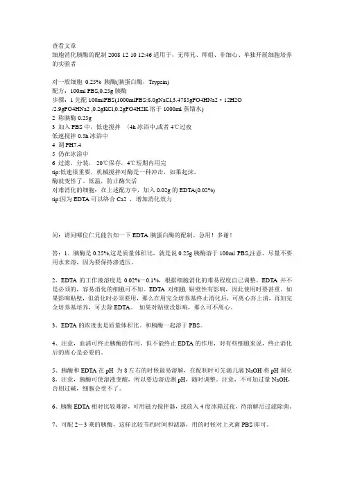
查看文章细胞消化胰酶的配制2008-12-10 12:46适用于,无师兄、师姐、非细心、单独开展细胞培养的实验者对一般细胞0.25% 胰酶(胰蛋白酶,Trypsin)配方:100ml PBS,0.25g胰酶步骤:1先配100mlPBS(1000mlPBS:8.0gNaCl,3.4785gPO4HNa2·12H2O/2.9gPO4HNa2 ,0.2gKCl,0.2gPO4H2K溶于1000ml蒸馏水)2 称胰酶0.25g3 加入PBS中,低速搅拌〈4h冰浴中,或者4℃过夜低速搅拌0.5h冰浴中4 调PH7.45 仍在冰浴中6 过滤,分装,-20℃保存,4℃短期内用完tip:低速很重要,机械搅拌对酶是一种冲击,如果起沫,酶就变性了。
低温,防止酶失活对难消化的细胞:在上述配方中,加入0.02g的EDTA(0.02%)tip:因为EDTA可以络合Ca2 ,增加消化效力问:请问哪位仁兄能告知一下EDTA-胰蛋白酶的配制。
急用!多谢!答:1、胰酶是0.25%,这是质量体积比,就是说0.25g胰酶溶于100ml PBS,注意,尽量不要用水来溶,因为要保持渗透压。
2、EDTA的工作液溶度是0.02%-0.1%,根据细胞消化的难易程度自己调整。
EDTA并不是必须的,容易消化的细胞可不加。
EDTA对细胞贴壁性有影响,因此使用时要甚重。
如果影响贴壁,但消化时必须要用,那么在用完全培养基终止消化后,可离心弃上清,再加完全培养基培养,可去除EDTA。
如果对贴壁没影响,那么可不离心。
3、EDTA的浓度也是质量体积比。
和胰酶一起溶于PBS。
4、注意,血清可终止胰酶的作用,但不能终止EDTA的作用,对有些细胞来说,终止消化后的离心是必要的。
5、胰酶和EDTA在pH 为8左右的时候最易溶解,在配制时可先滴几滴NaOH将pH调至8,注意,胰酶可使溶液变酸,所以要边溶边测pH,随时调整。
注意,不可加过量NaOH,否则过碱,细胞会受不了。
201404210936_美国药典34版胰蛋白酶质量标准Trypsin-USP34
Crystallized Trypsin» Crystallized Trypsin is a proteolytic enzyme crystallized from an extract of the pancreas of healthy bovine or porcine animals, or both. When assayed as directed herein, it contains not less than 2500 USP Trypsin Units in each mg, calculated on the dried basis, and not less than 90.0 percent and not more than 110.0 percent of the labeled potency.[NOTE—Determine the suitability of the substrates and check the adjustment of the spectrophotometer by performing the Assay using USP Crystallized Trypsin Reference Standard. ]Packaging and storage— Preserve in tight containers, and avoid exposure to excessive heat.USP R EFERENCE STANDARDS11—USP Trypsin Crystallized RSSolubility test— An amount, equivalent to 500,000 USP Trypsin Units, is soluble in 10 mL of water and in 10 mL of saline TS.M ICROBIAL ENUMERATION TESTS61 and T ESTS FOR SPECIFIED MICROORGANISMS62— It meets the requirements of the tests for absence of Staphylococcus aureus, Pseudomonas aeruginosa, and Salmonella species.L OSS ON DRYING731— Dry it in vacuum at 60 for 4 hours: it loses not more than5.0% of its weight.R ESIDUE ON IGNITION281: not more than 2.5%.Limit of chymotrypsin—0.067 M Phosphate buffer , pH 7.0—Dissolve 4.54 g of monobasic potassium phosphate in water to make 500 mL of solution. Dissolve 4.73 g of anhydrous dibasic sodium phosphate in water to make 500 mL of solution. Mix 38.9 mL of the monobasic potassium phosphate solution with 61.1 mL of dibasic sodium phosphate solution. Adjust dropwise, if necessary, with dibasic sodium phosphate solution to a pH of 7.0.Substrate solution— Dissolve 23.7 mg of N-acetyl-L-tyrosine ethyl ester, suitable for use in determining chymotrypsin, in about 50 mL of 0.067 M Phosphate buffer, pH 7.0 with warming. When cool, dilute with additional pH 7.0 buffer to 100 mL. (Substrate solution may be stored in the frozen state and used after thawing; it is important, however, to freeze immediately after preparation.)Crystallized Trypsin solution— Dissolve a sufficient quantity of Crystallized Trypsin, accurately weighed, in 0.0010 N hydrochloric acid to obtain a solution containing 650 USPTrypsin Units per mL.Procedure— Conduct the test in a suitable spectrophotometer equipped to maintain atemperature of 25 ± 0.1 in the cell compartment. Determine the temperature in the reaction cell before and after the measurement of absorbance to ensure that the temperature does not change by more than 0.5. Pipet 200 µL of 0.0010 N hydrochloric acid and 3.0 mL of the Substrate solution into a 1-cm cell. Place this cell in the spectrophotometer, and adjust the instrument so that the absorbance reads 0.200 at 237 nm. Pipet 200 µL of Crystallized Trypsin solution into another 1-cm cell, add 3.0 mL of the Substrate solution, and place the cell in the spectrophotometer. [NOTE—This order of addition is to be followed. ] At the time the Substrate solution is added, start a stopwatch, and read the absorbance at 30-second intervals for not less than 5 minutes. Repeat the procedure on the same dilution at least once. Absolute absorbance values are of less importance than the constancy of the rate of change of absorbance. If the rate of change does not remain constant for at least 3 minutes, repeat the run, and if necessary, use a lower concentration. The duplicate run at the same dilution should match the first run in rate of absorbance change. Determine the average absorbance change per minute, using only the values within the 3-minute portion of the curve where the rate of absorbance is constant. Plot a curve of absorbance against time. One USP Chymotrypsin Unit is the activity causing a change in absorbance of 0.0075 per minute under the conditions specified in this test. Calculate the number of USP Chymotrypsin Units per mg of Crystallized Trypsin taken by the formula:(A2A1) / (0.0075TW)in which A2 is the absorbance straight-line initial reading, A1is the absorbance straight-line final reading, T is the elapsed time, in minutes, between the initial and final readings, and W is the weight, in mg, of Crystallized Trypsin in the volume of solution used in determining the absorbance. Not more than 50 USP Chymotrypsin Units per 2500 USP Trypsin Units is found, indicating the presence of not more than approximately 5% of chymotrypsin.Assay—0.067 M Phosphate buffer , pH 7.6—Dissolve 4.54 g of monobasic potassium phosphate in water to make 500 mL of solution. Dissolve 4.73 g of anhydrous dibasic sodium phosphate in water to make 500 mL of solution. Mix 13 mL of the monobasic potassium phosphate solution with 87 mL of the anhydrous dibasic sodium phosphate solution.Substrate solution— Dissolve 85.7 mg of N-benzoyl-L-arginine ethyl ester hydrochloride, suitable for use in assaying Crystallized Trypsin (see N OTE), in water to make 100 mL. Dilute 10 mL of this solution with 0.067 M Phosphate buffer, pH 7.6 to 100 mL. Determine the absorbance of this solution, in a 1-cm cell, at 253 nm, in a suitable spectrophotometerequipped with thermospacers to maintain a temperature of 25 ± 0.1, using water as the blank. By the addition of 0.067 M Phosphate buffer , pH 7.6, or of the Substrate solution before dilution, adjust the absorbance so that it measures not less than 0.575 and not more than 0.585. Use this Substrate solution within 2 hours.Crystallized Trypsin solution — Dissolve a sufficient quantity of Crystallized Trypsin,accurately weighed, in 0.0010 N hydrochloric acid to obtain a solution containing about 50 to 60 USP Trypsin Units per mL.Procedure — Pipet 200 µL of 0.0010 N hydrochloric acid and 3.0 mL of the Substratesolution into a 1-cm cell. Place this cell in a spectrophotometer, and adjust the instrument so that the absorbance reads 0.050 at 253 nm. Pipet 200 µL of Crystallized Trypsin solution , containing 10 to 12 USP Trypsin Units, into another 1-cm cell, add 3.0 mL of Substrate solution , and place the cell in the spectrophotometer. At the time the Substrate solution is added, start a stopwatch, and read the absorbance at 30-second intervals for 5 minutes. Repeat the procedure on the same dilution at least once. Plot a curve ofabsorbance against time, and use only those values that form a straight line to determine the activity of the Crystallized Trypsin. If the rate of change does not remain constant for at least 3 minutes, repeat the run, and if necessary, use a lower concentration. One USP Trypsin Unit is the activity causing a change in absorbance of 0.003 per minute under the conditions specified in this Assay. Calculate the number of USP Trypsin Units per mg taken by the formula:(A 1 A 2) / (0.003TW )in which A 1 is the absorbance straight-line final reading, A 2 is the absorbance straight-line initial reading, T is the elapsed time, in minutes, between the initial and final readings, and W is the weight, in mg, of Crystallized Trypsin in the volume of solution used indetermining the absorbances.Auxiliary Information — Please check for your question in the FAQs before contactingUSP. Topic/Question Contact Expert Committee Monograph Fouad Atouf, Ph.D.Senior Scientific Liaison1-301-816-8365(BIO12010) Monographs - Biologics and Biotechnology 1Reference Standards RS Technical Services1-301-816-8129rstech@61Radhakrishna STirumalai, Ph.D.Principal Scientific Liaison1-301-816-8339(GCM2010) General Chapters - Microbiology 62Radhakrishna STirumalai, Ph.D. (GCM2010) General Chapters -MicrobiologyPrincipal Scientific Liaison1-301-816-8339USP34–NF29 Page 4535Pharmacopeial Forum: Volume No. 32(3) Page 779。
胰蛋白酶

基本信息
物化性质
胰蛋白酶是从牛、猪、羊的胰脏提取,纯化获得的结晶,再制成的冻干制剂。易溶于水,不溶于三氯甲烷、 乙醇、乙醚等有机溶剂。在pH1.8时,短时间煮沸几乎不失活;在碱溶液中加热则变性沉淀,Ca2+有保护和激活 作用,胰蛋白酶的等电点为pH10.1。
牛胰蛋白酶原有229个氨基酸组成,含6对二硫键,其氨基酸排列顺序和晶体结构已被阐明。在肠激酶活自身 催化下,酶原的N末端赖氨酸与异亮氨酸残基之间的肽键被水解,释放出来缬-天-天-天-天-赖6肽,生成有活性 的胰蛋白酶。牛的胰蛋白酶氨基酸残基223个,分子量23800 ,活性部位的丝氨酸残基是不可缺少的丝氨
胰蛋白酶能使痰、血凝块溶化变稀,易于引流排痰,加速创面愈合净化,促进肉芽组织增生,而不损伤正常 组织,临床上有消炎消肿功能。
其他
细胞培养
胰蛋白酶的作用是使细胞间的蛋白质水解从而使细胞离散。不同的组织或者细胞对胰酶的作用反应不一样。 胰酶分散细胞的活性还与其浓度、温度和作用时间有关,在 pH为 8.0、温度为 37℃时,胰酶溶液的作用能力最 强。使用胰酶时,应把握好浓度、温度和时间,以免消化过度造成细胞损伤。因 Ca2+、 Mg2+和血清、蛋白质可 降低胰酶的活性,所以配制胰酶溶液时应选用不含 Ca2+、 Mg2+的 BSS,如: D-Hanks液。终止消化时,可用 含有血清培养液或者胰酶抑制剂终止胰酶对细胞的作用。
1.用于脓胸、血胸、外科炎症、溃疡、创伤性损伤、娄管等所产生的局部水肿、血肿、脓肿。 2.用于呼吸道疾患溶解黏痰和脓性痰。 3.用于治疗毒蛇咬伤,曾试用于竹叶青、银环蛇、眼镜蛇、蝮蛇等毒蛇咬伤的各型病人800余例,均获治愈。
1.肝肾出血、出血倾向及结核性脓肿患者禁用。 2.不可用于急性炎症及出血空腔中。
胰蛋白酶-EDTA溶液
胰蛋白酶-EDTA溶液(0.25%:0.02%)产品简介:胰蛋白酶(Trypsin)是由胰脏产生没有活性的胰蛋白酶原分泌到小肠后,小肠内的肠肽酶会活化该酶原,形成胰蛋白酶。
胰蛋白酶的特点还在于已经活化的胰蛋白酶,能够继续活化更多胰蛋白酶原,这种过程即自动催化。
胰蛋白酶在小肠工作,它会将蛋白质水解为肽,进而分解为氨基酸,其最适温度约为37℃。
Leagene的 Trypsin-EDTA solution,含0.25%胰酶、0.02%EDTA。
该溶液经过过滤除菌,可以直接用于培养细胞的消化,或者一些组织的消化。
该产品通常室温消化1min 左右就可以消化下大多数贴壁细胞。
主要成分:主要由0.25%胰酶、0.02%EDTA等组成,不含酚红,过滤除菌。
自备材料:1、微量移液器2、PBS、Hanks液或无血清培养液3、显微镜4、离心机操作步骤(仅供参考):1、贴壁细胞的消化①吸除培养液,用无菌的PBS、Hanks液或无血清培养液洗涤细胞一次,以去除残余的血清。
②加入少量Trypsin-EDTA solution,略盖过细胞即可,室温放置0.5~2min,不同的细胞消化时间有所不同。
③显微镜下观察,细胞明显收缩,并且肉眼观察培养器皿底部发现细胞的形态发生明显的变化;或者用枪吹打细胞发现细胞刚好可以被吹打下来。
此时吸除胰酶细胞消化液。
加入含血清的完全细胞培养液,吹打下细胞,即可直接用于后续实验。
④如果发现消化不足,则加入Trypsin-EDTA solution重新消化。
⑤如果发现细胞消化时间过长,未及吹打细胞,细胞已经有部分直接从培养器皿底部脱落,直接用胰酶细胞培养液把细胞全部吹打下来。
1000~2000g离心1min,沉淀细胞,尽量去除胰酶细胞消化液后,加入含血清的完全培养液重新悬浮细胞,即可用于后续实验。
2、组织的消化不同的组织需要消化的时间相差很大,通常以消化后可以充分打散组织为宜。
注意事项:1、尽量减少反复冻融的次数,以免失效。
胰蛋白酶 Trypsin
胰蛋白酶Trypsin (Parenzyme) 为蛋白酶的一种,EC3.4.21.4。
在脊椎动物中,作为消化酶而起作用。
在胰脏是作为酶的前体胰蛋白酶原而被合成的。
作为胰液的成分而分泌,受肠激酶,或胰蛋白酶的限制分解成为活化胰蛋白酶,是肽链内切酶,它能把多肽链中赖氨酸和精氨酸残基中的羧基侧切断。
它不仅起消化酶的作用,而且还能限制分解糜蛋白酶原、羧肽酶原、磷脂酶原等其它酶的前体,起活化作用。
是特异性最强的蛋白酶,在决定蛋白质的氨基酸排列中,它成为不可缺少的工具。
牛的胰蛋白酶氨基酸残基223个,分子量为23300,活性部位的丝氨酸残基是不可缺少的丝氨酸蛋白酶。
除存在于脊椎动物外,还存在于蚕、海盘车、蝲姑、放线菌等范围广泛的生物体中。
另外与高等动物的血液凝固和炎症等有关的凝血酶、纤溶酶、舒血管素等蛋白酶在化学结构和特异性等方面与胰蛋白酶具有密切的关系,可以认为这些酶是从共同的祖先酶在进化过程中分化而来的。
胰糜蛋白酶与弹性蛋白酶在结构和催化机制方面也具有密切关系,但其特异性则完全不同。
胰蛋白酶系自牛、羊或猪胰中提取的一种蛋白水解酶。
中国药品标准规定按干燥品计算,每1mg 的效价不得少于2500单位。
由牛、羊、猪胰脏提取而得的一种肽链内切酶,只断裂赖氨酸或精氨酸的羧基参与形成的肽键。
白色或米黄色结晶性粉末。
溶于水,不溶于乙醇、甘油、氯仿和乙醚。
分子量24 000,pI 10.5,最适pH值7. 8~8.5左右。
pH值2~3是稳定的。
pH值5以上易自溶失活。
pH>9.0不可逆失活。
Ca2+对酶活性有稳定作用;重金属离子、有机磷化合物、DFP、天然胰蛋白酶抑制剂对其活性有强烈抑制。
临床用于抗炎消肿,工业上用于皮革制造、生丝处理、食品加工等。
作用与用途本品为蛋白质水解酶,能选择地水解蛋白质中由赖氨酸或精氨酸的羧基所构成的肽链,能消化溶解变性蛋质,对未变性的蛋白质无作用,因此,能使脓、痰液、血凝块等分解、变稀,易于引流排除,加速创面净化,促进肉芽组织新生,此外还有抗炎症作用。
胰蛋白酶比活力测定的实验操作
胰蛋白酶比活力测定的实验操作胰蛋白酶(trypsin)是一种重要的消化酶,用于蛋白质的降解。
测定胰蛋白酶的比活力可以评估其酶活性及纯度。
下面是测定胰蛋白酶比活力的实验操作步骤。
实验所需材料:1.胰酶溶液2. Brain Heart Infusion (BHI)培养基3. 丙氨酰-L-苯丙氨酰-L-色氨酸对(L-Tyr-L-Phe)4.0.1M氢氧化钠(NaOH)溶液5.0.1M硫酸(H2SO4)溶液实验步骤:1.准备酶样品:取适量的胰酶溶液,可以是商业制备的纯胰蛋白酶或者胰腺提取物。
2. 酶样品稀释:将胰酶溶液与BHI培养基按照一定比例混合,使得最终浓度在0.5-1.2mg/mL之间。
3.加入底物:向每个试管中加入20μL的丙氨酰-L-苯丙氨酰-L-色氨酸对底物。
4.孵育反应:将试管置于37℃恒温培养箱中,在设定的时间内进行酶催化反应。
5.终止反应:用加入2NNaOH溶液终止酶反应,使底物水解停止。
6.酶解产物的提取:将反应液离心,取上清液转移到新的离心管中。
实验测量方法:1. 分光光度计法:利用胰酶催化底物水解产生的对分光光度计可见吸收的产物进行测定。
用1cm光程的比色皿,将底物水解产物的吸光度读数记录下来。
2. pH差比色法:用pH差比色法测量胰酶反应液中产生的酪氨酸分解产物。
黄色小麦胚芽酪氨酸(Tyr)的醛缩反应,其产物具有红色或紫色,在526nm处吸收峰。
通过比较不同样品的吸收度可以得出胰酶比活力。
数据处理:1.分光光度计法:根据底物水解产物的吸光度,绘制标准吸光度-底物浓度曲线,利用该曲线确定底物浓度,进一步计算胰酶的比活力。
2.pH差比色法:计算样品吸收值,将其与标准吸光度曲线相对应的底物浓度进行比较,计算胰酶的比活力。
注意事项:1.操作要严格遵守无菌技术,以避免细菌或其他污染物对实验结果的干扰。
2.实验过程中可以选择不同的底物和测量方法,以适应实验目的和条件。
3.实验操作需谨慎,避免接触眼睛和口腔,注意个人安全。
无酶细胞消化液(含酚红)
无酶细胞消化液(含酚红)简介:胰蛋白酶(Trypsin)是由胰脏产生没有活性的胰蛋白酶原分泌到小肠后,小肠内的肠肽酶会活化该酶原,形成胰蛋白酶。
胰蛋白酶经常用于动物组织的培养细胞消化或者一些组织的消化,但对细胞有的潜在损害,尤其是在37℃环境下危害较大。
Leagene 无酶细胞消化液(含酚红)(Non-enzy Cell Detach Solution)其特点是:1、作用温和;2、对细胞的损伤和破坏极小,不影响细胞生物学特性,是肿瘤细胞的极好细胞脱壁方法;3、可以在血清存在的情况下进行消化,该消化液适用于消化肿瘤、脑、肝、肾、肺组织等,尤其适用于上皮组织。
组成:操作步骤(仅供参考):1、贴壁细胞的消化①吸除培养液,用无菌PBS、培养液洗涤细胞1次。
②加入少量Non-enzy Cell Detach Solution(含酚红),略盖过细胞即可(一般按细胞的有效体积的10倍添加)。
③室温放置,如置于37℃脱壁反应会加速,直到细胞完全脱壁,不同的细胞消化时间有所不同。
④加入细胞培养液或5倍体积PBS 缓冲液终止反应。
如果发现消化不足,则加入Non-enzy Cell Detach Solution(含酚红)重新消化。
⑤离心,沉淀细胞,弃上清,尽量去除Non-enzy Cell Detach Solution(含酚红),加入含血清的完全培养液重新悬浮细胞,即可用于后续实验。
2、组织的消化①PBS、培养液清洗组织1次,用无菌的刮勺或茶匙将剪碎的组织碎片转移至合适容器。
②按组织有效体积的5~10倍,加入Non-enzy Cell Detach Solution(含酚红)。
③置于37℃作用,无需振荡,不同的细胞消化时间有所不同。
对于较难分解的肿瘤细胞,可作用5天或更长时间,但应重新用消化液悬浮。
④吹打组织碎片数次,释放松散的细胞,轻轻晃动培养瓶或皿。
⑤离心,沉淀细胞,弃上清,尽量去除Non-enzy Cell Detach Solution(含酚红),加入含编号名称CC0123StorageNon-enzy Cell Detach Solution(含酚红)100ml -20℃使用说明书1份血清的完全培养液重新悬浮细胞,即可用于后续实验。
roche cooktail蛋白酶成分
roche cooktail蛋白酶成分罗氏鸡尾酒(Roche鸡尾酒)是一种用于生物科学研究的常用试剂盒,其中包含多种蛋白酶。
蛋白酶是一类能够加速蛋白质降解的酶类分子,它们具有多种不同的功能和特性。
以下将介绍Roche鸡尾酒中常见的蛋白酶成分。
1.胰蛋白酶(Trypsin):胰蛋白酶是一种消化酶,由胰腺分泌,用于在消化过程中分解蛋白质。
在Roche鸡尾酒中,胰蛋白酶被用于体外细胞培养中,用于细胞的离析和传代。
它能够通过剪切蛋白质的肽键来降解蛋白质分子。
2.胰蛋白酶抑制剂(Trypsin Inhibitor):胰蛋白酶抑制剂是一种能够抑制胰蛋白酶活性的分子。
在Roche鸡尾酒中,胰蛋白酶抑制剂被用于停止胰蛋白酶的活性,从而控制蛋白质降解的过程。
3.蛋白激酶抑制剂(Protein Kinase Inhibitors):蛋白激酶抑制剂是一类能够抑制蛋白激酶活性的分子。
在Roche鸡尾酒中,蛋白激酶抑制剂被用于研究细胞信号传导的过程。
它们能够干扰蛋白激酶的活性,从而影响细胞内信号传递通路。
4.磷酸酶抑制剂(Phosphatase Inhibitors):磷酸酶是一类能够去磷酸化蛋白质的酶。
在Roche鸡尾酒中,磷酸酶抑制剂用于抑制磷酸酶活性,从而保持蛋白质磷酸化状态。
5.蛋白酶抑制剂混合物(Protease Inhibitor Cocktail):这是Roche鸡尾酒中的一个重要成分,它由多种蛋白酶抑制剂组成,能够抑制多种蛋白酶的活性。
蛋白酶抑制剂混合物能够保护蛋白质免受蛋白酶降解的影响,从而保持蛋白质的稳定性。
Roche鸡尾酒中的这些蛋白酶成分在生物科学研究中发挥着重要作用。
它们能够帮助研究人员保护蛋白质的完整性,提高实验的可靠性和准确性。
另外,这些蛋白酶成分还能够用于调节细胞内的信号传导过程,从而揭示细胞生物学的各个方面。
需要注意的是,Roche鸡尾酒中的蛋白酶成分不能用于食品加工或药品制造。
它们仅限于科学研究中的使用,需要严格遵守实验室安全操作规程。
胰蛋白酶的提取
胰蛋白酶的提取原理和方法步骤2010-03-29 14:21:31 来源:易生物实验浏览次数:404 网友评论0 条在动物胰脏中除了存在胰蛋白酶外,还有另外两种与胰蛋白酶性质相似的蛋白水解酶:胰凝乳蛋白酶和弹性蛋白酶。
在胰蛋白酶提取过程中,三者彼此很难分开。
需采用合适的方法进一步分离纯化。
关键词:胰蛋白酶trypsin[原理]在动物胰脏中除了存在胰蛋白酶外,还有另外两种与胰蛋白酶性质相似的蛋白水解酶:胰凝乳蛋白酶和弹性蛋白酶。
在胰蛋白酶提取过程中,三者彼此很难分开。
需采用合适的方法进一步分离纯化。
从动物胰脏中提取胰蛋白酶,一般是用稀酸将胰腺细胞中含有的胰蛋白酶原提取出来,然后根据等电点沉淀的原理将提取液的pH值调至酸性(pH3.0左右),使大量的酸性蛋白沉淀出来。
经硫酸铵分级盐析将胰蛋白酶原、胰凝乳蛋白酶原和弹性蛋白酶原沉淀。
沉淀物经水溶解并调至pH8.0,用极少量的胰蛋白酶将胰蛋白酶原激活,同时溶液中的胰凝乳蛋白酶原和弹性蛋白酶原也被激活,三种酶原相互作用过程如下:也可采用从胰脏中提取出胰蛋白酶原,利用合适的提取溶液的pH值促使胰蛋白酶原部分自溶,并通过溶液中Ca2+的环境,使胰蛋白酶原被开始自溶的少量具有活性的胰蛋白酶所激活,转变为具有活性的胰蛋白酶(胰凝乳蛋白酶原及弹性蛋白酶原亦同时被胰蛋白酶激活成有活性的酶)。
激活后的酶溶液再进一步分离纯化。
[试剂和器材]1、试剂(1)pH2.5~3.0乙酸化的水溶液(2)10%(体积分数)乙酸(3)2mol/L硫酸(4)固体硫酸铵(5)无水CaCl2(6)结晶胰蛋白酶(7)25%乙醇溶液(含0.015mol/LHCl 、0.05mol/LCaCl2)(8)95%(体积分数)乙醇(9)5mol/LNaOH(10)丙酮2、器材(1)胰脏(2)组织捣碎机(3)离心机(4)磁力搅拌器(5)透析袋、20目筛网、纱布、水浴锅(6)烧杯、量筒、刻度吸管、试管、玻璃漏斗(7)布氏漏斗、抽滤瓶、温度计、滴管、玻璃搅棒、纱布、pH试纸等[方法和步骤]方法一1、胰蛋白酶原的提取取新鲜胰脏约150g,剥去结缔组织和脂肪,取净重100g,切成碎块,在组织捣碎机中捣碎,并加入2倍体积预冷的pH2.5~3.0 乙酸化水溶液,制成匀浆。
- 1、下载文档前请自行甄别文档内容的完整性,平台不提供额外的编辑、内容补充、找答案等附加服务。
- 2、"仅部分预览"的文档,不可在线预览部分如存在完整性等问题,可反馈申请退款(可完整预览的文档不适用该条件!)。
- 3、如文档侵犯您的权益,请联系客服反馈,我们会尽快为您处理(人工客服工作时间:9:00-18:30)。
货号: QS2303 规格:50管/48样胰蛋白酶(Trypsin)试剂盒说明书
紫外分光光度法
注意:正式测定之前选择2-3个预期差异大的样本做预测定。
测定意义:
胰蛋白酶选择性水解变性蛋白质中由赖氨酸或精氨酸的羧基所构成的肽链,是一种重要的消化酶。
此外,胰蛋白酶还广泛应用于脓胸、血胸、外科炎症、溃疡、创伤性损伤等所产生的局部水肿、血肿及脓肿等的辅助治疗。
测定原理:
胰蛋白酶催化水解BAEE的酯键,生成BA,BA在253nm处有吸收峰,通过测定253nm 吸光度增加速率,即可计算出胰蛋白酶的活性。
自备实验用品及仪器:
紫外分光光度计、台式离心机、水浴锅、可调式移液器、1mL石英比色皿、研钵、冰和蒸馏水。
试剂组成和配制:
试剂一:液体50mL×1瓶,4℃保存。
试剂二:粉剂×1瓶,4℃避光保存。
临用前加5mL蒸馏水充分溶解。
试剂三:液体50mL×1瓶,4℃保存。
粗酶液提取:
组织样品:按照组织质量(g):试剂一体积(mL)为1:5~10的比例(建议称取约0.1g组织,加入1mL试剂一)冰浴匀浆,8000g,4℃离心10min,取上清,即粗酶液。
测定操作:
1. 分光光度计预热30 min,调节波长到253 nm,蒸馏水调零。
2. 试剂二置于37℃水浴预热30min。
3. 空白管:取1mL石英比色皿,加入10μL蒸馏水,100μL试剂二,900μL试剂三,迅速混匀于253nm测定0s和60s的吸光度A1和A2,△A空白= A2-A1。
4. 测定管:取1mL石英比色皿,加入10μL粗酶液,100μL试剂二,900μL试剂三,迅速混匀于253nm测定0s和60s的吸光度A3和A4,△A测定= A4-A3。
胰蛋白酶活性计算公式:
(1) 按照蛋白浓度计算
活性单位定义:37℃每毫克蛋白质每分钟催化253nm处吸光值增加1为1个酶活单位。
胰蛋白酶(U/mg prot)= (△A测定-△A空白) ×V反总÷(Cpr×V1)÷T
=101×(△A测定-△A空白) ÷Cpr
Cpr:粗酶液蛋白质浓度(mg/mL),需要另外测定;V1:加入反应体系中粗酶液体积(mL),10μL=0.01 mL;V反总:反应总体积,1.01mL;T:反应时间(min),1min。
(2)按照样本质量计算
活性单位定义:37℃每克组织每分钟催化253nm处吸光值增加1为1个酶活单位。
胰蛋白酶(U/g鲜重)= (△A测定-△A空白) ×V反总÷(W×V1÷V2)÷T
第1页,共2页
=101×(△A测定-△A空白) ÷W
W:组织质量(g);V1:加入反应体系中粗酶液体积(mL),10μL=0.01 mL;V2:粗酶液总体积(mL),1 mL;V反总:反应总体积,1.01mL;T:反应时间(min),1min。
注意事项:
1.预实验保证吸光值变化在0.01~0.1之间。
2.配制好的试剂二4℃保存,7天内使用完毕。
第2页,共2页。