invitrogen AnnexinV PI双染试剂盒说明书中文
AnnexinV+PI说明书
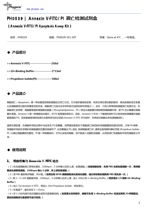
Alexa Fluor® 488 annexin V/Dead Cell Apoptosis Kit with Alexa®Fluor 488 annexin V and PI for Flow Cytometry Catalog no. V13241IntroductionApoptosis is a carefully regulated process of cell death that occurs as a normal part ofdevelopment. Inappropriately regulated apoptosis is implicated in disease states, such asAlzheimer’s disease and cancer. Apoptosis is distinguished from necrosis, or accidental celldeath, by characteristic morphological and biochemical changes, including compaction andfragmentation of the nuclear chromatin, shrinkage of the cytoplasm, and loss of membraneasymmetry.1-5 In normal live cells, phosphatidyl serine (PS) is located on the cytoplasmicsurface of the cell membrane. However, in apoptotic cells, PS is translocated from theinner to the outer leaflet of the plasma membrane, thus exposing PS to the external cellularenvironment.6 In leukocyte apoptosis, PS on the outer surface of the cell marks the cell forrecognition and phagocytosis by macrophages.7,8 The human anticoagulant, annexin V, isa 35–36 kDa Ca 2+-dependent phospholipid-binding protein that has a high affinity for PS.9Annexin V labeled with a fluorophore or biotin can identify apoptotic cells by binding to PSexposed on the outer leaflet.10The Alexa Fluor® 488 annexin V/Dead Cell Apoptosis Kit with Alexa® Fluor 488 annexinV and PI for flow cytometry provides a rapid and convenient assay for apoptosis. The kitcontains recombinant annexin V conjugated to one of our best and brightest fluorophores,the Alexa Fluor® 488 dye, to provide the maximum sensitivity. Alexa Fluor® 488 dye isan almost perfect spectral match to fluorescein (FITC), but it creates brighter and morephotostable conjugates.Table 1.Contents and storage information.In addition, the kit includes a ready-to-use solution of the red-fluorescent propidium iodide (PI) nucleic acid binding dye. PI is impermeant to live cells and apoptotic cells, but stains dead cells with red fluorescence, binding tightly to the nucleic acids in the cell. After staining a cell population with Alexa Fluor® 488 annexin V and PI in the provided binding buffer, apoptotic cells show green fluorescence, dead cells show red and green fluorescence, and live cells show little or no fluorescence (Figure 1). These populations can easily be distinguished using a flow cytometer with the 488 nm line of an argon-ion laser for excitation.We have optimized this assay using Jurkat cells, a human T-cell leukemia clone, treated with camptothecin to induce apoptosis. Some modifications may be required for use with other cell types. Because no single parameter defines apoptosis in all systems, we strongly suggest using a combination of different measurements for reliable detection of apoptosis. Refer to our website at for a wide selection of products for apoptosis research.Figure 1. Jurkat cells (T-cell leukemia, human) treated with 10 μM camptothecin for four hours (bottom panel) or untreated (as control, top panel). Cells were then treated with the reagents in the kit, followed by flow cytometric analysis. Note that the camptothecin-treated cells (bottom panel) have a higher percentage of apoptotic cells (indicated by an “A”) than the basal level of apoptosis seen in the control cells (top panel). L = live cells, D = dead cells.Before StartingMaterials Required but NotProvided• Samples (appropriate sample concentrations range from 2 × 10⁵ to 1 × 10⁶ cells/mL)• Inducing agent• Phosphate buffered saline (PBS)• Deionized waterCaution Propidium iodide is a potential mutagen; use appropriate precautions when handling thisreagent.Experimental ProtocolsWe have optimized this assay using Jurkat cells treated with camptothecin to induceapoptosis. Some modifications may be required for use with other cell types.Flow CytometryInduce apoptosis in cells using the desired method. Prepare a negative control by incubating1.1cells in the absence of inducing agent.Harvest the cells after the incubation period and wash in cold phosphate-buffered saline1.2(PBS).Prepare 1X annexin-binding buffer. For example, for ~10 assays, add 1 mL 5X annexin-1.3binding buffer (Component C) to 4 mL deionized water.Prepare a 100 μg/mL working solution of PI by diluting 5 μL of the 1 mg/mL PI stock solution1.4(Component B) in 45 μL 1X annexin-binding buffer.Store the unused portion of this working solution for future experiments.Re-centrifuge the washed cells (from step 1.2), discard the supernatant and resuspend the1.5cells in 1X annexin-binding buffer.Determine the cell density and dilute in 1X annexin-binding buffer to ~1 × 10⁶ cells/mL,preparing a sufficient volume to have 100 μL per assay.Add 5 μL Alexa Fluor® 488 annexin V (Component A) and 1 μL 100 μg/mL PI working1.6solution (prepared in step 1.4) to each 100 μL of cell suspension.Incubate the cells at room temperature for 15 minutes.1.71.8After the incubation period, add 400 μL 1X annexin-binding buffer, mix gently and keep thesamples on ice.As soon as possible, analyze the stained cells by flow cytometry, measuring the fluorescence1.9emission at 530 nm (e.g., FL1) and >575 nm (e.g., FL3). The population should separate intothree groups: live cells show only a low level of fluorescence, apoptotic cells show greenfluorescence, and dead cells show both red and green fluorescence (see Figure 1).Confirm the flow cytometry results by viewing the cells under a fluorescence microscope,using filters appropriate for fluorescein (FITC) and tetramethylrhodamine (TRITC) or TexasRed® dye.MicroscopyThis protocol was developed using Jurkat cells treated with camptothecin to induce apoptosisand may be adapted for adherent cell lines.Induce apoptosis in cells using the desired method. Prepare a negative control by incubating2.1cells in the absence of the inducing agent.After the incubation period, wash the cells in cold PBS.2.2Prepare 1X annexin-binding buffer. For example, to make 1 mL 1X buffer, add 200 μL 5X2.3annexin-binding buffer (Component C) to 800 μL deionized water.Prepare a 100 μg/mL working solution of PI by diluting 5 μL 1 mg/mL PI stock solution2.4(Component B) in 45 μL 1X annexin-binding buffer.Store the unused portion of this working solution saved for future experiments.2.5Re-centrifuge the washed cells (from step 2.2), discard the supernatant and resuspend thecells in 1X annexin-binding buffer.Determine the cell density and dilute in annexin-binding buffer to ~1 × 10⁶ cells/mL,preparing a sufficient volume for deposition on a slide.2.6Add 5–25 μL of the annexin V conjugate (Component A) and 1–2 μL of the 100 μg/mL PI working solution (prepared in step 2.4) to each 100 μL of cell suspension. Higherconcentrations of the annexin V conjugate tend to produce better results; the optimalstaining concentration needs to be determined empirically.2.7Incubate the cells at room temperature for 15 minutes.Wash the cells with 1X Annexin-Binding buffer.2.8Deposit the cells onto slides, mount them using the desired method and observe the2.9fluorescence using appropriate filters.The cells should separate into three groups: live, apoptotic, and dead. Live cells show onlyweak annexin V staining of the cellular membrane, while apoptotic cells show a significantlyhigher degree of surface labeling. Dead cells show both membrane staining by annexin V andstrong nuclear staining from the propidium iodide.References1. Immunol Cell Biol 76, 1 (1998);2. Cytometry 27, 1 (1997);3. J Pharmacol Toxicol Methods 37, 215 (1997);4. FASEB J 9, 1277 (1995);5. Am J Pathol 146, 3 (1995);6. Cytometry 31, 1 (1998);7. J Immunol 148, 2207 (1992);8. J Immunol 151, 4274 (1993);9. J Biol Chem 265, 4923 (1990); 10. Blood 84, 1415 (1994).Contact InformationMolecular Probes, Inc.29851 Willow Creek RoadEugene, OR 97402Phone: (541) 465-8300Fax: (541) 335-0504Customer Service:6:00 am to 4:30 pm (Pacific Time)Phone: (541) 335-0338Fax: (541) 335-0305probesorder@Toll-Free Ordering for USA:Order Phone: (800) 438-2209Order Fax: (800) 438-0228Technical Service:8:00 am to 4:00 pm (Pacific Time)Phone: (541) 335-0353Toll-Free (800) 438-2209Fax: (541) 335-0238probestech@Invitrogen European Headquarters Invitrogen, Ltd.3 Fountain DriveInchinnan Business ParkPaisley PA4 9RF, UKPhone: +44 (0) 141 814 6100Fax: +44 (0) 141 814 6260Email: euroinfo@Technical Services: eurotech@ For country-specific contact information, visit .Further information on Molecular Probes products, including product bibliographies, is available from your local distributor or directly from Molecular Probes. Customers in Europe, Africa and the Middle East should contact our office in Paisley, United Kingdom. All others should contact our Technical Service Department in Eugene, Oregon.Molecular Probes products are high-quality reagents and materials intended for research pur p os e s only. These products must be used by, or directl y under the super v ision of, a tech n ically qual i f ied individual experienced in handling potentially hazardous chemicals. Please read the Material Safety Data Sheet pro v id e d for each prod u ct; other regulatory considerations may apply.Limited Use Label License No. 223: Labeling and Detection TechnologyThe purchase of this product conveys to the buyer the non-transferable right to use the purchased amount of the product and compo-nents of the product in research conducted by the buyer (whether the buyer is an academic or for-profit entity). The buyer cannot sell or otherwise transfer (a) this product (b) its components or (c) materials made using this product or its components to a third party or oth-erwise use this product or its components or materials made using this product or its components for Commercial Purposes. The buyer may transfer information or materials made through the use of this product to a scientific collaborator, provided that such transfer is not for any Commercial Purpose, and that such collaborator agrees in writing (a) to not transfer such materials to any third party, and (b) to use such transferred materials and/or information solely for research and not for Commercial Purposes. Commercial Purposes means any activity by a party for consideration and may include, but is not limited to: (1) use of the product or its components in manufacturing; (2) use of the product or its components to provide a service, information, or data; (3) use of the product or its components for therapeutic, diagnostic or prophylactic purposes; or (4) resale of the product or its components, whether or not such product or its components are resold for use in research. Invitrogen Corporation will not assert a claim against the buyer of infringement of the above patents based upon the manufacture, use or sale of a therapeutic, clinical diagnostic, vaccine or prophylactic product developed in research by the buyer in which this product or its components was employed, provided that neither this product nor any of its components was usedin the manufacture of such product. If the purchaser is not willing to accept the limitations of this limited use statement, Invitrogen is willing to accept return of the product with a full refund. For information on purchasing a license to this product for purposes other than research, contact Molecular Probes, Inc., Business Development, 29851 Willow Creek Road, Eugene, OR 97402, Tel: (541) 465-8300. Fax: (541) 335-0354.Several Molecular Probes products and product applications are covered by U.S. and foreign patents and patents pending. All names con-t ain i ng the des i g n a t ion ® are reg i s t ered with the U.S. Patent and Trade m ark Office.Copyright 2010, Molecular Probes, Inc. All rights reserved. This information is subject to change without notice.Product List Current prices may be obtained from our website or from our Customer Service Department.Cat. no. Product Name Unit Size V13241 Alexa Fluor® 488 annexin V/Dead Cell Apoptosis Kit with Alexa® Fluor 488 annexin V and PI *for flow cytometry* *50 assays* . . . . . . . 1 kit。
Annexin V-FITC 凋亡检测试剂盒操作方法及步骤说明书

Annexin V-FITC凋亡检测试剂盒说明:细胞凋亡是细胞的基本特征之一,它在机体的胚胎发育、组织修复、内环境的稳定等方面起着十分重要的作用。
Annexin Ⅴ是一种分子量为35-36kD的Ca2+依赖性磷脂结合蛋白,能与细胞凋亡过程中翻转到膜外的磷脂酰丝氨酸(Phosphatidylserine,PS)高亲和力特异性结合。
以标记了FITC的Annexin Ⅴ作为荧光探针,利用流式细胞仪或荧光显微镜可检测细胞凋亡的发生。
Propidium iodide(PI)是一种核酸染料,它不能透过完整的细胞膜,但在凋亡中晚期的细胞和死细胞,PI能够透过细胞膜而使细胞核红染。
将Annexin Ⅴ与PI匹配使用,可以将凋亡早期的细胞和晚期的细胞以及死细胞区分开来。
产品组成(50/25次反应):· Annexin V-FITC 500ul/250ul(20ug /ml)· Binding Buffer 40ml/20 ml· Propidium Iodide (PI) 250ul/125ul(50ug /ml)保存条件: 2-8℃储存,有效期一年。
注意事项:1.为保证得到理想的实验结果,在使用此试剂盒之前请认真阅读该注意事项;2.试剂(尤其是小瓶装的试剂)在开盖前请短暂离心,将盖内壁上的液体甩至管底,避免开盖时液体洒落;3.Propidium iodide(PI)能通过皮肤吸收,对眼睛有刺激作用;4.此试剂盒仅供科研使用,不宜用于临床诊断;5.本试剂盒中提供的PBS为随试剂赠送,并非试剂盒的真正组成成分;细胞的洗涤同常规方法。
操作注意要点:1.整个操作过程动作要尽量轻柔,勿用力吹打细胞,尽量在4℃下操作;2.反应完毕后请尽快检测,因为细胞凋亡是一个动态的过程,反应1小时后荧光强度就开始衰变;3.Annexin V-FITC和Propidium iodide是光敏物质,在操作时请注意避光;4.成功的检测凋亡受以下几种因素的影响,如细胞类型、细胞膜上PS的密度、发生凋亡时PS翻转的比例、诱导细胞凋亡的方法、所用试剂、诱导凋亡的时间等,把这些影响因素进行优化对实验成功是非常必要的;5.在细胞洗涤的最后一步,请尽量将上清弃净,以免PBS残留,有可能会影响实验结果;6.PI染色时间过长有可能造成检测的凋亡率偏高,建议首先进行Annexin V-FITC染色,上机前5分钟再加入PI染色。
AnnexinV凋亡试剂盒操作步骤

请在使用前仔细阅读说明书Annexin V,FITC 凋亡检测试剂盒(100次)产品名称货号规格储存条件运输条件Annexin V,FITC结合物AD01-10 100次×1 0-5℃,避光(切勿冻存) 室温PI Solution AD02-05 50次×2 0-5℃,避光(切勿冻存) 室温10×Annexin V Binding Buffer AD03-05 50次×2 0-5℃,避光(切勿冻存) 室温*注:规格中的每“次”是以细胞浓度1×10⁶ cells/ml计算产品描述细胞凋亡是指为维持有机体内环境稳定,由基因控制的细胞自主的有序的死亡。
正常情况下任何细胞在形成过程中发生的异常都会通过凋亡消除。
例如体内的癌细胞增长为肿瘤的过程会受细胞凋亡的引导而被抑制。
然而在抑癌基因p53出现问题时,凋亡就不会诱导发生,从而导致癌细胞的不断增长。
细胞凋亡可以通过细胞形态的变化或生物化学的变化来检测。
目前常用的指标有caspase活性变化、DNA碎片、磷脂酰丝氨酸的外翻等。
Annexin V染色的细胞可以用于检测细胞凋亡早期的细胞膜变化。
在细胞凋亡早期,膜磷脂酰丝氨酸由脂膜内侧翻向外侧。
Annexin V 是一种分子量为35~36kD的Ca2+依赖性磷脂结合蛋白,与磷脂酰丝氨酸有高度亲和力,可通过细胞外侧暴露的磷脂酰丝氨酸与凋亡早期细胞的胞膜特异性结合,因此Annexin V 被作为检测细胞早期凋亡的灵敏指标之一。
用绿色荧光FITC标记的Annexin V 通过流式细胞仪或荧光显微镜可以检测到细胞凋亡的发生。
碘化丙啶(Propidium Iodide, PI)是一种核酸染料,PI只能透过凋亡晚期和死细胞的细胞膜,因此Annexin V和PI结合使用,可以区分凋亡早晚期的细胞及死细胞。
所需的设备和材料-合适量程的移液枪-样品和诱导剂-细胞培养用6,12,24,96孔板-PBS、去离子水-流式细胞仪或荧光显微镜。
ANNEXIN V-FITC PI 凋亡检测试剂盒说明书

第1页,共3页ANNEXINV-FITC/PI 凋亡检测试剂盒说明书注意:本产品试剂浓度发生变化,使用前请仔细阅读说明书。
货号:CA1020规格:20T/50T /100T保存:2-8°C ,避光保存,有效期1年。
产品说明:细胞凋亡早期改变发生在细胞膜表面,这些细胞膜表面的改变之一是磷脂酰丝氨酸(PS)从细胞膜内转移到细胞膜外,使PS 暴露在细胞膜外表面。
PS 是一种带负电荷的磷脂,正常主要存在于细胞膜的内面,在细胞发生凋亡时细胞膜上的这种磷脂分布的不对称性被破坏而使PS 暴露在细胞膜外。
Annexin V 具有易于结合到磷脂类如PS 的特性,对PS 有高度的亲和性。
因此,该蛋白可充当一敏感的探针检测暴露在细胞膜表面的PS 。
PS 转移到细胞膜外不是凋亡所独特的,也可发生在细胞坏死中。
两种细胞死亡方式间的差别是在凋亡的初始阶段细胞膜是完好的,而细胞坏死在其早期阶段细胞膜的完整性就破坏了。
因此,可以采用Annexin V 与PI 双染的方法,通过流式检测细胞早期凋亡。
操作步骤:1.细胞样品的准备:a)对于贴壁细胞:小心收集细胞培养液到一离心管内备用。
用不含EDTA 的胰酶消化细胞,至细胞可以被轻轻用移液管或枪头吹打下来时,加入前面收集的细胞培养液,吹打下所有的贴壁细胞,并轻轻吹散细胞。
再次收集到离心管内。
1000rpm 左右离心5min ,沉淀细胞。
对于特定的细胞,如果细胞无法完全离心至离心管底,可以适当延长离心时间或稍稍加大离心力。
小心吸除上清,可以残留约50µl 左右的培养液,以避免吸走细胞。
加入约1ml 4℃预冷的PBS ,重悬细胞,再次离心沉淀细胞,小心吸除上清。
b)对于悬浮细胞:1000rpm 左右离心5min ,沉淀细胞。
对于特定的细胞,如果细胞无法完全离心至离心管底,可以适当延长离心时间或稍稍加大离心力。
小心吸除上清,可以残留约50µl 左右的培养液,以避免吸走细胞。
kga101 n凯基annexin vegfp细胞凋亡检测试剂盒说明书
凯基Annexin V-EGFP细胞凋亡检测试剂盒(Annexin V-EGFP Apoptosis Detection Kit)Cat number:KGA For Research Use OnlyStore at4℃ for one yearExpire date:一、 试剂盒说明在正常细胞中,磷脂酰丝氨酸(PS)只分布在细胞膜脂质双层的内侧,而在细胞凋亡早期,细胞膜中的磷脂酰丝氨酸(PS)由脂膜内侧翻向外侧。
Annexin V是一种分子量为35~36kD的Ca2+依赖性磷脂结合蛋白,与磷脂酰丝氨酸有高度亲和力,故可通过细胞外侧暴露的磷脂酰丝氨酸与凋亡早期细胞的胞膜结合。
因此Annexin V被作为检测细胞早期凋亡的灵敏指标之一。
将Annexin V进行荧光素(EGFP、FITC)标记,以标记了的Annexin V作为荧光探针,利用荧光显微镜或流式细胞仪可检测细胞凋亡的发生。
与FITC的绿色荧光信号相比,EGFP的绿色荧光信号具有信号强,不易淬灭,稳定性高等优点,故本试剂盒采用EGFP作为荧光标记探针。
碘化丙啶(Propidium Iodide, PI)是一种核酸染料,它不能透过完整的细胞膜,但对凋亡中晚期的细胞和死细胞,PI能够透过细胞膜而使细胞核染红。
因此将Annexin V与PI匹配使用,就可以将处于不同凋亡时期的细胞区分开来。
本试剂盒可应用于培养细胞凋亡检测(不推荐用于检测组织样本)。
二、 试剂盒组份组份Cat: KGA10110 assays Cat: KGA10220 assaysCat: KGA10350 assaysCat: KGA104100 assays储存条件AnnexinV-EGFP 50μL 100μL 250μL 500μL 4℃避光Propidium Iodide 50μL 100μL 250μL 500μL 4℃避光Binding Buffer 5 mL 10.0 mL 25 mL 50 mL 4℃三、 试剂盒以外自备仪器和试剂流式细胞仪或荧光显微镜、低速离心机、微量移液器1.5m L Microtube、载玻片、盖玻片(荧光显微镜观察需用)、PBS、不含EDTA的胰酶消化液四、 使用注意事项1.微量试剂取用前请离心集液。
Annexin V-FITCPI细胞凋亡检测试剂盒
Annexin V-FITC/PI双染细胞凋亡检测试剂盒上海优宁维生物科技有限公司一、概述在正常细胞中,磷脂酰丝氨酸只分布在细胞膜脂质双层的内侧,细胞发生凋亡最早期,膜磷脂酰丝氨酸(PS)由脂膜内侧翻向外侧,这一变化早于细胞皱缩、染色质浓缩、DNA片断化和细胞膜的通透性增加等凋亡现象。
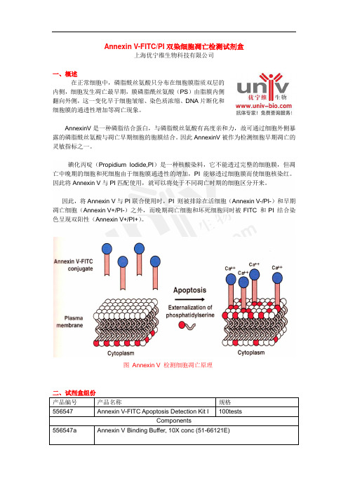
AnnexinV是一种磷脂结合蛋白,与磷脂酰丝氨酸有高度亲和力,故可通过细胞外侧暴露的磷脂酰丝氨酸与凋亡早期细胞的胞膜结合。
因此AnnexinV被作为检测细胞早期凋亡的灵敏指标之一。
碘化丙啶(Propidium Iodide,PI)是一种核酸染料,它不能透过完整的细胞膜,但凋亡中晚期的细胞和死细胞由于细胞膜通透性的增加,PI能够透过细胞膜而使细胞核染红。
因此将Annexin V与PI匹配使用,就可以将处于不同凋亡时期的细胞区分开来。
因此,将Annexin V与PI联合使用时,PI 则被排除在活细胞(Annexin V-/PI-)和早期凋亡细胞(Annexin V+/PI-)之外,而晚期凋亡细胞和坏死细胞同时被FITC 和PI 结合染色呈现双阳性(Annexin V+/PI+)。
图Annexin V 检测细胞凋亡原理三、实验步骤贴壁细胞需用0.25%的胰酶消化。
注意过度消化可损伤细胞。
在消化时可加2%的BSA 可防止消化过度。
如果用含EDTA的胰酶消化时,注意必须彻底清除EDTA:在标记前用1×PBS或1×bindingbuffer洗涤,清除EDTA,以免残余的EDTA与Ca2+螯合,影响Annexin V的结合。
(1)用去离子水将10×Binding Buffer稀释成1×Binding Buffer;(2)细胞收集。
悬浮细胞收集:离心5分钟;贴壁细胞:用不含EDTA的胰酶消化收集后(注:胰酶消化时间不宜过长,否则会影响细胞膜上磷脂酰丝氨酸与Annexin V-FITC的结合),于室温2000rpm离心5~10分钟,收集细胞;(3)细胞洗涤:用预冷1×PBS(4℃)重悬细胞一次,2000rpm离心5~10分钟,洗涤细胞;(4)加入300μL 的1×Binding Buffer 悬浮细胞;(5)Annexin V-FITC标记:加入5μL的Annexin V-FITC混匀后,避光,室温孵育15分钟;(6)PI标记:上机前5分钟再加入5μL的PI染色。
AnnexinV-FITCPI双染法细胞凋亡检测试剂盒操作说明

AnnexinV-FITCPI双染法细胞凋亡检测试剂盒操作说明细胞凋亡是细胞的基本特征之一,它在机体的胚胎发育、组织修复、内环境的稳定等方面起着十分重要的作用。
在细胞凋亡过程中,细胞内膜的磷酯酰丝氨酸外翻到细胞外膜,而Annexin V(膜联蛋白V)对其有天然的高亲和能力,故可以用FITC标记(绿色荧光)的Annexin V来检测细胞凋亡,但本方法的缺点是不能区分早期和晚期的凋亡细胞。
早期和晚期的凋亡细胞的一个重要区别在于细胞膜的完整性(凋亡早期细胞没有丧失膜的完整性),故可以用碘化丙啶(PropidiumIodide,PI)来染色加以区分。
图源:网络Annexin V-FITC/PI双染法细胞凋亡检测试剂盒特点:1. 即开即用,不需要专门花时间准备各种成分。
2. 整合了Annexin V-FITC和PI检测法的优点,提高了凋亡细胞检测的灵敏度和特异性,本方法是目前极为灵敏的细胞凋亡检测方法。
3. 跟流式细胞仪、荧光显微镜或其它荧光检测兼容。
Annexin V-FITC/PI双染法细胞凋亡检测试剂盒操作步骤:(一)获得目的基因1、通过PCR方法:以含目的基因的克隆质粒为模板,按基因序列设计一对引物(在上游和下游引物分别引入不同的酶切位点),PCR循环获得所需基因片段。
2、通过RT-PCR方法:用TRIzol法从细胞或组织中提取总RNA,以mRNA为模板,逆转录形成cDNA第一链,以逆转录产物为模板进行PCR循环获得产物。
(二)构建重组表达载体1、Annexin V-FITC/PI双染法细胞凋亡检测试剂盒载体酶切:将表达质粒用限制性内切酶(同引物的酶切位点)进行双酶切,酶切产物行琼脂糖电泳后,用胶回收Kit或冻融法回收载体大片段。
2、PCR产物双酶切后回收,在T4DNA连接酶作用下连接入载体。
(三) 获得含重组表达质粒的表达菌种1、将连接产物转化大肠杆菌DH5α,根据重组载体的标志(抗Amp 或蓝白斑)作筛选,挑取单斑,碱裂解法小量抽提质粒,双酶切初步鉴定。
PH0539-Annexin V-FITCPI 凋亡检测试剂盒使用手册
PH0539|Annexin V-FITC/PI凋亡检测试剂盒(Annexin V-FITC/PI Apoptosis Assay Kit)货号:PH0539规格:PH0539-50|50T存储:Store at4℃,一年有效。
◆产品组分>>Annexin V-FITC--------------------250ul>>10×Binding Buffer-----------------2*13ml>>Propidium Iodide(PI)--------------500ul◆产品简介细胞凋亡(Apoptosis)是一种由基因控制的细胞自主死亡方式。
它与组织器官的发育,机体正常生理功能的维持,某些疾病的发生发展以及细胞癌变过程均有着密切的关系。
细胞凋亡已成为生命学科极为活跃的研究领域之一。
此外,介绍几种常用的细胞凋亡检测方式。
在细胞凋亡的早期,细胞膜表面的磷脂酰丝氨酸(Phosphatidylserine,PS)将会从细胞膜内侧转移到细胞膜外侧,使PS充分暴露在细胞膜外表面。
Annexin V是一种磷脂结合蛋白,对PS有高度的亲和力。
因此,Annexin V作为一个敏感的探针可以有效地检测暴露在细胞膜表面的PS。
目前普遍使用的检测方法是将荧光标记后的Annexin V-FITC作为探针,利用流式细胞仪来检测细胞凋亡。
值得注意的是,在细胞坏死的过程中也会发生PS的暴露。
但两者的差别在于细胞凋亡的初始阶段细胞膜结构是完好的,只有PS转移,而细胞坏死则在早期阶段细胞膜的完整性就破坏了,从而暴漏出PS。
因此,检测细胞凋亡时,通常会使用颜料碘化丙啶(Propidium Iodide,PI)以确立细胞膜的完整性。
PI是一种核酸染料,它可以染色死细胞,但不能进入完整的活细胞,从而将凋亡性细胞和坏死性细胞区分开来。
◆使用说明1、细胞收集与Annexin V-FITC结合(1)在完成细胞凋亡诱导处理后,1500rpm,5分钟离心后弃上清,收集细胞。
- 1、下载文档前请自行甄别文档内容的完整性,平台不提供额外的编辑、内容补充、找答案等附加服务。
- 2、"仅部分预览"的文档,不可在线预览部分如存在完整性等问题,可反馈申请退款(可完整预览的文档不适用该条件!)。
- 3、如文档侵犯您的权益,请联系客服反馈,我们会尽快为您处理(人工客服工作时间:9:00-18:30)。
流式细胞仪
1,使用特定的方法诱导细胞凋亡,设置一个没有处理的control组
2,在一定孵育期后收获细胞,用冰冷的PBS清洗
3,准备1X的annexin结合液,例如,对于10个实验来说,加1ml 5X annexin 结合液(组分C)到4ml 去离子水中
4,准备一个100ug/ml的PI工作液,例如,通过稀释5ul 1mg/ml的PI储存液(组分B)到45ul 1X annexin 结合液中。
没有使用的这部分工作液用于以后的实验。
5,再次离心2步骤中洗过的细胞,弃上清用1X annexin 结合液重悬。
调整细胞密度,用1X的annexin 结合液稀释到大约1x10⁶/ml,为每个实验准备100ul 足够的体积。
6,加5ul Alexa Fluor 488 annexin V(组分A)和1ul 100ug/ml PI工作液(4步骤中准备的)到每100ul的细胞悬液中。
7,室温孵育细胞15min.
显微镜观察
1,使用特定的方法诱导细胞凋亡,设置一个没有处理的control组
2,在一定孵育期后收获细胞,用冰冷的PBS清洗
3,准备1X的annexin结合液,例如,配置1ml,加200ul 5X annexin 结合液(组分C)到800ul去离子水中
4,准备一个100ug/ml的PI工作液,例如,通过稀释5ul 1mg/ml的PI储存液(组分B)到45ul 1X annexin 结合液中。
没有使用的这部分工作液用于以后的实验。
5,再次离心2步骤中洗过的细胞,弃上清用1X annexin 结合液重悬。
调整细胞密度,用1X的annexin 结合液稀释到大约1x10⁶/ml,为每个实验准备足够的体积。
6,加5-25ul的annexin V缀合物(组分A)和1-2ul(100ug/ml)PI工作液到100ul 的细胞悬液中。
高浓度的annexinV缀合物会产生较好的结果;最佳染色浓度需要凭借经验
7,室温孵育细胞15min
8,1X annexin 结合液清洗细胞
9,用一个合适方法使细胞固定在载玻片上,用一个适当的滤镜观察荧光效果。
细胞应该被分成3组:活细胞,凋亡细胞和死细胞。
活细胞在细胞膜上有微弱的annexin V染色,而凋亡细胞在膜上有一个显著亮度,死亡细胞在膜上有annexin染色和核上的PI染色。