Radiation Safety
Radiation Safety
Health Effects of Radiation Exposure
Externally-contaminated people should be checked with radiation meters and given on-scene emergency care ASAP.
Health Effects of Radiation Exposure
Skin
Health Effects of Radiation Exposure
Depending on the level of exposure, radiation can pose a health risk. It can adversely affect individuals directly exposed as well as their descendants. Radiation can affect cells of the body, increasing the risk of cancer or harmful genetic mutations that can be passed on to future generations; or, if the dosage is large enough to cause massive tissue damage, it may lead to death within a few weeks of exposure.
Internally-contaminated people must be given medical care for injuries but there is little you can do to treat radiation exposures .
04.1 Radiation Safety
Radiation Basics
Units of Measurement
• Dose Equivalent – Measures the biological significance of the absorbed radiation – Each type of radiation has its own biological effect H=DxQxN
HSE Training 2005
Personnel Protection
Personal Dosimeter usage
• Causes of non-occupational high readings – Carrying badge in same compartment with sources during transport – Storing badge near radiation sources at facility or at home – Sending badge through airport X-ray machine – Having the badge on during medical treatment – Washing the badge should have little effect on its measurements
HSE Training 2005
Radiation Basics
Sources of Radiation
Alpha Particles
Beta Particles
Gamma Rays
• Unstable atoms give up energy (Radiation) as they try to seek a more stable state • The radiated energy can take several forms – Alpha radiation which consists of two protons and two neutrons. Alpha particles have limited penetrating power and can be stopped by a piece of paper – Beta radiation consists of particles similar to electrons. They travel at the speed of light and have more penetrating power than Alpha particles. Beta radiation can be stopped by your skin – Gamma radiation consists of electromagnetic waves and has considerable penetrating power, up to several inches Training 2005 HSE of steel.
(2) RADIATION SAFETY
Table 1. The people died and injured in the atomic bomb explosion in (广岛) (广岛) Japan inside 2 km
Distance from explosion (km) 0 ~ 0.5 0.6 ~ 1 1.1 ~ 1.5 1.6 ~ 2 Total Rate of die (%) 98.4 90.0 45.5 22.6 56.5 Rate of die in the day (%) 90.4 59.4 19.6 11.1 39.8
12
辐射实践
定义:与辐射相关的,增加了受照 剂量的人类活动. 实践的照射类型:职业照射,医疗 照射,公众照射
13 13
辐射干预
减少人类辐射实践过程中受照剂量 的一切措施,包括移开辐射源,改 变辐射途径或减少受照个人.
14 14
辐射防护目的
防止确定性效应的发生, 减少随机性效应的诱发.
15 15
23
7. Documentation (文件规范) 文件规范) 8. Allocation of responsibilities (岗位责任) 岗位责任) 9. Training and retraining (培训) 培训) 10. Licensing and regulations (规范许可) 规范许可) 11. Auxiliary systems (辅助系统) 辅助系统)
8 8
辐射敏感组织
性腺:主要遗传影响,性腺自身辐射敏感性 并不高,辐射致生殖细胞肿瘤未见报导. 红骨髓: >0.5Sv/a照射,主要诱发白血病. >0.5Sv/a照射,主要诱发白血病. 骨:主要骨表面上皮细胞,骨本身对辐射并 不敏感. 肺:辐射致肺癌主要源于内照射(Rn,Pu),外 肺:辐射致肺癌主要源于内照射(Rn,Pu),外 照射致肺癌概率与白血病类似.
1.辐射与健康安全 Radiation and
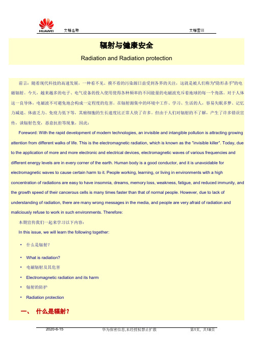
辐射与健康安全Radiation and Radiation protection前言:随着现代科技的高速发展,一种看不见、摸不着的污染源日益受到各界的关注,这就是被人们称为“隐形杀手”的电磁辐射。
今天,越来越多的电子、电气设备的投入使用使得各种频率的不同能量的电磁波充斥着地球的每一个角落。
对于人体这一良导体,电磁波不可避免地会构成一定程度的危害。
在辐射源集中的环境中工作、学习、生活的人,容易失眠多梦、记忆力减退、体虚乏力、免疫力低下等,其癌细胞的生长速度比正常人快了许多。
但由于人们对辐射的不了解,产生了许多错误宣传,谈辐射色变,恶意抗拒等现象,因此:Foreword: With the rapid development of modern technologies, an invisible and intangible pollution is attracting growing attention from different walks of life. This is the electromagnetic radiation, which is known as the "invisible killer". Today, due to the application of more and more electronic and electrical devices, electromagnetic waves of various frequencies and different energy levels are in every corner of the earth. Human body is a good conductor, and it is unavoidable for electromagnetic waves to cause certain harm to it. People working, learning, or living in environments with a high concentration of radiations are easy to have insomnia, dreams, memory loss, weakness, fatigue, and reduced immunity, and the growth speed of their cancerous cells is many times faster than that of normal people. However, due to lack of understanding of radiation, there are many wrong messages in the media, and people are very afraid of radiation and maliciously refuse to work in such environments. Therefore:本期宣传我们一起来学习以下内容:In this issue, we will learn the following together:·什么是辐射?·What is radiation?·电磁辐射及其危害·Electromagnetic radiation and its harm·辐射的防护·Radiation protection一、什么是辐射?I. What Is Radiation?自然界中的一切物体,都以电磁波的形式时刻不停地向外传送热量,这种传送能量的方式称为辐射。
Radiation Safety Manual 辐射安全手册英文版
Radiation Safety Manual FOR NURSING STAFFRev. June 2009Table of ContentsRadioactivity and Radiation 3 Radiation Protection at UVa Health System 3 Medical Radiation Sources 5 Radiation Producing Equipment 5 Radioactive Materials 5 Basic Radiation Safety Procedures 5 Dose Limits/Monitoring Requirements 6 Typical Exposure Levels During X-ray Exams 6 Radiation and Risk 7 Fetal Protection Policy 7 Guidelines for Nursing Personnel WorkingWith Brachytherapy (Implant) Patients 9 Guidelines for Nursing Personnel WorkingWith Prostate Brachytherapy Patients 11 Guidelines for Nursing Personnel WorkingWith Eye-Plaque Brachytherapy Patients 12 Guidelines for Nursing Personnel WorkingWith P-32 Patients 14 Guidelines for Nursing Personnel WorkingWith I-131 Therapy Patients 15 Guidelines for Nursing Personnel WorkingWith Y-90 Microsphere Brachytherapy Patients 16 Guidelines for Nursing Personnel Working inRadiographic/Fluoroscopic Procedures 17 Important! Reporting of Unusual or Unsafe Conditions 18 Radiation Protection Quiz 19 Worker Acknowledgement 21Radioactivity and RadiationAll matter in our environment is made of atoms. Most atoms we encounter on Earth are stable. Some atoms, however, are unstable, giving off energy in the form of radiation in order to reach a stable state. These atoms are said to be radioactive. An example is the radionuclide, Carbon-14, produced in the atmosphere when cosmic rays interact with stable nitrogen atoms. When a Carbon-14 atom undergoes radioactive decay, it gives off radiation in the form of a beta particle and then becomes a stable nitrogen atom once again. The existence of Carbon-14 in all living things enables archaeologists to date ancient artifacts.There are small amounts of naturally occurring radioactive substances in soil, rocks, plants, animals, and in our own bodies, all of which give off radiation. Large amounts of radiation are present in outer space and a small portion of this radiation penetrates the atmosphere. This low level of naturally occurring radiation is known as background radiation.Radiation can only be detected by specially designed instruments. Radiation may pass through an object, or it may be absorbed and cause changes at the site of absorption. Radiation is known to cause cancer and birth defects in animals and humans. The risk of radiation damage is related to the amount of radiation absorbed by an individual. With the amounts of radiation encountered by employees of the UVa Health System, the risk is very small.Radiation is useful in medicine because of its ability to penetrate tissue, allowing imaging and non-surgical treatment of internal structures. However, radiation may produce harmful biological effects. Observations of exposed human populations and animal experimentation indicate that exposure to low levels of radiation over a period of years may lead to a slight increase in the incidence of cancer and leukemia. Exposures to high levels of radiation produce the same effects faster and may also cause hair loss, skin burns, radiation sickness or even death. Radiation may also increase the risk of genetic abnormalities.Radiation Protection at UVa Health SystemTo minimize the biological effects of radiation, special rules and regulations are set forth for individuals occupationally exposed to radiation. The amount of radiation received by persons exposed occupationally should not exceed the dosages specified in the 10 CFR Part 20 of the Code of Federal Regulations, the Virginia State Regulations For Protection Against Radiation and the UVa Radiation Safety Guide.There is, in general, minimal external radiation hazard to hospital personnel from procedures involving radiation. Depending on your specific job duties, you may or may not be c lassified as a “radiation worker” and may or may not be required to wear personnel monitoring devices. All X-ray equipment operators are considered “radiation workers” and most require personnel monitoring. The need for personnel monitoring is determined by the likelihood of receiving exposures in excess of certain regulatory limits and by the recommendations of groups such as JCAHO (Joint Commission on Accreditation of Healthcare Organizations). Adherence to guidelines contained in this manual will help all X-ray equipment operators and radiology staff members keep their exposures as low as reasonably achievable (ALARA), and for moststaff members should reduce radiation exposures to levels allowable for individual members of public or in some cases, to levels indistinguishable from natural background.Radiation protection support services are provided for UVa Health System by the UVa Office of Environmenal Health and Safety (OEHS). These services include the oversight and administration of the personnel monitoring program, area surveys and in-service training of hospital workers. X-ray equipment inspections are performed by staff in Radiological Physics. Questions regarding the radiation protection program should be directed to the Radiation Safety Officer (RSO) at 982-4911. Radiation Safety can be reached after normal working hours at pager 923-5047.Medical Radiation SourcesSources of radiation are used at UVa for diagnosis, therapy and research. The most likely places to find radiation sources are in Radiology,Nuclear Medicine, Nuclear Cardiology, Radiation Oncology and certain hospital laboratories. However, mobile radiographic and fluoroscopic units are used frequently throughout the hospital, and many nuclear medicine patients retain radiopharmaceuticals for days or weeks after their procedures are complete. It has also become more common for specialty departments (such as GI Procedures, Urology, OR, etc.) to possess and operate their own diagnostic X-ray equipment. Thus, radiation may be encountered virtually anywhere within the health care system.Radiation Producing EquipmentIn diagnostic radiography, X-rays are produced when high-energy electrons collide with a metal target in an X-ray tube. X-rays are produced only when the machine is activated. The patient does not become radioactive from exposure to X-rays.In diagnostic fluoroscopy, X-ray images are viewed on a video monitor rather than on film. Fluoroscopy procedures are the largest source of occupational radiation exposure in medicine. Fluoroscopy is used to study moving structures, and to assess positioning during surgical and radiographic procedures. The portable fluoroscopy unit is often referred to as a "c-arm.” All X-ray machines are “registered” with the state radiat ion protection regulatory agency, the Virginia Department of Health’s Radiological Health Program.In radiation therapy, linear accelerators (powerful electron and X-ray beam machines) are used for the treatment of cancer. The energy of the X-ray radiation produced by these units is 10 to 100 times that of a diagnostic X-ray machine. Linear accelerators may treat with either X-rays or electrons. Registration and operation of linear accelerators is regulated by the State Radiological Health Program.Radioactive MaterialsRadionuclides like Tc-99m, I-131, P-32, Ir-192, Cs-137, I-125 and Y-90, are frequently used in hospitals. For example, I-131 is used as a diagnostic aid in the evaluation of thyroid function and also as a therapeutic agent in the treatment of thyroid disease. It can be expected that in the future many new uses for radioactive materials will be found in medicine. Most of these radioactive materials are used under a broadscope byproduct material license issued to UVa by the U.S. Nuclear Regulatory Commission. The UVa Radiation Safety Committee oversees and approves all use of radioactive materials at the institution as required by the license agreement. The Radiation Safety section of the Office of Environmental Health and Safety ac ts as an “agent” for this committee, managing the radiation protection program.Diagnostic Radiopharmaceuticals–Some of the radionuclides used in Nuclear Medicine and Nuclear Cardiology for diagnostic procedures emit gamma rays, which are a penetrating radiation, like X-rays. It is this penetrating quality that allows images of internal structures to be obtained. These radionuclides remain in the patient after the study is over, but have short half-lives, so the patient and the people around him or her are not exposed for a long period of time. A half-life is the time it takes to reduce the radioactivity of a substance by half. Diagnostic radiopharmaceuticals have half-lives from six hours to eight days. Short half-life nuclides may be intensely radioactive,but for a short period of time. After about 10 half-lives, the radioactivity is reduced to near background levels.Although radiation exposures may arise from the radiation emitted by radionuclides in patients, by accidental contamination of skin with radioactive materials, or by accidental ingestion of these materials (possibly through smoking or eating when hands are contaminated), there is, in general, no radiation hazard to hospital staff from these sources in patients who have received diagnostic or tracer doses of radioactive materials. No special precautions are needed in caring for them, and there are no restrictions on patient activities or contacts with other people.Therapeutic Radionuclides- When therapeutic radiopharmaceuticals or sealed sources are used, relatively large doses are involved. The patient can become a significant source of radiation exposure to staff, family and visitors. When procedures require that radiation precautions be put into effect, a radiation sign and a precaution sheet will be posted on or near the door to the patient's room.Laboratory Use of Radionuclides- Research and medical laboratories often use radionuclides that emit beta particles and low-energy gamma rays. Beta particles are not nearly as penetrating as gamma rays or X-rays. Weak and moderate energy betas will not even penetrate the skin. The most important safety precaution for most of these radionuclides is to keep the material from contaminating the skin thereby avoiding the possibility of ingestion or absorption.Because radiation cannot be seen or felt, the 3-blade radiation symbol shown below is used to alert you to the presence of radiation and/or radioactive material.Containers of radioactive material and rooms where radioactive materials are stored or used, are posted with the following label, which when in use is magenta or black with a yellow background: Rooms or areas where radiation-producing equipment is used are posted with the following sign, also magenta or black with a yellow background:Rooms where significant radiation exposure can occur from radioactive sealed sources in patients undergoing brachytherapy, or from patients receiving radiopharmaceutical therapy are posted with a “Caution – RAD IATION ISOLATION” sign like below. Radiation safety precautions are provided on these signs and include caregiver and visitor instructions, stay times, and Radiation Safety emergency contact information.Basic Radiation Safety ProceduresThe radiation protection program is guided by the concept of keeping radiation exposure As Low As Reasonably Achievable (ALARA). The ALARA concept is based on the assumption that any radiation dose, no matter how small, can have some adverse effect. At UVa, radiation exposure of all individuals routinely working with sources of radiation is monitored with a Luxel®OSL (Optically Stimulated Luminescence) dosimeter. The dosimeters are changed out and analyzed on a monthly or quarterly frequency. Action levels have been set by the Radiation Safety Committee at 10% of the regulatory limits. These levels trigger investigations to determine if the exposures were actually as low as reasonably achievable. If not, recommendations are made to ensure that future exposures are ALARA. Under the ALARA program, every reasonable means of lowering exposure is used. Radiation exposure can be minimized by utilizing three basic principles:1)Time: Shorter exposure time means a lower dose.2)Distance: Doubling the distance from a radiation source means one-fourth thedose rate. Tripling the distance gives one-ninth the dose rate.3)Shielding:The use of appropriate shielding greatly reduces the dose rate.Wearing a lead apron when performing a fluoroscopy procedure is one example. The shield material used and its thickness depend on the type and strength of the source of radiation.Remember that radiation cannot be seen or felt, but can be detected with radiation survey meters.Radioactive Spills: When a spill of radioactive material is encountered, do not clean it up. Isolate the area and notify the Radiation Safety Officer. Remember that small droplets may have splashed away from the spill. If liquid is running, try to contain it with a paper towel or other absorbent material, taking care not to contaminate yourself. All persons involved in a spill should be monitored for contamination before they are released.Loose sources or seeds: If a radioactive source or seed has become dislodged from the patient, do not touch the source! Remove all unnecessary personnel, and call Radiation Safety and Radiation Oncology. Try to get the source to the corner of the room via remote handling tool, yardstick, etc. Isolate the area and notify the Radiation Safety Officer.Dose Limits/Monitoring RequirementsThe average annual dose of a UVa Health System radiation worker is about 100 millirem (millirem or rem is a measure of radiation dose in the human body). A radiation worker is required to be monitored if he/she is likely to receive in excess of 10% of the dose limits. Those dose limits are:Whole Body: 5 rem/yearSkin/Extremities: 50 rem/yearLens of Eye: 15 rem/yearFetus: 500 milli rem/gestation (9 months)NOTE: 1 rem = 1,000 milliremPer regulations, an individual member of the public is allowed only 100 millirem per year from all licensed and registered radiation activities. Keep in mind, however, that the average U.S. citizen receives about 360 millirem of radiation each year from “background” radiation sources and medical procedures. (That’s the equivalent of about 36 chest X-rays!)Typical Exposure Levels During X-ray ExaminationsAn individual located four feet from the patient's bed at the time that radiographic exposure using a 14 x 17 image receptor is made, may typically receive about 0.010 millirem. To receive 500 millirem, one would have to remain at that distance for 50,000 X-ray exposures. An individual located four feet from a patient undergoing fluoroscopy may typically receive about 0.50 millirems per minute while the machine is "on". To receive 500 millirem, one would have to be in the location for 15-20 hours with the machine operating.Since radiation decreases rapidly with distance, the further one is from the patient during the actual X-ray examination, the smaller the exposure.Radiation and RiskEffects of large doses of radiation are well-documented and understood from the study of groups including atomic bomb survivors, radiation accident victims, radiation therapy patients, and early radiation researchers. The effects of the very low doses of radiation expected among workers in the hospital setting are difficult to observe and, therefore, are not as well understood. When a large dose of radiation is increased to an even larger dose, the adverse effects become greater or more prevalent. This dose vs. effect relationship can be thought of as linear, with confirmed and documented effects beginning at a certain “threshold” level of radiation dose.But since this “threshold” level is far greater than any allowable occupational dose, how is the risk of occupational radiation exposure assessed? Although the effects of very low doses of radiation are not t ruly known, health physicists “extend” w hat is known about the health effects of higher doses of radiation down to “zero” dose. In other words, any radiation dose is assumed to have some effect. Most scientists believe that this is a conservative model of the risk. With the amounts of radiation encountered by employees in the UVa Health System, the risk is very small. Consider that for very low doses of radiation the effect of most concern is cancer. If every member of a population of 1 million were to receive 10 millirem of radiation (average film chest X-ray), it is possible that 5 additional cancer deaths would be observed. Remember however, that out of this population of 1 million, about 200,000 (20%) will die of cancer, making these few additional cancer deaths statistically impossible to detect. Additionally, according to the Biological Effects of Ionizing Radiation (BEIR) committee, the risk of cancer death is 0.08% per rem for doses received rapidly (acute) and might be 2 times (0.04% per rem) less than that for doses received over a long period of time (chronic).It’s important to keep in mind that all activities carry some element of risk. For example, flying in an airplane, driving a car, smoking cigarettes, eating certain foods, and drinking alcoholic beverages are everyday activities that carry some risk. Many of us are willing to accept the risk from these activities.Fetal Protection PolicyRecent studies have shown that the risk of childhood leukemia and other cancers increases if the mother experienced a significant radiation exposure during pregnancy. The National Academy of Sciences has reported that the incidence of leukemia among children from birth to 10 years of age could rise from 3.7 cases in 10,000 children to 5.6 cases in 10,000 children if the children were exposed to 1 rem (1000 millirems) of radiation before birth. The Academy has also estimated that an equal number of other types of cancers could result from this level of radiation. Although other studies have shown a much smaller effect from radiation, each woman should be aware of any possible risk so that she can take steps she thinks are appropriate to protect her offspring.UVa has adopted a policy to protect the fetus/embryo of pregnant employees exposed to ionizing radiation in their work. Radiation protection regulations limit the occupational dose to pregnant women to 500 millirems over the course of the pregnancy if the worker declares her pregnancy in writing to the employer. Thisvalue is one-tenth of the permissible annual exposure established for adults. To help put this in perspective, the average annual dose from natural radiation sources is approximately 360 millirem.If an employee decides to declare her pregnancy, she should notify her supervisor and contact the OEHS Radiation Safety Office to discuss possible precautions to limit radiation exposure. Declaration of Pregnancy forms are available from Radiation Safety, 982-4911. The Radiation Safety Officer will review work assignments and radiation exposure history, and may recommend limitations in work assignment if necessary. Dosimeters will be assigned, with radiation exposures to be reviewed monthly. If radioactive materials are used, the employee may also be placed on a periodic bioassay program.Guidelines for Nursing PersonnelWorking with Brachytherapy (Implant) PatientsIn brachytherapy, small, sealed sources of radiation (typically Cs-137 or Ir-192), are positioned near the patient’s cancer site using special, surgically-implanted catheters. While these catheters are implanted in the operating room, the radiation sources themselves are inserted into these catheters in the patient’s room.Radiation exposure is the primary concern when working with brachytherapy patients. Exposure levels at the patient’s bedside can be as high as 50 to 100 millirem/hour. Contamination is not of concern as the radiation sources are “sealed.” No radioactivity is retained inside the patient once these sources are removed. Cs-137 sources are nickel-coated, approximately 20 mm long and 3 mm wide, and sometimes have an eyelet on one end that is color-coded. Ir-192 “seeds” are stainless steel encapsulated, only 3 mm long and 0.2 mm wide, and come in nylon strands with up to 12 seeds spaced typically 1 cm apart.The following guidelines should be observed when working with brachytherapy patients:∙Individuals that provide routine care for these patients will typically be issued a radiation dosimeter.∙Always wear your personnel monitoring badge (dosimeter) when attending the patient. Wear the badge between your waist and collar and make sure that the badge worn is the one issued in your name for the current monitoring period. Do not share badges with other workers. Do not take your badge home with you. When you are not wearing your dosimeter, store it in a controlled area away from all radiation sources.∙Provide all necessary care, but try to minimize time spent with the radioactive patient. Try to work behind mobile shields whenever possible, and work no closer to patient than necessary.∙Observe nursing stay times and instructions on the “Caution Radiation” sign s and instruction sheets posted by the door to the patient’s room. Read and observe any Radiation Safety instructions written in the patient’s chart.∙If you are the primary conta ct for matters of the patient’s care, be prepared to answer questions from other nurses, physicians, technical staff members, and visitors. Note the following:∙Other hospital staff members are allowed in patient room if stay times and other instructions are observed. (Exception: Personnel who do not routinely work with radiation therapy patients may not be required to wear a personnel monitoring device. Contact Radiation Safety with questions.)∙Visitors are permitted provided the visitor stay times and instructions are followed as posted by the room doorway. Visitor stay times are dependent upon the activity of the implant and generally are posted for a visitor distance of 2 meters or greater from the patient. The mobile shields should not be moved by visitor(s). Visitors under 18 and pregnant visitors are not permitted.∙Housekeeping and Dietary staff are not permitted in brachytherapy patient rooms. No room items are to be removed without clearance from radiation safety or medical physics personnel responsible for the implanted sources.∙Brachytherapy patients are to stay in pre-selected shielded rooms only.∙The room and patient are not to be released until Radiation Safety approved personnel perform a clearance survey.If a source becomes dislodged from the patient:-Do not touch the source! If possible, use a broom or some long handling tool to move it toa room corner-Remove all unnecessary personnel from source area and call Radiation Safety and Radiation Oncology.-If possible, using long tweezers, place source in the shielded container normally provided and located in the patient’s room along a far wall-If you can not get the source into the shielded container, try to get the source to the corner of the room via remote handling tool, yardstick, broom, etc.-Do not leave source near patient or attempt to re-insert source in patientNotify Radiation Safety and Radiation Oncology if a source becomes dislodged, or if there is a medical emergency (including patient death).Guidelines for Nursing PersonnelWorking with Prostate Seed ImplantBrachytherapy PatientsProstate brachytherapy is a minimally invasive procedure that implants small radioactive pellets (called seeds) that are about the size of a grain of rice into the prostate where they emit very low energy radiation, which is primarily absorbed in the treatment area immediately surrounding the seeds. Needles containing the seeds are inserted through the skin of the perineum. The radioactive material within the seeds gives off localized radiation for a number of months. Typically 50-100 seeds containing Pd-103 or I-125 are permanently implanted. These patients usually are administered an amount of radioactive material that is below regulatory release limits, and under normal conditions, patients can go home. For certain reasons, these patients may be hospitalized in a private room. No specific room is required for prostate brachytherapy treatments. The following guidelines should be observed: If Radiological Physics or EHS has not placed signs on the patient room or chart, call 4-5421 or 2-4911 for assistance and further instructions.∙The use of personnel monitoring badges (dosimeters) is not necessary when attending these patients. Radiation exposure from these patients is very low. ∙Provide all necessary care, but try to minimize time spent with patient, and work no closer to patient than necessary. ∙ Primary hazard: Some seeds may be lost through urination. If the patient does not have a urinary catheter in place, any seed(s) that are lost through urination can be flushed. If a patient does have a catheter and catheter bag in place, the bag should be visually inspected for seeds. If a seed is found follow these procedures:Do not attempt to remove the seed. Immediately notify EHS-Radiation Safety or the Radiation Oncology physicist for disposalThe following nursing guidelines apply:∙Observe nursing instructions if posted with the “Radiation” sign and any Radiation Safety instructions in the patient’s chart . ∙ If you are the primary contact for matters of the patient’s care, be prepared to answer questions from other nurses, physicians, technical staff members and visitors. Note the following:∙ Other hospital staff members and visitors are allowed in patient room if instructions are observed.∙ Housekeeping and Dietary staff are not permitted in prostate brachytherapy patient rooms. No room items are to be removed without clearance from Radiation Safety. It is especially important to hold the catheter and bag for a radiation survey before disposal.∙The room is not to be released until Radiological Physics (4-5421 days) or EHS (PIC 3454 evenings) performs a clearance survey. Notify the radiation oncology physicist at 4-5421, Environmental Health & Safety(Radiation Safety) and Radiation Oncology resident on-call if there is a medical emergency (including patient death).Guidelines for Nursing Personnel Working with Eye-Plaque Brachytherapy PatientsA radioactive plaque is a small, dish-shaped gold capdevice that contains radioactive sources. Ophthalmicplaque brachytherapy is the most commonly used "eye-sparing" treatment for choroidal melanoma. Eye-plaquescome in various sizes, typically between 10 and 20 mmdiameter. Eye-plaques contain rice-sized, radioactiveiodine-125 seeds that emit low energy photons. Thesephotons are effectively blocked by the gold of the plaquecreating a directional source. The radioactive eye-plaqueis sewn onto the eye so that it covers the intraoculartumor shadow, plus a 2-3 mm "free-margin." With theplaque in place, radiation is continuously delivered over a4-day period, and then the plaque is removed.Noradioactivity is retained inside the patient once these sources are removed.Patients with eye plaques are typically outpatients. A lead eye patch is placed over the plaque to reduce the radiation emitted from the plaque. The patch remains in place over the plaque during the treatment period and may be removed briefly to provide necessary care. The patient is instructed to go straight home and remain there for the duration of treatment. The patient is instructed when to return for plaque removal. A yellow wristband describing the type and amount of radioiodine in the plaque must be worn at all times.If the patient is an inpatient, the following guidelines should be observed when working with eye-plaque patients:∙The use of personnel monitoring badges (dosimeters) is not necessary when attending these patients.∙The patient may not leave the room while the radioactive implant is in place. The lead eye patch is to remain in place over the plaque during treatment (may be removed briefly to provide necessary care).∙Provide all necessary care, but try to minimize time spent with patient, and work no closer to patient than necessary.∙Observe nursing instructions posted with the “Radiation” sign and any radiation safety instructions written in the patient chart.∙As primary contact for matters of the patient’s care, be prepared to answer questions from other nurses, physicians, technical staff members, and visitors. Note the following:Eye plaque patients receive written patient instructions from Radiation Oncology.Other hospital staff members are allowed in patient room if instructions are observed.Visitors are also permitted provided the visitor stay times (when applicable) and instructions are followed as posted with the “Radiation” sign. All visitors are to remain a distance of 2 meters (6 feet) from the patient. Visitors under 18 and pregnant visitors are not permitted.Housekeeping and Dietary staff are not permitted in eye-plaque brachytherapy patient rooms.No room items (except food trays) are to be removed until cleared by Radiation Safety. Items that will be monitored include linens, patient gowns/clothing, and regular & medical waste.∙No specific room is required for eye-plaque treatments; however, these procedures are to be performed on a floor where the nursing staff is trained in handling such radioactive implant patients.。
国际电离辐射防护与辐射安全基本标准 英文缩写

国际电离辐射防护与辐射安全基本标准英文缩写English:The international standard for ionizing radiation protection and radiation safety is commonly abbreviated as ICRP, which stands for the International Commission on Radiological Protection. Founded in 1928, the ICRP is an independent organization that develops recommendations and guidance on radiological protection. Its fundamental principles revolve around the justification of practices, optimization of protection, and application of dose limits to ensure that radiation exposure is kept as low as reasonably achievable (ALARA). The ICRP regularly reviews scientific evidence to update its recommendations, taking into account advancements in technology and changes in understanding about the effects of radiation on human health. These recommendations are influential globally, shaping national regulations and standards for radiation protection and safety across various industries, including medicine, nuclear power, and industrial applications. The ICRP also collaborates with other international organizations such as the International Atomic Energy Agency (IAEA) and the World Health Organization (WHO) to promote consistent approaches to radiation protection worldwide,ensuring the safety of workers, the public, and the environment from the harmful effects of ionizing radiation.中文翻译:国际电离辐射防护与辐射安全的国际标准通常缩写为ICRP,代表着国际辐射防护委员会。
NASA Radiation Safety 辐射安全中英文培训2013 03
名称 对人无害剂量
耐受剂量
耐受剂量 耐受剂量 耐受剂量 最大允许剂量 最大允许剂量 最大允许剂量 最大允许当量 有效剂量 有效剂量
放射事故认定 Radiationthe accident assertion
受照人员及部位 Radiation dosage of personnel and location
应尽量远离放射源。
The dosage inverse to the square of distance, i.e. the dosage at 2m to the radioactive source is ¼ of the dosage at 1m. That’s to say, operators should keep away from the radioactive source as far as working condition permits.
数值
胶片照射7分钟 未能曝光时
皮肤红斑剂量 1/100 0.2R(伦琴)/天
换算成每天 100mSv/天 2mSv/天 2mSv/天
1934年 1936年
1950年 1959年 1965年 1977年 1990年 2002年
英国X射线与镭防护委员会 英国X射线与镭防护委员会 ICRP(国际放射防护委员会) ICRP ICRP ICRP ICRP ICRP
transportable type, X-Ray • 移动式: γ ( Ir-192半衰期75天 Se-75 半衰期120天)
transportable type, γ -Ray ( half-life of Ir-192 is 75 days, half-life of Se-75 is 120 days)
医学辐射防护学课件 15医用辐射事故的预防..已
3.较大辐射事故。 是指Ⅲ类放射源丢失、被盗、失控,或者放射 性同位素和射线装置失控导致9人以下(含9人) 急性重度放射病、局部器官残疾。
4.一般辐射事故。 是指Ⅳ类、Ⅴ类放射源丢失、被盗、失控,或 者放射性同位素和射线装置失控导致人员受到 超过年剂量限值的照射。
(二)辐射事故的管理
环境保护主管部门负责辐射事故的应急响应、 调查处理和定性定级工作,协助公安部门监控 追缴丢失、被盗的放射源; 公安部门负责丢失、被盗放射源的立案侦查 和追缴; 卫生主管部门负责辐射事故的医疗应急。
1. 外照射事故中的人员的处理
在外照射事故中,了解吸收剂量在体内 的分布对预后判断和医学处理方法的选择 非常重要。
受照人员病情的初步估计可根据其早期 症状、血象变化并参照其他物理计量的估 算,有条件者可进行外周血淋巴细胞染色 体畸变分析和淋巴细胞微核试验。
事故受照人员的处理方法
1. 全身或全身等效剂量<0.1Gy者可作一般 医学检查,确定是否需要治疗;
3.介入治疗医务人员放射损伤事故
经眼科确诊的两位介入医师及两位护士的眼 晶体损伤病例,医院均使用了床上管介入设备, 由于放射防护设施不到位,操作室内工作人员 所受的散射线水平相对较高。由于分次照射或 者长期曝光,估计这两所医院介入人员眼晶体 的剂量在不到4年的时间里已经超过了足以导 致细胞炎性及毒性反应的辐射阈剂量。
005245gy1985加速器主管加速器技术人员在机器经常自动停机工作不正常情况下进行检修检修后不检验辐射输出量擅自切断安全联锁改用手工操作致使连续2d发生超剂量照射直接或加速13名患者死亡1988x射线机对机器进行试验因摄片限时故障成持续曝光状态15min球管冒烟后才发现4人受照全身照射最大者15gy1989后装机用于妇科宫腔肿瘤治疗的一台后装机由于源与源辫的连接不合格未修复治疗中源掉落机房内无剂量报警仪25名患者和12名医务人员受到照射医务人员受到最大照射者047gy60co治疗机检修中照射事故概况序号发生年份受照的主要原因照射性质和健康后果197060co治疗机故障外请两人维修用手触摸裸源111014bq引起局部受照2人均截肢1972非专业人员检修60co治疗机1人患急性放射病1983非专业人员检修60co治疗机941013bq6次2人均患急性放射2001源出入不畅一维修工误使用压缩气管并碰到手动开关60co源1221014bq被气动系统打出当时不知情当看到源指示杆后立即手动开关弹起源回储存位受到照射时间约10s1人受到相当于全身均匀照射154gy二医用辐射事故典型案例1x射线诊断机故障所致人员受超剂量照射事1988年4月30日某医院4人在对x射线诊断机进行试验时因摄片限时装置发生故障成持续曝光状态15min后见管球冒烟立即停机
RADIATION SAFETY MANUAL 辐射安全手册英 文版
RADIATION SAFETY MANUALRevision 6. 16 June 2014Radiation Safety Manual 0 of 34 Document Number SD002.3ContentsPage Introduction 31General 42Normal Operating Procedures 52.1 Basic Principles of Control 52.2 General Procedures for Working with Radioactive Materials 52.3 Methods of Protection 52.3.1 The External Radiation Hazard 62.3.2 The Internal Radiation Hazard 72.4 Procedure for the Procurement and Use of Radioactive 82.5 Scanning Electron Microscopes – MST Department 92.6 PHILIPS X-Ray Diffraction Spectroscope – MST and CES Dept. 92.7 Dual Energy X-Ray Absorptiometer DXA 102.8 Sealed Radioactive Sources – Physics Department 102.9 Unsealed Radioactive Sources – CES Dept. 112.10 Security and Safety of Licensed Items 123 Emergency Operating Procedures133.1 Personal Contamination133.2 Emergency Contact Details (out of hours) 134 Monitoring for Radiation and Contamination144.1 Monitoring the Workplace 144.2 Personal Radiation Monitoring 144.3 Portable Radiation Monitoring Equipment 154.4 Portable Surface Contamination Monitoring Equipment 154.5 Wipe Testing of Sealed Sources 154.6 Maintenance Procedures for the Irradiating Equipment 164.7 Monitoring of I-125 Contamination 164.8 Wipe-testing of Sealed I-129 Source 184.9 Storage and disposal of Radioactive Waste 19I-125 Laboratory Safety Poster 215 Radiological Protection Administration 225.1 Role of the Heads of Department 225.2 Role of the Radiological Protection Advisor 225.3 Role of the Radiological Protection Officer 225.4 Role of the Departmental Radiological Protection Supervisors 225.5 Administration for Radiological Protection at the University of Limerick 236 Intervention Plan and Emergency Plan for Radiological Accidents6.1 Introduction 266.2 Purpose of plan 266.3 Organisational structure and responsibilities 266.4 Team responsibilities 296.5 Crisis scenarios involving radioactive sources or X-ray apparatus 306.6 Call out procedures 32References 33 TablesTable 1 Main types of ionising radiation‟s and their hazardsIntroductionThe Statutory requirements for the protection of workers and general public against the danger of radiation are based on the following three fundamental principles:-(a) every activity resulting in an exposure to ionising radiation shall be justifiedby the advantages which it produces;(b) all exposures shall be as low as reasonably achievable (the ALARAprinciple);(c) the dose equivalent to individuals shall not exceed the prescribed limits.For the purposes of this manual radioactive materials can be classified into two groups - unsealed dispersible radioactive material and sealed sources. The following definitions apply:-(a) a radioactive substance is any substance which is suspected to have anactivity concentration greater than the value stated in SI 125 of 2000;(b) a sealed source is a radioactive material wholly bonded within a solidinactive material or encapsulated in a receptacle so that no leakage canoccur during storage or foreseeable conditions of use (Refer to ISO 2919);(c) unsealed dispersible radioactive material is radioactive material notconsidered to be a sealed source.Work with radioactive substances is perhaps more stringently controlled than some aspects of chemical research, and this is reflected in Irish legislation for the handling of radioactive materials.1. General1.1 The University of Limerick academic Departments shall comply with theconditions of the RPII License in the control and use of radioactive substances and irradiating apparatus.1.2 The Radiation Safety Manual is for the use and guidance of all Universitystaff for whom it is relevant. The provisions of the Radiation Safety Manual are to supplement the requirements of the RPII Licence and shall be observed in full.2 Normal Operating Procedures2.1 Basic Principles of ControlThere are three main principles of control against the internal radiationhazard:(a) containment;(b) cleanliness;(c) use of the least toxic radioactive material that is suitable, andthe minimum quantities in all experiments.The two methods of containment of operations most widely used are partial containment by means of fume cupboards, and complete containment by means of glove boxes.2.2 General Procedures for Working with Radioactive MaterialsThe following working practices and procedures should be adopted:(a) the laboratory bench should be maintained in a tidy andorderly state;(b) there should be no unnecessary accumulation of radioactivematerials;(c) any surface contamination arising during an operation shouldbe cleaned-up immediately;2.3 Methods of ProtectionEvery attempt must be made to limit the degree of exposure achieved through working with radioactive materials or radiation sources. Table 1 lists the types of radiation which may be encountered. Their range in air varies with their nature and energy and gives rise to two types of radiological hazard - internal and external.Radiation type Main hazard topersonnelProtection Alpha particles Internal ContainmentBeta particles Internal andexternal skin dose Containment, local shielding and exposure timeGamma & X-rays External Distances, shielding andexposure timeNeutrons External Special shielding and exposuretimeTable 1Main types of ionising radiation’s and their hazards2.3.1 The External Radiation HazardThe basic methods of protection against external radiation are:-(a) restriction of the strength of every source to the minimumnecessary for the task in hand;(b) the use of the maximum amount of distance between thesource and the operator, compatible with the satisfactory andsafe performance of the work;(c) restriction of the period of exposure to the minimumcompatible with safe working;(d) the use of suitable shielding.The protection necessary in any particular situation to ensure that doses are kept below the relevant limit may be achieved by a combination of these methods.(b) The Use of DistanceThe intensity of radiation from a radioactive source decreaseswith increasing distance. For a point source (and where thedimensions of the source are small compared with thedistance from the source to the point of interest), the doserate is inversely proportional to the square of the distance, i.e.,by doubling the distance the dose rate is reduced by a factorof 4 and so on. For example, the gamma dose rate from a 1GBq cobalt-60 source decreases with increasing distance asfollows.at 1 cm ~ 3.5 Sv h-1at 10 cm ~35 mSv h-1at 100 cm ~350 Sv h-1Radioactive sources should therefore never be handled withbare hands, or with gloved hands unless the thickness of theglove is sufficient to reduce the radiation to reasonable levels.(c) The Use of TimeThe acceptable dose must be kept both within statutory doselimits and ALARA - As Low as Reasonably Achievable.Exposure to high dose rates calls for careful pre-planning,and sometimes for 'dummy' runs. 'On the job' discussions ina radiation field should be avoided.(d) The Use of ShieldingBeta Radiation. The most suitable shielding materials forbeta radiation are sheets of light metals such as aluminium orPerspex. The absorption of beta particles in matter gives riseto bremsstrahlung radiation (electromagnetic radiationresulting from the retardation of charged particles). Forsources of energetic beta radiation, a combination of Perspexand lead makes the best shielding material.Gamma rays cannot be completely absorbed by a shield; theyare only reduced in intensity. Of course, any degree ofattenuation is possible if the shield is made thick enough. Theapproximate thicknesses of various materials required toattenuate 1 MeV gamma rays by a factor of 10 are:LEAD IRON CONCRETE WATER3.5cm 6cm 20cm 40cm(These figures refer to broad beam geometry)2.3.2 The Internal Radiation HazardRoutes of Entry:(a) Ingestion. Contamination on surfaces may lead to ingestion ofactivity through the mouth. Control is based on a combination of rules and procedures and strict laboratory discipline, e.g. in the correct use and removal of gloves, correct monitoring procedures after working in contaminated areas, and no eating, smoking, drinking, or applying make-up in contamination-controlled areas. (b) Inhalation.Work carried out in a laboratory or workshop can beaccompanied by the formation of airborne contamination. Theassessment of the significance of radioactive airbornecontamination is a difficult problem due to the influence of manyfactors such as breathing characteristics (rate of breathing, whetherthe individual breathes through the nose or the mouth etc.), the size,shape and density and the chemical properties of the airborneparticles (which will affect lung deposition and subsequentmetabolism), and the ventilation pattern in the working area. Controlis largely based on proper containment and ventilation coupled withcorrect working discipline. Before a job is carried out considerationmust be given to the possibility of airborne contamination.(c) Absorption. Radioactive contamination may penetrate the skin bydiffusion through the skin barrier or via cuts and wounds.Radioactive materials deposited on the skin and absorbed throughthe skin may subsequently disperse via the blood stream. Organicsolvents are particularly dangerous in that they can penetrate theskin easily. In general, however, the skin forms an efficient barrier tocontamination. Control is based largely on correct laboratorydiscipline and techniques, e.g. when using solvents suitable glovesshould be worn.2.4 Procedure for the Procurement and Use of Radioactive Materialsand Irradiating Apparatus.Permission to acquire and to use licensable items may be granted bythe Head of Department after consultation with the RadiologicalProtection Officer (RPO) and the Departmental RadiologicalProtection Supervisor (DRPS).Faculty who wish to acquire licensable items must make anapplication in writing to the RPO. The application for acquisitionshould be submitted sufficiently in advance of the anticipated needto permit time to review the application and evaluate it withoutdelaying the proposed project. Approval to use licensable itemscarries with it the responsibility for the faculty member to ensure thatappropriate safety measures are established, appropriate recordsare kept, and that only properly trained personnel are permitted towork with radioactive materials and irradiating apparatus.All other incoming shipments of licensable items (transfers, gifts,samples, replacements, etc.) must be approved by the RPO prior toreceipt.2.5 Scanning Electron Microscopes - Material Science andTechnology DepartmentOnly the named operators (Dr M Pomeroy, Ms G Hanrahan, Ms P Olsthoorn) may use the Jeol 840 SEM. Post graduate students, researchers and external operators may use the JEOL 35 SEM provided permission is acquired from the named operators.All named operators, academic staff, postgraduate students, researchers and external operators using the scanning electron microscopes must meet the following requirements:a) a personnel dosimeter badge must be worn at all times, this canbe ordered through G Hanrahan or P Olsthoorn in the SEMLaboratory;b) training must be arranged with the named operators;c) booking and access must be cleared with the named operators;d) Jeol 840 and Jeol 35 operating instructions must be followed;e) correct handling procedures must be followed when re-filling theSEM 840 dewer with liquid nitrogen;f) the laboratory must be left in a clean and tidy state;g) post graduate students and research personnel who operate theJeol 35 after hours must ensure that the laboratory door is lockedwhen the instrument is unattended;h) any faults should be reported immediately to any of the namedoperators;i) good laboratory practices and procedures must be followed at alltimes. Failure to comply with the above regulations will result inrefusal in permission to operate the Jeol 840 and Jeol 35Scanning Electron Microscope.2.6 PHILIPS X-Ray Diffraction & Philips Sequential XRFSpectrometer’s – MST and CES Dept.The named operators (R Hutchison, C Considine and N Coleman),academic and technical staff, postgraduate and research studentsmay use the above equipment provided the following requirementsare met:a) anyone requiring to use the equipment must get permission fromeither of the DRPSs (Prof. M Pomeroy or Ms M Munroe, or CConsidine);b) a personal dosimeter badge must be worn at all times, this canbe ordered through the DRPSs or named operators;c) training must be arranged with the named operators;d) booking and access must be cleared with the named operators;e) any instructions originating from the DRPSs and the namedoperators must be followed;f) the User Sheet must be filled in for each sample analysed;g) the laboratory must be left in a clean and tidy state;h) any faults should be reported to the named operators, or DRPSs(M Munroe or N Coleman).Failure to comply with the above regulations will result in yourpersonal dosimeter badge being withdrawn and access to thelaboratory denied for one month.2.7 Dual Energy X-Ray Absorptiometer DXAPrior to use refer to the document …RADIATION SAFETY PROCEDURES - Dual Energy X-Ray Absorptiometer DXA‟ issued by the RPA (J Upton) on March 2006 (copies available from P Thornton or P Jakeman) and to Section H of the RPII Licence.2.8 Sealed Radioactive Sources – Physics DepartmentLaboratory experiments, which use radioactive sources, may only be used with the knowledge and permission of the DRPS for the Physics Department Dr. M Laugier. When using sealed radiation sources (i.e. Griffin & George specimens) the following procedures should be followed:a) working area must be kept free from all clutter whileexperimentation is in progress;b) sources are to be handled with a forceps only;c) sources must be pointed away from the user and other personnelin the vicinity;d) dosimeter and white coat to be worn when performing anynuclear experiments;e) at the completion of the experiment sources are to be placed inthe original lead containers and returned to the DRPS or MQuinn;f) wash hands thoroughly after use.The DRPS (Physics Department) will control the stored radioactive sources and he will perform an inventory check of theChamber contents, at monthly intervals. The results of the inventory check will be recorded in the “Physics Department RadioactiveSources Inventory Log”.2.9 Unsealed Radioactive Sources (Custody Only) – CESDepartment2.9.1 General Requirements for Laboratories Containing RadioactiveMaterialsAccess to storage facilities should be properly marked withappropriate radiation warning signs. The radioactive materialsshould be placed in the lead lined shielded chest in a lockablecabinet. Surfaces on which radioactive sources are to be usedshould be easily cleanable and sharp corners, crevices andangles should be minimised.2.9.2 Storing Lead Lined Chest in the CES store (A3 009B)The responsibility for the control of the store is vested with theChemical and Environmental Sciences (CES) Department.The DRPS for the CES Department (M Munroe) will control thestored radioactive sources and will perform an inventory check ofits contents at monthly intervals. The results of the inventorycheck will be recorded on an inventory log. The radioactivespecimens shall be kept in store which is of solid construction, itis lockable and is provided with the following:a) adequate space for equipment access;b) arrangements for the easy segregation of radioactivesubstances with due regard for easy identification andremoval;c) should be safe, secure, fire proof, and easy todecontaminate;d) entrance should be clearly marked and conditions of entrydisplayed;2.9.3 Supervision of Lead Lined ChestAccess to the chest will only be allowed when the followingconditions are met:a) a white coat, gloves and a personal dosimeter must be wornat all times;b) training must be arranged through the DRPS wherenecessary;c) operational procedures and instructions concerning anyactivities involving radioactive sources must be understoodprior to commencement of activities;d) all samples and items for storage must be clearly labelledshowing type, date generated, amount of radiation, ownerand, where applicable, disposal date;e) the addition or removal of any radioactive specimen must berecorded in the Inventory Log;f) Store with the radiation chest should be kept clean and tidy;g) glove-disposal and hand wash facilities should be used whenwork is completed.2.10 Security and Safety of Licensed ItemsAll departments who control licensed items shall:a) prevent, so far as is reasonably possible, the loss or theft ofany licensed item or the unauthorised removal from itsassigned location;b) carry out monthly visual checks of licensed items and, whereappropriate, a radiation survey shall be carried out at monthlyintervals. The departments must keep records of thesechecks;c) departments shall inform the RPO (P Thornton) immediatelyof any loss or theft of a licensed item; and,d) inform the Chief Fire Officer of the location of licensed itemsannually.Section 33 Emergency Operating Procedures3.1 Personal Contamination(a) Wash: Wet the skin thoroughly and apply detergent. Do notuse abrasives, highly alkaline soaps or organicsolvents. Work up a full lather and keep it wet.Wash contaminated area 2-3 minutes, being carefulnot to spread the contamination. In the case ofextensive contamination promptly use anemergency shower.(b) Rinse: Thoroughly rinse with lukewarm water(c) Monitor: Test the effectiveness of the procedure using a survey meter, if applicable.(d) Repeat: Repeat procedure 3-4 times, using a soft brush ifnecessary, being careful to avoid irritation to theskin.(e) ProtectSkin: Apply lanolin or other hand cream to prevent chapping.3.2 Emergency Contact Details (out of hours)Name W.Tel W.Fax H.Tel Mobile Philip ThorntonSafety Officer / RPO061 202239 061 202595 061 340030 086 8351374 UL Security (24 Hours) 061 203333Radiological ProtectionInstitute of Ireland01 269 7766Gardai, Fire Brigade &Ambulance999 or 112Section 44 Monitoring for Radiation and Contamination(a) Personal monitors which, as their names implies, are carried on theperson and hence give a measurement of the radiation or aircontamination to which the person is exposed;(b) Portable monitors, usually battery operated, can be moved fromplace to place as the need arises. They are used in particular forcarrying out detailed measurements at various positions duringspecific operations, and also for carrying out routine surveys;4.1 Monitoring in the WorkplaceSuitable monitoring equipment must be obtained prior to startingwork and must be available during the work period. If there is anydoubt regarding the equipment to be used the DepartmentalRadiological Protection Supervisor (DRPS) or the RadiologicalProtection Officer must be consulted.The following points should be borne in mind:(a) During active handling operations (e.g. fume cupboards orglove boxes) monitoring should be carried out frequently aswork proceeds, and at the end of a working session prior toleaving the laboratory;(b) gloved hands and laboratory coats and other parts whichmight become contaminated must be monitored;4.2 Personal Radiation MonitoringThermoluminescent Dosimeters (TLDs)Thermoluminescent materials such as lithium fluoride (LiF) releaselight when they are heated after exposure to beta or gammaradiation and can therefore be used for the measurement of dose.TLDs are particularly useful when measuring extremity dose, orwhen rapid results are required. The badges are processed by theRPII who retain records of the employees‟ exposure history toionising radiations.The SEM Technicians (G Hanrahan/P Olsthoorn) are issued with theappropriate number of dosimeters and a list of dosimeter numbersand wearers names. A corresponding number of holders will also beissued with the first consignment. At the end of the monitoringperiod (normally four weeks) the RPII will provide a replacementconsignment of dosimeters. The SEM Technician will return the firstconsignment of dosimeters as soon as, but not before the second consignment is received, the dosimeter holder being retained. This procedure is repeated at the end of the second monitoring period and so on.The RPII will normally issue routine reports within two weeks of receipt of dosimeters. …Urgent‟ reports, such as in cases of suspected overexposure of personnel will, on request, be issued on the same day that the dosimeter is received by the RPII. The Head of Department, the Radiological Protection Officer (RPO) and the Department Radiological Protection Supervisor (DRPS) must be immediately informed of all cases of suspected overexposure and where a dosimeter reading of 0.5 mSv per month is recorded. In keeping with the Regulations (SI 125 of 2000) which require that records of doses to exposed workers be kept at the University, the reports will be sent to the University Safety Officer for review and storage. The reports will be kept for 30 years.4.3 Portable Radiation Monitoring EquipmentThese instruments use ionisation chambers, Geiger-Muller counters, proportional counters or scintillation counters as the detector according to the type and range of the ionising radiation to be measured. A wide selection of battery operated monitors are available for the measurement of dose rates in the range from natural background of about 0.1 Sv h-1to 50 Sv h-1. Energy response, monitor range, and se nsitivity to ionising radiation‟s are important factors which should be taken into account before selecting and using a particular monitor in an ionising radiation environment.4.4 Portable Surface Contamination Monitoring EquipmentThese instruments use a Geiger-Muller counter, scintillation counter, or proportional counter detector coupled to a suitable counting rate meter.The Safety Officer has acquired a Scintillation Mini-Monitor Series 900 with a type 44A probe attachment. The instrument should be operated as per the manufacturers instructions. The monitor and probe are calibrated by the manufacturer and are to be re-calibrated at least every 12 months.4.5 Wipe-Testing of Sealed SourcesAll sealed sources must be wipe-tested at least once every 24 months or more frequently dependent upon the conditions and usage to which the source is subject. The laboratory wipe test procedure is as follows:(a) Wear disposable gloves when performing wipe tests tominimise the spread of contamination from surfaces wiped tofingers;(b) use 1" diameter filter paper, moistened with alcohol (i.e.Sterets Pre-Injection Swabs Stock No. 00766691 arerecommended by the RPII);(c) apply light pressure to the swab, contacting 100cm2 (4" x 4"square) of the surface to be tested;(d) each wipe sample should be counted with an assay systemsensitive to the isotopes used in the area tested. This testingwill normally carried out by the RPII;(e) surfaces for which wipe test results exceed 185 DPM/100cm2must be immediately decontaminated and re-wipe tested;(f) (e) above must be repeated as necessary until wipe testresults are less than 185 DPM/100cm2.4.6 Maintenance Procedure for Irradiating ApparatusAll irradiating apparatus are to be maintained and serviced according to the manufacturers instructions by trained and authorised personnel. Records of maintenance and servicing procedures are to be retained by the responsible Department.4.7 Monitoring for I-125 Contamination (PESS DEPT. ONLY)Personal Thermoluminescent Dosimeters (TLDs)are supplied by the RPII and issued to all persons engaged in research with I-125.Each person is responsible for wearing his/her personal monitor and preserving the integrity of the monitor as follows:(a) TLDs are stored in the designated holding cabinet when not inuse;(b) TLDs are worn attached to the laboratory coat at the trunkwhilst working in the designated radiation area;(c) TLDs are replaced by the RPII after the designatedmonitoring period (normally 4-8 weeks) and the exposurehistory recorded by the DPRS.(d) The RPII will normally issue routine reports within two weeksof receipt of dosimeters. …Urgent‟ reports, such as in cases ofsuspected overexposure of the personnel will, on request, beissued on the same day that the dosimeter is received by theRPII. The Head of Department, the Radiological ProtectionOfficer (RPO) and the Department Radiological ProtectionSupervisor (DRPS) must be immediately informed of allcases of suspected overexposure and where a dosimeterreading of 0.5mSv per month is recorded. In keeping with theRegulations (SI No. 43 of 1991) which require that records ofdoses to exposed workers be kept at the University, thereports will be sent to the University Safety Officer for reviewand storage. The reports will be kept for thirty years.4.7.1 Monitoring I-125 contamination in PG-038The Biochemisty Laboratory PG-038 has a Scintillation Mini-MonitorSeries 900 with a type 44A probe attachment available for thispurpose. The instrument, which is currently on loan to the PESSDepartment, should be operated as per the manufacturer‟sinstructions. The monitor and probe are calibrated by themanufacturer and are to be recalibrated at least every 12 months.Monitoring of potential I-125 contamination must be undertaken priorto starting work and following the completion of work involving theuse of I-125 products. The monitor must also be available to the userduring any work period. If there is any doubt regarding the equipmentto be used the Department Radiological Protection Supervisor(DRPS) or the Radiological Protection Officer must be consulted.4.7.2 Laboratory Surface I-125 Contamination SurveyThe designated area of the Biochemistry laboratory (PG-038) whereunsealed gamma sources are used or stored is to be scanned with asurface meter to detect significant sources of contamination. Sites ofmeasurement are depicted in Figure 1 below. All exposure readingsare recorded and audited by the DPRS.Surveyed surfaces, which have exposures significantly abovebackground, should be included in the wipe tests to evaluatewhether removable contamination exists.During active handling operations (e.g. fume cupboards or gloveboxes) monitoring should be carried out frequently as work proceeds,and at the end of a working session prior to leaving the laboratory;Gloved hands and laboratory coats and other parts that mightbecome contaminated must be monitored.Fridge - Fume Hood & Gamma Counter BenchSite 1 & 2 Sink – Site 3, 4 & 5 -Site 6 Site 7 & 8。
辐射事故应急预案英文版
I. IntroductionThis Radiation Accident Emergency Plan is formulated to ensure the effective prevention and control of radiation accidents, minimize the impact on human health and the environment, and provide a scientific and standardized response to radiation accidents. This plan applies to all radiation accidents that may occur in our organization, including but not limited to the loss, theft, or uncontrolled release of radioactive sources, and accidents involving the transportation and disposal of radioactive substances.II. Scope of ApplicationThis plan applies to all personnel, facilities, and activities related to radiation sources and radioactive substances within our organization. It includes but is not limited to the following:1. The use, storage, and transportation of radioactive sources and radiation devices;2. The production, use, and disposal of radioactive substances;3. The emergency response to radiation accidents;4. The post-accident investigation and evaluation.III. Emergency Principles1. People-oriented: Prioritize the protection of human health and safety.2. Prevention first: Take proactive measures to prevent radiation accidents.3. Unity of leadership: Establish a unified command and coordination mechanism.4. Scientific response: Implement scientific and standardized emergency response measures.5. Protection of the environment: Minimize the impact of radiation accidents on the environment.IV. Organization and Responsibilities1. Emergency Response Organization: Establish an emergency response organization responsible for the coordination and implementation of emergency response measures.2. Emergency Response Team: Form an emergency response team composed of relevant personnel with professional knowledge and skills.3. Responsibilities of the Emergency Response Organization:a. Receive and analyze radiation accident reports;b. Implement emergency response measures;c. Coordinate with relevant departments and units;d. Conduct emergency training and exercises;e. Carry out post-accident investigation and evaluation.V. Emergency Measures1. Initial Response:a. Secure the accident scene to prevent the spread of radiation;b. Evacuate personnel from the affected area;c. Establish an emergency command center;d. Provide medical assistance to affected personnel.2. Containment and Control:a. Implement measures to contain the spread of radiation;b. Control the release of radioactive substances;c. Conduct decontamination and isolation of affected areas.3. Medical Assistance:a. Provide medical treatment for affected personnel;b. Conduct health monitoring of exposed individuals;c. Provide psychological counseling for affected personnel.4. Environmental Protection:a. Conduct environmental monitoring and assessment;b. Implement measures to restore the affected environment;c. Report the environmental impact of the accident to relevant authorities.VI. Post-Accident Investigation and Evaluation1. Conduct a thorough investigation of the cause of the accident;2. Analyze the effectiveness of emergency response measures;3. Identify areas for improvement and implement corrective actions;4. Report the investigation results to relevant authorities.VII. Implementation and Training1. This plan shall be implemented in accordance with the provisions of relevant laws and regulations;2. Regular emergency training and exercises shall be conducted to ensure the effectiveness of emergency response measures;3. All personnel involved in radiation work shall receive appropriate training on radiation safety and emergency response.This Radiation Accident Emergency Plan aims to provide a comprehensive and standardized framework for the prevention and control of radiation accidents. By following this plan, we can effectively minimize the impact of radiation accidents on human health and the environment.。
- 1、下载文档前请自行甄别文档内容的完整性,平台不提供额外的编辑、内容补充、找答案等附加服务。
- 2、"仅部分预览"的文档,不可在线预览部分如存在完整性等问题,可反馈申请退款(可完整预览的文档不适用该条件!)。
- 3、如文档侵犯您的权益,请联系客服反馈,我们会尽快为您处理(人工客服工作时间:9:00-18:30)。
Acute vs. Chronic Effects
Length of Effect
Acute Chronic
Length of Exposure
Health Effects of Radiation Exposure
People exposed to external sources of radiation do not pose contamination problems.
Internally-contaminated people must be given medical care for injuries but there is little you can do to treat radiation exposures .
Radiological Control
X-rays
---X-rays and gamma rays have essentially the same properties, but differ in origin; I.e., X-rays are emitted from processes outside the nucleus, while gamma rays originate inside the nucleus. They are generally lower in energy and therefore less penetrating than gamma rays. A few millimeters of lead can stop medial X-rays.
Stages of Acute Radiation Syndrome
R 800
600 400 200 0
nausea, white white blooddeath within within death nausea, blood vomiting cells affected weeks or days or days vomiting cells affected weeks
Radiological Control
---A L A R A ---A
Time
Distance
Shielding
Radiological Control
---A L A R A ---A Through use of proper equipment, procedures and controls:
Gamma
---Gamma rays can easily pass completely through the human body or be absorbed by tissue, thus constituting a radiation hazard for the entire body. Several meters of concrete or a few centimeters of lead may be required to stop the more energetic gamma rays.
Ionizing Radiation Hazards
Alpha
— Alpha particles can be stopped completely by a sheet of paper. These particles are usually completely absorbed by the outer dead layer of the human skin and, so, alpha emitting radioisotopes are not a hazard outside the body. However, they can be very harmful if they are ingested or inhaled.
(The type of shielding will depend on the type of radiation. For example something as simple as paper will shield alpha radiation, but you may need 15 centimeters of lead to shield against gamma radiation.)
– Transportation and Storage – Permit To Work – Radiation Area – Radiography Safety Procedure
Incident and Injury
NonNon-Ionizing Radiation
A form of electromagnetic energy that does not have enough energy to ionize atoms.
Radiological Control
---Transportation and Storage --All radiation sources entering LOP site or F1 must get approval by JGC HSE manager. Hazardous Material Entry Pass must be applied. Transportation Safety Storage area and facilities must approved by JGC HSE Manager and Local Authorities. Meet relevant regulations for radiation source storage
Beta
---Beta particles travel appreciable distances in air, but can be reduced or stopped by a layer of clothing or by a few millimeters of a substance such as aluminum. Some beta particles are capable of penetrating the skin and causing radiation damage; however, as with alpha emitters, beta emitters are generally more hazardous when inhaled or ingested.
Minimizing Time -- Decreasing the time spent near a radioactive source decrease the exposure. Maximizing Distance – Increasing the distance between the radioactive source and the worker reduces exposure Using Shielding – A shielding material, such as lead, is placed between the worker and the source, reducing exposure.
Ultraviolet (UV) radiation High energy visible light Infrared (IR) radiation
Microwave radiation Radio frequency (RF) radiation Lasers
Ionizing Radiation
Mouth Pharynx Esophagus Liver Stomach Large Intestine Small Intestine Digestive System
Routes of Exposure: Inhalation
Pharynx Trachea
Bronchii Bronchiole
Respiratory System
Routes of Exposure: Direct Contact
Chemicals can enter directly through the skin... Epidermis …or through hair follicles
Dermis
Subcutaneous Tissue
Any electromagnetic or particle radiation capable of producing ions when it interacts with atoms and molecules. Particle Radiation alpha (α ) α beta ( β ) neutrons Electromagnetic gamma ( γ ) x-rays The above will be used on CSPC LOP project.
---A L A R A ---A
To reduce our exposure to radiation, the concept of ALARA is practiced. ALARA means keeping exposure to radiation As Low As Reasonably Achievable.
Health Effects of Radiation Exposure
Externally-contaminated people should be checked with radiation meters and given on-scene emergency care ASAP.
Health Effects of Radiation Exposure
CSPC NANHAI PETROCHEMICALS PROJECT (LOP PROCESS UNIT)