ZEISS荧光体视显微镜
图森TCC-3.3ICE-N评测报告参考模板

图森TCC-3.3ICE-N CCD与成像软件TSView7测评方策(论坛ID:阿猫)平时在实验室接触的显微成像设备大多是德国或日本的,几乎没有接触过国产的显微成像设备。
在很多人眼中,“国产”就是低端,廉价,质量差的代名词,所以很想了解一下国货的质量到底如何,都说与进口货有差距,差距在哪,到底有多大。
于是我就报名参加了这次活动,希望能从中找出一些答案。
在此感谢图森公司和中国显微图像网让我们有这么好的一个试用机会。
本次测试的是图森图像公司提供330万像素显微荧光用冷CCD:TCC-3.3ICE-N及附带的图森图像软件。
本文主要分CCD图片拍摄和软件图片处理两部分进行测评,本次测试的显微镜有Zeiss Imager A1和Z1,Leica 205 FA,Nikon MZ800,分别测试了图森相机在生物荧光显微镜与体视显微镜的适用情况,通过多种动植物及微生物样品测试了图森相机在明场,DIC,相差和荧光观察时的成像质量。
正好Imager A1和Z1显微镜有两个接口,在明场,DIC,相差和荧光观察时都使用了Zeiss AxioCam HRc相机进行了拍照对比,采用的像素分辨率是供选择中的,与TCC-3.3ICE-N(2048×1536)最相近的2776×2080。
由于两款相机的价格相差好几倍,所以以下的对比测评是摒弃相机价格的纯技术比较,性价比的问题后面我也会谈到。
相机对比测评时,尽可能保证拍摄条件一致,采用的均是图片中某一小区域100%原始大小进行比较,且图片均未经过后期处理,原图出片(比较软件图像处理功能时除外)。
相机外观与软件操作界面简介第一眼看到图森这款CCD的时候,比想象中的要小一点,不过拿在手里面感觉还是挺有分量的。
相机结构设计紧凑,采用的是C型接口,通用性很高,可以很方便地搭载在我们实验室现有的各种显微镜上(图1)。
正上方有个很可爱的小风扇,用于散热,让我联想起显卡上的小风扇。
Zeiss 激光扫描共聚焦显微镜 操作手册

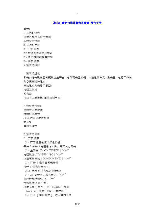
Zeiss 激光扫描共聚焦显微镜操作手册目录:1 系统得组成系统组成及光路示意图实物照片说明2 系统得使用2、1 开机顺序2、2 软件得快速使用说明2、3 显微镜得触摸屏控制2、4 关机顺序3 系统得维护1 系统得组成激光扫描共聚焦显微镜系统主要由:电动荧光显微镜、扫描检测单元、激光器、电脑工作站及各相关附件组成。
系统组成及光路示意图:电脑工作站激光器电动荧光显微镜扫描检测单元实物照片说明:电动荧光显微镜扫描检测单元CO2 培养系统控制器激光器电脑工作站2 系统得使用2、1 开机顺序(1)打开稳压电源(绿色按钮)等待2 分钟(电压稳定)后,再开其它开关(2)主开关[ MAIN SWITCH ]“ON”电脑系统[ SYSTEMS/PC ]“ON”扫描硬件系统[ PONENTS ]“ON”(3)打开[ 电动显微镜开关]打开[ 荧光灯开关](注:具有5 档光强调节旋钮)(4)Ar 离子激光器主开关“ON”顺时针旋转钥匙至“—”预热等待约15分钟,将激光器[ 扳钮] 由“Standby”扳至“Laser run”状态,即可正常使用(5)打开[ 电脑开关],进入操作系统注:键盘上也具有[ 电脑开关]2、2 软件得快速使用说明(1)电脑开机进入操作系统界面后,双击桌面共聚焦软件ZEN 图标(2)进入ZEN 界面,弹出对话框:“Start System”——初始化整个系统,用于激光扫描取图、分析等。
“Image Processing”——不启动共聚焦扫描硬件,用于已存图像数据得处理、分析。
(3)软件界面:1 功能界面切换:扫描取图(Acquisition)、图像处理(Processing)、维护(Maintain) (注:Maintain仅供Zeiss专业工程师使用)2 动作按钮;3 工具组(多维扫描控制);4 工具详细界面;5 状态栏;6 视窗切换按钮;7 图像切换按钮;8 图像浏览/预扫描窗口;9 文档浏览/处理区域;10 视窗中图像处理模块动作按钮:Single ——扫描单张图片、并在图像预览窗口显示。
Zeiss 激光扫描共聚焦显微镜 操作手册

Zeiss 激光扫描共聚焦显微镜操作手册目录:1 系统的组成系统组成及光路示意图实物照片说明2 系统的使用2.1 开机顺序2.2 软件的快速使用说明2.3 显微镜的触摸屏控制2.4 关机顺序3 系统的维护1 系统的组成激光扫描共聚焦显微镜系统主要由:电动荧光显微镜、扫描检测单元、激光器、电脑工作站及各相关附件组成。
系统组成及光路示意图:电脑工作站激光器电动荧光显微镜扫描检测单元实物照片说明:电动荧光显微镜扫描检测单元CO2 培养系统控制器激光器电脑工作站2 系统的使用2.1 开机顺序(1)打开稳压电源(绿色按钮)等待2 分钟(电压稳定)后,再开其它开关(2)主开关[ MAIN SWITCH ]“ON”电脑系统[ SYSTEMS/PC ]“ON”扫描硬件系统[ COMPONENTS ]“ON”(3)打开[ 电动显微镜开关]打开[ 荧光灯开关](注:具有5 档光强调节旋钮)(4)Ar 离子激光器主开关“ON”顺时针旋转钥匙至“—”预热等待约15分钟,将激光器[ 扳钮] 由“Standby”扳至“Laser run”状态,即可正常使用(5)打开[ 电脑开关],进入操作系统注:键盘上也具有[ 电脑开关]2.2 软件的快速使用说明(1)电脑开机进入操作系统界面后,双击桌面共聚焦软件ZEN 图标(2)进入ZEN 界面,弹出对话框:“Start System”——初始化整个系统,用于激光扫描取图、分析等。
“Image Processing”——不启动共聚焦扫描硬件,用于已存图像数据的处理、分析。
(3)软件界面:1 功能界面切换:扫描取图(Acquisition)、图像处理(Processing)、维护(Maintain)(注:Maintain仅供Zeiss专业工程师使用)2 动作按钮;3 工具组(多维扫描控制);4 工具详细界面;5 状态栏;6 视窗切换按钮;7 图像切换按钮;8 图像浏览/预扫描窗口;9 文档浏览/处理区域;10 视窗中图像处理模块动作按钮:Single ——扫描单张图片、并在图像预览窗口显示。
蔡司体视显微镜SteREO Discovery.V20

蔡司体视显微镜SteREO Discovery.V20
品牌:卡尔·蔡司 型号:SteREO Discovery.V20
制造商:德国卡尔蔡司公司 经销商:北京普瑞赛司仪器有限公司
产地:德国 联系方式:800-890-0660
技术参数:
1、总放大倍率: 4.7x-1312.5x
2、变倍比为:20:1
3、工作距离为10-253mm,
4、最大行程:340mm
5、调焦精度:350nm,
6、实际视场范围:0.18-48.7mm
7、冷光源: 15V150W ( 24V250W)
8、可扩展性:可配图像分析系统(数码相机、摄像头、图像分析软件)
仪器介绍:
采用系统控制器(SyCop)和人机交互控制器(H.I.P)控制显微镜的所有动作(自动倍数切换、自动聚焦、自动色温),自动化程度高,操作极为方便、舒适。
用于产品的宏观检验,能够检验断口内的裂纹(挤压裂纹、淬火裂纹、铸造裂纹)、裂口、纵向裂纹、焊和不良、疏松、氧化膜、气孔等宏观缺陷。
图像质量: 提供最高反差、最高衬度、最高分辨率的三维图像
光学系统: 国际最先进标准的Telescope(CMO)光学原理设计
显微观察新技术(蔡司专利): 防眩光技术
设计理念: 人机工程学原理, 模块化设计和开放式结构,保留了所有选择项,便于日后的升级和功能增强
使用寿命:60年
原产地:德国(国内无合资厂)。
ZEISS 倒置荧光显微镜操作手册-Axio Observer D1——【蔡司安装】

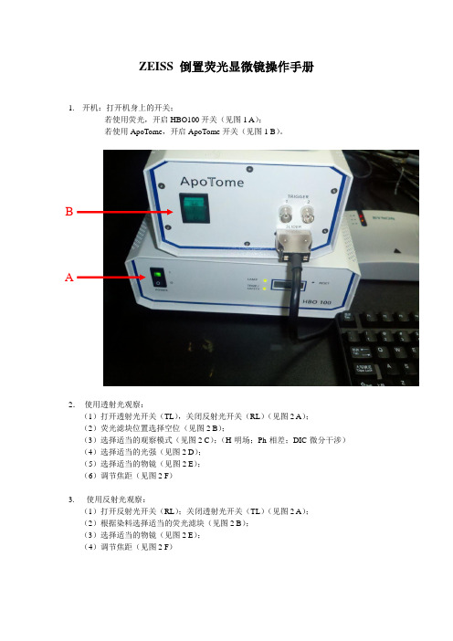
ZEISS 倒置荧光显微镜操作手册
1.开机:打开机身上的开关;
若使用荧光,开启HBO100开关(见图1 A);
若使用ApoTome,开启ApoTome开关(见图1 B)。
B
A
2.使用透射光观察:
(1)打开透射光开关(TL),关闭反射光开关(RL)(见图2 A);
(2)荧光滤块位置选择空位(见图2 B);
(3)选择适当的观察模式(见图2 C);(H-明场;Ph-相差;DIC-微分干涉)(4)选择适当的光强(见图2 D);
(5)选择适当的物镜(见图2 E);
(6)调节焦距(见图2 F)
3. 使用反射光观察:
(1)打开反射光开关(RL);关闭透射光开关(TL)(见图2 A);
(2)根据染料选择适当的荧光滤块(见图2 B);
(3)选择适当的物镜(见图2 E);
(4)调节焦距(见图2 F)
4. 利用相机获取图像:
(1)手动调节显微镜光路至相机(见图3 A )
(2)利用Axio Vision 软件进行拍摄(具体应用参加Axio Vision 操作手册)
D A
B C
E F A
5. 显示屏各项参数意义:
(1)物镜信息 (见图4A ):包含放大倍数(如20X )、数值孔径(如0.3)、物镜属性(如
LD-长工作距离;A Pln-平场;Ph1-相差)
(2)总放大倍数(见图4B ):如200X
(3)透射光开关(见图4C ):如ON 或OFF
(4)反射光开关(见图4D ):如○或●
(5)荧光滤块位置(见图4E ):如05 AF 430
E D C B A。
体式显微镜研究级Stemi 2000-C

天,进的显微更加总体 广泛 研户的层次产品1、2、3、4、技术品牌:卡尔制造商:德产地:德ZEISS 一ZEISS 仍致的显微技术微镜与以往加环保。
体描述:体视显微镜泛地应用于材研究级体式显的极高的认可次的检验和科品特点:采用国际上图像。
完美的3D宽视场光学 超长工作距术参数:研究尔·蔡司德国卡尔蔡国 百多年的骄致力于为用术,我们正在往相比,它们镜又称“实体显材料宏观表面显微镜Stemi 可和良好的口科研需求。
上最先进的D 图像,在学观察,S 距离,Stem 究级体式显蔡司公司骄人历史从用户研发最具在为全世界们的成像质显微镜”或“立面观察、失效2000-C 是蔡碑。
其配置的的Telescope 在整个变焦范temi 2000-mi 2000-C 显微镜St 型号:经销商联系方发明世界上具创造力的的用户开拓量更好、效立体显微镜”分析、断口分蔡司公司的经的灵活性和广e(CMO)光学范围内都能-C 能够为您能够为您提temi 200Stemi 20商:北京普瑞方式:800-上首台显微的显微镜系列拓一条探索微效率更高、机,是一种具有分析等工业领经典显微镜,广泛的适用性学原理设计能提供清晰的您提供最大提供最高可00-C00-C 瑞赛司仪器890-0660镜开始。
一列产品。
通微观世界的机械性能更有正像立体感领域。
在各领域的应性能够满足各计,为用户提的无失真的大118mm 的视可达286mm 的器有限公司 一个世纪后的通过我们不断的道路。
今天更加稳定,并感地显微镜,应用中获得了各领域用户的提供最锐利的图像 视场范围。
的工作距离的今断改天的并且被了客不同利的离。
1、总2、最3、基4、冷5、可产品1、 2、3、4、 5、6、 7、 8、总放大倍率:最大工作距离基本物体视场冷光源:8V2可扩展性:可品应用:材料检测:组织结构、 微电子技术 半导体行业像。
医学技术:要求在高倍 药物:检 琉璃纤维技成像,保证 法医学:织用于鉴定真 文物修复:求具有高分1.95X-250X 离为286mm 场直径为35m 20W (12V7可配图像分析:检测材料失效分析术领域:在业:芯片刻:检测模制倍下观察,测双折射蛋技术:涂层证高分辨率织物、头发真实情况。
Zeiss 激光扫描共聚焦显微镜 操作手册

Zeiss 激光扫描共聚焦显微镜操作手册目录:1 系统的组成系统组成及光路示意图实物照片说明2 系统的使用2.1 开机顺序2.2 软件的快速使用说明2.3 显微镜的触摸屏控制2.4 关机顺序3 系统的维护1 系统的组成激光扫描共聚焦显微镜系统主要由:电动荧光显微镜、扫描检测单元、激光器、电脑工作站及各相关附件组成。
系统组成及光路示意图:电脑工作站激光器电动荧光显微镜扫描检测单元实物照片说明:电动荧光显微镜扫描检测单元CO2 培养系统控制器激光器电脑工作站2 系统的使用2.1 开机顺序(1)打开稳压电源(绿色按钮)等待2 分钟(电压稳定)后,再开其它开关(2)主开关[ MAIN SWITCH ]“ON”电脑系统[ SYSTEMS/PC ]“ON”扫描硬件系统[ COMPONENTS ]“ON”(3)打开[ 电动显微镜开关]打开[ 荧光灯开关](注:具有5 档光强调节旋钮)(4)Ar 离子激光器主开关“ON”顺时针旋转钥匙至“—”预热等待约15分钟,将激光器[ 扳钮] 由“Standby”扳至“Laser run”状态,即可正常使用(5)打开[ 电脑开关],进入操作系统注:键盘上也具有[ 电脑开关]2.2 软件的快速使用说明(1)电脑开机进入操作系统界面后,双击桌面共聚焦软件ZEN 图标(2)进入ZEN 界面,弹出对话框:“Start System”——初始化整个系统,用于激光扫描取图、分析等。
“Image Processing”——不启动共聚焦扫描硬件,用于已存图像数据的处理、分析。
(3)软件界面:1 功能界面切换:扫描取图(Acquisition)、图像处理(Processing)、维护(Maintain)(注:Maintain仅供Zeiss专业工程师使用)2 动作按钮;3 工具组(多维扫描控制);4 工具详细界面;5 状态栏;6 视窗切换按钮;7 图像切换按钮;8 图像浏览/预扫描窗口;9 文档浏览/处理区域;10 视窗中图像处理模块动作按钮:Single ——扫描单张图片、并在图像预览窗口显示。
蔡司研究级倒置万能材料显微镜Axio Observer A1m

研究级倒置万能材料显微镜Axio Observer A1m品牌:卡尔·蔡司 型号:Axio Observer A1m制造商:德国卡尔蔡司公司 经销商:北京普瑞赛司仪器有限公司产地:德国 联系方式:800-890-0660ZEISS一百多年的骄人历史从发明世界上首台显微镜开始。
一个世纪后的今天,ZEISS仍致力于为用户研发最具创造力的显微镜系列产品。
通过我们不断改进的显微技术,我们正在为全世界的用户开拓一条探索微观世界的道路。
今天的显微镜与以往相比,它们的成像质量更好、效率更高、机械性能更加稳定,并且更加环保。
总体描述:金相学主要指借助光学(金相)显微镜和体视显微镜等对材料显微组织、低倍组织和断口组织等进行分析研究和表征的材料学科分支,既包含材料显微组织的成像及其定性、定量表征,亦包含必要的样品制备、准备和取样方法。
其主要反映和表征构成材料的相和组织组成物、晶粒(亦包括可能存在的亚晶)、非金属夹杂物乃至某些晶体缺陷(例如位错)的数量、形貌、大小、分布、取向、空间排布状态等。
金相学的兴起给金属材料研究带来了历史性的变革,而蔡司长久以来一直致力于金相显微镜的研发与应用,并将金相学的科研水平推向一个又一个高点。
完美旗舰—Axio Observer A1m 2007年面世。
Axio observer A1m是卡尔蔡司成功的Axiovert 200mat机型的延伸,AxioObserver A1m她将CARLZEISS所特有的高质量成像与优越的机械稳定性完美结合。
Axio Oberver A1m 无论是成像质量、操作舒适性,还是机械稳定性方面匀改写了世界倒置式研究级显微镜的新标准。
每个细节都符合材料领域特殊的光学要求。
她以最尖端的光学技术和最完美的制造工艺延续着卡尔•蔡司成功的传奇故事!产品特点:1、采用世界上最优秀的无限远双重色彩校正及反差增强型(ICCS)光学系统,为用户提供最锐利的图像。
2、6位功能观察转盘,为您提供最高的升级空间。
- 1、下载文档前请自行甄别文档内容的完整性,平台不提供额外的编辑、内容补充、找答案等附加服务。
- 2、"仅部分预览"的文档,不可在线预览部分如存在完整性等问题,可反馈申请退款(可完整预览的文档不适用该条件!)。
- 3、如文档侵犯您的权益,请联系客服反馈,我们会尽快为您处理(人工客服工作时间:9:00-18:30)。
M i c r o s c o p y f r o m C a r l Z e i s sSteREO Lumar.V12The New FluorescenceBrilliant fluorescencein stereomicroscopyFluorescence Carl Zeiss: FluoresScienceFluorescence: the basis of many modern microscopic techniques.Fluorescent proteins in particular have become one of the key methods. New fluorescence applications enable increasingly differentiated observation in laser scanning microscopy and light microscopy, where stereomicroscopic fluorescence applications have gained enormous significance.Developing such microscopes and imaging systems is a science in itself. At Carl Zeiss we have committed ourselves to this challenge with uncompromising dedication and extensive know-how. After all, when you are working at the boundary to the invisible, you can't make any compromises. To give your best, you need the best tools possible, tools• with the highest efficiency• with the most innovative technologies• with the most powerful imaging systems.From the very beginning Carl Zeiss has set high standards in light and in confocal laser scanning microscopy – with internationally leading techno-logies for fluorescence. Our focus on this key tech-nique for the life science research has a name –Carl Zeiss: FluoresScience. This is the Zeiss seal of quality and our pledge that the best fluorescence tools for the life sciences will be made by Carl Zeiss, both today and in the future.Tupaia brain.Multiple fluorescenceObjective: NeoLumar S 1.5xMagnification 150x*Specimen: Prof.E.Fuchs,S.Bauch,Primatenzentrum Göttingen,Germany* Actual viewing magnification.Magnification 23x*Magnification 80x*Magnification 150x*ContentsSteREO Lumar.V12 – Expanding the Boundaries3Applications4Pioneering Work6Fluorescence Optics8Ergonomics and Operation10Operation Concept: SyCoP12Illumination and Contrasting Techniques14Imaging System16Systems Overview182Objective NeoLumar S 1.5x5OpticPioneering Work from Carl ZeissAs the heart of a microscope, the objectives deter-mine the optical performance of the entire system –particularly in the case of fluorescence microscopy.Carl Zeiss, a leader in fluorescence microscopy, has invested its entire know-how and experience in the development of new fluorescence objectives for SteREO Lumar.V12. Now you can profit from this know-how: with NeoLumar S, you have two objec-tives at your disposal both setting new standards in fluorescence applications.Fluorescence highlights:Objectives for greater brillianceThe basis for the best fluorescence in stereo-microscopy is provided by two objectives designed especially for SteREO Lumar.V12. With resolution of 0.6 mm, the NeoLumar S 1.5x objective is ideal for overviews and documentation. The NeoLumar S 0.8x objective with its free working distance of 80 mm is particularly suitable for over-views and specimen preparation. You benefit from uncompromising excellence in optics for every application.Observing much more:Resolution in large specimen fieldsThis new optical quality is based on other impor-tant components in addition to the objectives –for example, the luminous zoom and the stray-light minimizing tubes for eyepieces with a 23 mm field of view. Together they make the SteREO Lumar.V12 microscope system the new standard in fluorescence stereomicroscopy and give you unrivalled illumination with exceedingly high reso-lution and exceptional contrast in the largest of specimen fields.Microstructured substrate immune fluorescent-stained with adherent cells.Objective: NeoLumar S 1.5x Magnification: 150x*Specimen: Prof.M.Bastmeyer,Dr.D.LehnertFriedrich-Schiller-Universität Jena,GermanyClearly resolved for the first time using a stereo-microscope with UV excitation: distances between the single points of 5 µm (left) and 1 µm (right).7Specially developed for fluorescence stereomicroscopy: thetwo objectives NeoLumar S 0.8x and NeoLumar S 1.5x –unrivalled in luminosity and UV transmission.Magnification 150x*SteREO Lumar.V12 withNeoLumar S 1.5x objective8Shining ExamplesNew freedom in illumination: Light zoom HiLiteFor optimal fluorescence excitation, the integrated light zoom with lamp mount and 4x filter mount (HiLite) is automatically coupled to the observation zoom. A remarkable option: the light zoom can be detached from the observation zoom. TheDeveloped for conventional light fluorescence microscopy, SteREO Lumar.V12 offers first-class optical illumination systems These systems feature even illumination of specimens at all magnifi-cations, and an independent zoom for optimal adaptation of fluorescence excitation light to the specimen field selected. In addition, continuous automation makes the handling of filters particu-larly easy.advantage of the free light zoom: it is possible to individually increase the brightness of selected specimen structures at low resolutions – thereby mobilizing the last reserves of light. And at high resolutions the light intensity can be reduced, thus ensuring deblurred specimens.Fluorescence filter sets: Found in the filter wheelFor simplification and acceleration of fluorescence:the filter wheel can accept up to four differentfluorescence filter sets. Each set consists of an excitation filter and two emission filters.Fluorescence is easy: the filter wheels are inserted into the microscope and automatically recognized (Push&Slide). Changing of the filter sets is motor-ized.Changing the filter wheelFilter wheelsThe filter wheel in SteREO Lumar.V12 offers room for 4 filter sets.Every filter set consists of an excitation filter and 2 emission filters The filter wheel can be completely removed,enabling you to change filter sets easily.SteREO Lumar.V12 – the powerful fluorescence microscope from Carl Zeiss.In the picture: AFR for automatic filter recognitionEffective, error-free work with different filters – a must in modern fluorescence stereomicroscopy. The solution from Carl Zeiss: AFR (Automatic Filter Recognition). SteREO Lumar.V12 recognizes the filter via an integrated color sensor. SyCoP informs you about the available filter sets. In addition, it shows you the filter set currently in the beam path, together with all related spectral data.Push&Slide HIP SteREO Lumar.V12 –the powerful fluorescence microscope from Carl Zeiss.No longer necessary to turn the knob : The HIP (Human Interface Panel) replaces the conventional knob onthe stereomicroscope.You can now zoom at any speed you select.Important optical data such as magnification,specimen field diameter,maximum resolution possible or depth of field of the current setting are visible on adisplay.9101.Stability, Precision and Room for ManipulationExtreme precision: Motorized focusingFast and highly sensitive – the newly developed high-quality mechanics of the motor focusing are as accurate as clockwork. Using HIP (Human Interface Panel), the focus can now be rapidly set and precisely reproduced – if desired, via the fine setting in 350 nm steps! This amazing precision can even be attained with equipment weighing upto 17 kg across a 340 mm travel range.As the optical performance of modern stereomicros-copes grows, so do the technical demands on the mechanics of such systems. The stand and focusing equipment play a major role here. Stability, sturdin-ess and sufficient room in the specimen field are as important as rapid and reproducible focusing on the specimen – adapted to the individual application.Intelligent tool: The focus managerFocusing on a specimen have been enormously simplified: After objective is changed, a corre-sponding speed profile is automatically set for focusing – sensitive for high-resolution objectives,fast for low resolution. In addition, you can work with fine focus in steps of 350 nm. An electronic specimen protection prevents damage to your specimens.New standard: The Z-measure-mentWith its sturdy stand design and precise motoriza-tion of its focus setting, SteREO Lumar.V12 ensu-res that the position of the microscope to the spe-cimen can be directly displayed. The Z-position is set to zero at the press of a button, the next spe-cimen detail will be focused , and the difference is displayed with precision of +/- 30 µm.HIPAmple room in the specimen space: the unusual stand construction with decentralized profile S column.11StandGear rodRoom for objectives: Generous working areas and preciseAmple room for objects and manipulation: with its large scratch-resistant stage plate (250 x 410mm), SteREO Lumar.V12 provides you with generous specimen space. Gliding, rotating, ball-and-socket, mechanical and scanning stages can be easily mounted and attached via an interface (120 mm in diameter). The light and highly sensi-tive adjustable gliding stage is ideal for higher Ready for the motorized future:The CANBUS29 systemSteREO Lumar.V12 was developed as a platform for the motorized and digitalized fluore-scence stereomicroscopy of the future. Its open CANBUS29 system "understands" and integrates all motorized components.HIP (Human Interface Panel) replaces the conventional focusing knobs The current Z-position is always 340 mm precise focusing – new wear-resistant plastics make this possible.Motorized focusing of the S column – platform for further motorized CANBUS29-controlled components.SyCoP – A Revolutionary Operating ConceptConcentrating on the essentials:SyCoPFreely positionable, SyCoP can be operated equal-ly well by right-handed or left-handed users.Instead of lifting your eyes countless times toselect and control settings, changes and manipu-lations, you can operate the microscope "bytouch" – and keep your eyes and your concentra-tion on the specimen.Touchscreen Keys JoyStickSpecially designed for stereomicroscopy, SyCoP(Sy stem Co ntrol P anel) combines joystick, buttonsand touch screen in the convenient design of a com-puter mouse, enabling easy handling of increasinglycomplex operations. With SyCoP you can control allessential functions of a microscope fast, with preci-sion and reproducibly, without lifting your eyes fromthe eyepiece. This innovative and mobile operatingunit makes it possible for you to reach new heightsof perfection in automated microscopy. This is partic-ularly important in complicated processes such as flu-orescence microscopy, where you should be devotingmost of your attention to the specimen – and not tooperating your microscope.A new feeling in microscopy:Joystick for focus and zoomZoom in by joystick in the east-west direction,focus in the north-south – the two most commonprocesses in microscopy can now easily be con-trolled via one operating element, saving you timeand unnecessary movement. In addition, you cancontrol light or quickly switch on illumination andcontrasting techniques via fixed buttons. Thetouch screen has two functions: as a display fordata and information and as a further controlpanel to "switch on" additional functions of thestereomicroscope.More automation: Future optionswith SyCoPSyCoP is also an option for the future. Its openconcept allows new functions and upgrades allmotorized accessories to be added in the future.13SyCoPtime this information – normally difficult for the user of a stereomicroscope to access – can now be read. Another improvement: the brightness of the display can be adjusted, even turned off, making it equally suitable for work in lighted and darke-ned rooms.Now complex stereomicroscopic systems can be easily operated using only one hand.Reliably and error-free – and without taking your eyes off the specimen.Perfect information: Supplying facts to the microscopeA further innovation in microscopy: SyCoP informs its users at a glance about all important optical parameters of the current setting, such as total magnification, visible specimen field, maximum resolution possible, and depth of field. For the firstIntelligent Light ManagementFor the first time in stereomicroscopy there is a self-aligning HBO fluorescence illumination. This uniqueillumination guarantees consistently reproducibleresults, avoiding the necessity of time-consumingadjustments. The intelligent integration of fiber-opticillumination components for reflected and transmit-ted light guarantees the highest possible contrastand optimal illumination, from the overview down tothe last detail.Self-aligning: The HBO lampPrerequisite for strong signals in fluorescence: thenew HBO lamp in SteREO Lumar.V12, exclusivefrom Carl Zeiss. Thanks to the new self-adjustingconstruction, the HBO burner aligns itself auto-matically every time it is switched on. This resultsin a stable, optimum setting during the entire lifeof the lamp, ensuring perfect illumination of thefield of view and consistently excellent fluores-cence results.Imaging in oblique transmitted-light brightfieldfor counter-contrasting of transparent and fluorescentspecimens.Illumination and coReflected light Transmitted light3-day-old Zebra fishRed fluorescence: motor cerebral nervesGreen fluorescence: motor and sensory cerebral nervesObjective: NeoLumar S 1.5xMagnification: 150x*Specimen: Prof.M.Bastmeyer,Dr.M.Marx,Friedrich-Schiller-UniversitätJena,Germany.reproduzierbar abspeichern.Kontrastprogramm:Diese modulare, nachrüstbare Einrichtung istgeeignet für Hellfeld, Schrägbeleuchtung undDunkelfeld. Mit ihren zusätzlichen Spiegel-Freiheitsgraden bietet sie eine verbesserte schiefeBeleuchtung auch bei hohen Vergrößerungen,was den Informationsgehalt der Bilder steigert.Die besonders große Arbeitsfläche erleichtert dieArbeit beim Screening von Petrischalen und ande-ren Kulturgefäßen.Practice-oriented alternatives:Powerful cold-light illuminationsourcesTwo powerful cold-light sources provide a valuablealternative to illumination and contrasting equip-ment for reflected and transmitted light: Schott KL1500 LCD and Schott KL 2500 LCD. The latter hasthe higher performance and can be controlled viaSyCoP, with its easy-to-find buttons for selectingand regulating light. The Light Manager guaran-tees that specimens are evenly lit throughout thezoom range while a memory control ensures thatuser settings are stored and reproducible.A good contrast: Transmittedlight equipment SThis modular, retrofittable equipment is ideal forbrightfield, oblique illumination and darkfield.With its additional flexibility to move the mirror, itoffers improved oblique illumination, even at highmagnifications. As a result, the images containmore information. The exceptionally large wor-king area simplifies the screening of petri dishesand other culture vessels.ntrasting techniquesLampThe HiLite excitation beam path zoom can be detached from the observationzoom and controlled separately.Advantage: reduced intensity of the excitationlight during the observation of intensely lit specimens at high magnifications.Unique: the illumination of the specimen field remains homogeneous.HiLite attached to the observation zoom results in stray light.ContrastingFrom Steromicroscope to System:Fluorescence ImagingPowerful from the start:The basic program from AxioVisionThe entry-level version of AxioVision provides you with a high-performance tool featuring an impres-sive wealth of functions. The benefits for you:software control of your instrument, easy storage of microscope parameters, automatic recall of sca-ling factors, and easy configuration of individual steps.The system concept of stereomicroscopy is brought to perfection by AxioVision, the software for digital microscope systems. In addition to a unique modular structure that fulfills the demands of current micros-copy, it features attractive options for expansion and upgrades. AxioVision integrates microscope control,image acquisition, image processing as well as image analysis, management and archiving into a single system. There is a precise software solution for every requirement which can be easily and inexpensively expanded – from powerful entry models for new-comers right up to systems for advanced users.Team work: AxioVision and digi-tal camera systemsAxioVision has interfaces for standard technolo-gies which enable the use of a wide range of cameras – from simple consumer models to scien-tific ones. Digital cameras from the Carl Zeiss camera family offer additional advantages.The AxioCam family: Flexible spe-cialists for every needThe AxioCAm family: Flexible specialists for every needCarl Zeiss offers a wide range of digital cameras in various performance class. With their high dyna-mics of 12 or 14 bits, the monochrome camerasfeature optimal resolution and high sensitivity,particularly with weak fluorescence specimens.The color cameras provide excellent color repro-duction and outstanding resolution – in addition to up to 12 megapixels per color channel without any loss through color interpolation. The cameras are Peltier-cooled and protect specimens through fast shutter synchronization. A particular highlight is the fast live image, even with long exposure times.12Skeletal preparation of a newborn mouse Red staining: calcified bone tissue Blue staining: cartilage tissues Transmitted-light,brightfieldSteREO Discovery.V12,Objective PlanApo S 0,63x AxioCam MRc5,AxioVision (Module Panorama)Magnification 5x*Sample:Dr.Kenji Imai,M.D.,PhD.GSF-National Research Center for Environment and HealthNeuherberg,GermanyVisible quality: Functions for image acquisitionNo single work station is like the other. In addi-tion, your requirements change with the growing and diverse demands of your work. With its wealth of image acquisition functions, AxioVision can easily meet these challenges, enabling you to put together exactly what you need for best results. Even simple two-dimensional imaging benefits from the automatically assigned image formats and setting information that are stored together with the image. These reproducible con-ditions are essential for correct image comparison.Life in focus: Modules for the observation of living organismsOriented towards the requirements of biomedical laboratories, powerful and easy to operate:AxioVision has aquisition modules for multichan-nel and time-lapse fluorescence images. With a mechanical or electronic specimen stage, easily assembled images can also be created with the new modules MosaiX and Panorma. You get out-standing resolution while maintaining image overview.Measurement, documentation,archiving: AxioVsion for the ana-lysis and management of imagesFor individual or routine measurements of your specimens: AxioVision provides you with the tools you need to analyze image information - whether interactively or automatically. Modules to archive image, text and graphic, simplify information and accelerate data management. You can catalogue images, assign categories and keywords and add annotations and comments. Meta data from the images are automatically taken over, displayed,exported and processed.Fig.1Worm C.elegans on an Agar plate.Oblique illumination in transmitted light-brightfield Objective: Neolumar S 1.5x Magnification: 150x*Fig.2Desmid algae Closterium (Desmidiales)UV excitationObjective: Neolumar S 1.5x Magnification: 150x*Specimen: T.Friedl,Sammlung von Algenkulturen,Universität Göttingen,Germany18System overview19ObjectivesCarl Zeiss Microscopy GmbH 07745 Jena, Germany microscopy@ www.zeiss.de/stereo-lumarEyepiecesTechnical dataP r i n t e d o n e n v i r o n m e n t -f r i e n d l y p a p e r , b l e a c h e d w i t h o u t t h e u s e o f c h l o r i n e .S u b j e c t t o c h a n g e .46-0007 e 09.2004* Free working distance。
